Báo cáo y học: "Anti-inflammatory and antiarthritic effects of piperine in human interleukin 1β-stimulated fibroblast-like synoviocytes and in rat arthritis models" pps
Bạn đang xem bản rút gọn của tài liệu. Xem và tải ngay bản đầy đủ của tài liệu tại đây (1.78 MB, 9 trang )
Open Access
Available online />Page 1 of 9
(page number not for citation purposes)
Vol 11 No 2
Research article
Anti-inflammatory and antiarthritic effects of piperine in human
interleukin 1β-stimulated fibroblast-like synoviocytes and in rat
arthritis models
Jun Soo Bang
1
*, Da Hee Oh
1
*, Hyun Mi Choi
1
, Bong-Jun Sur
2
, Sung-Jig Lim
3
, Jung Yeon Kim
4
,
Hyung-In Yang
5
, Myung Chul Yoo
6
, Dae-Hyun Hahm
2
and Kyoung Soo Kim
1
1
East-West Bone & Joint Research Institute, East-West Neo Medical Center, Kyung Hee University, 149 Sangil-dong, Gangdong-gu, Seoul, Republic
of Korea
2
Acupuncture and Meridian Science Research Center, Kyung Hee University, Hoeggidong, Dongdaemoon-gu, Seoul, Republic of Korea
3
Department of Pathology, East-West Neo Meidcal Center, Kyung Hee University, 149 Sangil-dong, Gangdong-gu, Seoul, Republic of Korea
4
Department of Pathology, Inje University Sanggye Paik Hospital, Sanggye 7 dong 761-7, Nowon-gu, Seoul, Republic of Korea
5
Department of Internal Medicine, East-West Neo Medical Center, Kyung Hee University, 149 Sangil-dong, Gangdong-gu, Seoul, Republic of Korea
6
Department of Orthopedic Surgery, East-West Neo Medical Center, Kyung Hee University, 149 Sangil-dong, Gangdong-gu, Seoul, Republic of
Korea
* Contributed equally
Corresponding author: Dae-Hyun Hahm, Kyoung Soo Kim,
Received: 30 Dec 2008 Revisions requested: 9 Feb 2009 Revisions received: 4 Mar 2009 Accepted: 30 Mar 2009 Published: 30 Mar 2009
Arthritis Research & Therapy 2009, 11:R49 (doi:10.1186/ar2662)
This article is online at: />© 2009 Bang et al.; licensee BioMed Central Ltd.
This is an open access article distributed under the terms of the Creative Commons Attribution License ( />),
which permits unrestricted use, distribution, and reproduction in any medium, provided the original work is properly cited.
Abstract
Introduction The objective of this study was to determine the
anti-inflammatory, nociceptive, and antiarthritic effects of
piperine, the active phenolic component in black pepper extract.
Methods The in vitro anti-inflammatory activity of piperine was
tested on interleukin 1β (IL1β)-stimulated fibroblast-like
synoviocytes derived form patients with rheumatoid arthritis. The
levels of IL6, matrix metalloproteinase (MMPs), cyclo-oxygenase
2 (COX-2), and prostaglandin E2 (PGE
2
) were investigated by
ELISA and RT-PCR analysis. The analgesic and antiarthritic
activities of piperine were investigated on rat models of
carrageenan-induced acute paw pain and arthritis. The former
were evaluated with a paw pressure test, and the latter by
measuring the squeaking score, paw volume, and weight
distribution ratio. Piperine was administrated orally to rats at 20
and 100 mg/kg/day for 8 days.
Results Piperine inhibited the expression of IL6 and MMP13
and reduced the production of PGE
2
in a dose dependant
manner at concentrations of 10 to 100 μg/ml. In particular, the
production of PGE
2
was significantly inhibited even at 10 μg/ml
of piperine. Piperine inhibited the migration of activator protein
1 (AP-1), but not nuclear factor (NF)κB, into the nucleus in IL1β-
treated synoviocytes. In rats, piperine significantly reduced
nociceptive and arthritic symptoms at days 8 and 4,
respectively. Histological staining showed that piperine
significantly reduced the inflammatory area in the ankle joints.
Conclusions These results suggest that piperine has anti-
inflammatory, antinociceptive, and antiarthritic effects in an
arthritis animal model. Thus, piperine should be further studied
with regard to use either as a pharmaceutical or as a dietary
supplement for the treatment of arthritis.
Introduction
Rheumatoid arthritis is characterized by chronic proliferative
synovitis, inflammatory immune cell infiltration into the synovial
fluid and cartilage destruction [1]. Proliferative fibroblast-like
synoviocytes (FLSs) play crucial roles in both the propagation
of inflammation and joint damage because they produce a
great amount of proinflammatory mediators such as matrix
metalloproteinses (MMPs), interleukin (IL)6, IL8 and prostag-
landin E
2
(PGE
2
) [2].
Thus, anti-inflammatory agents are administrated as long-term
treatments for patients with rheumatoid arthritis. However,
ELISA: enzyme-linked immunosorbent assay; FLS: fibroblast-like synoviocytes; H&E: hematoxylin and eosin; IL: interleukin; MMP: matrix metallopro-
teinase; OA: osteoarthritis; RA: rheumatoid arthritis; WDR: weight distribution ratio.
Arthritis Research & Therapy Vol 11 No 2 Bang et al.
Page 2 of 9
(page number not for citation purposes)
anti-inflammatory agents carry the risk of gastrointestinal toxic-
ity; thus, their use is limited. In an attempt to avoid adverse gas-
trointestinal effects, a new generation of non-steroidal anti-
inflammatory drugs (NSAIDs) was developed that selectively
inhibited cyclo-oxygenase (COX)-2 selective inhibitors (for
example, celecoxib, rofecoxib, and valdecoxib) [3]. Celecoxib
and valdecoxib appear to show satisfactory cardiovascular
safety, however, rofecoxib was withdrawn from the market due
to cardiovascular toxicity. However, side effects remain one of
the problems for long-term use; thus, there is a need for anti-
inflammatory drugs with less severe side effects. In addition,
recent interest in alternative treatments for arthritis [4,5] has
promoted their use in the US, but scientific evidence of antiar-
thritic efficacy is lacking.
Black pepper (Piper nigrum) is commonly used as a spice in
human diets, but it is also used as a medicine, a preservative,
and a perfume in many Asian countries. An extract of the active
phenolic component, piperine, is well known to provide bene-
ficial physiological effects [6]. It stimulates the digestive
enzymes of pancreas, protects against oxidative damage, low-
ers lipid peroxidation, and enhances the bioavailability of a
number of therapeutic drugs. In addition, its anti-inflammatory
activities have been demonstrated in rat models of carra-
geenan-induced rat paw edema, cotton pellet-induced granu-
loma, and a croton oil-induced granuloma pouch [7].
Constituents of the piper species have shown in vitro inhibi-
tory activity against the enzymes responsible for leukotriene
and prostaglandin biosynthesis, 5-lipoxygenase and COX-1,
respectively [8]. These effects of piperine seem to be benefi-
cial for inflammatory diseases that are accompanied by severe
pain; for example, rheumatoid arthritis.
The excellent therapeutic properties of piperine have been
demonstrated in various cell types [9-12]. Nevertheless, little
is known about the effect of piperine on the production of
proinflammatory mediators in FLSs. Furthermore, to our knowl-
edge, its antiarthritic efficacy has never been evaluated. In this
study, the anti-inflammatory effects of piperine were tested in
IL1β-stimulated rheumatoid arthritis fibroblast-like synovio-
cytes derived from patients with rheumatoid arthritis. Its antiar-
thritic efficacy was evaluated in animal models of experimental
arthritis.
Materials and methods
Cell culture and reagents
All in vitro experiments were carried out with fibroblast-like
synoviocytes derived from patients with rheumatoid arthritis
(RA). After obtaining informed consent, synovial tissues were
collected from RA patients. They met the 1987 American Col-
lege of Rheumatology (ACR) criteria for the diagnosis of RA
and had been treated with non-biological disease-modifying
antirheumatic drugs (DMARDs) and were underwent thera-
peutic joint surgery. FLSs were isolated as described previ-
ously [13] and grown in Dulbecco's Modified Eagle Medium
(DMEM, low glucose) (Gibco-BRL, Grand Island, NY, USA)
supplemented with 10% (v/v) fetal bovine serum (FBS; Gibco-
BRL) and 1 × antibiotic-antimycotic (Gibco-BRL). After the
cells had grown to confluence, they were split at a 1:4 ratio.
FLS passages 3 to 6 from three patients were used for all
experiments. Piperine, prednisolone, corn oil and carrageenan
were obtained from Sigma-Aldrich Korea (Young-In, Korea).
Celecoxib was purchased in the form of the commercial drug,
Celebrex (capsules; Pfizer Korea, Seoul, Korea).
Semiquantitative RT-PCR
FLSs (2.5 × 10
5
cells) were cultured overnight in 60 mm
dishes containing 2 ml of media. Cells were incubated with
serum-free media for 2 h and new serum-free media was
replaced just prior to the addition of piperine and cultured for
24 h. Supernatants were collected for ELISA and the cells
were used for semiquantitative RT-PCR. Trizol was used to
extract total RNA from the cells. Complementary DNA was
synthesized from 1 μg of total RNA in a 20 μl reverse transcrip-
tion reaction mixture. For semiquantitative PCR, aliquots of
cDNA were amplified in a 25 μl PCR mixture according to the
protocol provided by the manufacturer (TaKaRa Bio, Kyoto,
Japan), as described previously [13]. The PCR conditions for
the MMPs, IL6, and COX-2 were as follows: 30 to 33 cycles
of 95°C for 45 s, 55 to 60°C for 45 s, and 72°C for 45 s. PCR
products were subjected to electrophoresis on 1.5% agarose
gels containing ethidium bromide, and the bands were visual-
ized under ultraviolet (UV) light.
ELISA
Synovial cells (2.5 × 10
5
cells/60 mm dish/2 ml serum-free
media) were treated with various concentrations of piperine
30 minutes prior to IL1β stimulation. Conditioned media was
collected 24 h later. Briefly, FLS cultures were centrifuged and
the supernatants were collected and analyzed for IL6, PGE
2
,
MMP1, and MMP13 with an ELISA kit (R&D Systems, Minne-
apolis, MN, USA). For mRNA analysis, the cells were lysed and
total RNA was extracted. The mRNA levels of IL6 and COX-2
were measured by semiquantitative RT-PCR analysis. The
COX-2 protein expression was measured by western blot.
Three independent experiments were performed in duplicate.
Each experiment was performed using synovial cells from dif-
ferent patients. The collected supernatants were analyzed for
IL6, PGE
2
, MMP1 and MMP13 using commercial kits (ELISA;
R&D Systems). For the measurement of transcription factors,
nuclear factor (NF)κB and activator protein 1 (AP-1), in the
nucleus, FLSs were seeded (5 × 10
6
cells) into 100 mm
dishes and grown to 80% confluence. The cells were serum-
starved overnight and stimulated by IL1β (10 ng/ml) for 90
minutes in the presence or absence of piperine. Subsequently,
the cells were washed twice in phosphate-buffered saline
(PBS) and treated with lysis buffer and the extraction of tran-
scription factors from the nucleus was performed according to
the manufacturer's protocol (Active Motif, Seoul, Korea).
Available online />Page 3 of 9
(page number not for citation purposes)
Western blot analysis
FLSs cultured (2.5 × 10
5
cells) in 60 mm dishes were serum-
starved overnight and stimulated by IL1β (10 ng/ml) for 10 or
30 minutes in the presence or absence of piperine. The cells
were subsequently washed twice in PBS and treated with 50
μl of lysis buffer (20 mM Tris-Cl pH 8.0, 150 mM NaCl, 1 mM
ethylenediaminetetraacetic acid (EDTA), 1% Triton X-100, 20
μg/ml chymostatin, 2 mM phenylmethylsulfonyl fluoride
(PMSF), 10 μM leupeptin, and 1 mM 4-(2-aminoethyl)benze-
nesulfonyl fluoride (AEBSF)). As described previously [13],
the samples were separated using 12% SDS-PAGE, and
were then transferred to Hybond-ECL membranes (Amer-
sham, Arlington Heights, IL, USA). The membranes were first
blocked with 6% non-fat milk dissolved in Tris-buffered saline/
Tween (TBST) buffer (10 mM Tris-Cl pH 8.0, 150 mM NaCl,
0.05% Tween 20). The blots were then probed with various
rabbit polyclonal antibodies for inhibitor of κB (IκB)α, p-ERK1/
2, p-P38, p-Jun N-terminal kinase (JNK) and β-actin (Cell Sig-
naling Technology, Beverly, MA, USA) diluted 1:1,000 in TBS
at 4°C for overnight, and incubated with 1:1,000 dilutions of
goat anti-rabbit IgG secondary antibody coupled with horse-
radish peroxidase. The blots were developed using the ECL
method (Amersham). For re-probing, the blots were incubated
in the stripping buffer (100 mM 2-mercaptoethanol, 2% SDS,
62.5 mM Tris-HCl pH 6.7) at 50°C for 30 minutes with occa-
sional agitation.
Histological assessment of inflammation
The rats were killed after 9 days of carrageenan and control
treatments. Immunohistochemical staining was performed to
determine the degree of immune cells infiltration into the joints.
Rat ankles were dissected, fixed in 10% formalin, dehydrated
through a graded ethanol series, cleared in xylene, and proc-
essed for embedding in paraffin wax with routine protocols. A
microtome was used to cut 4 μm-thick sections that were sub-
sequently stained with hematoxylin and eosin (H&E) stain. The
degree of inflammation was evaluated on a scale from 0 to 5
by three different pathologists that had been blinded to the
treatments. The scale was defined as follows: 0 = no inflam-
mation, 1 = mild inflammation, 2 = mild/moderate inflamma-
tion, 3 = moderate inflammation, 4 = moderate/severe
inflammation, and 5 = severe inflammation.
Rat models of paw hyperalgesia and arthritis
Sprague-Dawley 5-week-old to 6-week-old male rats, pur-
chased from SLC (Shizuoka, Japan), were used in this study.
All animals were maintained in plastic cages at 21 to 24°C
under a 12 h light/dark cycle and were given free access to
pellet food and water. They were adapted for at least 1 week
prior to the start of the experiment. All subjects were habitu-
ated to the behavioral test chambers and handled with special
care to minimize stress. All methods were approved by the Ani-
mal Care and Use Committee of Kyung Hee University. All pro-
cedures were conducted in accordance with the Guide for the
Care and Use of Laboratory Animals, published by the Korean
National Institute of Health.
To induce paw hyperalgesia, rats were given an intraplantar
injection of 1% carrageenan (0.1 ml) in the posterior right paw
as described previously [14]. After 3 hr of the injection, the
pain threshold was measured using a paw pressure analgesia
instrument (UGO-BASIL Biological Research Apparatus,
Comerio-Varese, Italy) for the Randall-Selitto test paw. A total
of 10 rats were studied per group and the test was performed
blind. Rats were starved overnight and piperine was evaluated
at doses of 20 and 100 mg/kg. Piperine dissolved in corn oil
was fed orally 1 h before carrageenan injection. To evaluate
paw hyperalgesia, we measured the tolerance to increasing
mild pressure on the affected paw between a flat surface and
a blunt pointer of the instrument, as manufacturer's protocols.
The effects of piperine were compared to the effects of Cele-
brex (Pfizer), a selective COX-2 inhibitor (100 mg/kg).
The carrageenan-induced arthritic rat model was prepared as
described previously [15]. Animals were briefly anesthetized
with 3% isoflurane in a mixed N
2
O/O
2
gas. Arthritic inflamma-
tion was induced by a single injection of 3% carrageenan sus-
pended in 100 μl of pyrogen-free sterile saline, into the left
tibiotarsal ankle joint. The effects of piperine were compared
to the effects of prednisolone (10 mg/kg), a corticosteroid.
To evaluate the arthritic progression of carrageenan-induced
arthritis in the rat, three different parameters were measured:
paw volume, squeaking score in the ankle flexion test, and
weight distribution ratio (WDR). These were considered
behavioral indicators of carrageenan-induced arthritis and
checked daily for 9 days. With progression of arthritis, redness
and swelling of the ankle joints and arthritic pain started to
appear and reached a maximum on day 1 after the carra-
geenan injection. At that time, piperine and prednisolone dis-
solved in corn oil was administrated orally once a day for 8
days.
The paw volumes were measured using a digital plethysmom-
eter (UGO-BASIL Biological Research Apparatus), as
described by Kwon et al. [16]. Paw volumes were expressed
as relative values to that of day 0 when carrageenen was
injected. The ankle flexion test involved gentle flexion and
extension of the carrageenan-injected ipsilateral hind limb, as
described by Kwon et al. [16]. This elicited vocalizations
(squeaking) that were scored on a scale (squeaking score) as
a measure of hyperalgesia. The procedure of flexion and exten-
sion were repeated 10 times in every 5 s and the rating of 0
(null) or 1 (vocalization) was given to each hind limb. This test
was performed only once a day in each animal. The WDR is a
ratio of the percentage of weight carried on each hind leg in
which the weight-bearing forces of both hind limbs were
measured with an incapacitance meter (UGO-BASIL Biologi-
cal Research Apparatus), as previously described by Hwang
Arthritis Research & Therapy Vol 11 No 2 Bang et al.
Page 4 of 9
(page number not for citation purposes)
et al. [15]. To evaluate arthritic pain, the rat was placed in the
test box of an incapacitance meter in which a slanted plank is
located. The bearing force of each hind limb was quantified by
two mechanotransducers, separately placed below the two
hind limbs: one is normal and the other is the arthritic limb. The
bearing force of each hind limb was estimated as a 5-s aver-
age, and the mean bearing force was calculated from four sep-
arate estimations. The WDR percentage was calculated as
percentage WDR = 100 × (weight borne by ipsilateral limb/
total weight borne by both limbs). The WDR of the hind paws
in the normal group was 50:50 (data not shown), indicating
that 50% of the weight was carried in each hind paw. As the
pain and swelling of the ankle progressed due to induction of
arthritis, the balance of weight was disrupted, resulting in a
reduction of the WDR in the arthritic leg. All behavioral tests
were performed blinded.
Statistical analysis
The in vitro experimental data are expressed as the mean ±
standard error of the mean (SEM) of three independent exper-
iments. The in vivo experimental data are presented as the
mean ± SEM. The differences between groups were assessed
by repeated analysis of variance (ANOVA), followed by the
Tukey "honestly significantly different" (HSD) post hoc analy-
sis. The degree of inflammation observed in H&E stained sec-
tions was compared between groups with the Mann-Whitney
test. Differences were considered significant at P < 0.05.
Results
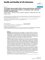
Effect of piperine on FLS production of inflammatory
mediators
To test the anti-inflammatory efficacy of piperine, FLSs were
stimulated with IL1β at 10 ng/ml in the presence or absence
of piperine. The addition of IL1β significantly increased the
production of IL6 and PGE
2
compared to that of controls (no
IL1β). The addition of piperine greatly inhibited the IL6 and
PGE
2
response to IL1β in dose-dependent manner (Figure 1).
Piperine also inhibited both the protein and mRNA expression
levels of IL6 and COX-2. In particular, piperine inhibited the
production of PGE
2
more potently than the production of IL6.
Interestingly, piperine inhibited the expression of the COX-2
protein more significantly than the COX-2 mRNA.
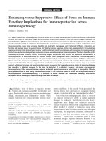
Next, we tested whether piperine inhibited the expression of
the extracellular matrix degradation enzymes (MMPs). MMP1
and MMP13 play an important role in degrading cartilage in
IL1β-stimulated FLSs. We found that piperine inhibited
MMP13 expression at both the protein and mRNA levels, but
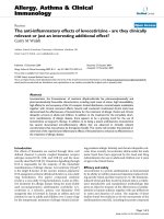
not MMP1 (Figure 2). To understand the molecular mecha-
nisms underlying piperine inhibition of IL6, COX-2 and MMP
expression, we investigated the MAP kinase and IκB kinase
signaling pathways by western blot (Figure 3a). Interestingly,
piperine did not significantly affect the IκB kinase signaling
pathway or the MAP kinase mediated phosphorylation of JNK
and P38; however, piperine slightly inhibited the MAP kinase
mediated phosphorylation of ERK1/2. In addition, piperine
reduced the level of AP-1 that migrated to the nucleus (in
response to IL1β) in a dose dependent manner, but did affect
Figure 1
Effect of piperine on the production of proinflammatory mediatorsEffect of piperine on the production of proinflammatory mediators. (a) ELISA results show that piperine inhibited the production of interleukin (IL)6
and prostaglandin E
2
(PGE
2
) in IL1β-stimulated fibroblast-like synoviocytes (FLSs) in a dose-dependent manner. (b) Piperine effects on IL6 and
cyclo-oxygenase (COX)-2 mRNA expression measured by semiquantitative RT-PCR. (c) Piperine effects on COX-2 protein expression measured by
western blot. Experiments were performed with synovial cells derived from patients with rheumatoid arthritis. Values are expressed ± standard error
of the mea (SEM). ***P < 0.001 vs IL1β treated group without piperine.
Available online />Page 5 of 9
(page number not for citation purposes)
the levels of NFκB in nucleus (Figure 3b). This suggested that
piperine inhibition of the ERK1/2 signaling pathway blocked
the migration of AP-1 into the nucleus.
Analgesic effect of piperine in carrageenan-induced paw
hyperalgesia
Because piperine significantly inhibited the production of
PGE
2
and the protein levels of COX-2, we tested whether pip-
Figure 2
Effect of piperine on the production of extracelluar matrix degradation enzymes (matrix metalloproteinases (MMPs))Effect of piperine on the production of extracelluar matrix degradation enzymes (matrix metalloproteinases (MMPs)). (a) ELISA results show that pip-
erine inhibited the production of MMP13, but not MMP1 proteins, in interleukin (IL)1β-stimulated fibroblast-like synoviocytes (FLSs) in a dose
dependent manner. (b) mRNA levels were measured by semiquantitative RT-PCR. Experiments were performed with synovial cells derived from
patients with rheumatoid arthritis. Values are expressed ± standard error of the mean (SEM). ***P < 0.001 vs IL1β treated group without piperine.
Figure 3
Effects of piperine on signaling pathways and transnuclear migrationEffects of piperine on signaling pathways and transnuclear migration. (a) Interleukin (IL)1β-stimulated fibroblast-like synoviocytes (FLSs) treated with
piperine were analyzed by western blot. Piperine treatment did not inhibit the degradation of inhibitor of κB (IκB)α, but slightly inhibited the phospho-
rylation of extracellular-regulated kinase (ERK)1/2 in the MAP kinase signaling pathways was slightly inhibited in the presence of piperine. (b) The
nuclear levels of nuclear factor (NF)κB and activator protein 1 (AP-1) were measured by ELISA detecting p65 and c-FOS from nuclear extracts,
respectively, on an ELISA. Piperine inhibited the level of AP-1 in the nucleus, but not NFκB levels. Values are expressed ± standard error of the mean
(SEM). ***P < 0.001 vs IL1β treated group without piperine or piperine alone.
Arthritis Research & Therapy Vol 11 No 2 Bang et al.
Page 6 of 9
(page number not for citation purposes)
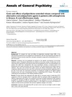
erine had antinociceptive effects in a rat model of carra-
geenan-induced paw hyperalgesia. We found in paw pressure
tests that rats treated with piperine could tolerate higher pres-
sures on the affected paw (Figure 4a). The efficacy at 100 mg/
kg was better than that of celecoxib, and at a dose of 20 mg/
kg, piperine showed a mild analgesic effect.
Antiarthritic effect of piperine on the carrageenan-
induced arthritis rat model
To demonstrate the in vivo antiarthritic effect of piperine, the
efficacy of piperine was tested in a rat model of carrageenan-
induced arthritis. The piperine (100 mg/kg) group showed a
significant reduction in paw volume compared to the vehicle-
treated arthritic group (Figure 4). At this dose, piperine
showed almost the same efficacy as prednisolone (10 mg/kg),
which was used as a positive control. Piperine also provided a
mild antiedema effect at 20 mg/kg, although it was not statis-
tically significant.
The vocalizations caused by flexion or extension of the
inflamed ankle reached a maximum point on day 1 after the
carrageenan injection and was sustained at a maximum level in
untreated rats through the end of the experiment (Figure 4c).
In the100 mg/kg piperine treated group, the number of vocal-
izations started to decrease at 5 days post-carrageenan injec-
tion. At 20 mg/kg, piperine exhibited little analgesic effect.
Next, we measured the analgesic effect of piperine (20 and
100 mg/kg) on the weight distributed on the hind paws
(WDR) of rats with carrageenan-induced arthritis in one paw
(Figure 4d). Before the carrageenan injection (day 0), the
mean WDR did not differ significantly among the experimental
groups (the WDR was 50:50, thus controls carried 50% of the
weight on each hind paw). However, significant changes in the
ratio were observed on day 1 after the carrageenan injection,
and the weight carried by the affected leg in the vehicle-
treated arthritic group (CON) reached 20% at day 9. Distinct
recovery of WDR was observed in groups that received 20
and 100 mg/kg piperine on days 8 and 9, despite the statisti-
cally insignificant analgesic effect of 20 mg/kg piperine.
Figure 4
Analgesic and antiarthritic effects of piperine in rat models of paw edema and arthritic ankleAnalgesic and antiarthritic effects of piperine in rat models of paw edema and arthritic ankle. (a) Piperine showed analgesic effects in carrageenan-
induced paw edema. The y axis indicated the pressure (g) that was tolerated before the rat exhibited signs of pain. Arthritic symptoms were meas-
ured by (b) relative paw volume, expressed as a function of the unaffected paw (100%). (c) The ankle flexion pain score (a value of 0 represents no
indication of pain); and (d) the weight distribution ratio (a value of 50% indicated that weight was equally distributed between the two hind paws).
The results indicated that piperine had antiarthritic effects. Con = control mice, Cele = celecoxib (100 mg/kg), PI-20/PI-100 = pierine at 20/100
mg/kg, Pre = prednisolne. Values are expressed ± standard error of the mean (SEM). *P < 0.05, **P < 0.01, ***P < 0.001 vs control group.
Available online />Page 7 of 9
(page number not for citation purposes)
Anti-inflammatory effect of piperine by histological
evaluation
To evaluate the anti-inflammatory effects of piperine, samples
of the ankle joints from each experimental group were exam-
ined by H&E staining. We found that the group that received
piperine (100 mg/kg) had significantly smaller areas of lym-
phocyte infiltration into the joints compared to the corn oil
treated group (Figure 5). The degree of inflammation in five
specimens was evaluated by three different pathologists. The
scores indicated that piperine significantly reduced the inflam-
mation induced by carrageenan (Figure 5b).
Discussion
Anti-inflammatory drugs used for treating chronic inflammatory
diseases such as rheumatoid arthritis are typically prescribed
long term to properly control the disordered immune system.
Thus, there is a strong need to develop safe and effective
drugs for the long-term use. Many groups have studied non-
steroidal anti-inflammatory small molecules that were derived
from natural sources with the aim of developing new treat-
ments for clinical use [17]. For example, curcumin is a
polyphenolic compound derived from the dietary spice, tur-
meric. Recently, curcumin has been shown to possess diverse
pharmacological properties, including anti-inflammation, anti-
proliferation, and antiangiogenesis. Currently, curcumin is in
phase I of clinical trials [18].
Piperine is also a promising natural source with potential for
clinical use. Piper longum Linn. has been used in Asia as a nat-
ural treatment for poor peripheral blood circulation [19]. Piper
longum Linn. and Piper nigrum Linn. are conventionally used
as immune enhancers in Indian traditional medicine [20].
Therefore, piperine has been proven effective indirectly, but its
mechanism of action remains unknown. In the present study,
we evaluated the anti-inflammatory and antiarthritic effects of
piperine to determine whether it had therapeutic potential for
the treatment of arthritis.
We found that piperine significantly inhibited the production of
two important proinflammatory mediators, IL6 and PGE
2
, in
IL1β-stimulated human FLS. This result was consistent with
other studies that showed potent anti-inflammatory effects in
other systems. The inhibition of PGE
2
production is important
due to its central role in triggering pain. In addition, MMP1 and
Figure 5
Histological evaluation of the anti-inflammatory effects of piperineHistological evaluation of the anti-inflammatory effects of piperine. Paraffin sections of rat ankles were stained with hematoxylin and eosin (H&E). (a)
Histopathological analysis showed that piperine (100 mg/kg) significantly inhibited ankle inflammation. Each photo is representative of five speci-
mens for each group (original magnification × 100). The insets are enlargements of the regions outlined in black, and show the infiltrates at a magni-
fication of × 200. (b) The degree of inflammation was evaluated on a scale from 0 to 5 by three pathologists that were blinded to the treatments.
Values are expressed ± standard error of the mean (SEM). *P < 0.05 vs corn oil treated group.
Arthritis Research & Therapy Vol 11 No 2 Bang et al.
Page 8 of 9
(page number not for citation purposes)
MMP13 collagenases play dominant roles in RA and osteoar-
thritis because they are the rate-limiting components of the
collagen degradation process. The significant inhibition of
MMP13 expression is particularly important because it
degrades a wide range of collagenous and non-collagenous
extracellular matrix macromolecules and is remarkably active
against collagen type II, the predominant collagen in cartilage.
To our knowledge, this is the first report to show that piperine
inhibited the expression of MMP13 in IL1β-stimulated FLSs.
We also investigated the molecular mechanisms underlying
piperine inhibition. We found that piperine did not significantly
inhibit the activation of MAP kinase or IκB kinase signaling
pathways. At the maximum concentration tested (100 μg/ml),
piperine slightly inhibited the phosphorylation of ERK1/2 stim-
ulated by IL1β. Piperine also inhibited the activation of the tran-
scription factor AP-1, but not NFκB, in our system. However,
a previous study in B16F-10 melanoma cells showed that pip-
erine was able to inhibit the activation of several transcription
factors, including NFκB, c-FOS, cAMP response element
binding (CREB) and activating transcription factor 2 (ATF-2).
Accordingly, in that study, it significantly reduced the produc-
tion of IL1β, tumor necrosis factor (TNF)α, IL6, and granulo-
cyte-macrophage colony stimulating factor (GM-CSF) [21].
We used two animal models to evaluate the in vivo analgesic
or antiarthritic effects of piperine. We found that piperine (100
mg/kg) effectively improved the symptoms of arthritic diseases
with an effect comparable to prednisolone, although at 20 mg/
kg, piperine did not have a significant analgesic effect in the
arthritic animal model.
One of the major drawbacks of the current study was the large
amount of piperine administered. Though the effects of 100
mg/kg piperine were therapeutic, several other studies have
shown in vivo effects with doses below 50 mg/kg [22-24].
Furthermore, in this study, the effects reached significance at
8 or 9 days. Thus, the potency of piperine was relatively weak
compared to 10 mg/kg prednisolone, which showed signifi-
cant effects at 4 or 5 days. The in vivo toxicity of a 100 mg/kg
dose piperine has not been tested; however, rats did not
exhibit any adverse effects and they survived throughout the
experiments. Nevertheless, piperine was shown to have immu-
notoxicological effects in mice at a dose of 2.25 mg/kg [25].
Piperine is also known to enhance the bioavailability of some
drugs by inhibiting drug metabolism or by increasing absorp-
tion [18,24,26]. Thus, piperine may prove to be useful on com-
bination treatments with other drugs. For example, a
combination of gallic acid and piperine reduced beryllium-
induced hepatorenal dysfunction and the associated oxidative
stress [22]. In addition, a synergistic effect of piperine was
demonstrated in a clinical study that tested the pharmacoki-
netics of nevirapine, a potent non-nucleoside inhibitor of HIV-
1 reverse transcriptase [27]. The combination therapy was
well tolerated, with few or no clinical adverse effects, and the
mean maximum plasma concentration of nevirapine was
increased when combined with piperine. In another clinical
study, piperine was shown to increase the plasma levels of
coenzyme Q10 [28]. Therefore, piperine may improve the ther-
apeutic effect or lower the dose requirements of other drugs
when administrated with DMARDs as a therapeutic drug or
dietary supplement. In addition, combinations of DMARDs
with piperine may reduce the side effects of DMARDs.
Conclusions
For the first time, we have demonstrated that piperine has
antirheumatic effects in animal models and anti-inflammatory
effects on IL1β-stimulated FLSs. Our results suggest that pip-
erine has potential as a therapeutic drug or dietary supple-
ment. Thus, further investigations should focus on the
development of piperine analogues that have potent efficacy
and few adverse effects.
Competing interests
The authors declare that they have no competing interests.
Authors' contributions
KSK and DHH participated in the data analysis and the design
of the study, and drafted the manuscript. JSB, DHO, HMC,
BJS, JYK, and SJL performed the experiments. MCY and HIY
provided the synovium from patients and participated in the
design of the study. All authors read and approved the final
manuscript.
Acknowledgements
This work was supported by a research grant from the Korean Ministry
of Health & Welfare (03-PJ9-PG6-SO01-002).
References
1. Lee YA, Kim JY, Hong SJ, Lee SH, Yoo MC, Kim KS, Yang HI: Syn-
ovial proliferation differentially affects hypoxia in the joint cav-
ities of rheumatoid arthritis and osteoarthritis patients. Clin
Rheumatol 2007, 26:2023-2029.
2. Mor A, Abramson SB, Pillinger MH: The fibroblast-like synovial
cell in rheumatoid arthritis: a key player in inflammation and
joint destruction. Clin Immunol 2005, 115:118-128.
3. Zhang B, He XL, Ding Y, Du GH: Gaultherin, a natural salicylate
derivative from Gaultheria yunnanensis: towards a better non-
steroidal anti-inflammatory drug. Eur J Pharmacol 2006,
530:166-171.
4. Gaby AR: Alternative treatments for rheumatoid arthritis.
Altern Med Rev 1999, 4:392-402.
5. Jacobs JWG, Kraaimaat FW, Bijlsma JW: Why do patients with
rheumatoid arthritis use alternative treatments? Clin Rheuma-
tol 2001, 20:192-196.
6. Srinivasan K: Black pepper and its pungent principle-piperine:
a review of diverse physiological effects. Crit Rev Food Sci
Nutr 2007, 47:735-748.
7. Mujumdar AM, Dhuley JN, Deshmukh VK, Raman PH, Naik SR:
Anti-inflammatory activity of piperine. Jpn J Med Sci Biol 1990,
43:95-100.
8. Stohr JR, Xiao PG, Bauer R: Constituents of Chinese Piper spe-
cies and their inhibitory activity on prostaglandin and leukot-
riene biosynthesis in vitro. J Ethnopharmacol 2001,
75:133-139.
9. Matsuda D, Ohte S, Ohshiro T, Jiang W, Rudel L, Hong B, Si S,
Tomoda H: Molecular target of piperine in the inhibition of lipid
Available online />Page 9 of 9
(page number not for citation purposes)
droplet accumulation in macrophages. Biol Pharm Bull 2008,
31:1063-1066.
10. Lee CS, Han ES, Kim YK: Piperine inhibition of 1-methyl-4-phe-
nylpyridinium-induced mitochondrial dysfunction and cell
death in PC12 cells. Eur J Pharmacol 2006, 537:37-44.
11. Chu CY, Chang JP, Wang CJ: Modulatory effect of piperine on
benzo[a]pyrene cytotoxicity and DNA adduct formation in V-79
lung fibroblast cells. Food Chem Toxicol 1994, 32:373-377.
12. Singh J, Reen RK, Wiebel FJ: Piperine, a major ingredient of
black and long peppers, protects against AFB1-induced cyto-
toxicity and micronuclei formation in H4IIEC3 rat hepatoma
cells. Cancer Lett 1994, 86:195-200.
13. Kim KS, Park EK, Ju SM, Jung HS, Bang JS, Kim C, Lee YA, Hong
SJ, Lee SH, Yang HI, Yoo MC: Taurine chloramine differentially
inhibits matrix metalloproteinase 1 and 13 synthesis in inter-
leukin-1β stimulated fibroblast-like synoviocytes. Arthritis Res
Ther 2007, 9:R80.
14. Yoo EA, Kim SD, Lee WM, Park HJ, Kim SK, Cho JY, Min W, Rhee
MH: Evaluation of antioxidant, antinociceptive, and anti-inflam-
matory activities of ethanol extracts from Aloe saponaria Haw.
Phytother Res 2008, 22:1389-1395.
15. Hwang HJ, Lee HJ, Kim CJ, Shim I, Hahm DH: Inhibitory effect of
amygdalin on lipopolysaccharide-inducible TNF-alpha and IL-
1β mRNA expression and carrageenan-induced rat arthritis. J
Microbiol Biotechnol 2008, 18:1641-1647.
16. Kwon YB, Lee JD, Lee HJ, Han HJ, Mar WC, Kang SK, Beitz AJ,
Lee JH: Bee venom injection into an acupuncture point
reduces arthritis associated edema and nociceptive
responses. Pain 2001, 90:271-280.
17. Chrubasik JE, Roufogalis BD, Chrubasik S: Evidence of effective-
ness of herbal antiinflammatory drugs in the treatment of
painful osteoarthritis and chronic low back pain. Phytother Res
2007, 21:675-683.
18. Anand P, Kunnumakkara AB, Newman RA, Aggarwal BB: Bioa-
vailability of curcumin: problems and promises. Mol Pharm
2007, 4:807-818.
19. Iwashita M, Saito M, Yamaguchi Y, Takagaki R, Nakahata N: Inhib-
itory effect of ethanol extract of
Piper longum L. on rabbit
platelet aggregation through antagonizing thromboxane A2
receptor. Biol Pharm Bull 2007, 30:1221-1225.
20. Pathak N, Khandelwal S: Cytoprotective and immunomodulat-
ing properties of piperine on murine splenocytes: an in vitro
study. Eur J Pharmacol 2007, 576:160-170.
21. Pradeep CR, Kuttan G: Piperine is a potent inhibitor of nuclear
factor-kappaB (NF-kappaB), c-Fos, CREB, ATF-2 and proin-
flammatory cytokine gene expression in B16F-10 melanoma
cells. Int Immunopharmacol 2004, 4:1795-1803.
22. Zhao JQ, Du GZ, Xiong YC, Wen YF, Bhadauria M, Nirala SK:
Attenuation of beryllium induced hepatorenal dysfunction and
oxidative stress in rodents by combined effect of gallic acid
and piperine. Arch Pharm Res 2007, 30:1575-1583.
23. Vijayakumar RS, Nalini N: Piperine, an active principle from Piper
nigrum, modulates hormonal and apo lipoprotein profiles in
hyperlipidemic rats. J Basic Clin Physiol Pharmacol 2006,
17:71-86.
24. Selvendiran K, Banu SM, Sakthisekaran D: Oral supplementation
of piperine leads to altered phase II enzymes and reduced
DNA damage and DNA-protein cross links in benzo(a)pyrene
induced experimental lung carcinogenesis. Mol Cell Biochem
2005, 268:141-147.
25. Dogra RK, Khanna S, Shanker R: Immunotoxicological effects of
piperine in mice. Toxicology 2004, 196:229-236.
26. Scholz S, Williamson G: Interactions affecting the bioavailabil-
ity of dietary polyphenols in vivo. Int J Vitam Nutr Res 2007,
77:224-235.
27. Kasibhatta R, Naidu MU: Influence of piperine on the pharma-
cokinetics of nevirapine under fasting conditions: a ran-
domised, crossover, placebo-controlled study. Drugs R D
2007, 8:383-391.
28. Badmaev V, Majeed M, Prakash L: Piperine derived from black
pepper increases the plasma levels of coenzyme Q10 follow-
ing oral supplementation. J Nutr Biochem 2000, 11:109-113.