Báo cáo y học: "Inflammation predicts accelerated brachial arterial wall changes in patients with recent-onset rheumatoid arthritis" pptx
Bạn đang xem bản rút gọn của tài liệu. Xem và tải ngay bản đầy đủ của tài liệu tại đây (245.82 KB, 7 trang )
Open Access
Available online />Page 1 of 7
(page number not for citation purposes)
Vol 11 No 2
Research article
Inflammation predicts accelerated brachial arterial wall changes
in patients with recent-onset rheumatoid arthritis
Suad Hannawi
1
, Thomas H Marwick
2
* and Ranjeny Thomas
1
*
1
Diamantina Institute, University of Queensland, Princess Alexandra Hospital, Ipswich Road, Woolloongabba, Queensland 4102, Australia
2
Department of Medicine, University of Queensland, Princess Alexandra Hospital, Ipswich Road, Woolloongabba, Queensland 4102, Australia
* Contributed equally
Corresponding author: Ranjeny Thomas,
Received: 17 Dec 2008 Revisions requested: 14 Jan 2009 Revisions received: 16 Mar 2009 Accepted: 6 Apr 2009 Published: 6 Apr 2009
Arthritis Research & Therapy 2009, 11:R51 (doi:10.1186/ar2668)
This article is online at: />© 2009 Hannawi et al.; licensee BioMed Central Ltd.
This is an open access article distributed under the terms of the Creative Commons Attribution License ( />),
which permits unrestricted use, distribution, and reproduction in any medium, provided the original work is properly cited.
Abstract
Introduction Patients with recent-onset rheumatoid arthritis
(RA) have impaired brachial artery endothelial function
compared with controls matched for age, sex and
cardiovascular risk factors. The present study examined
endothelium-dependent (flow-mediated dilatation (FMD)) and
independent (glyceryl trinitrate (GTN)-mediated dilatation
(GMD)) structural responses in early RA patients, and
determined progress over one year.
Methods Brachial artery FMD and GMD and carotid intima
media thickness (cIMT) were studied using ultrasound in 20
patients diagnosed with early RA in whom symptoms had been
present for less than 12 months, and in 20 control subjects
matched for age, sex and established cardiovascular risk
factors. FMD and GMD were re-assessed after 12 months in RA
patients and the change in each parameter was calculated. Data
were analysed by univariate regression.
Results Mean FMD and GMD were significantly lower in early
RA patients at baseline than in controls, but each parameter
significantly improved in one year. FMD and GMD responses
were positively associated with each other. Patients' age, C-
reactive protein (CRP) level and cIMT at baseline and CRP level
at one year, were negatively associated with change in brachial
responses in one year.
Conclusions Patients with recent-onset RA have altered
brachial artery responses signifying both functional and
structural abnormalities. However, early control of inflammation
may reduce arterial dysfunction and thus the tendency for
atherosclerotic progression.
Introduction
Patients with rheumatoid arthritis (RA) experience cardiovas-
cular (CV) events more often than expected [1] and their mor-
tality attributable to CV causes is increased [2]. Furthermore,
ischaemic heart disease (IHD) has been found to occur about
10 years earlier in patients with RA compared with a popula-
tion of patients with osteoarthritis matched for classical CV
risk factors. Indeed, it has been suggested that RA itself is an
independent risk factor for IHD [3]. The similarities which exist
between the inflammatory/immunological reaction in RA and
atherosclerosis raise the possibility that inflammatory mecha-
nisms responsible for synovial lesions might also involve the
vessel wall to facilitate the development of atherosclerotic
lesions [4].
Although atherosclerosis can manifest as overt CV disease, it
can be detected at an earlier stage by recognition of abnormal
endothelial function and elevated carotid intima media thick-
ness (cIMT) as measured by ultrasound. These measures cor-
relate closely with direct measurement of local and systemic
atherosclerotic burden in studies of pathology and with clinical
CV endpoints [5,6]. Ultrasonographic assessment of the com-
mon carotid artery is a feasible, reliable, valid and cost-effec-
tive method for both population studies and clinical trials of
atherosclerosis progression and regression [7]. We showed
that cIMT is significantly higher in patients presenting with
early RA than in controls matched for age, sex and CV risk fac-
tors [8].
cIMT: carotid intima-media thickening; CRP: C-reactive protein; CV: cardiovascular; DMARDs: disease-modifying anti-rheumatic drugs; ESR: eryth-
rocyte sedimentation rate; FMD: flow mediated dilatation; GMD: GTN-mediated dilatation; GTN: glyceryl trinitrate; HAQ: health assessment ques-
tionnaire; HCQ: hydroxychloroquine; IHD: ischaemic heart disease; MTX: methotrexate; NO: nitric oxide; RA: rheumatoid arthritis; RF: rheumatoid
factor; SD: standard deviation; SSZ: sulfasalazine; TNF: tumour necrosis factor.
Arthritis Research & Therapy Vol 11 No 2 Hannawi et al.
Page 2 of 7
(page number not for citation purposes)
Endothelial function can be assessed by ultrasound as flow-
mediated vasodilatation of the brachial artery in response to
increased vessel wall shear stress and mediated by nitric oxide
(NO) release by endothelial cells [9,10]. Brachial artery
vasodilatation can also be assessed by exogenous NO after
sublingual glyceryl trinitrate (GTN) [11]. Post-GTN vasodilata-
tion is considered endothelium-independent and predomi-
nantly mediated by smooth muscle. The dose-response curve
for GTN-mediated vasodilatation has been examined in sub-
jects with proven coronary artery disease and in healthy con-
trols [12]. Patients with coronary disease had significantly
reduced GTN-mediated vasodilatation, with the greatest dif-
ference observed with lower doses of GTN, suggesting that
atherosclerosis is associated with functional abnormalities of
both endothelium and vascular smooth muscle cells [12].
A previous study of 10 patients with early RA demonstrated
impaired flow-mediated endothelial function, with improve-
ment over six months of therapy [13]. Moreover, infliximab ther-
apy improved endothelial-dependent vasodilatation,
apparently through direct endothelial, rather than systemic
effects [14]. In women without RA, treatment-associated
improvement in endothelium-dependent vasodilatation has
been shown to decrease the risk of subsequent CV events
[15]. In longstanding RA, endothelial dysfunction is predicted
by the C-reactive protein (CRP) level [15-19]. Other acute and
chronic inflammatory states have also been shown to impair
endothelial function [20-22]. In the current study, we studied
a group of early RA patients, in whom cIMT had also been
determined, and investigated the progression of endothelium-
dependent and -independent brachial arterial function after
one year.
Given the relationship of inflammation severity to atheroscle-
rotic burden, as measured by cIMT and carotid plaque, at pres-
entation with early RA [23], we hypothesised that inflammatory
indices would predict arterial responses. Therefore, a group of
conventional CV risk factors and indices of RA inflammatory
disease were analysed at baseline and one year to determine
their relationship to changes in brachial artery response.
Materials and methods
RA patients
All 31 study participants met the American College of Rheu-
matology 1987 revised criteria for the classification of RA [24].
All the patients were enrolled from 2004 to 2005 with a diag-
nosis of RA with symptom duration less than 12 months.
Patients attended an early RA clinic regularly, and received
combination methotrexate (MTX), sulfasalazine (SSZ) and
hydroxychloroquine (HCQ) [25], unless contraindicated, after
diagnosis and active disease were confirmed. After diagnosis,
patients were treated according to a response-driven step-up
algorithm, as previously described [26], with the aim of achiev-
ing clinical remission [27-29]. Intra-articular but not oral corti-
costeroids were used to control inflammation in addition to
disease-modifying antirheumatic drugs (DMARDs), as
required.
At one year, 52% were taking MTX, SSZ and HCQ, 6% MTX
and SSZ, 16% MTX and HCQ, 10% SSZ and HCQ and 6%
SSZ only, and 10% refused treatment. An additional 20
healthy subjects were recruited from the community, who
could be matched for age, sex and CV risk factors against 20
of the early RA subjects. We matched against a database of
more than 1000 individuals recruited in a primary prevention
setting. The primary match was with the age and gender, after
which we matched on number and type of risk factors on a cat-
egorical basis. Although exact blood pressure or lipid levels
were not matched, we were able to match for hypertension
and hyperlipidaemia, in addition to smoking status in almost all
the cases.
Study procedure
The study was approved by the human research ethics com-
mittee at Princess Alexandra Hospital, Woolloongabba, and all
subjects provided written informed consent. CV risk factors
were ascertained among RA patients at baseline, as previously
described [8]. RA disease activity parameters and laboratory
measurements were assessed as previously described, at
baseline and 12 months. Flow-mediated dilatation (FMD),
GTN-mediated dilatation (GMD), cIMT and plaque were meas-
ured soon after the diagnosis of RA was confirmed; FMD and
GMD were repeated after 12 months and as described [8].
Vascular function testing
Brachial artery flow and GMD testing was undertaken within
one to four weeks after diagnosis and before commencement
of DMARDs. Individual blood pressure and heart rate
remained constant during the testing. No subject had ultra-
sound evidence of brachial artery atherosclerotic plaque.
Ultrasonography was performed by a trained investigator (SH)
who was unaware of the subject's clinical data, using a high-
resolution ultrasound machine, according to the International
Brachial Artery Reactivity Task force Guidelines [30]. Two
patients were taking statins, one was taking an angiotensin-
converting enzyme inhibitor and one patient was taking both.
Although all four patients were taking a once daily dose of their
medication, they were asked to postpone this medication until
after the vascular ultrasound. No patient was taking aspirin or
GTN treatment.
Subjects rested in a supine position in a quiet, dark, tempera-
ture-controlled room. A pneumatic cuff was placed around the
upper forearm distal to the segment of brachial artery, scanned
in longitudinal section 2 to 10 cm above the antecubital
crease. Focus, depth and gain were individually set to optimise
images of the lumen/arterial wall interface. A baseline scan
was recorded for two minutes, followed by induction of hyper-
aemia by cuff inflation to 240 mmHg for four minutes. The FMD
scan commenced 30 seconds before release of the cuff, and
Available online />Page 3 of 7
(page number not for citation purposes)
continued for one minute afterwards. A second baseline scan
was recorded 10 minutes later. The coefficient of variation (cv)
was 0.18 for FMD. A tablet of GTN was administered sublin-
gually in a standardised manner and recording was continued
for a further four minutes for GMD. The cv was 0.17 for GMD.
Measurement of the brachial artery diameter was synchro-
nised with the R wave of the electrocardiogram, to avoid pos-
sible errors resulting from artery pulsation. FMD and GMD
were expressed as the relative increase in brachial artery diam-
eter during hyperaemia:
cIMT was measured using carotid duplex scanning and auto-
mated software as previously described [31]. The cv for cIMT
was 0.2.
Statistical analysis
In a preliminary evaluation, continuous variables were tested
for normality of distribution; transformations were applied for
non-normally distributed variables. Variables with normal distri-
bution were expressed as the mean ± standard deviation (SD)
and categorical variables as percentages. Differences
between the FMD and GMD in RA and control groups were
compared using the two-sample (independent) Student's t-
test for normally distributed data. Log transformations were
applied to non-normally distributed data. Associations of FMD
and GMD with age, continuous CV variables and disease
activity variables were evaluated using linear regression analy-
sis. Brachial reactivity improvement, inflammatory and RA dis-
ease activity markers and lipid profile were summarised using
mean values for the baseline measures and compared with the
mean values at one year using paired Student's t-tests. The dif-
ferences between the mean values were examined by two-
sample Student's t-tests. Linear regression analysis examined
univariate correlations, and two-sided values of P < 0.05 were
regarded as statistically significant. For all the analyses, Stata
9/SE (Stata Corp, College Station, TX, USA) statistical soft-
ware was used [8].
Results
Clinical features
Thirty-one patients presenting with RA within 12 months of
symptom onset (Table 1) were treated and followed clinically
for one year. Of the patients, 90% were Caucasian, 3% were
Australian aboriginal and 6% Asian. Four patients had previ-
ous CV events, including one male with a previous transient
ischaemic attack at 52 years of age, two males with a history
of angina at the ages of 48 and 64 years, and one patient with
myocardial infarction (MI) at age 48 years. Twenty of these RA
patients could be matched for age, sex and CV risk factors
against 20 healthy control subjects from a database of more
than 1000 individuals recruited in a primary prevention setting.
Baseline cIMT, FMD and GMD in 20 patients with early
RA and 20 matched controls
The mean age at the time of diagnosis was 45 years (range 23
to 64 years), with male patients significantly older than females
(51.2 ± 9.2 years vs. 40.2 ± 10.7 years, P = 0.03). The mean
duration of RA symptoms at the time of scanning was 6 ± 3
months (range 1 to 12 months). Mean FMD and GMD of the
brachial artery were significantly lower in RA patients than in
controls (Figure 1a). We repeated the analysis after removing
patients with previous CV events and their matched controls,
and the results did not differ (FMD 5 ± 4 vs. 11 ± 7, P = 0.001
and GMD 11 ± 6 mm vs. 17 ± 8 mm, P = 0.02).
FMD and GMD annual change and determinants of
progression
The annual changes in FMD and GMD measured by ultra-
sound were determined. FMD and GMD improved significantly
over one year (Figure 1b). Univariate analyses were carried out
to determine CV risk factors or inflammatory factors which
might contribute to these changes in vascular parameters in
patients with early RA over the first year. Improvements in FMD
and GMD over one year were correlated. Age, baseline CRP
and baseline cIMT were negatively correlated with the change
in both FMD and GMD (Table 2). CRP level at one year was
also negatively associated with the change in FMD and GMD.
Patients who presented with high CRP levels at RA disease
onset were more likely to continue with high CRP levels by one
year (data not shown, P < 0.03). These data suggest that
endothelial dysfunction is reversible with falling levels of CRP.
In support of this idea, erythrocyte sedimentation rate (ESR)
level at baseline exhibited a similar negative relationship with
FMD progression, and rheumatoid factor (RF) positive patients
were less likely to improve FMD (Table 2). There were no sig-
nificant associations of FMD with health assessment question-
naire (HAQ), gender, smoking history, blood pressure or lipid
levels. In contrast, history of smoking ever and smoking pack
year history were negatively correlated with change in GMD,
consistent with the utility of GMD as a structural measure of
vascular disease (Table 2). There were no significant associa-
tions of GMD with HAQ, ESR, blood pressure, high-density
lipoprotein or triglyceride levels.
Discussion
An increasing body of evidence indicates that atherosclerosis
shares similarities with other inflammatory/autoimmune dis-
eases; indeed, there are surprising similarities between the
inflammatory response observed in atherosclerosis and RA
[32-34]. The current study hypothesised that the severity of
systemic inflammation would be associated with progression
of arterial dysfunction in RA. Patients with RA are at signifi-
cantly higher risk of death than an age-matched population
[35] with CV disease as a major cause of increased mortality
[1], and discovery of early interventions in RA disease that
might influence atherosclerotic progression are essential to
determine approaches that could in turn reduce the
100 ×− −[( /post hyperaemia diameter basal diameter) basal diametter].
Arthritis Research & Therapy Vol 11 No 2 Hannawi et al.
Page 4 of 7
(page number not for citation purposes)
subsequent risk of CV mortality. Of importance, the current
studies clearly demonstrate the effect of early DMARD inter-
vention, and the effect of reduction in CRP on vascular
endothelial dysfunction and GTN-mediated brachial arterial
function during the first year of RA. Furthermore, while each
vascular measurement indicates a different aspect of vascular
dysfunction – FMD is a measure of endothelial function [9,10]
and GMD of smooth muscle damage [12] – each was revers-
ible with treatment, and annual changes in each measure were
correlated.
Arterial compliance is contributed by the arterial media layer,
with abnormalities resulting from combined endothelial and
smooth muscle damage [36]. Reduction in CRP in patients
with RA was associated with substantial and significant
improvement in both endothelium-dependent and endothe-
lium-independent skin microvascular dysfunction [37,38]. A
similar improvement in FMD and GMD occurred after treat-
ment of early RA patients in the present study.
Disease activity and functional impairment are predictive of
mortality in RA. Thus, if effective therapy could be introduced
prior to the development of arterial damage, outcome could be
improved. It has been demonstrated extensively that early ther-
apeutic intervention with anti-rheumatic drugs improves the
prognosis of RA [39], and that disease duration is a significant
Table 1
Patient details at baseline and one year
Week 0
(mean ± SD)
Week 52 P value
Demographic details
Male:female 19:12
Mean age (range, years) 54 (23 to 78)
Body mass index (range, kg/m
2
) 28 (17 to 45)
Rheumatoid arthritis characteristics
Disease duration, mean (range, months) 1.8 (1 to 12)
Tender joint count (out of 53) 18 (14) 7.6 (8.1) < 0.001
Swollen joint count (out of 44) 15 (8.9) 5.0 (9.3) < 0.001
Health assessment questionnaire score (maximum disability 24) 3.3 (3.5) 1.8 (2.7) 0.006
Physician's global assessment of disease activity (maximum 100) 37 (27) 26 (28) 0.013
Joint pain (VAS, maximum 100) 51 (32) 24 (26) < 0.001
Rheumatoid factor level 281 (438) 98 (127) 0.011
Disease activity score, 4 v 4.3 (1.6) 2.6 (1.4) < 0.001
Rheumatoid factor positive, n (%) 21 (68%) 17 (55%)
Cardiovascular risk factors
History of ever smoking, n (%) 20 (65%)
History of current smoking, n (%) 8 (26%)
History of hypertension, n (%) 6 (19%)
History of diabetes mellitus, n (%) 3 (10%)
History of hyperlipidaemia, n (%) 2 (6.5%)
History of myocardial infarction, n (%) 1 (3.2%)
History of angina, n (%) 2 (6.5%)
History of stroke and/or transient ischaemic attack, n (%) 1 (3.2%)
Family history of cardiovascular disease, n (%) 6 (19%)
Laboratory values
Erythrocyte sedimentation rate (mm/hour) 40 (25) 21 (16) < 0.001
C-reactive protein (mg/L, normal < 6) 27 (25) 9.3 (12) < 0.001
Bold values are statistically significant. SD = standard deviation; VAS = visual analogue scale.
Available online />Page 5 of 7
(page number not for citation purposes)
determinant of response to therapy [40]. The strategy of com-
bined DMARDs in early RA shows a beneficial effect [41,42].
We previously demonstrated that patients with early RA had
significantly higher average cIMT values and more plaque than
healthy controls matched for CV risk factors. cIMT was pre-
dicted by age and CRP level at first presentation with RA. A
recent small study demonstrated that baseline cIMT in patients
with established RA predicted development of CV events over
the next five years [43]. Our results suggest that baseline
CRP, ESR and RF may be useful prognostic markers for CV
disease along with non-invasive measures of vascular function,
including cIMT, FMD and GMD.
The lack of improvement in traditional CV risk factors in RA
patients despite the improvement in FMD and GMD, which
were both positively correlated with improvement in CRP, fur-
ther implicates inflammation in arterial function impairment.
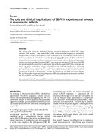
Figure 1
Brachial dilatation of 20 patients with early RA and matched controls, and of 31 patients with early RA at baseline and one yearBrachial dilatation of 20 patients with early RA and matched controls,
and of 31 patients with early RA at baseline and one year. Flow-medi-
ated and GTN-mediated dilatation of the brachial artery was measured
using ultrasound as described in the methods. Results presented as
mean ± standard deviation. Flow mediated vasodilatation, 100 × (post-
hyperaemic diameter-basal diameter)/basal diameter, GTN-mediated
dilatation, 100 × (post-GTN diameter-basal diameter)/basal diameter.
(a) RA patients are compared with healthy controls matched for age,
sex and CV risk factors. (b) RA patients are compared at baseline and
after one year of treatment with anti-rheumatic drugs. FMD = flow-medi-
ated dilatation; GMD = GTN-mediated dilatation; GTN = glyceryl trini-
trate; RA = rheumatoid arthritis.
Table 2
Univariate analysis of the relationship between change in flow-
mediated dilatation and GTN-mediated dilatation, features of
RA and CV risk factors in 31 patients with early RA
R
2
coeff P
FMD:
RF presence - - 0.037
Log CRP level
At week 0 0.173 -0.946 0.020
At week 52 0.319 -1.700 0.001
ESR
At week 0 0.142 -1.451 0.037
At week 52 0.001 -0.065 0.91
Change in GMD 0.428 0.487 < 0.001
Carotid IMT at baseline 0.196 -10.22 0.013
Age 0.346 -0.133 < 0.001
Post menopausal status 0.002
GMD:
Log CRP level
At week 0 0.214 -0.394 0.006
At week 52 0.431 -0.693 < 0.001
Change in FMD 0.428 0.880 < 0.001
Carotid IMT at baseline 0.232 -3.89 0.007
Age 0.206 -0.035 0.012
Pack year history 0.173 -0.148 0.022
Ever smoking 0.02
LDL
At week 0 0.173 -0.561 0.086
At week 52 0.352 -0.552 0.015
Parameters showing statistically significant relationships are
included. Bold values are statistically significant.
CRP = C-reactive protein; CV = cardiovascular; ESR = erythrocyte
sedimentation rate; FMD = flow-mediated dilatation; GMD = GTN-
mediated dilatation; GTN = glyceryl trinitrate; IMT = intima-media
thickening; LDL = low-density lipoprotein; RA = rheumatoid arthritis;
RF = rheumatoid factor.
Arthritis Research & Therapy Vol 11 No 2 Hannawi et al.
Page 6 of 7
(page number not for citation purposes)
Moreover, it is likely that endothelial function in RA primarily
depends on the control of inflammatory activity rather than the
specific treatment, as demonstrated by improvement with a
variety of effective anti-rheumatic agents [16,44-46]. MTX was
shown to reduce overall mortality by 60% primarily by reduc-
ing mortality from coronary heart disease [47]. The mechanism
by which MTX provides CV protection might be explained at
least in part by suppression of systemic inflammation.
Although endothelial dysfunction has been reported in RA
patients treated with long-term MTX [48], the improvement of
endothelial function in our patients might be explained by early
suppression of inflammation compared with the treatment of
established RA patients with chronic inflammatory effects on
vessels. In another study, reversal of endothelial dysfunction in
patients with long-standing severe RA was only transient in
response to TNF inhibitors, despite good control of systemic
inflammation, suggesting that structural vascular changes
might preclude more prolonged effects [45]. A recent demon-
stration that abnormal FMD was associated with longer dis-
ease duration in patients with established RA also supports
the concept that endothelial dysfunction progresses and
becomes less reversible over time [49].
Traditional CV risk factors identified from epidemiological
studies such as hyperlipidaemia, smoking and older age may
interact to damage the endothelium and smooth muscle in
asymptomatic patients in the same way as they are known to
interact to determine the risk of clinical CV endpoints [50]. Our
data demonstrate that improvement in GMD, a marker of
endothelial and smooth muscle dysfunction, after one year of
treatment of early RA was significantly negatively associated
with pack year history of smoking. Cigarette smoking
increases oxidative stress because of low circulating levels of
antioxidants and increased levels of oxygen-derived free radi-
cals and lipid peroxides that degrade NO, leading to endothe-
lial dysfunction [51,52]. Thus, smoking history may have a
specific impact on the capacity of the endothelium and smooth
muscle to respond to the anti-inflammatory effects of treat-
ment. In a study of patients with longstanding RA, where those
with a history of CV disease or CV risk factors including smok-
ing were excluded, GMD in the RA patients was no different
to that of age- and sex-matched controls or of the healthy con-
trol subjects in the current study [49]. These data suggest the
hypothesis that inflammatory arthritis in the absence of CV risk
factors does not promote arterial smooth muscle damage.
Conclusions
This study shows that inflammation severity is closely associ-
ated with functional and structural arterial wall changes in
patients with early RA. Early control of inflammation is associ-
ated with improved arterial function which may reduce athero-
sclerotic progression.
Competing interests
The authors declare that they have no competing interests.
Authors' contributions
SH, TM and RT were involved in conception, design, acquisi-
tion, analysis, interpretation of data and drafting and revising
the manuscript: All authors read and approved the final
manuscript.
Acknowledgements
Supported by grants from the PA Hospital Foundation, Australian Rotary
Health Research Fund and a scholarship from the Ministry of Higher
Education, United Arab Emirates. RT is supported by Arthritis Queens-
land. We thank Matthew Brown for helpful discussions. There are no
commercial affiliations.
References
1. del Rincon ID, Williams K, Stern MP, Freeman GL, Escalante A:
High incidence of cardiovascular events in a rheumatoid
arthritis cohort not explained by traditional cardiac risk factors.
Arthritis Rheum 2001, 44:2737-2745.
2. Wolfe F, Mitchell DM, Sibley JT, Fries JF, Bloch DA, Williams CA,
Spitz PW, Haga M, Kleinheksel SM, Cathey MA: The mortality of
rheumatoid arthritis. Arthritis Rheum 1994, 37:481-494.
3. Banks M, Flint J, Bacon P, Kitas G: Rheumatoid arthritis is an
independent risk factor for ischaemic heart disease. Arthritis
Rheum 2000, 1909:S358.
4. Lutzky V, Hannawi S, Thomas R: Cells of the synovium in rheu-
matoid arthritis. Dendritic cells. Arthritis Res Ther 2007, 9:219.
5. Pignoli P, Tremoli E, Poli A, Oreste P, Paoletti R: Intimal plus
medial thickness of the arterial wall: a direct measurement
with ultrasound imaging. Circulation 1986, 74:1399-1406.
6. Lorenz MW, Markus HS, Bots ML, Rosvall M, Sitzer M: Prediction
of clinical cardiovascular events with carotid intima-media
thickness: a systematic review and meta-analysis. Circulation
2007, 115:459-467.
7. Salonen JT, Salonen R: Ultrasound B-mode imaging in observa-
tional studies of atherosclerotic progression. Circulation 1993,
87:II56-65.
8. Hannawi S, Haluska B, Marwick TH, Thomas R: Atherosclerotic
disease is increased in recent onset rheumatoid arthritis: a
critical role for inflammation. Arthritis Res Ther 2007, 9:R116.
9. Raitakari OT, Seale JP, Celermajer DS: Impaired vascular
responses to nitroglycerin in subjects with coronary
atherosclerosis. Am J Cardiol 2001, 87:217-219. A218.
10. Joannides R, Haefeli WE, Linder L, Richard V, Bakkali EH, Thuillez
C, Luscher TF: Nitric oxide is responsible for flow-dependent
dilatation of human peripheral conduit arteries in vivo. Circu-
lation 1995, 91:1314-1319.
11. Corretti MC, Anderson TJ, Benjamin EJ, Celermajer D, Charbon-
neau F, Creager MA, Deanfield J, Drexler H, Gerhard-Herman M,
Herrington D, Vallance P, Vita J, Vogel R: Guidelines for the ultra-
sound assessment of endothelial-dependent flow-mediated
vasodilation of the brachial artery: a report of the International
Brachial Artery Reactivity Task Force. J Am Coll Cardiol 2002,
39:257-265.
12. Adams MR, Robinson J, McCredie R, Seale JP, Sorensen KE,
Deanfield JE, Celermajer DS: Smooth muscle dysfunction
occurs independently of impaired endothelium-dependent
dilation in adults at risk of atherosclerosis. J Am Coll Cardiol
1998, 32:123-127.
13. Bergholm R, Leirisalo-Repo M, Vehkavaara S, Makimattila S, Task-
inen MR, Yki-Jarvinen H: Impaired responsiveness to NO in
newly diagnosed patients with rheumatoid arthritis. Arterio-
scler Thromb Vasc Biol 2002, 22:1637-1641.
14. Gonzalez-Juanatey C, Testa A, Garcia-Castelo A, Garcia-Porrua C,
Llorca J, Gonzalez-Gay MA: Active but transient improvement of
endothelial function in rheumatoid arthritis patients undergo-
ing long-term treatment with anti-tumor necrosis factor alpha
antibody. Arthritis Rheum 2004, 51:447-450.
15. Modena MG, Bonetti L, Coppi F, Bursi F, Rossi R: Prognostic role
of reversible endothelial dysfunction in hypertensive post-
menopausal women. J Am Coll Cardiol 2002, 40:505-510.
Available online />Page 7 of 7
(page number not for citation purposes)
16. Hansel S, Lassig G, Pistrosch F, Passauer J: Endothelial dysfunc-
tion in young patients with long-term rheumatoid arthritis and
low disease activity. Atherosclerosis 2003, 170:177-180.
17. Vaudo G, Marchesi S, Gerli R, Allegrucci R, Giordano A, Siepi D,
Pirro M, Shoenfeld Y, Schillaci G, Mannarino E: Endothelial dys-
function in young patients with rheumatoid arthritis and low
disease activity. Ann Rheum Dis 2004, 63:31-35.
18. Cardillo C, Schinzari F, Mores N, Mettimano M, Melina D, Zoli A,
Ferraccioli G: Intravascular tumor necrosis factor alpha block-
ade reverses endothelial dysfunction in rheumatoid arthritis.
Clin Pharmacol Ther 2006, 80:275-281.
19. Hingorani AD, Cross J, Kharbanda RK, Mullen MJ, Bhagat K, Taylor
M, Donald AE, Palacios M, Griffin GE, Deanfield JE, MacAllister RJ,
Vallance P: Acute systemic inflammation impairs endothelium-
dependent dilatation in humans. Circulation 2000,
102:994-999.
20. Raza K, Thambyrajah J, Townend JN, Exley AR, Hortas C, Filer A,
Carruthers DM, Bacon PA: Suppression of inflammation in pri-
mary systemic vasculitis restores vascular endothelial func-
tion: lessons for atherosclerotic disease? Circulation 2000,
102:1470-1472.
21. Lima DS, Sato EI, Lima VC, Miranda F Jr, Hatta FH: Brachial
endothelial function is impaired in patients with systemic
lupus erythematosus. J Rheumatol 2002, 29:292-297.
22. Sari I, Okan T, Akar S, Cece H, Altay C, Secil M, Birlik M, Onen F,
Akkoc N: Impaired endothelial function in patients with anky-
losing spondylitis. Rheumatology (Oxford) 2006, 45:283-286.
23. Carroll L, Hannawi S, Marwick T, Thomas R: Rheumatoid arthri-
tis: links with cardiovascular disease and the receptor for
advanced glycation end products. Wien Med Wochenschr
2006, 156:42-52.
24. Arnett FC, Edworthy SM, Bloch DA, McShane DJ, Fries JF, Cooper
NS, Healey LA, Kaplan SR, Liang MH, Luthra HS, et al.: The Amer-
ican Rheumatism Association 1987 revised criteria for the
classification of rheumatoid arthritis. Arthritis Rheum 1988,
31:315-324.
25. O'Dell JR, Haire CE, Erikson N, Drymalski W, Palmer W, Eckhoff
PJ, Garwood V, Maloley P, Klassen LW, Wees S, Klein H, Moore
GF: Treatment of rheumatoid arthritis with methotrexate
alone, sulfasalazine and hydroxychloroquine, or a combination
of all three medications. N Engl J Med 1996, 334:1287-1291.
26. Proudman SM, Keen HI, Stamp LK, Lee AT, Goldblatt F, Ayres OC,
Rischmueller M, James MJ, Hill CL, Caughey GE, Cleland LG:
Response-driven combination therapy with conventional dis-
ease-modifying antirheumatic drugs can achieve high
response rates in early rheumatoid arthritis with minimal glu-
cocorticoid and nonsteroidal anti-inflammatory drug use.
Semin Arthritis Rheum 2007, 37:99-111.
27. Roberts LJ, Cleland LG, Thomas R, Proudman SM: Early combi-
nation disease modifying antirheumatic drug treatment for
rheumatoid arthritis. Med J Aust 2006, 184:122-125.
28. Saunders SA, Capell HA, Stirling A, Vallance R, Kincaid W, McMa-
hon AD, Porter DR: Triple therapy in early active rheumatoid
arthritis: a randomized, single-blind, controlled trial comparing
step-up and parallel treatment strategies. Arthritis Rheum
2008, 58:1310-1317.
29. Kooij SM van der, Allaart CF, Dijkmans BA, Breedveld FC: Innova-
tive treatment strategies for patients with rheumatoid arthritis.
Curr Opin Rheumatol 2008, 20:287-294.
30. Raitakari OT, Celermajer DS: Flow-mediated dilatation. Br J Clin
Pharmacol 2000, 50:397-404.
31. Stata corp: Stata statistical software: Release 9.0. College sta-
tion, Texas: Stata corp. LP; 2005.
32. Wal AC van der, Piek JJ, de Boer OJ, Koch KT, Teeling P, Loos CM
van der, Becker AE: Recent activation of the plaque immune
response in coronary lesions underlying acute coronary
syndromes. Heart 1998, 80:14-18.
33. Pasceri V, Yeh ET: A tale of two diseases: atherosclerosis and
rheumatoid arthritis. Circulation 1999, 100:2124-2126.
34. Ross R: Atherosclerosis – an inflammatory disease. N Engl J
Med 1999, 340:115-126.
35. Gabriel SE, Crowson CS, Kremers HM, Doran MF, Turesson C,
O'Fallon WM, Matteson EL: Survival in rheumatoid arthritis: a
population-based analysis of trends over 40 years. Arthritis
Rheum 2003, 48:54-58.
36. Van Doornum S, McColl G, Jenkins A, Green DJ, Wicks IP:
Screening for atherosclerosis in patients with rheumatoid
arthritis: comparison of two in vivo tests of vascular function.
Arthritis Rheum 2003, 48:72-80.
37. Datta D, Ferrell WR, Sturrock RD, Jadhav ST, Sattar N: Inflamma-
tory suppression rapidly attenuates microvascular dysfunction
in rheumatoid arthritis. Atherosclerosis 2007, 192:391-395.
38. Galarraga B, Khan F, Kumar P, Pullar T, Belch JJ: C-reactive pro-
tein: the underlying cause of microvascular dysfunction in
rheumatoid arthritis. Rheumatology (Oxford) 2008,
47:1780-1784.
39. Quinn MA, Emery P: Window of opportunity in early rheumatoid
arthritis: possibility of altering the disease process with early
intervention. Clin Exp Rheumatol 2003, 21:S154-157.
40. Anderson JJ, Wells G, Verhoeven AC, Felson DT: Factors predict-
ing response to treatment in rheumatoid arthritis: the impor-
tance of disease duration. Arthritis Rheum 2000, 43:22-29.
41. Landewe RB, Boers M, Verhoeven AC, Westhovens R, Laar MA
van de, Markusse HM, van Denderen JC, Westedt ML, Peeters AJ,
Dijkmans BA, Jacobs P, Boonen A, Heijde DM van der, Linden S
van der: COBRA combination therapy in patients with early
rheumatoid arthritis: long-term structural benefits of a brief
intervention. Arthritis Rheum 2002, 46:347-356.
42. Puolakka K, Kautiainen H, Mottonen T, Hannonen P, Korpela M,
Julkunen H, Luukkainen R, Vuori K, Paimela L, Blafield H, Hakala M,
Leirisalo-Repo M: Impact of initial aggressive drug treatment
with a combination of disease-modifying antirheumatic drugs
on the development of work disability in early rheumatoid
arthritis: a five-year randomized followup trial. Arthritis Rheum
2004, 50:55-62.
43. Gonzalez-Juanatey C, Llorca J, Martin J, Gonzalez-Gay MA:
Carotid intima-media thickness predicts the development of
cardiovascular events in patients with rheumatoid arthritis.
Semin Arthritis Rheum 2009, 38:366-371.
44. Maki-Petaja KM, Booth AD, Hall FC, Wallace SM, Brown J,
McEniery CM, Wilkinson IB: Ezetimibe and simvastatin reduce
inflammation, disease activity, and aortic stiffness and
improve endothelial function in rheumatoid arthritis. J Am Coll
Cardiol 2007, 50:852-858.
45. Bosello S, Santoliquido A, Zoli A, Di Campli C, Flore R, Tondi P,
Ferraccioli G: TNF-alpha blockade induces a reversible but
transient effect on endothelial dysfunction in patients with
long-standing severe rheumatoid arthritis. Clin Rheumatol
2008, 27:833-839.
46. Gonzalez-Juanatey C, Llorca J, Vazquez-Rodriguez TR, Diaz-Varela
N, Garcia-Quiroga H, Gonzalez-Gay MA: Short-term improve-
ment of endothelial function in rituximab-treated rheumatoid
arthritis patients refractory to tumor necrosis factor alpha
blocker therapy. Arthritis Rheum 2008, 59:1821-1824.
47. Choi HK, Hernan MA, Seeger JD, Robins JM, Wolfe F: Methotrex-
ate and mortality in patients with rheumatoid arthritis: a pro-
spective study. Lancet 2002, 359:1173-1177.
48. Gonzalez-Juanatey C, Llorca J, Testa A, Revuelta J, Garcia-Porrua
C, Gonzalez-Gay MA: Increased prevalence of severe subclini-
cal atherosclerotic findings in long-term treated rheumatoid
arthritis patients without clinically evident atherosclerotic
disease. Medicine (Baltimore) 2003, 82:407-413.
49. Kerekes G, Szekanecz Z, Der H, Sandor Z, Lakos G, Muszbek L,
Csipo I, Sipka S, Seres I, Paragh G, Kappelmayer J, Szomjak E,
Veres K, Szegedi G, Shoenfeld Y, Soltesz P: Endothelial dys-
function and atherosclerosis in rheumatoid arthritis: a multi-
parametric analysis using imaging techniques and laboratory
markers of inflammation and autoimmunity. J Rheumatol
2008, 35:398-406.
50. Celermajer DS, Sorensen KE, Spiegelhalter DJ, Georgakopoulos
D, Robinson J, Deanfield JE: Aging is associated with endothe-
lial dysfunction in healthy men years before the age-related
decline in women. J Am Coll Cardiol 1994, 24:471-476.
51. Murohara T, Kugiyama K, Ohgushi M, Sugiyama S, Yasue H: Cig-
arette smoke extract contracts isolated porcine coronary
arteries by superoxide anion-mediated degradation of EDRF.
Am J Physiol 1994, 266:H874-880.
52. Sanderson KJ, van Rij AM, Wade CR, Sutherland WH: Lipid per-
oxidation of circulating low density lipoproteins with age,
smoking and in peripheral vascular disease. Atherosclerosis
1995, 118:45-51.