Báo cáo y học: "Post-translational aging of proteins in osteoarthritic cartilage and synovial fluid as measured by isomerized aspartate" pot
Bạn đang xem bản rút gọn của tài liệu. Xem và tải ngay bản đầy đủ của tài liệu tại đây (1.56 MB, 9 trang )
Open Access
Available online />Page 1 of 9
(page number not for citation purposes)
Vol 11 No 2
Research article
Post-translational aging of proteins in osteoarthritic cartilage and
synovial fluid as measured by isomerized aspartate
Jonathan B Catterall
1
*, Daniel Barr
2
*, Michael Bolognesi
3
, Robert D Zura
3
and Virginia B Kraus
2
1
Department of Medicine, Duke University, 1102 Duke North, Durham, NC 27710, USA
2
School of Medicine, Duke University, 125 Davison Building, Durham, NC 27710, USA
3
Department of Surgery, Duke University, 7690 HAFS Building, Hospital North, Durham, NC 27710, USA
* Contributed equally
Corresponding author: Virginia B Kraus,
Received: 15 Sep 2008 Revisions requested: 24 Oct 2008 Revisions received: 20 Feb 2009 Accepted: 16 Apr 2009 Published: 16 Apr 2009
Arthritis Research & Therapy 2009, 11:R55 (doi:10.1186/ar2675)
This article is online at: />© 2009 Catterall et al.; licensee BioMed Central Ltd.
This is an open access article distributed under the terms of the Creative Commons Attribution License ( />),
which permits unrestricted use, distribution, and reproduction in any medium, provided the original work is properly cited.
Abstract
Introduction Aging proteins undergo non-enzymatic post-
translational modification, including isomerization and
racemization. We hypothesized that cartilage with many long-
lived components could accumulate non-enzymatically modified
amino acids in the form of isomerized aspartate and that its
liberation due to osteoarthritis (OA)-related cartilage
degradation could reflect OA severity.
Methods Articular cartilage and synovial fluid were obtained
from 14 randomly selected total knee arthroplasty cases (56 to
79 years old) and non-arthritis cartilage from 8 trauma cases (51
to 83 years old). Paired lesional cartilage and non-lesioned OA
cartilage were graded histologically using a modified Mankin
system. Paired cartilage and synovial fluids were assayed for
isomerized aspartate, phosphate-buffered saline/EDTA
(ethylenediaminetetraacetic acid) extractable
glycosaminoglycans, and total protein. Macroscopically normal
non-lesioned OA cartilage was separated into superficial and
deep regions when cartilage thickness was at least 3 mm (n =
6).
Results Normalized to cartilage wet weight, normal cartilage
and deep non-lesioned OA cartilage contained significantly (P <
0.05) more isomerized aspartate than superficial non-lesioned
OA cartilage and lesioned cartilage. Synovial fluid isomerized
aspartate correlated positively (R
2
= 0.53, P = 0.02) and
glycosaminoglycans correlated negatively (R
2
= 0.42, P = 0.04)
with histological OA lesion severity. Neither synovial fluid
isomerized aspartate nor glycosaminoglycans nor total protein
correlated with histological scores of non-lesioned areas.
Conclusions We show for the first time that human cartilage
and synovial fluid contain measurable quantities of an
isomerized amino acid and that synovial fluid concentrations of
isomerized aspartate reflected severity of histological OA.
Further assessment is warranted to identify the cartilage
proteins containing this modification and to assess the
functional consequences and biomarker applications of this
analyte in OA.
Introduction
As proteins age, they undergo non-enzymatic post-transla-
tional modifications leading to accumulation of these modifica-
tions in long-lived proteins that potentially can alter both their
structure and their properties. In the intracellular milieu, non-
enzymatic protein modifications can be repaired or the protein
replaced [1]. However, in extracellular proteins whose turno-
ver is slow, non-enzymatic modifications can accumulate in a
time-dependent manner. This build-up of age-related changes
can be used as a biological clock, allowing the ages of pro-
teins to be determined [2,3]. The rate of amino acid modifica-
tion is influenced by local conditions such as pH [4,5],
temperature [6], and protein structure and conformation [7-
10] but is also dependent on the amino acid itself [1]. As these
changes may bring about structural alterations, they are not
necessarily biologically silent as evinced by the association of
racemized and isomerized amino acids in human tissues with
a variety of disease states, including cataract formation, Paget
disease of bone, Alzheimer disease, and UV radiation-induced
skin damage [11]. The formation of isomerized aspartate (Iso-
EDTA: ethylenediaminetetraacetic acid; GAG: glycosaminoglycan; IsoAsp: isomerized aspartate; OA: osteoarthritis; PBS: phosphate-buffered saline;
PIMT: protein-isoaspartyl-methyl-transferase.
Arthritis Research & Therapy Vol 11 No 2 Catterall et al.
Page 2 of 9
(page number not for citation purposes)
Asp) within matrix proteins could also interfere with the normal
turnover of cartilage as certain proteases, including matrix
metalloproteinase-3 and some caspases, are unable to cleave
their substrate if an IsoAsp is present in the target sequence
[12]. Nothing at present is known of the biological effects of
these amino acid changes in cartilage, thus representing a sig-
nificant knowledge gap.
As part of cartilage homeostasis, cartilage proteins are contin-
ually degraded and replaced by the chondrocytes. This matrix
turnover happens most rapidly in the vicinity of the cells [13]
and is greatly reduced in the interterritorial matrix that is further
removed from the cells. The proof of this concept so far has
been demonstrated for the proteoglycan component of carti-
lage [14]. As there is very little protein turnover within the inter-
territorial matrix, the collagen found within this region is
believed to be the longest lived and so the most susceptible
to age-related post-translational damage. Turnover also varies
with distance from the articular cartilage surface [14], age
[15], and between different cartilage matrix molecules. For
instance, proteoglycans such as aggrecan turn over much
more quickly (3 to 25 years) [2] than the collagen molecules
(half-life of 100 to 400 years) [16,17]. In this study, we hypoth-
esized that osteoarthritis (OA)-related cartilage degradation
would increase the liberation of aged protein fragments con-
taining IsoAsp from cartilage into synovial fluid and that syno-
vial fluid IsoAsp content would reflect OA-related cartilage
turnover and degradation state. To evaluate this hypothesis,
we analyzed synovial fluid and matched cartilages from the
same individuals as well as normal age-matched cartilages for
IsoAsp, protein, and phosphate-buffered saline (PBS)/ethylen-
ediaminetetraacetic acid (EDTA) extractable glycosaminogly-
can (GAG) (representing already fragmented and readily
solubilized protein components with GAG chains) [18]. We
found that synovial fluid levels of IsoAsp reflected severity of
cartilage histological degeneration.
Materials and methods
Cartilage and synovial fluid collection
Waste articular cartilage and synovial fluid were obtained from
14 cases of randomly selected total knee arthroplasties per-
formed to alleviate symptoms of OA (Table 1). Normal non-
arthritic control samples were obtained from trauma patients
who showed no signs of OA as determined by the surgeon
and macroscopic inspection of the specimens. Samples were
collected under Institutional Review Board approval as waste
surgical specimens. Cartilage specimens were immediately
washed four times with PBS with 0.02% sodium azide (no
Ca
2+
, no Mg
2+
) (pH 7.2) (hereafter PBS) to remove body flu-
ids, scored for severity of cartilage degradation according to
the Collins grade [19], and cut into strips as either lesioned or
non-lesioned OA cartilage based on location and macro-
scopic appearance (Figure 1a). When non-lesioned OA carti-
lage was at least 3 mm thick, a section was divided in half
lengthwise to produce separate superficial and deep portions
of equal thickness (n = 6 of the 14). Strips for histological anal-
ysis were painted with 20% (vol/vol) black Indian ink (Sanford
Corporation, Bellwood, IL, USA) as previously described [20]
to facilitate differentiation of superficial and deep regions. All
specimens were processed and frozen at -80°C within 8 hours
of surgery. Synovial fluid was centrifuged (8°C, 3,500 g, 5 min-
utes), and the supernatant was aliquoted and frozen at -80°C
within 2 hours of surgery. Due to viscosity, all synovial fluid
samples were treated with 5 U/mL of the hyaluronan-specific
Streptomyces hyaluronidase (Sigma-Aldrich, St. Louis, MO,
USA) overnight at 37°C prior to further analysis.
Extraction of highly soluble proteins from cartilage
Extraction of soluble proteins from cartilage was performed
with minor modifications according to the method of Vilim and
colleagues [21]. Cartilage sections were frozen in liquid nitro-
gen, pulverized, weighed, and mixed with 1.5 mL of PBS/100
mg pulverized cartilage with 10 mM EDTA and protease inhib-
itor cocktail (Pierce, Rockford, IL, USA) overnight at 4°C.
Quantities of up to 0.12 g of pulverized cartilage were used for
extraction. Extracts were cleared by centrifugation (8°C,
15,000 g, 30 minutes) and dialyzed in 3,500-kDa cutoff cas-
settes (Pierce, Rockford, IL, USA) against PBS at 4°C for 24
hours with buffer changes at 2 and 5 hours. Extracts were then
frozen at -80°C until further analysis.
Determination of protein content
Total protein was determined using the commercially available
BCA™ Protein Assay Kit in accordance with the instructions of
the manufacturer (Pierce, Rockford, IL, USA).
Determination of isomerized aspartate content
IsoAsp content was measured using the commercially availa-
ble ISOQUANT
®
IsoAspartate Detection Kit (Promega Corpo-
ration, Madison, WI, USA) in accordance with the radioactive
detection protocol of the manufacturer. Of note, this kit ena-
bled detection of two forms of aspartate, isomerized aspartate
(
L-β-Asp) and racemized aspartate (D-α-Asp), but does not
recognize the
D-β-Asp racemized form.
Determination of glycosaminoglycan content
GAG content of cartilage extracts and synovial fluid was quan-
tified by dye-binding assay with dimethylmethylene blue
(Sigma-Aldrich) as previously described [22].
Histological section preparation and evaluation
Cartilage sections, previously stained with Indian ink, were cut
into 3-mm-thick pieces and embedded in Tissue Tek OCT
Compound (Sakura Finetek USA, Inc., Torrance, CA, USA)
using Peel-A-Way Disposable Embedding Molds (Poly-
sciences, Inc., Warrington, PA, USA). Molds were wrapped in
aluminum foil and stored at -80°C until cryosectioning (8-μm-
thick sections), and sections were stored in standard slide
boxes wrapped in aluminum foil. At least one section per spec-
imen was stained with 0.04% toluidine blue dye in 0.1 M
Available online />Page 3 of 9
(page number not for citation purposes)
sodium acetate at pH 4. Each section was graded blinded to
specimen location and pairing using a modified Mankin grad-
ing system [23].
Statistical methods
Statistical analyses were performed using GraphPad Prism
version 4.02 (GraphPad Software, Inc., San Diego, CA, USA).
To meet assumptions of normality, linearity, and homoscedas-
ticity essential to methods of linear regression modeling, we
logarithmically transformed IsoAsp, GAG, and protein values
and used the D'Agostino and Pearson omnibus normality test
to confirm a normal distribution. Results for different cartilage
locations were evaluated using the Wilcoxon signed rank test.
All variances quoted are standard errors of the mean.
Results and Discussion
Sample characteristics
OA samples were collected from a total of 14 patients at the
time of knee joint arthroplasty. The samples originated from a
cohort of eight men and six women with a mean (standard
deviation) age of 69.8 (8.9) years (range 56 to 88 years: 29%
were 56 to 64 years old and 71% were at least 65 years old).
Based on Collins grades, the majority of specimens had
severe degradation at the lesion site: grade 2 (n = 1), grade 3
(n = 2), and grade 4 (n = 9). Cartilage from the lesion and non-
lesioned areas had modified Mankin scores ranging from 3 to
12 and from 0 to 8, respectively (Table 1), with the lesional car-
tilage having a significantly higher (P < 0.0005) mean modi-
fied Mankin score (Figure 1b). The six non-lesioned OA
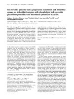
Figure 1
Characterization of cartilage specimensCharacterization of cartilage specimens. (a) Schematic of the cartilage sampling locations at the lesion and remote from the lesion yielding cartilage
from deep and superficial non-lesioned areas. (b) Comparison of modified Mankin scores for both lesioned and non-lesioned osteoarthritis (OA) car-
tilage. Representative toluidine blue-stained cartilage histological sections show the proteoglycan content of (c) non-lesioned superficial (NLS) and
non-lesioned deep (NLD) OA cartilage and (d) lesioned OA cartilage.
Arthritis Research & Therapy Vol 11 No 2 Catterall et al.
Page 4 of 9
(page number not for citation purposes)
cartilage specimens at least 3 mm in thickness had modified
Mankin scores ranging from 3 to 5 (individuals 62 ± 9 years
old, range 56 to 77 years). We also collected eight surgical
waste cartilages from patients with acute joint trauma to serve
as age-matched non-arthritic control samples (individuals 69.3
± 11.3 years, range 51 to 83 years). Representative histolog-
ical sections from non-lesioned and lesioned OA cartilage
regions are presented in Figure 1c, d, demonstrating more
intense GAG staining in the deep non-lesioned OA cartilage
regions and less intense staining in lesioned cartilage (Figure
1d) compared with the superficial non-lesioned OA cartilage
regions.
Change in isomerized aspartate with age in
osteoarthritis and non-osteoarthritis trauma cartilage
extracts
To investigate age-related changes in cartilage, we deter-
mined IsoAsp levels in cartilage extracts from both OA and
non-arthritic trauma cartilage (Figure 2). There was a strong
negative correlation of IsoAsp with age (R
2
= 0.70, P = 0.009)
in non-arthritic trauma cartilage. There was no association of
IsoAsp with age in OA lesion cartilage (R
2
= 0.008, P = 0.78)
or non-lesioned OA cartilage (R
2
= 0.002, P = 0.9). The strong
negative correlation of IsoAsp with age in non-arthritic carti-
lage can be explained, as shown previously [24], by the recip-
rocal rise in these cartilages of the very long-lived racemized
aspartate (
D-β-Asp) which is not recognized by the protein-iso-
aspartyl-methyl-transferase (PIMT) assay [24]. We further
explored this through an examination of deep and superficial
cartilage and synovial fluid content of IsoAsp.
Isomerized aspartate varied by cartilage location and
degeneration status
We investigated IsoAsp, GAG, and protein content per carti-
lage wet weight in the patients (n = 6) for whom we had mac-
roscopically normal OA cartilage obtained remote from the
lesion with a minimum cartilage depth of 3 mm. Cartilage
extracts were prepared from the deep and superficial halves of
these specimens and compared with cartilage extracts pre-
pared from non-OA control cartilages selected prior to extrac-
tion to provide the most appropriate age-matched samples
(OA 62 ± 9 years of age, n = 6; non-OA 69 ± 11 years of age,
n = 8) available. Significantly more IsoAsp and GAG were in
the deep non-lesioned OA cartilage compared with the super-
ficial non-lesioned OA cartilage (Table 2 and Figure 3). There
was no significant difference between the deep and superficial
regions for total protein. OA lesion cartilage was comparable
to the superficial OA non-lesioned cartilage for all of the ana-
lytes measured and significantly different from deep non-
lesioned OA cartilage for IsoAsp (P < 0.05). Although differ-
ences between superficial and deep regions were apparent,
there was no significant difference comparing the overall mean
data for all 14 of the OA lesional and matched OA non-lesional
cartilages. This result demonstrated that even macroscopically
Table 1
Sample characteristics
Specimen Age, years Gender Collins grade Modified Mankin grade Samples
Lesion Non-lesioned OA cartilage Cartilage Synovial fluid
1 74 Female 4 8 - L/NL Yes
2 67 Male 3 10 8 L/NL Yes
369Male27 3 L/NLS-NLDYes
4 56 Female 3 12 3 L/NLS-NLD No
576Male48 5 L/NLYes
677Male4 - 3 L/NLS-NLDNo
759Male45 2 L/NLYes
8 79 Female 4 10 0 L/NL Yes
975Male48 5 L/NLS-NLDYes
10 62 Male 4 12 2 L/NLS-NLD Yes
11 60 Male 4 12 5 L/NLS-NLD Yes
12 69 Female 4 3 0 L/NL Yes
13 66 Female 4 - 2 L/NL No
14 88 Female 4 11 5 L/NL No
L, lesion; NL, non-lesioned osteoarthritis cartilage; NLD, deep non-lesioned osteoarthritis cartilage; NLS, superficial non-lesioned osteoarthritis
cartilage; OA, osteoarthritis.
Available online />Page 5 of 9
(page number not for citation purposes)
Figure 2
Age relationship of isomerized aspartate (IsoAsp) levels in cartilage extractsAge relationship of isomerized aspartate (IsoAsp) levels in cartilage
extracts. (a) IsoAsp levels in cartilage extracts of non-osteoarthritis
(non-OA) trauma cartilages. (b) IsoAsp levels in cartilage extracts from
non-lesioned OA areas. (c) IsoAsp levels in cartilage extracts from OA
lesion areas. NS, not significant.
Figure 3
Comparisons of the analyte concentrations in cartilage extractComparisons of the analyte concentrations in cartilage extract. Mean
(standard error of the mean) concentrations of (a) isomerized aspartate
(IsoAsp), (b) protein, and (c) glycosaminoglycan (GAG) in different
regions within osteoarthritis (OA) cartilage and non-arthritic cartilage.
Analytes were normalized to gram of wet weight cartilage. Statistical
significance was determined using the Wilcoxon signed rank test, and
results represent six individual patients. Using all 14 available cartilage
extracts, we found no significant differences between the non-lesional
and lesional cartilage extracts but did observe significantly higher (P <
0.05) levels of protein in the extracts of non-OA cartilage compared
with any OA cartilage samples.
Arthritis Research & Therapy Vol 11 No 2 Catterall et al.
Page 6 of 9
(page number not for citation purposes)
normal appearing OA cartilage is not, on the whole, signifi-
cantly different than lesional cartilage. We therefore examined
normal age-matched specimens of cartilage derived from
acute trauma and without pre-existing OA by history or visual
inspection. Normal cartilage had higher mean IsoAsp and pro-
tein and lower mean concentrations of PBS/EDTA extractable
GAG than OA cartilages (Table 2). Significant differences (P
< 0.001 to 0.05) were found between normal cartilage IsoAsp
and superficial non-lesioned OA cartilage, between normal
cartilage protein and any OA cartilages, and between normal
cartilage GAG and any OA cartilages.
Synovial fluid alterations associated with osteoarthritis
lesion severity
To understand the effect of OA lesion on synovial fluid constit-
uents, we measured synovial fluid IsoAsp, protein, and GAG
concentrations and compared these values with the amount of
damage at the lesion as determined by the modified Mankin
score. All synovial fluid values were log-transformed to obtain
a Gaussian distribution before linear regression analysis (Fig-
ure 4). Synovial fluid IsoAsp concentration demonstrated a
significant positive correlation with OA lesional histological
severity (R
2
= 0.40, P = 0.039). Total synovial fluid protein
showed a similar but non-significant (R
2
= 0.23, P = 0.16)
positive correlation with cartilage damage. However, synovial
fluid GAG levels decreased significantly with lesion severity
(R
2
= 0.42, P = 0.04). These data are in agreement with our
PBS/EDTA GAG data as more GAG was extractable from the
non-OA cartilage than the lesional cartilage, suggesting that
more advanced OA has less GAG available for release into the
synovial fluid. There were no significant correlations of these
synovial fluid analytes with histological scores for the non-
lesioned regions: IsoAsp (R
2
= 0.029, P = 0.66), protein (R
2
= 0.2, P = 0.22), or GAG (R
2
= 5 × 10
-5
, P = 0.996). As for
OA cartilage, there was no significant correlation between
patient age and levels of IsoAsp (R
2
= 0.08, P = 0.41), protein
(R
2
= 0.01, P = 0.80), or GAG (R
2
= 0.02, P = 0.65) (data not
shown).
Conclusions
This study is the first to demonstrate the presence of a modi-
fied amino acid, in the form of IsoAsp, in OA-affected human
cartilage and synovial fluid and to examine associations
between IsoAsp content and the degree of OA-related tissue
damage. We found significantly more IsoAsp in normal carti-
lage and the deeper cartilage zones than the superficial non-
lesioned OA cartilage and lesional regions. IsoAsp in cartilage
is an intermediate non-enzymatic alteration prior to conversion
to racemized aspartate. The PIMT enzyme does not detect the
main racemized form (
D-β-Asp). Thus, the decline in IsoAsp
from a tissue or body fluid could be due to conversion to a
racemized form or due to loss from tissue. With these data, we
show that it is due to loss from the tissue and in proportion to
lesion severity. Our previous work demonstrated that racemi-
zation is not increased in OA non-lesioned cartilage relative to
normal cartilage [24], which is also supportive of our conclu-
sion that IsoAsp in synovial fluid represents catabolism of mol-
ecules of intermediate age. The relative accumulation of
IsoAsp in the deep regions of cartilage that was comparable
to control cartilage shows, as suspected, that the superficial
zone is more actively turning over than the deep region. An
alternative explanation may be that IsoAsp accumulates due to
steric hindrance from intact matrix and matrix interaction, pre-
venting the conversion of aspartate to the
D-β-Asp racemized
form. Due to the steady age-related increase in aspartate
racemization in normal cartilage previously observed by us and
others, we believe this is unlikely [24-26].
The amount of readily extractable GAG from the deep regions
of non-lesioned OA cartilage contrasted with the very small
amounts of readily extractable GAG from normal cartilage.
This suggests that the cartilage in these deep, albeit non-
lesioned, areas of an OA cartilage is not normal but rather that
there has been previous proteolysis of GAG-bearing proteins
that are readily solubilized with the addition of PBS/EDTA
alone. Our data are consistent with previous results showing
that cartilage aggrecan degradation proceeds in a two-state
manner in rheumatoid arthritis [27]. That study showed that, at
early stages, the GAG-containing regions are lost from carti-
lage and the amount of GAG in synovial fluid declines with
Table 2
Mean analyte concentrations in cartilage extracts
Cartilage region Number IsoAsp μM/g cartilage ± SEM Protein μg/g cartilage ± SEM GAG μg/g cartilage ± SEM
Normal 8 7,092 ± 1,568 65,092 ± 18,017 743 ± 102
Lesioned OA 14 4,942 ± 719 15,281 ± 552
a
4,463 ± 1,053
b
Non-lesioned OA 14 7,439 ± 1,273 17,926 ± 1,134
c
2,911 ± 657
b
Superficial 6 2,453 ± 315
c
11,266 ± 818
c
3,626 ± 921
Deep 6 3,999 ± 665
d
16,894 ± 3,105
c
7,316 ± 1,279
a, d, e
a
P < 0.001 compared to normal cartilage;
b
P < 0.01 compared to normal cartilage;
c
P < 0.05 compared to normal cartilage;
d
P < 0.05 compared
to superficial cartilage;
e
P < 0.05 compared to osteoarthritis (OA) lesion cartilage. GAG, glycosaminoglycan; IsoAsp, isomerized aspartate; SEM,
standard error of the mean.
Available online />Page 7 of 9
(page number not for citation purposes)
increasing disease severity. This is exactly the result we find.
The authors of that study further found that the hyaluronan-
binding (G1) domain of aggrecan is retained early in the dis-
ease process and then finally released to synovial fluid and so
follows a reciprocal pattern to GAG loss. We did not measure
hyaluronan-binding region fragments. We also know that
aggrecan plays a protective role in preventing degradation of
collagen fibrils [28]. With increasing loss of proteoglycan,
there is increasing degradation of collagen and other matrix
components [29], and this is entirely compatible with our
results showing a tendency to increased protein loss to the
synovial fluid with increasing lesion severity and loss of GAG
from the cartilage. The fact that IsoAsp also increased with dis-
ease severity suggests that the trend to increased synovial
fluid protein is not in fact derived from repair but rather prima-
rily from cartilage degradation.
Age-related modifications of lesional cartilage were compara-
ble to superficial non-lesioned OA cartilage. Deep cartilage
extracts had more GAG than found in the superficial cartilage,
a result corroborated by histological observations. Moreover,
the observation that there was reduced GAG present in the
lesional extracts was consistent with previous studies [30-34].
All of these data agree in principle with the molecular biology
data of Fukui and colleagues [35], who observed that cartilage
erosion leads the newly exposed chondrocytes now at the sur-
face to take on the gene expression profiles of the superficial
cartilage chondrocytes.
While our study has added significantly to the body of knowl-
edge concerning the effects of OA on human cartilage and
synovial fluid contents, several important questions require fur-
ther investigation. The unique ability of synovial fluid IsoAsp to
reflect OA lesion severity makes it an attractive disease
marker; however, its full potential cannot be realized without
identification of the primary IsoAsp-containing molecule(s).
That knowledge would allow the calculation of the modified-to-
unmodified molecule ratio and provide a potential marker to
facilitate clinical treatment and understanding, analogous to
the markers HbA1C in diabetes and αCTX-I/βCTX-1 in Paget
disease of bone [36]. Furthermore, the role of IsoAsp in the
pathophysiology of OA deserves consideration and further
investigation based on the paradigm provided by another form
of modifications, racemization, that has been shown to desta-
bilize the collagen triple helix [37].
The data presented here are consistent with our belief that
'aged' neo-epitopes found in the older zones of cartilage have
the potential to be important biomarkers of OA. We believe
that non-progressive OA represents a balance of catabolic
and anabolic processes and that most of the proteins released
as part of cartilage turnover will be the recently synthesized
proteins, which will have greatly reduced levels of age-related
protein modifications such as IsoAsp. However, in active OA
progression, catabolism will outpace anabolism and destruc-
Figure 4
Association of synovial fluid analytes and osteoarthritis severityAssociation of synovial fluid analytes and osteoarthritis severity. Associ-
ation of synovial fluid (a) isomerized aspartate (IsoAsp), (b) protein, and
(c) glycosaminoglycan (GAG) with the modified Mankin scores of
lesional cartilage. Analytes were natural logarithm (LN)-transformed to
produce a normal distribution as determined by the D'Agostino and
Pearson omnibus normality test. Results represent 10 separate individ-
ual patients for whom both cartilage and synovial fluid were available
(Table 1). NS, not significant; SF, synovial fluid.
Arthritis Research & Therapy Vol 11 No 2 Catterall et al.
Page 8 of 9
(page number not for citation purposes)
tion of the deeper cartilage zones will occur, leading to the
release of these age-modified proteins. We believe that these
aged molecules may have the potential to more accurately pre-
dict active progressive cartilage destruction as their levels will
be independent of the confounding factor of increased synthe-
sis, which can occur during active repair.
In summary, we have demonstrated for the first time that (a)
proteins in the deeper zones of cartilage contain more age-
related post-translational modifications in the form of the
isomerized amino acid IsoAspartate and (b) synovial fluid lev-
els of the age-related protein post-translational modification
IsoAspartate correlated with increased cartilage damage. Our
finding that protein modification, specifically IsoAsp, reflects
severity of cartilage damage suggests that age-related post-
translational protein modifications have the potential to serve
as disease activity and progression biomarkers in OA.
Competing interests
The authors declare that they have no competing interests.
Authors' contributions
JBC and DB contributed equally to the experimental design,
the preparation of the manuscript, and the statistical analysis.
MB and RDZ coordinated and organized the primary sample
collection and critically evaluated both the study design and
the manuscript. VBK conceived and designed the study,
supervised the project, and assisted in both the statistical
analysis and the manuscript preparation and editing. All
authors read and approved the final manuscript.
Acknowledgements
We wish to thank Janet Huebner for assistance with histology. This
study was supported by a Eugene A. Stead student research scholar-
ship (to DB), National Institutes of Health (NIH)/National Institute of
Arthritis and Musculoskeletal and Skin Diseases grant UO1 AR050898,
and NIH/National Institute on Aging grant Pepper OAIC P30
AG028716.
References
1. Cloos PA, Christgau S: Non-enzymatic covalent modifications
of proteins: mechanisms, physiological consequences and
clinical applications. Matrix Biol 2002, 21:39-52.
2. Maroudas A, Bayliss MT, Uchitel-Kaushansky N, Schneiderman R,
Gilav E: Aggrecan turnover in human articular cartilage: use of
aspartic acid racemization as a marker of molecular age. Arch
Biochem Biophys 1998, 350:61-71.
3. Robinson NE, Robinson AB: Molecular Clocks: Deamidation of
Asparaginyl and Glutaminyl Residues in Peptides and Proteins
London, ON, Canada: The Althouse Press; 2004.
4. Brennan TV, Clarke S: Spontaneous degradation of polypep-
tides at aspartyl and asparaginyl residues: effects of the sol-
vent dielectric. Protein Sci 1993, 2:331-338.
5. Oliyai C, Borchardt RT: Chemical pathways of peptide degrada-
tion. VI. Effect of the primary sequence on the pathways of
degradation of aspartyl residues in model hexapeptides.
Pharm Res 1994, 11:751-758.
6. Ohtani S, Ito R, Arany S, Yamamoto T: Racemization in enamel
among different types of teeth from the same individual. Int J
Legal Med 2005, 119:66-69.
7. Fujii N, Momose Y, Harada K: Kinetic study of racemization of
aspartyl residues in model peptides of alpha A-crystallin. Int J
Pept Protein Res 1996, 48:118-122.
8. Fujii N, Momose Y, Ishii N, Takita M, Akaboshi M, Kodama M: The
mechanisms of simultaneous stereoinversion, racemization,
and isomerization at specific aspartyl residues of aged lens
proteins. Mech Ageing Dev 1999, 107:347-358.
9. Potter SM, Henzel WJ, Aswad DW: In vitro aging of calmodulin
generates isoaspartate at multiple Asn-Gly and Asp-Gly sites
in calcium-binding domains II, III, and IV. Protein Sci 1993,
2:1648-1663.
10. Sandmeier E, Hunziker P, Kunz B, Sack R, Christen P: Spontane-
ous deamidation and isomerization of Asn108 in prion peptide
106–126 and in full-length prion protein. Biochem Biophys Res
Commun 1999, 261:578-583.
11. McCudden CR, Kraus VB: Biochemistry of amino acid racemi-
zation and clinical application to musculoskeletal disease.
Clin Biochem 2006, 39:1112-1130.
12. Bohme L, Bar JW, Hoffmann T, Manhart S, Ludwig HH, Rosche F,
Demuth HU: Isoaspartate residues dramatically influence sub-
strate recognition and turnover by proteases. Biol Chem 2008,
389:1043-1053.
13. Handley C, McQuillan D, Campbell M, Bolis S: Steady-state
metabolism in cartilage explants. In Articular Cartilage Bio-
chemistry Edited by: Kuettner K, Schleyerbach R, Hascall V. New
York: Raven Press; 1986:163-179.
14. Mok SS, Masuda K, Hauselmann HJ, Aydelotte MB, Thonar EJ:
Aggrecan synthesized by mature bovine chondrocytes sus-
pended in alginate. Identification of two distinct metabolic
matrix pools. J Biol Chem 1994, 269:33021-33027.
15. Sandy JD, Plaas AH: Age-related changes in the kinetics of
release of proteoglycans from normal rabbit cartilage
explants. J Orthop Res 1986, 4:263-272.
16. Maroudas A, Palla G, Gilav E: Racemization of aspartic acid in
human articular cartilage. Connect Tissue Res 1992,
28:161-169.
17. Verzijl N, DeGroot J, Thorpe SR, Bank RA, Shaw JN, Lyons TJ,
Bijlsma JW, Lafeber FP, Baynes JW, TeKoppele JM: Effect of col-
lagen turnover on the accumulation of advanced glycation end
products. J Biol Chem 2000, 275:39027-39031.
18. Vilim V, Krajickova J: Proteoglycans of human articular cartilage.
Identification of several populations of large and small prote-
oglycans and of hyaluronic acid-binding proteins in succes-
sive cartilage extracts. Biochem J 1991, 273:579-585.
19. Collins D: The Pathology of Articular and Spinal Diseases London,
UK: Edward Arnold & Co; 1949.
20. Richardson CD, Bae WC, Fazeli B, Filvaroff EH, Sah RL: Quanti-
tative characterisation of osteoarthritis in the guinea pig.
Paper presented at: Orthopaedic Research Society 47th Annual
Meeting; 25–28 February 2001; San Francisco, CA, USA .
21. Vilim V, Lenz ME, Vytasek R, Masuda K, Pavelka K, Kuettner KE,
Thonar EJ: Characterization of monoclonal antibodies recog-
nizing different fragments of cartilage oligomeric matrix pro-
tein in human body fluids. Arch Biochem Biophys 1997,
341:8-16.
22. Chandrasekhar S, Esterman MA, Hoffman HA: Microdetermina-
tion of proteoglycans and glycosaminoglycans in the presence
of guanidine hydrochloride. Anal Biochem 1987, 161:
103-108.
23. Carlson CS, Loeser RF, Purser CB, Gardin JF, Jerome CP: Oste-
oarthritis in cynomolgus macaques. III: effects of age, gender,
and subchondral bone thickness on the severity of disease. J
Bone Miner Res 1996, 11:1209-1217.
24. Stabler TV, Byers SS, Zura RD, Kraus VB: Amino acid racemiza-
tion reveals differential protein turnover in osteoarthritic artic-
ular and meniscal cartilages. Arthritis Res Ther 2009, 11:R34.
25. Ohtani S, Matsushima Y, Kobayashi Y, Yamamoto T: Age estima-
tion by measuring the racemization of aspartic acid from total
amino acid content of several types of bone and rib cartilage:
a preliminary account. J Forensic Sci 2002, 47:32-36.
26. Pfeiffer H, Mornstad H, Teivens A: Estimation of chronologic age
using the aspartic acid racemization method. I. On human rib
cartilage. Int J Legal Med 1995, 108:19-23.
27. Saxne T, Heinegard D: Synovial fluid analysis of two groups of
proteoglycan epitopes distinguishes early and late cartilage
lesions. Arthritis Rheum 1992, 35:385-390.
28. Pratta MA, Yao W, Decicco C, Tortorella MD, Liu RQ, Copeland
RA, Magolda R, Newton RC, Trzaskos JM, Arner EC: Aggrecan
Available online />Page 9 of 9
(page number not for citation purposes)
protects cartilage collagen from proteolytic cleavage. J Biol
Chem 2003, 278:45539-45545.
29. Melrose J, Fuller ES, Roughley PJ, Smith MM, Kerr B, Hughes CE,
Caterson B, Little CB: Fragmentation of decorin, biglycan, lum-
ican and keratocan is elevated in degenerate human menis-
cus, knee and hip articular cartilages compared with age-
matched macroscopically normal and control tissues. Arthritis
Res Ther 2008, 10:R79.
30. Brocklehurst R, Bayliss MT, Maroudas A, Coysh HL, Freeman MA,
Revell PA, Ali SY: The composition of normal and osteoarthritic
articular cartilage from human knee joints. With special refer-
ence to unicompartmental replacement and osteotomy of the
knee. J Bone Joint Surg Am 1984, 66:95-106.
31. Dumond H, Presle N, Terlain B, Mainard D, Loeuille D, Netter P,
Pottie P: Evidence for a key role of leptin in osteoarthritis.
Arthritis Rheum 2003, 48:3118-3129.
32. Horton WE Jr, Yagi R, Laverty D, Weiner S: Overview of studies
comparing human normal cartilage with minimal and
advanced osteoarthritic cartilage. Clin Exp Rheumatol 2005,
23:103-112.
33. Squires GR, Okouneff S, Ionescu M, Poole AR: The pathobiology
of focal lesion development in aging human articular cartilage
and molecular matrix changes characteristic of osteoarthritis.
Arthritis Rheum 2003, 48:1261-1270.
34. Sweet MB, Thonar EJ, Immelman AR, Solomon L: Biochemical
changes in progressive osteoarthrosis. Ann Rheum Dis 1977,
36:387-398.
35. Fukui N, Ikeda Y, Ohnuki T, Tanaka N, Hikita A, Mitomi H, Mori T,
Juji T, Katsuragawa Y, Yamamoto S, Sawabe M, Yamane S, Suzuki
R, Sandell LJ, Ochi T: Regional differences in chondrocyte
metabolism in osteoarthritis: a detailed analysis by laser cap-
ture microdissection. Arthritis Rheum 2008, 58:154-163.
36. Garnero P, Fledelius C, Gineyts E, Serre CM, Vignot E, Delmas
PD: Decreased beta-isomerization of the C-terminal telopep-
tide of type I collagen alpha 1 chain in Paget's disease of bone.
J Bone Miner Res 1997, 12:1407-1415.
37. Shah NK, Brodsky B, Kirkpatrick A, Ramshaw JA: Structural con-
sequences of D-amino acids in collagen triple-helical pep-
tides. Biopolymers 1999, 49:297-302.