Báo cáo y học: "Enhanced reactivity to pain in patients with rheumatoid arthritis" ppsx
Bạn đang xem bản rút gọn của tài liệu. Xem và tải ngay bản đầy đủ của tài liệu tại đây (273.06 KB, 9 trang )
Open Access
Available online />Page 1 of 9
(page number not for citation purposes)
Vol 11 No 3
Research article
Enhanced reactivity to pain in patients with rheumatoid arthritis
Robert R Edwards
1,2
, Ajay D Wasan
1
, Clifton O Bingham III
3
, Joan Bathon
3
,
Jennifer A Haythornthwaite
2
, Michael T Smith
2
and Gayle G Page
4
1
Department of Anesthesiology, Harvard Medical School, Brigham & Women's Hospital, 850 Boylston Street, Suite 302, Chestnut Hill, MA 02467,
USA
2
Department of Psychiatry, Johns Hopkins University School of Medicine, 600 N. Wolfe Street, Baltimore, MD 21287, USA
3
Division of Rheumatology, Johns Hopkins University School of Medicine, 5200 Eastern Avenue, MFL Suite 4100, Baltimore, MD 21224, USA
4
Johns Hopkins University School of Nursing, 525 N. Wolfe Street, Baltimore, MD 21287, USA
Corresponding author: Robert R Edwards,
Received: 16 Feb 2009 Revisions requested: 1 Apr 2009 Revisions received: 17 Apr 2009 Accepted: 4 May 2009 Published: 4 May 2009
Arthritis Research & Therapy 2009, 11:R61 (doi:10.1186/ar2684)
This article is online at: />© 2009 Edwards et al.; licensee BioMed Central Ltd.
This is an open access article distributed under the terms of the Creative Commons Attribution License ( />),
which permits unrestricted use, distribution, and reproduction in any medium, provided the original work is properly cited.
Abstract
Introduction Maladaptive physiological responses to stress
appear to play a role in chronic inflammatory diseases such as
rheumatoid arthritis (RA). However, relatively little stress
research in RA patients has involved the study of pain, the most
commonly reported and most impairing stressor in RA. In the
present study, we compared psychophysical and physiological
responses to standardized noxious stimulation in 19 RA patients
and 21 healthy controls.
Methods Participants underwent a single psychophysical
testing session in which responses to a variety of painful stimuli
were recorded, and blood samples were taken at multiple time
points to evaluate the reactivity of cortisol, interleukin-6 (IL-6),
and tumor necrosis factor-alpha (TNF-α) to the experience of
acute pain.
Results The findings suggest that RA patients display a fairly
general hyperalgesia to mechanical and thermal stimuli across
several body sites. In addition, while serum cortisol levels did not
differ at baseline or following pain testing in patients relative to
controls, the RA patients tended to show elevations in serum IL-
6 and demonstrated enhanced pain-reactivity of serum levels of
TNF-α compared with the healthy controls (P < 0.05).
Conclusions These findings highlight the importance of pain as
a stressor in RA patients and add to a small body of literature
documenting amplified responses to pain in RA. Future studies
of the pathophysiology of RA would benefit from the
consideration of acute pain levels when comparing RA patients
with other groups, and future trials of analgesic interventions in
RA patients may benefit from evaluating the effects of such
interventions on inflammatory activity.
Introduction
Multiple lines of investigation suggest that stress plays a sig-
nificant role in shaping the course of inflammatory diseases
such as rheumatoid arthritis (RA) [1-3]. Stress activates a cas-
cade of neurohumoral events, many of which may be dysregu-
lated in RA patients, including aspects of the hypothalamic-
pituitary-adrenal (HPA) axis, the autonomic nervous system,
and pro-inflammatory processes [1,3]. Dozens of studies over
the past several decades have evaluated the effect of multiple
types of stressors on the physiology and symptomatology of
patients with RA. Collectively, laboratory research has docu-
mented a maladaptively pro-inflammatory response to stress
among RA patients, with elevated stress-reactivity of factors
such as C-reactive protein (CRP) [4] and tumor necrosis fac-
tor-alpha (TNF-α) [5]. Moreover, a relative hypo-responsive-
ness of the autonomic nervous system and HPA system have
been observed in RA patients in response to mental stress as
well as a variety of physical stressors [1,3].
Much stress research in RA has been conducted outside of
the laboratory, and studies of naturally occurring stressors
have revealed that elevations of daily stress among RA
patients are associated with increases in musculoskeletal ten-
derness, interleukin-6 (IL-6) levels, and disease activity [6-9].
ANOVA: analysis of variance; BDI: Beck Depression Inventory; CPT: cold pressor task; CRP: C-reactive protein; DAS28: disease activity score using
28 joint counts; DMARD: disease-modifying antirheumatic drug; GCRC: general clinical research center; HPA: hypothalamic-pituitary-adrenal; HPTh:
heat pain threshold; IL-6: interleukin-6; i.v.: intravenous; MTX: methotrexate; PPTh: pressure pain threshold; RA: rheumatoid arthritis; SBP: systolic
blood pressure; SF-36: Short Form Health Survey-36; TNF: tumor necrosis factor.
Arthritis Research & Therapy Vol 11 No 3 Edwards et al.
Page 2 of 9
(page number not for citation purposes)
Interestingly, relatively little of this research has involved the
study of pain, the most commonly reported and most impairing
stressor in RA [10]. The experience of pain is generally asso-
ciated with enhanced release of pro-inflammatory cytokines,
which in turn sensitize the nervous system, promoting a further
amplification of pain transmission [11-14]. To date, a handful
of human studies have documented the presence of cytokine
reactivity to the application of calibrated noxious stimuli in
humans. Significant increases in pro-inflammatory cytokines
such as IL-6 have been observed following non-tissue-damag-
ing painful stimulation in healthy adults [15,16], patients with
juvenile RA [17], and patients with persisting low back pain
[18].
Given that RA patients experience persistent pain and chronic
inflammation, it is natural to inquire whether the inflammatory
response to the experience of pain itself is normal in RA.
Importantly, psychophysical studies indicate that, relative to
controls, RA patients exhibit lower pressure pain thresholds
(PPThs) and enhanced sensitivity to noxious stimuli across a
variety of anatomical sites, including both inflamed joints and
non-inflamed tissues [19-26], suggesting central amplification
of pain-related information. This enhancement of pain sensitiv-
ity appears to be magnified in individuals with RA of longer
duration [25].
To date, although it is well established that RA patients are
more behaviorally responsive to noxious stimulation relative to
non-arthritic controls, no studies have evaluated whether RA
patients show aberrant inflammation-related responses to the
experience of acute pain in a controlled laboratory setting. It is
important to evaluate the inflammatory response to noxious
stimulation among RA patients as daily pain is among their
most common and salient stressors. In the present project, we
focus on assessing IL-6, TNF-α, and cortisol reactivity to acute
painful stimulation in a sample of RA patients compared with
age- and gender-matched healthy controls.
Materials and methods
Participants
Participants were 19 treated RA patients and 21 generally
healthy controls, free from rheumatic disease. RA patients
were recruited via letters and flyers sent to patients of the
Johns Hopkins Arthritis Center, who were diagnosed with RA
using the American College of Rheumatology criteria [27];
controls were recruited through the posting of flyers and the
use of newspaper advertisements around the Baltimore com-
munity. All subjects provided informed consent, and the study
was approved by the Johns Hopkins Institutional Review
Board. None of the authors has any financial or other conflicts
of interest with regard to this study or its findings.
Inclusion criteria for the study (for RA patients) included RA as
the primary source of persistent pain; no current mood or anx-
iety disorder; no history of myocardial infarction or cardiovas-
cular disease; no history of peripheral neuropathy, Raynaud
syndrome, vasculitis, or peripheral vascular disease; no cur-
rent infection; no history of other autoimmune or rheumatic dis-
orders; and no recent history of substance abuse or
dependence. Subjects taking opioid, antidepressant, or ster-
oid medications were not included in the study. Pregnant
women were also not included in the study. Healthy controls
met all of the same criteria; in addition, they did not have RA or
other joint pain and were not taking any centrally acting medi-
cations. RA patients reported being on stable treatment regi-
mens for at least 1 month; those taking non-steroidal anti-
inflammatory medications were asked to abstain from using
them for 24 hours prior to the laboratory session.
Session protocol
All subjects provided verbal and written informed consent, and
all procedures were approved by an institutional review board.
Many of these procedures have been described previously
[16]. The setting for the study was a general clinical research
center (GCRC) based within a university hospital. Participants
arrived between 12 and 12:30 p.m.; they had previously been
requested to refrain from using over-the-counter medications
or caffeine, smoking, or performing other than mild exercise
prior to their arrival. To avoid interfering with RA treatment reg-
imens, participants were asked to take their RA medications as
prescribed. After informed consent and screening for eligibil-
ity, participants completed questionnaires for approximately
10 minutes. Questionnaires included a medical history form,
questions about current pain and current stress levels (rated
on 0-to-10 scales), the Beck Depression Inventory (BDI) [28],
and the Short Form Health Survey-36 (SF-36) [29]. Determi-
nation of eligibility for the study was made based on question-
naires and a medical history taken by a research nurse at the
GCRC.
Next, subjects were seated comfortably in a reclining chair and
an intravenous (i.v.) line was inserted in the left forearm by a
GCRC research nurse [17,30]. After i.v. placement and a 15-
minute period of rest, two baseline blood samples (10 mL),
separated by 5 minutes, were drawn. These two values were
averaged together in order to maximize stability of the baseline
estimates. Baseline systolic and diastolic blood pressures
were then recorded. Subsequently, participants underwent
the psychophysical pain testing procedures described below
(the duration of pain testing was approximately 45 minutes),
after which additional blood samples (10 mL) were taken at
several time points: immediately after testing and 15, 30, and
60 minutes after testing.
Psychophysical pain testing (45-minute session)
Mechanical pain thresholds were assessed first using a digital
pressure algometer (Somedic Production AB, Sollentuna,
Sweden). As in previous studies [19,21,23], we selected sev-
eral muscle/joint sites and bilaterally assessed PPThs. PPThs
were determined twice at each of the following sites on the
Available online />Page 3 of 9
(page number not for citation purposes)
right and left sides of the body in a randomized order: the belly
of the trapezius muscle, the metacarpophalangeal joint of the
thumb, and the quadriceps muscle, near the insertion of the
proximal patellar tendon. At each site, mechanical force was
applied using a 0.5-cm
2
probe covered with polypropylene
pressure-transducing material; pressure was increased at a
steady rate of 30 kPa/second until the subject indicated that
the pressure was 'first perceived as painful'.
Next, contact heat stimuli were delivered using a Medoc Ther-
mal Sensory Analyzer (TSA-2001; Medoc Ltd., Ramat Yishai,
Israel). Thermal assessment included sampling of heat pain
thresholds (HPThs) on the ventral forearm using an ascending
method of limits paradigm with a rate of rise of 0.5°C/second
[31]. Three trials of HPTh were performed first, followed by
four trials of suprathreshold heat stimulation. In brief, four
sequences of 10 rapid heat pulses were applied to the fore-
arm, similar to prior studies [32,33]. Within each sequence,
the procedure was as follows: from a 38°C baseline tempera-
ture, 10 successive thermal pulses were delivered. The rate of
rise and fall of the thermode temperature was 10°C/second,
and target temperatures were delivered for approximately 0.5
seconds each. The thermode remained in a fixed position dur-
ing administration of the 10 pulses and then was re-positioned
between sequences, with inter-sequence intervals of 2 min-
utes. Two different target temperatures (49°C and 51°C) were
used two times each in randomized order. Subjects verbally
rated the painfulness of each thermal pulse on a 0-to-100 (0 =
'no pain' and 100 = 'most intense pain imaginable') numeric
rating scale and then rated the painfulness of lingering after-
sensations 15 seconds after the stimuli had ceased [34,35].
Finally, responses to noxious cold were evaluated using a
repeated cold pressor task (CPT), involving immersion of the
right hand in a circulating cold water bath maintained at 4°C.
The CPT is the most commonly used method of pain induction
in the laboratory and has demonstrated clinical relevance
[36,37]. Several recent studies indicate that the CPT provokes
increases in cortisol and norepinepherine as well as producing
increases in pro-inflammatory cytokine production [16,17]. In
the present protocol, participants underwent a series of five
CPTs, with the first four consisting of serial immersions of the
right hand for 30 seconds, with 2 minutes between immer-
sions. The fifth and final CPT involved an immersion of the right
hand lasting until a participant reached pain tolerance (or a 3-
minute maximum). Participants rated the intensity of the cold
pain on a 0-to-100 scale ('no pain' to 'most intense pain imag-
inable') at the midpoint and conclusion of each CPT. Following
the final CPT, participants continued to relax in the chair as
subsequent blood samples were taken.
Physiological measures
Each blood sample (that is, two baseline samples, one sample
immediately after pain testing, then samples at 15, 30, and 60
minutes following the conclusion of pain testing) was col-
lected in a 10-mL tube and transported to the GCRC Core
Laboratory, where it was centrifuged, aliquoted, and stored in
a -80°C freezer for later assay. Serum cortisol was assessed
in duplicate using a radioimmunoassay (Diagnostic Systems
Laboratories, Inc., Webster, TX, USA), with a lower limit of
detection of 0.5 μg/dL, a sensitivity of 0.11 μg/dL, and an
intra-assay coefficient of variation of less than 10%. A stand-
ard high-sensitivity enzyme-linked immunosorbent assay (R&D
Systems, Minneapolis, MN, USA) was used to assess serum
levels of IL-6 in duplicate. This assay has a lower limit of detec-
tion of 0.16 pg/mL, a sensitivity of 0.04 pg/mL, and an intra-
assay coefficient of variation of less than 5%. Similarly, an
enzyme-linked immunosorbent assay from the same company
(R&D Systems) was used to assess serum levels of TNF-α in
duplicate. This assay has a lower limit of detection of 0.25 pg/
mL, a sensitivity of 0.06 pg/mL, and an intra-assay coefficient
of variation of less than 10%.
Data analysis
Simple between-group comparisons (RA patients compared
with controls) were made using analysis of variance (ANOVA).
Changes, across the two groups, in serum levels of cortisol, IL-
6, and TNF-α were evaluated using repeated measures
ANOVA. Inter-relationships among study variables were eval-
uated using Pearson correlations. All analyses were performed
using SPSS (SPSS Inc., Chicago, IL, USA).
Results
RA patients reported a mean time since diagnosis of 8.3 years
(standard deviation = 6.4 years). The mean disease activity
score using 28 joint counts (DAS28) for the sample was 3.1
± 1.4. In addition, the mean CRP level in RA patients was 3.3
± 3.9 μg/ml. These values suggest generally low to moderate
levels of disease activity, on average, in these patients and are
broadly consistent with other, larger US studies of treated RA
patients (for example, in [38], mean RA duration = 12.4 years,
mean DAS28 score = 3.7, and median CRP = 2.6 μg/ml).
RA patients did not differ (all P values of greater than 0.10)
from controls on demographic variables such as age (mean
age for RA patients = 51.7 ± 12.2 years and mean age for
controls = 50.3 ± 12.7 years), gender (58% women in the RA
group and 52% women in the control group), ethnicity (58%
in the RA group were white and 67% in the control group were
white), or education (mean years of education for RA patients
= 14.0 ± 2.7 and mean years of education for controls = 15.1
± 2.5). In addition, CRP levels in RA patients (mean = 3.3 ±
3.9) did not differ significantly from CRP levels in controls
(mean = 2.5 ± 3.5). Finally, resting systolic blood pressures
(SBPs) in the controls (mean = 122.8 ± 9.6 mmHg) did not
differ from SBPs in the RA patients (mean = 122.1 ± 18.8
mmHg). Similarly, diastolic blood pressures in the controls
(mean = 70.1 ± 6.0 mmHg) and RA patients (mean = 64.4 ±
10.7 mmHg) were similar (P > 0.10).
Arthritis Research & Therapy Vol 11 No 3 Edwards et al.
Page 4 of 9
(page number not for citation purposes)
All RA patients were receiving treatment for their disease,
though with significant variability in the treatment regimens.
The following is a summary of the disease-modifying antirheu-
matic drugs (DMARDs) taken by the 19 RA patients in this
study: methotrexate (MTX) monotherapy (n = 8), hydroxychlo-
rochloroquine monotherapy (n = 2), TNF antagonist mono-
therapy (n = 3), MTX + other non-biologic DMARD (n = 4),
and MTX + TNF antagonist (n = 2).
Questionnaires
In terms of questionnaire responses, RA patients did report
higher levels of current and recent pain and lower scores on
indices of health and physical functioning relative to the con-
trols (Table 1). Interestingly, patients and controls did not differ
on self-report of current stress levels or the SF-36 indices of
mental/emotional health. RA patients did endorse higher
scores on the BDI, although mean levels of depressive symp-
toms were low and within the normal range (that is, BDI scores
of less than 10 are generally considered subclinical) for both
groups.
Pain responses
Comparisons between RA patients and controls on measures
of psychophysical pain responses yielded statistically signifi-
cant (P ≤ 0.05) or near-significant differences on a number of
measures. RA patients had lower HPThs, lower mechanical
pain thresholds on the thumb, higher pain intensity ratings of
51°C heat stimuli and heat after-sensations, lower cold pain
tolerance, and higher cold pain ratings during the CPT tests.
Tendencies that did not reach the level of frank statistical sig-
nificance were noted for PPTh on the trapezius and heat pain
Table 1
Comparison of rheumatoid arthritis patients and controls on pain and questionnaire responses
RA patients
(n = 19)
Controls
(n = 21)
P value
Responses to noxious stimuli
HPTh, °C 41.4 ± 5.1 44.4 ± 4.5 0.05
PPTh on leg, kPa 665.5 ± 287.7 811.3 ± 400.1 0.19
PPTh on thumb, kPa 295.7 ± 141.6 395.7 ± 150.7 0.03
PPTh on trapezius, kPa 404.5 ± 160.7 536.2 ± 276.9 0.08
Cold pain rating (0 to 100) at midpoint 82.3 ± 12.6 65.4 ± 25.6 0.01
Cold pain rating (0 to 100) at conclusion 83.0 ± 12.4 67.7 ± 24.8 0.02
Cold pain tolerance, seconds 61.8 ± 54.1 111.8 ± 63.8 0.01
Heat pain rating at 49°C 74.4 ± 25.1 57.7 ± 34.7 0.09
Heat pain rating at 51°C 86.8 ± 16.2 68.2 ± 32.4 0.03
Painful heat after-sensations 16.8 ± 23.2 5.7 ± 9.4 0.05
Questionnaire data
Current pain (0 to 10) 3.2 ± 2.3 0.4 ± 0.3 < 0.001
Current stress (0 to 10) 2.2 ± 2.4 1.2 ± 1.9 0.17
Beck Depression Inventory score 7.0 ± 6.3 2.5 ± 3.0 0.01
SF-36, subscale score
General health 52.9 ± 20.3 84.0 ± 19.4 < 0.001
Physical functioning 42.1 ± 24.2 69.1 ± 26.7 0.002
Physical role 28.9 ± 31.5 86.9 ± 28.1 < 0.001
Bodily pain 44.2 ± 21.3 85.7 ± 23.7 < 0.001
Energy/fatigue 53.4 ± 18.4 67.6 ± 18.1 0.02
Mental health 80.7 ± 13.7 81.0 ± 14.5 0.95
Emotional role 84.2 ± 34.0 93.7 ± 22.7 0.30
Social functioning 70.4 ± 27.7 94.6 ± 7.5 0.001
Data are presented as mean ± standard deviation. HPTh, heat pain threshold; PPTh, pressure pain threshold; RA, rheumatoid arthritis; SF-36,
Short Form Health Survey-36.
Available online />Page 5 of 9
(page number not for citation purposes)
ratings in response to the 49°C stimuli. These data are pre-
sented in Table 1.
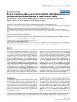
Physiological responses
Repeated measures ANOVAs were used to evaluate
between-group differences in levels of cortisol, IL-6, and TNF-
α over the course of the session. As the demographics of the
groups were similar, we did not control for age, gender, race,
or education, but SF-36 general health subscale scores were
entered as a covariate in order to statistically control for clear
group differences in perceived health. For measures of serum
cortisol, there was a strong main effect of time [F(4,34) = 8.3,
P < 0.01], but no significant main effect of group or group ×
time interaction (P > 0.1). For IL-6, there was also a main effect
of time [F(4,34) = 4.0, P < 0.01] as well as a trend for a main
effect of group [(F(1,37) = 3.2, P = 0.07]. On average, the RA
patients had serum IL-6 levels that tended to be higher than
those of the controls at every time point. The IL-6 data showed
no interaction between group × time. Finally, for the TNF-α
data, the main effects of time and group were qualified by a
significant interaction [F(4,34) = 3.3, P = 0.02]. Among the
RA patients, serum TNF-α increased significantly from base-
line following the pain testing (P < 0.05), whereas no signifi-
cant changes in TNF-α were observed in the controls.
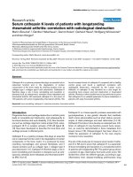
Cortisol, IL-6, and TNF-α data are depicted in Figure 1.
Although our sample of 19 RA patients is too small to permit
extensive investigation of the relationships between cytokine
responses to pain and clinical variables, we assessed correla-
tions of TNF-α and IL-6 responses with the SF-36 subscales
of bodily pain, energy/fatigue, and physical functioning. Within
the RA group, TNF-α levels were unrelated to bodily pain or
physical functioning but showed a tendency to relate to lower
levels of energy (or higher levels of fatigue): r = -0.43, P =
0.07. IL-6 levels were similarly associated with bodily pain (r =
-0.41, P = 0.08), energy/fatigue (r = -0.45, P = 0.06), and
physical functioning (r = -0.42, P = 0.08).
Discussion
The present findings are consistent with previous research
suggesting that RA patients exhibit reduced quality of life rela-
tive to controls [39-41]. Interestingly, though, these effects are
relatively specific in the present study to measures of pain and
physical functioning (that is, the RA and control groups did not
differ on the SF-36 subscales that evaluate mental health and
emotional functioning). Moreover, our findings complement
previous work indicating that individuals with RA are more sen-
sitive to a variety of modalities of noxious stimulation relative to
a healthy comparison group [19-26]. These data suggest that
RA patients display hyperalgesia to mechanical and thermal
stimuli at both disease-affected sites (that is, PPTh on the
thumb was lower in RA patients relative to controls) and many
non-joint sites (that is, on the skin of the forearm, HPThs were
lower and heat pain ratings were higher in RA patients). The
generalized nature of the enhanced sensitivity to pain
observed in these patients suggests alterations in pain
processing at the level of the central nervous system, as we
[42] and others [43,44] have hypothesized.
To our knowledge, this is the first investigation to report differ-
ences between RA patients and controls in physiological
responses to acute, standardized, non-tissue-damaging, nox-
ious stimulation. Although prior work had indicated that stress
is likely to play a significant role in the maladaptive functioning
of neuroendocrine and inflammatory processes in patients
with RA [1-3], the physiological perturbations associated with
pain perception had not previously been evaluated. The
present findings reveal that, in treated RA patients compared
with controls, acute pain induction is associated with eleva-
tions in serum TNF-α levels that last for at least 1 hour. These
data are consistent with the notion that the experience of pain
is associated with enhanced release of pro-inflammatory
cytokines, which in turn sensitize the nervous system, promot-
ing a further amplification of pain transmission [11-14]. While
several other human studies had documented the presence of
cytokine reactivity to the application of calibrated noxious stim-
uli [15,16,18], these results indicate that such reactivity (at
least for TNF-α) may be magnified in the context of RA. Stres-
sors such as pain activate a cascade of neurohumoral events,
many of which may be dysregulated in RA patients, who show
a maladaptively pro-inflammatory response to various types of
stress [4,5]. Moreover, a relative hypo-responsiveness of the
autonomic nervous system and HPA system have been
observed in RA patients [1,3,45,46], although we did not find
group differences in this study in the response of cortisol to
acute pain. The acute increase in cortisol following painful
stimulation is consistent with prior studies [47], but it is impor-
tant to note that stress responses in RA patients are complex
and vary as a function of the stimulus. For example, in contrast
to pain as a stressor, exercise stress does not induce cortisol
increases in either RA patients or controls [48]. However, an
insulin tolerance stress test resulted in a finding of hypocorti-
solemia among the RA patients relative to controls [49], and
similar results were obtained using a combined stressor of
exercise, cold pain, and mental stress [50]. Thus, rather than a
global generalized hypo-responsiveness of the HPA axis to
stress in RA, there appears to be a significant stimulus specif-
icity to stress response profiles.
The greater reactivity of TNF-α and the potentially chronic ele-
vations in IL-6 levels in RA patients are likely to have deleteri-
ous long-term consequences. TNF-α upregulates a number of
inflammatory processes, and the resulting inflammatory cas-
cade leads directly to joint-damaging events such as cartilage
breakdown and resorption of bone. In addition, IL-6 induces
muscle and joint hyperalgesia [51,52] and mediates the devel-
opment of injury-induced hyperalgesia [53]. Following surgery,
IL-6 levels are associated with postoperative pain [54-56] and
reduced functioning [57]. Even in this small sample of RA
patients, we find suggestive correlations of TNF-α and IL-6 lev-
Arthritis Research & Therapy Vol 11 No 3 Edwards et al.
Page 6 of 9
(page number not for citation purposes)
els with indices of fatigue, pain, and physical function. In the
future, longitudinal studies will likely be helpful in evaluating
potential causal links between cytokine reactivity to acute pain
and outcomes such as physical disability and joint damage. In
addition, larger-sample studies that can group RA patients as
a function of treatment (for example, using TNF antagonists
versus not) will be important in evaluating the role of differing
pharmacologic regimens in shaping these associations. It is
especially interesting that the present findings were observed
in a sample of treated RA patients with, on average, low to
moderate levels of disease activity and CRP levels that were
not different from the controls.
Some important limitations of this study will need to be
addressed in later research. We did not include a pain-free
control session and hence we cannot exclude the possibility
Figure 1
Changes in serum levels of (a) cortisol, (b) interleukin-6 (IL-6), and (c) tumor necrosis factor-alpha (TNF-α) over the course of the sessionChanges in serum levels of (a) cortisol, (b) interleukin-6 (IL-6), and (c) tumor necrosis factor-alpha (TNF-α) over the course of the session. Data are
presented as mean ± 95% confidence interval. RA, rheumatoid arthritis.
Available online />Page 7 of 9
(page number not for citation purposes)
that the elevated TNF-α reactivity in the RA patients was due
to factors other than pain. In addition, our measure of TNF-α
reactivity showed no sign of decline at our final assessment
point, 1 hour after the end of painful stimulation. Thus, we are
not able to determine the full time course of this reactivity to
pain and it is possible that the increases in TNF-α in the RA
patients continued over longer durations. It would also have
been desirable to obtain measurements, at the same time
points, on other factors that have been linked to pain
responses such as anti-inflammatory cytokines [58], catecho-
lamines [59], growth hormone [60], and blood pressure reac-
tivity (a useful index of sympathetic nervous system activation
in the context of pain responses [61,62]). Also lacking in this
study were any data on prior food consumption during the day
of testing. Although we standardized the time of day, the tim-
ing and content of a meal can influence basal cytokine levels
[63,64]. Future studies in this area may wish to more strin-
gently control for such factors. Finally, this cross-sectional
study does not have the capacity to determine the causal links
between RA disease processes and cytokine reactivity to pain.
It is possible, for example, that pre-existing individual differ-
ences in pro-inflammatory cytokine responses to acute stress,
perhaps conferred by genotype or early environmental experi-
ence, represent a risk factor for the development of RA or
other systemic inflammatory diseases. Alternatively, dysregula-
tion of stress responses may be solely a function of the dis-
ease itself. Additional longitudinal research methodologies will
be necessary to illuminate such questions.
In spite of these limitations, this study highlights the impor-
tance of pain and stress in patients with RA. It is important to
note that a handful of studies have suggested that, under non-
stress conditions, basal TNF-α levels may be comparable
between RA patients and controls [65,66]. In the present
investigation, we find that, at baseline, serum TNF-α does not
differ significantly between groups; it is only following the
stressor of acute pain that differences between RA patients
and controls emerge. Future studies of the pathophysiology of
RA would likely benefit from the consideration of such acute
stress and pain levels. Moreover, future clinical trials of analge-
sics in RA may provide opportunities to examine the effects of
pain-relieving treatment on inflammatory activity. Finally, in
future studies, the isolation of specific cell populations in
cytokine assays or the use of stimulation techniques that per-
mit quantification of cytokine production on a 'per-cell' basis
[5] would potentially provide valuable information about the
molecular and cellular processes that underpin these
observed findings.
Conclusions
Compared with controls, RA patients show elevations in pain
sensitivity in response to multiple stimulus modalities across
several body sites. In addition, RA patients display higher lev-
els of serum IL-6 and enhanced pain-reactivity of serum levels
of TNF-α. Abnormal pro-inflammatory responses to painful
stimulation may play a deleterious role in shaping the long-term
symptomatology of RA.
Competing interests
The authors declare that they have no competing interests.
Authors' contributions
RRE conceived of the study, analyzed the data, and drafted
the manuscript. ADW assisted with interpretation of results
and drafting of the manuscript. COB and JB participated in the
design and coordination of the study, assisted with patient
recruitment, and helped to draft the manuscript. JAH and MTS
participated in the conception and design of the study, over-
saw data collection, and assisted with data analysis and inter-
pretation. GGP assisted with conduct, analysis, and
interpretation of the assays. All authors read and approved the
final manuscript.
Acknowledgements
This work was supported by National Institutes of Health grant K23
AR051315 (to RRE) and by awards from the American College of Rheu-
matology (to RRE) and Arthritis Foundation (to RRE). These funding
bodies had no direct role in study design, data analysis, or the writing of
the manuscript. They provided salary support for RRE and salary for
research assistants involved in data collection.
References
1. Geenen R, van Middendorp H, Bijlsma JW: The impact of stres-
sors on health status and hypothalamic-pituitary-adrenal axis
and autonomic nervous system responsiveness in rheumatoid
arthritis. Ann N Y Acad Sci 2006, 1069:77-97.
2. Cutolo M, Straub RH: Stress as a risk factor in the pathogene-
sis of rheumatoid arthritis. Neuroimmunomodulation 2006,
13:277-282.
3. Straub RH, Baerwald CG, Wahle M, Janig W: Autonomic dys-
function in rheumatic diseases. Rheum Dis Clin North Am
2005, 31:61-75. viii.
4. Veldhuijzen van Zanten JJ, Ring C, Carroll D, Kitas GD: Increased
C reactive protein in response to acute stress in patients with
rheumatoid arthritis. Ann Rheum Dis 2005, 64:1299-1304.
5. Motivala SJ, Khanna D, FitzGerald J, Irwin MR: Stress activation
of cellular markers of inflammation in rheumatoid arthritis:
protective effects of tumor necrosis factor alpha antagonists.
Arthritis Rheum 2008, 58:376-383.
6. Urrows S, Affleck G, Tennen H, Higgins P: Unique clinical and
psychological correlates of fibromyalgia tender points and
joint tenderness in rheumatoid arthritis. Arthritis Rheum 1994,
37:1513-1520.
7. Davis MC, Zautra AJ, Younger J, Motivala SJ, Attrep J, Irwin MR:
Chronic stress and regulation of cellular markers of inflamma-
tion in rheumatoid arthritis: implications for fatigue. Brain
Behav Immun 2008, 22:24-32.
8. Zautra AJ, Hamilton NA, Potter P, Smith B: Field research on the
relationship between stress and disease activity in rheuma-
toid arthritis. Ann N Y Acad Sci 1999, 876:397-412.
9. Zautra AJ, Yocum DC, Villanueva I, Smith B, Davis MC, Attrep J,
Irwin M: Immune activation and depression in women with
rheumatoid arthritis. J Rheumatol 2004, 31:457-463.
10. Jakobsson U, Hallberg IR: Pain and quality of life among older
people with rheumatoid arthritis and/or osteoarthritis: a liter-
ature review. J Clin Nurs 2002, 11:430-443.
11. De Jongh RF, Vissers KC, Meert TF, Booij LH, De Deyne CS, Hey-
len RJ: The role of interleukin-6 in nociception and pain. Anesth
Analg 2003, 96:1096-1103. table.
12. Kidd BL, Photiou A, Inglis JJ: The role of inflammatory mediators
on nociception and pain in arthritis. Novartis Found Symp
2004, 260:122-133.
Arthritis Research & Therapy Vol 11 No 3 Edwards et al.
Page 8 of 9
(page number not for citation purposes)
13. Wieseler-Frank J, Maier SF, Watkins LR: Central proinflamma-
tory cytokines and pain enhancement. Neurosignals 2005,
14:166-174.
14. Thacker MA, Clark AK, Marchand F, McMahon SB: Pathophysiol-
ogy of peripheral neuropathic pain: immune cells and mole-
cules. Anesth Analg 2007, 105:838-847.
15. Lutgendorf SK, Logan H, Costanzo E, Lubaroff D: Effects of acute
stress, relaxation, and a neurogenic inflammatory stimulus on
interleukin-6 in humans. Brain Behav Immun 2004, 18:55-64.
16. Edwards RR, Kronfli T, Haythornthwaite JA, Smith MT, McGuire L,
Page GG: Association of catastrophizing with interleukin-6
responses to acute pain. Pain 2008, 140:135-144.
17. Voort C Roupe van der, Heijnen CJ, Wulffraat N, Kuis W, Kavelaars
A: Stress induces increases in IL-6 production by leucocytes
of patients with the chronic inflammatory disease juvenile
rheumatoid arthritis: a putative role for alpha(1)-adrenergic
receptors. J Neuroimmunol 2000, 110:223-229.
18. Geiss A, Rohleder N, Kirschbaum C, Steinbach K, Bauer HW,
Anton F: Predicting the failure of disc surgery by a hypofunc-
tional HPA axis: evidence from a prospective study on patients
undergoing disc surgery. Pain 2005, 114:104-117.
19. Dhondt W, Willaeys T, Verbruggen LA, Oostendorp RA, Duquet
W: Pain threshold in patients with rheumatoid arthritis and
effect of manual oscillations. Scand J Rheumatol 1999,
28:88-93.
20. Fredriksson L, Alstergren P, Kopp S: Pressure pain thresholds in
the craniofacial region of female patients with rheumatoid
arthritis. J Orofac Pain 2003, 17:326-332.
21. Gerecz-Simon EM, Tunks ER, Heale JA, Kean WF, Buchanan
WW: Measurement of pain threshold in patients with rheuma-
toid arthritis, osteoarthritis, ankylosing spondylitis, and
healthy controls. Clin Rheumatol 1989, 8:467-474.
22. Hendiani JA, Westlund KN, Lawand N, Goel N, Lisse J, McNearney
T: Mechanical sensation and pain thresholds in patients with
chronic arthropathies. J Pain 2003, 4:203-211.
23. Incel NA, Erdem HR, Ozgocmen S, Catal SA, Yorgancioglu ZR:
Pain pressure threshold values in ankylosing spondylitis.
Rheumatol Int 2002, 22:148-150.
24. Laursen BS, Bajaj P, Olesen AS, Delmar C, Arendt-Nielsen L:
Health related quality of life and quantitative pain measure-
ment in females with chronic non-malignant pain. Eur J Pain
2005, 9:267-275.
25. Leffler AS, Kosek E, Lerndal T, Nordmark B, Hansson P: Somato-
sensory perception and function of diffuse noxious inhibitory
controls (DNIC) in patients suffering from rheumatoid arthritis.
Eur J Pain 2002, 6:161-176.
26. Wendler J, Hummel T, Reissinger M, Manger B, Pauli E, Kalden JR,
Kobal G: Patients with rheumatoid arthritis adapt differently to
repetitive painful stimuli compared to healthy controls. J Clin
Neurosci 2001, 8:272-277.
27. Arnett FC, Edworthy SM, Bloch DA, McShane DJ, Fries JF, Cooper
NS, Healey LA, Kaplan SR, Liang MH, Luthra HS, et al.: The Amer-
ican Rheumatism Association 1987 revised criteria for the
classification of rheumatoid arthritis. Arthritis Rheum 1988,
31:315-324.
28. Beck AT, Steer RA: Internal consistencies of the original and
revised Beck Depression Inventory. J Clin Psychol 1984,
40:1365-1367.
29. Ware JE Jr, Sherbourne CD: The MOS 36-item short-form
health survey (SF-36). I. Conceptual framework and item
selection. Med Care 1992, 30:473-483.
30. al'Absi M, Wittmers LE, Ellestad D, Nordehn G, Kim SW, Kirsch-
baum C, Grant JE: Sex differences in pain and hypothalamic-
pituitary-adrenocortical responses to opioid blockade. Psy-
chosom Med 2004, 66:198-206.
31. Edwards RR, Haythornthwaite JA, Sullivan MJ, Fillingim RB: Cata-
strophizing as a mediator of sex differences in pain: differen-
tial effects for daily pain versus laboratory-induced pain. Pain
2004, 111:335-341.
32. Edwards RR, Fillingim RB: Effects of age on temporal summa-
tion of thermal pain: clinical relevance in healthy older and
younger adults. J Pain 2001, 2:307-317.
33. Robinson ME, Wise EA, Gagnon C, Fillingim RB, Price DD: Influ-
ences of gender role and anxiety on sex differences in tempo-
ral summation of pain. J Pain 2004,
5:77-82.
34. Staud R, Robinson ME, Price DD: Temporal summation of sec-
ond pain and its maintenance are useful for characterizing
widespread central sensitization of fibromyalgia patients. J
Pain 2007, 8:893-901.
35. Staud R, Koo E, Robinson ME, Price DD: Spatial summation of
mechanically evoked muscle pain and painful aftersensations
in normal subjects and fibromyalgia patients. Pain 2007,
130:177-187.
36. Edwards RR, Sarlani E, Wesselmann U, Fillingim RB: Quantitative
assessment of experimental pain perception: multiple
domains of clinical relevance. Pain 2005, 114:315-319.
37. Bisgaard T, Klarskov B, Rosenberg J, Kehlet H: Characteristics
and prediction of early pain after laparoscopic cholecystec-
tomy. Pain 2001, 90:261-269.
38. Giles JT, Bartlett SJ, Andersen RE, Fontaine KR, Bathon JM: Asso-
ciation of body composition with disability in rheumatoid
arthritis: impact of appendicular fat and lean tissue mass.
Arthritis Rheum 2008, 59:1407-1415.
39. Tugwell P, Idzerda L, Wells GA: Generic quality-of-life assess-
ment in rheumatoid arthritis. Am J Manag Care 2008, 14:234.
40. Bansback NJ, Anis AH, Marra CA: Patient reported outcomes for
rheumatoid arthritis: where are we and where are we going?
J Rheumatol 2008, 35:1482-1483.
41. Harrison MJ, Davies LM, Bansback NJ, Ingram M, Anis AH, Sym-
mons DP: The validity and responsiveness of generic utility
measures in rheumatoid arthritis: a review. J Rheumatol 2008,
35:592-602.
42. Edwards RR, Bingham CO III, Bathon J, Haythornthwaite JA: Cat-
astrophizing and pain in arthritis, fibromyalgia, and other rheu-
matic diseases. Arthritis Rheum 2006, 55:325-332.
43. Yunus MB: Role of central sensitization in symptoms beyond
muscle pain, and the evaluation of a patient with widespread
pain. Best Pract Res Clin Rheumatol 2007, 21:481-497.
44. Bliddal H, Danneskiold-Samsoe B: Chronic widespread pain in
the spectrum of rheumatological diseases. Best Pract Res Clin
Rheumatol 2007, 21:391-402.
45. Cutolo M, Sulli A, Pizzorni C, Craviotto C, Straub RH:
Hypotha-
lamic-pituitary-adrenocortical and gonadal functions in rheu-
matoid arthritis. Ann N Y Acad Sci 2003, 992:107-117.
46. Cutolo M, Sulli A, Pizzorni C, Secchi ME, Soldano S, Seriolo B,
Straub RH, Otsa K, Maestroni GJ: Circadian rhythms: glucocor-
ticoids and arthritis. Ann N Y Acad Sci 2006, 1069:289-299.
47. Greisen J, Hokland M, Grøfte T, Hansen PO, Jensen TS, Vilstrup
H, Tønnesen E: Acute pain induces an instant increase in natu-
ral killer cell cytotoxicity in humans and this response is abol-
ished by local anaesthesia. Br J Anaesth 1999, 83:235-240.
48. Kurtais Y, Tur BS, Elhan AH, Erdogan MF, Yalcin P: Hypotha-
lamic-pituitary-adrenal hormonal responses to exercise stress
test in patients with rheumatoid arthritis compared to healthy
controls. J Rheumatol 2006, 33:1530-1537.
49. Eijsbouts AM, Hoogen FH van den, Laan RF, Hermus AR, Sweep
CG, Putte LB van de: Hypothalamic-pituitary-adrenal axis activ-
ity in patients with rheumatoid arthritis. Clin Exp Rheumatol
2005, 23:658-664.
50. Dekkers JC, Geenen R, Godaert GL, Glaudemans KA, Lafeber FP,
van Doornen LJ, Bijlsma JW: Experimentally challenged reactiv-
ity of the hypothalamic pituitary adrenal axis in patients with
recently diagnosed rheumatoid arthritis. J Rheumatol 2001,
28:1496-1504.
51. Dina OA, Green PG, Levine JD: Role of interleukin-6 in chronic
muscle hyperalgesic priming. Neuroscience 2008,
152:521-525.
52. Brenn D, Richter F, Schaible HG: Sensitization of unmyelinated
sensory fibers of the joint nerve to mechanical stimuli by inter-
leukin-6 in the rat: an inflammatory mechanism of joint pain.
Arthritis Rheum 2007, 56:351-359.
53. Summer GJ, Romero-Sandoval EA, Bogen O, Dina OA, Khasar
SG, Levine JD: Proinflammatory cytokines mediating burn-
injury pain. Pain 2008, 135:98-107.
54. Geiss A, Varadi E, Steinbach K, Bauer HW, Anton F: Psychoneu-
roimmunological correlates of persisting sciatic pain in
patients who underwent discectomy. Neurosci Lett 1997,
237:65-68.
55. Lisowska B, Mas´liñski W, Maldyk P, Zabek J, Baranowska E: The
role of cytokines in inflammatory response after total knee
arthroplasty in patients with rheumatoid arthritis. Rheumatol
Int 2008, 28:667-671.
56. Lisowska B, Maldyk P, Kontny E, Michalak C, Jung L, Cwiek R:
Postoperative evaluation of plasma interleukin-6 concentra-
Available online />Page 9 of 9
(page number not for citation purposes)
tion in patients after total hip arthroplasty. Ortop Traumatol
Rehabil 2006, 8:547-554.
57. Miller RR, Cappola AR, Shardell MD, Hawkes WG, Yu-Yahiro JA,
Hebel JR, Magaziner J: Persistent changes in interleukin-6 and
lower extremity function following hip fracture. J Gerontol A
Biol Sci Med Sci 2006, 61:1053-1058.
58. Uceyler N, Eberle T, Rolke R, Birklein F, Sommer C: Differential
expression patterns of cytokines in complex regional pain syn-
drome. Pain 2007, 132:195-205.
59. Harden RN, Rudin NJ, Bruehl S, Kee W, Parikh DK, Kooch J, Duc
T, Gracely RH: Increased systemic catecholamines in complex
regional pain syndrome and relationship to psychological fac-
tors: a pilot study. Anesth Analg 2004, 99:1478-1485.
60. Schell E, Theorell T, Hasson D, Arnetz B, Saraste H: Stress
biomarkers' associations to pain in the neck, shoulder and
back in healthy media workers: 12-month prospective follow-
up. Eur Spine J 2008, 17:393-405.
61. Caceres C, Burns JW: Cardiovascular reactivity to psychologi-
cal stress may enhance subsequent pain sensitivity. Pain
1997, 69:237-244.
62. Edwards RR, Ness TJ, Fillingim RB: Endogenous opioids, blood
pressure, and diffuse noxious inhibitory controls: a preliminary
study. Percept Mot Skills 2004, 99:679-687.
63. Manning PJ, Sutherland WH, McGrath MM, de Jong SA, Walker
RJ, Williams MJ: Postprandial cytokine concentrations and
meal composition in obese and lean women. Obesity (Silver
Spring) 2008, 16:2046-2052.
64. Kallio P, Kolehmainen M, Laaksonen DE, Pulkkinen L, Atalay M,
Mykkänen H, Uusitupa M, Poutanen K, Niskanen L: Inflammation
markers are modulated by responses to diets differing in post-
prandial insulin responses in individuals with the metabolic
syndrome. Am J Clin Nutr 2008, 87:1497-1503.
65. Frode TS, Tenconi P, Debiasi MR, Medeiros YS: Tumour necrosis
factor-alpha, interleukin-2 soluble receptor and different
inflammatory parameters in patients with rheumatoid arthritis.
Mediators Inflamm 2002, 11:345-349.
66. Matsuzaki T, Nakajima A, Ishigami S, Tanno M, Yoshino S: Mirthful
laughter differentially affects serum pro- and anti-inflamma-
tory cytokine levels depending on the level of disease activity
in patients with rheumatoid arthritis. Rheumatology (Oxford)
2006, 45:182-186.