Báo cáo y học: "Hypoxia upregulates angiogenesis and synovial cell migration in rheumatoid arthritis" docx
Bạn đang xem bản rút gọn của tài liệu. Xem và tải ngay bản đầy đủ của tài liệu tại đây (1.1 MB, 11 trang )
Open Access
Available online />Page 1 of 11
(page number not for citation purposes)
Vol 11 No 3
Research article
Hypoxia upregulates angiogenesis and synovial cell migration in
rheumatoid arthritis
Mohammed A Akhavani
1,2
, Leigh Madden
1
, Ian Buysschaert
1,3
, Branavan Sivakumar
1,2
,
Norbert Kang
2
and Ewa M Paleolog
1
1
Kennedy Institute of Rheumatology, Imperial College Faculty of Medicine, Aspenlea Road, London W6 8LH, UK
2
Royal Free Hospital, Pond Street, London NW3 2QG, UK
3
Vesalius Research Center, VIB, Katholieke Universiteit Leuven, Campus Gasthuisberg, Herestraat 49, box 912, 9th floor, 3000 Leuven, Belgium
Corresponding author: Ewa M Paleolog,
Received: 10 Nov 2008 Revisions requested: 9 Jan 2009 Revisions received: 18 Mar 2009 Accepted: 8 May 2009 Published: 8 May 2009
Arthritis Research & Therapy 2009, 11:R64 (doi:10.1186/ar2689)
This article is online at: />© 2009 Akhavani et al.; licensee BioMed Central Ltd.
This is an open access article distributed under the terms of the Creative Commons Attribution License ( />),
which permits unrestricted use, distribution, and reproduction in any medium, provided the original work is properly cited.
Abstract
Introduction Rheumatoid arthritis (RA) is characterised by
invasion of cartilage, bone and tendon by inflamed synovium.
Previous studies in our laboratory have shown that hypoxia is a
feature of RA synovitis. In the present study, we investigated the
consequences of hypoxia on angiogenesis and synovial
fibroblast migration in RA.
Methods Synovial tissue was harvested from RA patients, and
synovial membrane cells were cultured under conditions either
of hypoxia (1% oxygen) or normoxia (21% oxygen). Protein
levels of matrix metalloproteinases (MMPs) and angiogenic
factors were measured, while RNA was extracted for PCR
quantification of MMPs/tissue inhibitors of MMP (TIMPs) and
angiogenic factors. Migration of RA synovial fibroblasts through
collagen, and the effect of RA synovial cell supernatants in an in
vitro angiogenesis assay, were utilised to determine the
functional relevance of changes in mRNA/protein.
Results We observed upregulation under hypoxic conditions of
MMPs responsible for collagen breakdown, specifically
collagenase MMP-8, and the gelatinases MMP-2 and MMP-9, at
both mRNA and protein levels. Increased MT1-MMP mRNA was
also observed, but no effect on TIMP-1 or TIMP-2 was detected.
RA fibroblast migration across collagen was significantly
increased under hypoxic conditions, and was dependent on
MMP activity. Furthermore, expression of angiogenic stimuli,
such as vascular endothelial growth factor (VEGF), and VEGF/
placental growth factor heterodimer, was also increased.
Crucially, we show for the first time that hypoxia increased the
angiogenic drive of RA cells, as demonstrated by enhanced
blood vessel formation in an in vitro angiogenesis assay.
Conclusions Hypoxia may be responsible for rendering RA
synovial lining proangiogenic and proinvasive, thus leading to
the debilitating features characteristic of RA.
Introduction
Rheumatoid arthritis (RA) is a chronic systemic inflammatory
disorder of unknown aetiology, characterised by altered cellu-
lar immunity. Importantly, RA synovium is characterised by an
abundance of blood vessels of different sizes [1-4]. Alterations
in angiogenic factors, as well as in endothelial cell turnover
and apoptosis, have been reported [5-7]. RA is also a disorder
in which matrix metalloproteinase (MMP) upregulation ulti-
mately results in destruction of articular cartilage and underly-
ing subchondral bone [8].
The microenvironment of the inflamed joint is characterised by
a low partial pressure of oxygen. Low oxygen tension measure-
ments were first recorded in the synovial fluid of patients with
RA [9], and subsequent studies demonstrated decreased oxy-
gen tension and glucose levels alongside raised carbon diox-
ide, lactate and acetate levels, consistent with anaerobic
metabolism [10,11]. More recently, our group has confirmed
using a sensitive microelectrode technique that synovium in
RA patients is more hypoxic than normal synovium [12]. We
observed that median synovial oxygen tension in patients with
RA was 6% (46 mmHg), compared with 10% (74 mmHg) in
Ct: threshold cycle; DMEM: Dulbecco's modified Eagle's medium; ELISA: enzyme-linked immunosorbent assay; FCS: foetal calf serum; HIF: hypoxia
inducible transcription factor; MMP: matrix metalloproteinase; PCR: polymerase chain reaction; PlGF: placental growth factor; RA: rheumatoid arthri-
tis; TIMP: tissue inhibitor of matrix metalloproteinase; VEGF: vascular endothelial growth factor.
Arthritis Research & Therapy Vol 11 No 3 Akhavani et al.
Page 2 of 11
(page number not for citation purposes)
patients without RA. Furthermore, we studied patients with RA
hand disease, since dorsal wrist swelling due to inflammation
of synovium surrounding the tendons of the hand is often the
first presentation of RA, and indeed up to 50% of patients with
tendon disease can show tenosynovial invasion into the ten-
don substance itself [13]. We documented that invasive teno-
synovium was significantly more hypoxic (median oxygen
tension 3%, 26 mmHg) than either noninvasive tenosynovium
or joint synovium in the same RA patients, suggesting that
hypoxia might be driving invasion of tendon by the synovial tis-
sue, and hence potentially promoting tendon rupture [12]. In
the same study, using in vitro synovial membrane cell cultures,
we demonstrated enhanced secretion of the proangiogenic
protein vascular endothelial growth factor (VEGF). While we
speculated that this may lead to augmented synovial angio-
genesis and/or tendon invasion, however, we were unable at
the time to confirm the functional relevance of these findings.
Although the full mechanism for tendon invasion remains
unknown, in addition to enhanced angiogenesis, altered
expression of MMP and/or the tissue inhibitors of MMP
(TIMPs) has been postulated as being responsible for the
increased collagen breakdown observed with tendon invasion.
The balance between MMP/TIMP is likely to influence cell inva-
sion, in the context of angiogenesis (via degradation of extra-
cellular matrix) and/or in terms of invasion by synovium of
underlying tissue such as cartilage, bone and tendon. There is
also emerging evidence that MMP may be modulated by alter-
ations in oxygen tension. In endothelial cells, prolonged
hypoxia enhanced expression of the gelatinase MMP-2 [14].
Breast cancer cells when cultured in hypoxia showed
increased secretion of another gelatinase, MMP-9 [15].
Hypoxia upregulated MMP-2 and MMP-9 activity in a variety of
adenocarcinoma cell lines and increased their invasiveness in
vitro [16]. Crucially, there is evidence that MMPs are regulated
by the hypoxia inducible transcription factor (HIF) pathway
[17-20]. The role hypoxia plays in regulation of the MMP/TIMP
balance in RA, and the in vivo relevance of such changes to
synovial cell migration, however, have not been investigated.
Previous studies have demonstrated that RA tenosynovial cul-
tures, obtained from patients undergoing wrist extensor teno-
synovectomy, produce more MMP-1, MMP-2, MMP-8 and
MMP-13 than matched encapsulating tenosynovium [21,22].
RA tenosynovium was subsequently reported more vascular
(assessed by measuring CD31 expression) than RA joint syn-
ovial lining [23], although the driving force behind such
changes remained unclear. Taken together with our demon-
stration that RA tenosynovium is more hypoxic than noninva-
sive synovium from the same patients [12], we hypothesised
that hypoxia drives angiogenesis and/or synovial invasion. In
the present study, we examined the functional relevance of in
vivo synovial hypoxia in terms of angiogenesis. Furthermore,
we examined the effect of hypoxia on MMP/TIMP expression,
and the consequences of changes in the MMP/TIMP balance
on migration through collagen by RA synovial fibroblasts.
Materials and methods
Patient recruitment and tissue culture
A total of 19 patients were recruited at Mount Vernon Hospital,
Northwood, Middlesex or at the Royal Free Hospital, Hamp-
stead, London. All patients met American College of Rheuma-
tology 1987 criteria for RA [24]. Full ethical approval was
granted for the project (Local Ethics Research Committee
EC2003-64). Preoperative informed consent was obtained in
all cases.
Operative procedures were carried out under general anaes-
thetic. Synovial tissue was harvested for the present study
from the following procedures: dorsal tenosynovectomy, flexor
tenosynovectomy or arthroplasty of the metacarpophalangeal
joints. Tissue was collected into DMEM (PAA Laboratories,
Coelbe, Germany) containing heat-inactivated 5% FCS (PAA
Laboratories) and was digested in DMEM containing 5% FCS,
1 g/l collagenase A (Boehringer Mannheim, Germany) and
0.15 g/l DNAse (Sigma, Poole, UK) [25]. The disaggregated
cells were filtered through nylon mesh, and were plated at 1 ×
10
6
/ml into 75 cm
2
culture flasks (BD Falcon, Leuven, Bel-
gium) under normoxic (21% oxygen) or hypoxic (1% oxygen)
conditions using an air-tight hypoxic incubator with inflow and
outflow valves (Wolf Laboratories Limited, York, UK). Oxygen
concentrations were continuously measured with a built-in
oxygen sensor and the percentage of oxygen was adjusted by
addition of nitrogen [12,26]. The 3-(4,5-dimethylthiazol-2-yl)-
2,5-diphenyltetrazolium bromide colorimetric assay was used
to ensure there was no loss of cell viability under hypoxia (data
not shown).
After 24 hours of incubation, supernatants were removed and
stored at -80°C for protein studies and functional assays,
while cellular RNA was extracted as described below.
Measurement of protein levels for angiogenic factors
and MMP/TIMP
Protein concentrations of VEGF/placental growth factor
(PlGF) heterodimer, MMP-2, MMP-8, MMP-9 and MMP-13
were measured using commercially available kits (R&D Sys-
tems, Abingdon, UK), according to the manufacturer's proto-
col. To measure VEGF and PlGF, plates were coated with
capture antibody for VEGF (1 μg/ml mouse monoclonal anti-
human VEGF; R&D Systems) or PlGF (4 μg/ml mouse mono-
clonal anti-human PlGF; R&D Systems). Anti-human VEGF
(200 ng/ml goat polyclonal biotinylated immunoglobulin) and
anti-human PlGF (60 ng/ml biotinylated goat polyclonal IgG)
detection antibodies were obtained from R&D Systems.
Bound PlGF or VEGF was detected using streptavidin-horse-
radish peroxidise (Amersham Life Sciences, Little Chalfont,
UK), followed by 3,3',5,5'-tetramethylbenzidine (Kirkegaard
and Perry Laboratories, Gaithersburg, MD, USA). The
Available online />Page 3 of 11
(page number not for citation purposes)
amounts of VEGF and PlGF were determined in relation to
recombinant human VEGF-165 and PlGF protein (R&D Sys-
tems).
Gene expression studies
To measure gene expression, total RNA was isolated using
TRIzol™ (Sigma-Aldrich, Poole, UK) followed by phenol/chlo-
roform extraction. To remove any potential DNA contamina-
tion, RNA was treated using DNAse with RNasin (Ambion Ltd,
Huntingdon, UK). Quantification of the RNA yield from each
sample was carried out at 260 nm on a spectrophotometer
(Genova, Jenway, Dunmow, UK). cDNA was synthesised
using dNTP and Moloney Murine Leukaemia Virus reverse
transcriptase (Promega, Southampton, UK).
Exon-spanning PCR primers (MWG, Ebersberg, Germany) for
quantitative PCR were designed using Primer 3 Software and
UCSC Genome Bioinformatics [26], and are presented in
Table 1.
For mRNA quantification, the ABI Prism 7700 Sequence
Detection System (Applied Biosystems, Foster City, CA, USA)
was used. The following materials were used in PCR reac-
tions: SYBR Green Jumpstart Taq ReadyMix (Sigma), contain-
ing dNTP (dATP, dCTP, dGTP, and dTTP), Taq DNA
Polymerase, Jumpstart Taq Antibody and SYBR Green I dye.
Data were analysed by determining the threshold cycle (Ct)
value and were normalised to an endogenous housekeeping
gene, acidic ribosomal protein, using the 2-
ΔΔCt
mathematical
model, where ΔΔCt = ΔCt (target sample) - ΔCt (reference
sample), and ΔCt is the mean Ct of triplicate reactions of the
target gene subtracted from the mean Ct of the housekeeping
gene (acidic ribosomal protein). Values were normalised to a
reference sample of pooled human cDNA. Validation of the 2-
ΔΔCt
method was carried out by analysing changes in ΔCt with
changes in input cDNA concentration. If the absolute value of
the slope was greater than 0.1, primers were re-designed and
revalidated.
Angiogenesis assay
Supernatants from normoxic or hypoxic cultures were centrifu-
gally concentrated at 10,000 × g for 90 minutes in Vivaspin 4
spin columns (molecular weight cutoff value of 5 kDa; Sarto-
rius, Epsom, UK) to remove any factors that could confound
the subsequent functional assay (such as changes in pH or
free radicals). The protein fraction was reconstituted in fresh
medium to the same volume as the initial sample. ELISA was
performed, as described above, on pre-spun and reconsti-
tuted samples, to ensure no loss of VEGF, PlGF or VEGF/
PlGF heterodimer. No VEGF, PlGF or VEGF/PlGF protein was
detected in the filtrates.
To measure angiogenesis, a commercially available kit was
used (AngioKit; TCS Cell Works, Buckingham, UK). Wells
were treated on day 0 with or without human recombinant
VEGF (2 ng/ml), or in the presence of culture supernatants.
Triplicate cultures were examined daily for cell morphology
and signs of growth, and medium changes were carried out on
days 1, 4, 7, and 9. On day 11, expression of CD31 was visu-
alised by staining with mouse anti-human CD31 antibody
(TCS Cell Works), followed by goat anti-mouse IgG alkaline
phosphatase and ρ-nitrophenol phosphate, and absorbance at
405 nm was measured. Subsequently, 5-bromo-4-chloro-3-
indolyl phosphate/nitroblue tetrazolium (TCS Cell Works)
Table 1
Exon-spanning PCR primers
Forward (5' to 3') Reverse (5' to 3') Product size (base pairs)
ARP CTCTGGAGAAACTGCTGCC TGTAGATGCTGCCATTGTCG 378
VEGF GTCTTCAAGGAGCGCTGGTTCTG TAGCCCGGTTCTACCTTCAG 354
PlGF GAAGCCGGAAGGAGGAGAC GTCTGTGGCCTTCTCTC 321
MMP-1 GGAGATCATCGGGACAACTC ACCGGACTTCATATGTCG 529
MMP-2 CAAGTGGTCCGTGTGAAGTATG CGTCATCGTAGTTGGCTGTG 496
MMP-3 GACAAAGGATACAACAGGGACC TATCAGAAATGGCTGCATCG 575
MMP-8 GAAGCCGGAAGGAGGAGAC GTCTGTGGCCTTCTCTC 321
MMP-9 CAAGGATGGGAAGTACTGGCG TCAACTCACTCCGGGAACTC 464
MMP-13 GATACGTTCTTACAGAAG GACAAATCATCTTCATCACC 496
MT1-MMP GTCTTCAAGGAGCGCTGGTTCTG TAGCCCGGTTCTACCTTCAG 488
TIMP-1 CTTGCTGCTCTACCTCCACC CTGCATTCACATTTGTTGTGC 548
TIMP-2 GAGTGCAAGATCACGCGCGCTGCC TGGGAGATCCCTAAGTGCTG 469
ARP, acidic ribosomal protein; MMP, matrix metalloproteinase; PlGF, placental growth factor; TIMP, tissue inhibitor of matrix metalloproteinase;
VEGF, vascular endothelial growth factor.
Arthritis Research & Therapy Vol 11 No 3 Akhavani et al.
Page 4 of 11
(page number not for citation purposes)
insoluble substrate was added until the tubules developed a
dark colour. Wells were washed and air-dried before capture
using a BH2 microscope (Olympus Optical, Japan) linked to a
KY-F55BE video camera (Victor Company, London, UK).
Synovial cell migration assay
To prepare rheumatoid fibroblasts, RA synovial membrane
cells were isolated as described above. After overnight incu-
bation, nonadherent cells were removed by changing the
media and cells were cultured for three passages before use.
For the migration assay, porcine collagen type IA (Cellmatrix I-
A, 3.0 mg/ml HCl solution, pH 3.0, from porcine tendon; Nitta
Gelatin, Osaka, Japan) was neutralised using 1 M NaOH to pH
8.0, and coated onto Boyden chambers (8.0 μm pore size; BD
Biosciences, Oxford, UK). A total of 20,000 fibroblasts were
added to the top chambers, while bottom chambers were filled
with RPMI containing 10% FCS. A universal MMP inhibitor
(10 μM N-((2R)-2-(hydroxamidocarbonylmethyl)-4-methylpen-
tanoyl)-
L-tryptophan methylamide, known as GM6001; Milli-
pore Ltd, Watford, UK) was added to both the bottom and top
chambers of selected inserts. Cultures were exposed to either
normoxia (21% oxygen) or hypoxia (1% oxygen) for 48 hours.
Following migration, inserts were removed and placed in stain-
ing solution (CytoSelect Cell Migration assay; Cell Biolabs
Inc., San Diego, CA, USA), and then inverted and photo-
graphed. Inserts were subsequently placed in extraction solu-
tion (CytoSelect Cell Migration assay; Cell Biolabs Inc.). The
resulting absorbance was measured spectrophotometrically
at 540 nm.
Statistical analyses
To compare two groups of paired data, Student's two-tailed t
test was used for normally distributed data and the Wilcoxon
signed rank test was used for nonparametric data. For three or
more normally distributed groups of data, one-way analysis of
variance with Bonferroni's multiple comparison test was used.
The GraphPad Prism 5 package was used (La Jolla, CA, USA).
Results
Increased synovial membrane angiogenic activity in
response to hypoxia
We have previously shown that VEGF protein is upregulated
by hypoxia in rheumatoid synovial cell cultures [12,27]. To fur-
ther investigate the effect of hypoxia, parallel cultures were
exposed to either 21% oxygen (normoxia) or 1% oxygen
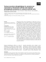
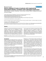
(hypoxia) for 24 hours. Our data show that hypoxia significantly
increased VEGF mRNA, with a median fold increase of 6.37
relative to matched normoxic cultures (P = 0.0001; Figure 1a).
In contrast, PlGF mRNA was significantly downregulated by
exposure to hypoxia. Median levels of PlGF mRNA following
24 hours of hypoxic culture were 48% of those observed
under normoxic conditions, with a decrease observed in 13/
19 RA cultures (P = 0.0207; Figure 1b). This was confirmed
at the protein level, in that hypoxia upregulated release of
VEGF protein (data not shown) but reduced levels of PlGF
protein in 13/15 culture supernatants (median release: nor-
moxia, 0.82 ng/ml; hypoxia, 0.36 ng/ml; P = 0.0006; Figure
1c). Release of PlGF/VEGF heterodimer, however, was
increased by hypoxia (median release: normoxia, 0.13 ng/ml;
hypoxia, 0.20 ng/ml; P = 0.0005; Figure 1d).
We subsequently examined the functional relevance of the
upregulation of proangiogenic factors. A total of six patients
were studied for this purpose. Following centrifugal concen-
tration, proteins were diluted in fresh medium prior to use in an
in vitro angiogenesis assay. To ensure that there was no loss
of protein through centrifugation of the RA cell culture super-
natants, ELISA was carried out on original supernatants, on
reconstituted protein fractions and on the aqueous phase
remaining after the centrifugation. These tests confirmed that
filtration did not significantly affect protein levels of VEGF,
PlGF or VEGF/PlGF (Spearman correlation coefficient for
comparison of levels in protein fractions versus original frac-
tions = 0.974, P < 0.0001; data not shown).
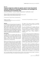
Our data show that normoxic synovial cell culture superna-
tants significantly upregulated angiogenesis in vitro. Addition
of hypoxic synovial cell culture supernatants, however, upreg-
ulated this angiogenic response still further, in comparison
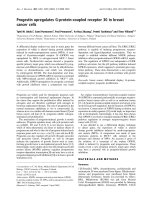
with the normoxic supernatants from the same patient. A typi-
cal experiment is illustrated in Figure 2a, which shows that
VEGF stimulated angiogenesis in vitro by 2.09 ± 0.21-fold
(mean ± standard deviation), compared with 1.68 ± 0.04-fold
and 2.21 ± 0.11-fold for normoxic and hypoxic supernatants,
respectively (both P < 0.001 versus unstimulated cells,
respectively). Furthermore, there was a significant (P < 0.001)
difference in the angiogenic response induced by hypoxic
supernatants relative to normoxic supernatants. These find-
ings were confirmed in synovial cell supernatants from a total
of six RA patients, all of which showed greater angiogenic
activity for hypoxic supernatants relative to normoxic superna-
tants (P = 0.0013; Figure 2b). Representative images show-
ing the morphology of CD31-positive tubule-like structures
obtained in response to either medium alone (Figure 2c),
VEGF (Figure 2d), normoxic RA synovial cell supernatants
(Figure 2e) or hypoxic RA synovial cell supernatants from the
same patient (Figure 2f) are shown.
Hypoxia enhances synovial cell migration through
collagen
In parallel to measuring angiogenic molecules, we examined
the effect of hypoxia on MMP/TIMP. Interestingly, in our exper-
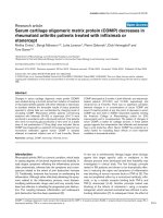
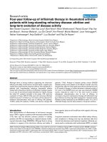
iments using RA synovial membrane cells, MMP-2 mRNA lev-
els were significantly upregulated in response to hypoxia, with
a median 1.75-fold increase (P = 0.006; Figure 3a). The
upregulation of MMP-2 mRNA was observed in 16/19 of RA
patients. Equally, there was an increase in MMP-2 protein.
Median expression of MMP-2 in normoxic conditions was 524
ng/ml (range 122 to 1,259 ng/ml), compared with 1,047 ng/
ml (range 358 to 1,877 ng/ml) under hypoxia. The upregula-
Available online />Page 5 of 11
(page number not for citation purposes)
tion was observed in 10/12 samples (P = 0.0019; Figure 3b).
A similar pattern was observed for MMP-9, with mRNA levels
upregulated by hypoxia in 11/16 samples by a median of 1.65-
fold (P = 0.0362; Figure 3c). MMP-9 protein expression in
normoxia was in the range 31 to 82 ng/ml (median 57 ng/ml),
compared with 32 to 127 ng/ml (median 69 ng/ml) under
hypoxic conditions (P = 0.0328; Figure 3d).
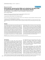
In terms of collagenase expression, MMP-8 levels were signif-
icantly upregulated by hypoxia. There was a 3.09-fold upregu-
lation of MMP-8 mRNA, which was observed in 9/10 patients
(P = 0.0039; Figure 4a). There was also an increase in MMP-
8 protein levels. MMP-8 expression under normoxic conditions
ranged from 0.14 to 2.38 ng/ml (median 0.52 ng/ml). For the
same samples cultured under hypoxic conditions, MMP-8 lev-
els varied from 0.17 to 2.80 ng/ml (median 1.00 ng/ml, P =
0.0266; Figure 4b). In contrast, when cultured under hypoxic
conditions, RA synovial cells exhibited significant downregula-
tion of MMP-13 mRNA (P = 0.0012; Figure 4c) and protein (P
= 0.0024; Figure 4d). For example, median MMP-13 protein
levels for normoxic cultures were 2.29 ng/ml, compared with
0.87 ng/ml under hypoxia. Overall, 12/14 patients showed
decreased MMP-13 mRNA levels, and 10/12 patients showed
decreased MMP-13 protein levels when cultured under
hypoxic conditions. Levels of another collagenase, MMP-1,
were unchanged (Table 2). Furthermore, in our study, MT1-
MMP mRNA was upregulated significantly by hypoxia (P <
0.01; Table 2), although the increase was relatively modest
(1.38-fold, range 0.11-fold to 1.99-fold). Finally, mRNA levels
of MMP-3, TIMP-1 and TIMP-2 did not change in response to
hypoxia (Table 2).
To study the potential effect of these changes in MMP/TIMP
on synovial invasiveness, we utilised a model in which fibrob-
lasts are cultured on a collagen matrix. We confirmed that
fibroblasts also upregulate MMP-2 (P = 0.0184 by paired t
test) and MT1-MMP (P = 0.0049 by paired t test; data not
shown). Interestingly, levels of MMP-8 also appeared unaf-
fected by hypoxia, in contrast to the RA synovial membrane
cells. To study cell migration, RA fibroblasts were placed in
wells coated with type-I collagen and were exposed to either
normoxia or hypoxia. We found a significant enhancement of
Figure 1
Hypoxia differentially modulates the angiogenic balanceHypoxia differentially modulates the angiogenic balance. Rheumatoid arthritis synovial cells were exposed to either 21% oxygen (normoxia) or 1%
oxygen (hypoxia) for 24 hours. mRNA levels of (a) vascular endothelial growth factor (VEGF) and (b) placental growth factor (PlGF) were measured
by quantitative PCR (n = 19). The change in threshold cycle (ΔCt) values was calculated for each mRNA using the 2-ΔΔ
Ct
method against the
housekeeping gene acidic ribosomal protein. The fold changes in mRNA levels were related to a reference sample (human cDNA). In parallel,
release of (c) PlGF and (d) VEGF/PlGF heterodimer was measured by ELISA of cell culture supernatants (n = 13 to 15). Data were analysed versus
normoxia by Wilcoxon signed rank test: *P < 0.05, ***P < 0.001.
Arthritis Research & Therapy Vol 11 No 3 Akhavani et al.
Page 6 of 11
(page number not for citation purposes)
cell migration through collagen under hypoxic conditions, with
a median increase in absorbance at 540 nm equivalent to
+43% (range +25% to +127%, P < 0.001 versus normoxia;
Figure 5). This increase in migration was observed for all six
patients used, and was evident when the stained cells were
studied under the microscope. Interestingly, cell migration was
significantly reduced when GM6001, a universal MMP inhibi-
tor, was used. This reduction was observed for both the nor-
moxic conditions (median levels 59% relative to response in
the absence of GM6001, P < 0.01) and hypoxic cell culture
conditions (median levels 58% relative to response in the
absence of GM6001, P < 0.01). Even in the presence of
GM6001, cells incubated under hypoxic conditions exhibited
significantly (P < 0.001) increased migration when compared
with the normoxic GM6001-blocked counterparts (Figure 5).
Figure 2
Rheumatoid arthritis synovial cells exposed to hypoxia express more proangiogenic activityRheumatoid arthritis synovial cells exposed to hypoxia express more proangiogenic activity. Rheumatoid arthritis (RA) synovial cells were exposed to
either 21% oxygen (normoxia) or 1% oxygen (hypoxia) for 24 hours. Cell supernatants were filtered, and the protein fraction was resuspended in
fresh medium. Angiogenesis in response to RA synovial cell supernatants was assessed after 11 days, using CD31 expression quantified by colori-
metric assay. (a) Representative data, with cells exposed to either vascular endothelial growth factor (VEGF) (2 ng/ml), RA synovial cell superna-
tants or no stimulus. Data are means of triplicate determinations, and were analysed by one-way analysis of variance: ***P < 0.001. (b) Comparison
of angiogenesis in response to normoxic and hypoxic RA synovial cell supernatants. Data are means of paired triplicate determinations for six sepa-
rate patients, and were analysed by paired t-test: **P < 0.01. (c) to (f) Representative images showing morphology of the formed tubes stained for
CD31 at day 11 (objective magnification, ×40): (c) untreated, (d) VEGF treated (2 ng/ml), (e) normoxic RA synovial cell supernatants and (f) hypoxic
RA synovial cell supernatants from the same patient.
Available online />Page 7 of 11
(page number not for citation purposes)
Discussion
RA primarily affects the synovial lining, resulting in destructive
changes to the joints and soft tissues, most commonly in the
hand and wrist. Approximately 50% of patients with RA have
tendon involvement, and tenosynovial proliferation can result in
tendon adhesions, scarring or rupture [28,29]. The mecha-
nism by which the tenosynovial lining causes tendon damage
is poorly understood, but is thought to involve alterations in the
MMP/TIMP balance. MMPs play a key role in the degradation
of extracellular matrix, as well as in intercellular communica-
tion, cell migration, tumour progression and angiogenesis
[8,30,31]. Jain and colleagues reported that RA tenosynovial
cells produce greater amounts of collagenase (MMP-1, MMP-
8 and MMP-13) and gelatinase (MMP-2) enzymes compared
with noninvasive tenosynovium [21,22]. Furthermore, our
group reported that invasive tenosynovium is more hypoxic
than noninvasive RA tenosynovium, or indeed than normal ten-
osynovium [12]. Intriguingly, hypoxia has been shown to
upregulate gelatinases (MMP-2 and MMP-9) [14,15], and to
enhance cancer cell invasiveness in vitro [16]. MMP-1 and
MMP-3 mRNA levels have been reported to be upregulated
under hypoxic conditions in RA fibroblasts [32]. Additionally,
MMP-13 has been identified as a hypoxia-induced gene in car-
cinoma cells [33].
The objective of our study was therefore to examine the poten-
tial in vivo consequences of hypoxia in RA in terms of synovial
invasion, by exposing RA synovial membrane cells to 1% oxy-
gen. In our study, hypoxia upregulated MMP-2, MMP-8 and
MMP-9, while significantly downregulating MMP-13, at both
mRNA and protein levels. This last finding was in keeping with
previously reported work, which showed a modest (although
not statistically significant) reduction in MMP-13 [23]. We
observed no effect of hypoxia on MMP-1 levels, in agreement
with published data [23], and no effect on MMP-3, TIMP-1,
and TIMP-2. In contrast, MT1-MMP was upregulated at the
mRNA level. While there have been previous reports of
increased MMP expression/activity in RA fibroblasts exposed
to hypoxia, particularly for MMP-1 and MMP-3 [32,34,35], our
studies have attempted to mimic RA synovial membrane milieu
by utilising total RA synovial membrane cells, which include
macrophages as well as fibroblasts, thus possibly explaining
the differences between these published data and our own.
Figure 3
Hypoxia modulates gelatinase expression by rheumatoid arthritis synovial cellsHypoxia modulates gelatinase expression by rheumatoid arthritis synovial cells. Rheumatoid arthritis synovial cells were exposed to either 21% oxy-
gen (normoxia) or 1% oxygen (hypoxia) for 24 hours. mRNA levels of (a) matrix metalloproteinase (MMP)-2 and (c) MMP-9 were measured by quan-
titative PCR (n = 16 to 18). The change in threshold cycle (ΔCt) values was calculated for each mRNA using the 2-ΔΔ
Ct
method against the
housekeeping gene acidic ribosomal protein. The fold changes in mRNA levels were related to a reference sample (human cDNA). In parallel,
release of (b) MMP-2 and (d) MMP-9 protein was measured by ELISA of cell culture supernatants (n = 12). Data were analysed versus normoxia by
Wilcoxon signed rank test (b, c) or paired t test (a, b) as appropriate: *P < 0.05, **P < 0.01.
Arthritis Research & Therapy Vol 11 No 3 Akhavani et al.
Page 8 of 11
(page number not for citation purposes)
To confirm the functional significance of these findings, we
investigated the effect of hypoxia on RA synovial cell migration,
and observed that hypoxia significantly increased migration of
RA fibroblasts through type-I collagen. To assess whether this
migration was dependent on MMP activity, we utilised the
MMP inhibitor GM6001, which inhibits MMP-1, MMP-2, MMP-
3, MMP-8 and MMP-9 [36]. Although migration was signifi-
cantly reduced by the use of GM6001 in both the normoxic
and hypoxic cell cultures, there were still a greater number of
cells migrating in the hypoxic cultures, when compared with
their normoxic counterparts. This suggests that cell migration
through collagen under hypoxic conditions may in part involve
MT1-MMP, which is not blocked by GM6001, as well as
GM6001-sensitive MMP. Significantly, there is emerging evi-
Figure 4
Hypoxia modulates collagenase expression by rheumatoid arthritis synovial cellsHypoxia modulates collagenase expression by rheumatoid arthritis synovial cells. Rheumatoid arthritis synovial cells were exposed to either 21% oxy-
gen (normoxia) or 1% oxygen (hypoxia) for 24 hours. mRNA levels of (a) matrix metalloproteinase (MMP)-8 and (c) MMP-13 were measured by
quantitative PCR (n = 10 to 14). The change in threshold cycle (ΔCt) values was calculated for each mRNA using the 2-ΔΔ
Ct
method against the
housekeeping gene acidic ribosomal protein. The fold changes in mRNA levels were related to a reference sample (human cDNA). In parallel,
release of (b) MMP-8 and (d) MMP-13 protein was measured by ELISA of cell culture supernatants (n = 12 to 13). Data were analysed versus nor-
moxia by Wilcoxon signed rank test: *P < 0.05, **P < 0.01.
Table 2
Effect of hypoxia on MMP/TIMP mRNA
MMP/TIMP Normoxia Hypoxia P value
MMP-1 (n = 19) 0.88 (0.23 to 3.58) 1.11 (0.14 to 6.77) 0.475
MMP-3 (n = 16) 0.38 (0.04 to 1.48) 0.21 (0.01 to 1.09) 0.224
MT1-MMP (n = 19) 1.71 (0.03 to 9.13) 2.45 (0.15 to 10.78) 0.009
TIMP-1 (n = 19) 1.31 (0.03 to 18.90) 2.68 (0.01 to 26.08) 0.131
TIMP-2 (n = 19) 2.57 (0.12 to 20.97) 2.20 (0.24 to 18.64) 0.457
Data presented as the median (range) and were analysed by Wilcoxon signed rank test. Rheumatoid arthritis synovial cells were exposed to either
21% oxygen (normoxia) or 1% oxygen (hypoxia) for 24 hours. mRNA levels of matrix metalloproteinase (MMP)-1, MMP-3, MT1-MMP, tissue
inhibitor of matrix metalloproteinase (TIMP)-1 and TIMP-2 were measured by quantitative PCR. The change in threshold cycle (ΔCt) values was
calculated using the 2-ΔΔ
Ct
method against the housekeeping gene acidic ribosomal protein. The fold changes in mRNA levels were related to a
reference sample (human cDNA).
Available online />Page 9 of 11
(page number not for citation purposes)
dence that MT1-MMP is regulated by HIF-2α [17,18]. We and
other workers have shown the presence of HIF-1 and HIF-2 in
RA synovium [12,37,38], reinforcing the concept that the RA
hypoxic milieu may promote alterations in MMP.
As well as affecting matrix degradation, hypoxia is likely to pro-
foundly modulate synovial angiogenesis, through the regula-
tion of angiogenic stimulators such as VEGF [3,4,7]. Co-
localisation of HIF-2α and VEGF emphasises the role of
hypoxia in the upregulation of angiogenesis in tenosynovitis
[12]. We therefore examined the effects of hypoxia on proan-
giogenic molecules – namely, VEGF, PlGF and VEGF/PlGF
heterodimer.
VEGF was used as the control for the present study, as previ-
ous studies from our laboratory have consistently shown
VEGF protein upregulation in RA synovial cultures under
hypoxic conditions [12,23]. In our study, when RA synovial
cells were cultured at 1% oxygen, these showed upregulation
of VEGF mRNA and protein levels in all samples. Upregulation
of VEGF mRNA under hypoxic conditions has been reported
previously for temporomandibular joint synoviocytes [39], but
we believe this is the first time that VEGF mRNA upregulation
in RA metacarpophalangeal synovial and tenosynovial cell cul-
tures has been reported. Furthermore, expression of both
PlGF mRNA and PlGF protein was significantly downregu-
lated. Interestingly, PlGF-deficient mice do not display major
vascular abnormalities, unlike mice lacking VEGF, suggesting
that this molecule is not essential during physiological angio-
genesis [40]. PlGF homodimers and PlGF/VEGF heterodim-
ers are present in the synovial fluid of patients with
inflammatory arthropathies, including RA [41]. PlGF has previ-
ously been reported to be induced by hypoxia in fibroblasts
[42]. In contrast, our data showed that PlGF was downregu-
lated by hypoxia, suggesting that hypoxia is probably not the
only regulator of PlGF expression in RA.
In cells co-expressing VEGF and PlGF mRNA, VEGF/PlGF
heterodimer protein is also expressed, and some of the biolog-
ical activities attributed to VEGF homodimers may be medi-
ated by VEGF/PlGF heterodimers [43]. Interestingly, our data
show that expression of VEGF/PlGF heterodimer in response
to hypoxia follows a similar trend to that of VEGF.
We have shown that certain proangiogenic factors are upreg-
ulated in RA synovial cell cultures under hypoxic conditions. To
investigate whether this translates to an effect on angiogen-
esis, we used supernatants of the cell cultures from which the
mRNA and protein data were obtained, and applied these in
an in vitro angiogenesis assay. Using this approach, we were
able to demonstrate for the first time that hypoxic RA synovial
cell cultures induced significantly more vessel outgrowth than
their normoxic counterparts, supporting our hypothesis that
hypoxia is likely to promote synovial angiogenesis.
Conclusions
In the present study we have demonstrated that RA synovial
cells cultured under hypoxic conditions show upregulation of
proangiogenic factors. Crucially, our data also show that
hypoxia increases the angiogenic drive of RA cells. Our data
additionally provide evidence that certain MMPs are upregu-
lated by hypoxia in RA synovial cells, and that this effect is
accompanied by an enhanced capacity of RA cells to migrate
through collagen.
Taken together, these data suggest that the hypoxic RA envi-
ronment promotes and upregulates angiogenesis in inflamed
RA synovium, making it proangiogenic and proinvasive. In the
context of RA, it seems likely that a disturbance in the balance
between MMP/TIMP brought about by tissue hypoxia may
determine whether the tenosynovium invades tendons, bone
or cartilage.
Competing interests
The authors declare that they have no competing interests.
Figure 5
Cell migration through type-I collagen is enhanced under hypoxic con-ditions: effect of matrix metalloproteinase inhibitionCell migration through type-I collagen is enhanced under hypoxic con-
ditions: effect of matrix metalloproteinase inhibition. Rheumatoid arthri-
tis fibroblast migration was investigated under normoxic (21% oxygen)
and hypoxic (1% oxygen) conditions over a 48-hour culture period. The
top chamber was coated with porcine type-I collagen, followed by the
addition of 20,000 fibroblasts per well. The chambers were subse-
quently placed in a 24-well culture plate, with the lower chambers con-
taining DMEM plus 10% FCS. A universal MMP inhibitor (GM6001; 10
μM) was added to both the bottom and top chambers of selected
inserts, before exposure to either normoxia or hypoxia. Migration from
six different patients, assayed in triplicate, is shown. Data presented as
the mean ± standard error of the mean and were analysed by repeated-
measures one-way analysis of variance, with Bonferroni's post hoc test
for multiple comparisons: **P < 0.01, ***P < 0.001.
Arthritis Research & Therapy Vol 11 No 3 Akhavani et al.
Page 10 of 11
(page number not for citation purposes)
Authors' contributions
MAA, NK and EMP designed the study. MAA, LM, IB and BS
carried out all of the experiments. NK and EMP oversaw the
project running and data analysis, and drafted the manuscript.
All authors read and approved the final manuscript.
Acknowledgements
The authors are grateful for the support of the Restoration of Appear-
ance and Function Trust (MAA and BS), the Royal College of Surgeons
of England (MAA and BS) and The Dunhill Medical Trust (MAA). IB is
supported by the IWT, Belgium. The Kennedy Institute of Rheumatology
is supported by the Arthritis Research Campaign of Great Britain. The
authors gratefully acknowledge the advice of Dr Yoshifumi Itoh
(Kennedy Institute of Rheumatology). We are grateful for support from
the NIHR Biomedical Research Centre funding scheme.
References
1. Fearon U, Griosios K, Fraser A, Reece R, Emery P, Jones PF, Veale
DJ: Angiopoietins, growth factors, and vascular morphology in
early arthritis. J Rheumatol 2003, 30:260-268.
2. Szekanecz Z, Koch AE: Mechanisms of disease: angiogenesis
in inflammatory diseases. Nat Clin Pract Rheumatol 2007,
3:635-643.
3. Larsen H, Akhavani MA, Raatz Y, Paleolog EM: Gene expression
studies to investigate disease mechanisms in rheumatoid
arthritis: does angiogenesis play a role? Curr Rheumatol Rev
2007, 3:243-251.
4. Khong TL, Larsen H, Raatz Y, Paleolog E: Angiogenesis as a
therapeutic target in arthritis: learning the lessons of the color-
ectal cancer experience. Angiogenesis 2007, 10:243-258.
5. Ceponis A, Konttinen YT, Imai S, Tamulaitiene M, Li TF, Xu JW,
Hietanen J, Santavirta S, Fassbender HG: Synovial lining,
endothelial and inflammatory mononuclear cell proliferation in
synovial membranes in psoriatic and reactive arthritis: a com-
parative quantitative morphometric study. Br J Rheumatol
1998, 37:170-178.
6. Walsh DA, Wade M, Mapp PI, Blake DR: Focally regulated
endothelial proliferation and cell death in human synovium.
Am J Pathol 1998, 152:691-702.
7. Sivakumar B, Harry LE, Paleolog EM: Modulating angiogenesis:
more vs less. JAMA 2004, 292:972-977.
8. Murphy G, Nagase H: Reappraising metalloproteinases in
rheumatoid arthritis and osteoarthritis: destruction or repair?
Nat Clin Pract Rheumatol 2008, 4:128-135.
9. Lund-Olesen K: Oxygen tension in synovial fluids. Arthritis
Rheum 1970, 13:769-776.
10. Treuhaft PS, McCarty DJ: Synovial fluid pH, lactate, oxygen and
carbon dioxide partial pressure in various joint diseases.
Arthritis Rheum 1971, 14:475-484.
11. Ahlqvist J: A hypothesis on the pathogenesis of rheumatoid
and other non-specific synovitides. IV A. The possible interme-
diate role of local hypoxia and metabolic alterations. Med
Hypotheses 1984, 13:257-302.
12. Sivakumar B, Akhavani MA, Winlove CP, Taylor PC, Paleolog EM,
Kang N: Synovial hypoxia as a cause of tendon rupture in rheu-
matoid arthritis. J Hand Surg (Am)
2008, 33:49-58.
13. Williamson SC, Feldon P: Extensor tendon ruptures in rheuma-
toid arthritis. Hand Clin 1995, 11:449-459.
14. Ben-Yosef Y, Lahat N, Shapiro S, Bitterman H, Miller A: Regula-
tion of endothelial matrix metalloproteinase-2 by hypoxia/
reoxygenation. Circ Res 2002, 90:784-791.
15. Canning MT, Postovit LM, Clarke SH, Graham CH: Oxygen-medi-
ated regulation of gelatinase and tissue inhibitor of metallo-
proteinases-1 expression by invasive cells. Exp Cell Res 2001,
267:88-94.
16. Ridgway PF, Ziprin P, Alkhamesi N, Paraskeva PA, Peck DH, Darzi
AW: Hypoxia augments gelatinase activity in a variety of ade-
nocarcinomas in vitro. J Surg Res 2005, 124:180-186.
17. Koochekpour S, Jeffers M, Wang PH, Gong C, Taylor GA,
Roessler LM, Stearman R, Vasselli JR, Stetler-Stevenson WG,
Kaelin WG Jr, Linehan WM, Klausner RD, Gnarra JR, Woude GF
Vande: The von Hippel – Lindau tumor suppressor gene inhib-
its hepatocyte growth factor/scatter factor-induced invasion
and branching morphogenesis in renal carcinoma cells. Mol
Cell Biol 1999, 19:5902-5912.
18. Petrella BL, Lohi J, Brinckerhoff CE: Identification of membrane
type-1 matrix metalloproteinase as a target of hypoxia-induci-
ble factor-2 alpha in von Hippel – Lindau renal cell carcinoma.
Oncogene 2005, 24:1043-1052.
19. Kondo S, Kubota S, Shimo T, Nishida T, Yosimichi G, Eguchi T,
Sugahara T, Takigawa M: Connective tissue growth factor
increased by hypoxia may initiate angiogenesis in collabora-
tion with matrix metalloproteinases. Carcinogenesis 2002,
23:769-776.
20. Munoz-Najar UM, Neurath KM, Vumbaca F, Claffey KP: Hypoxia
stimulates breast carcinoma cell invasion through MT1-MMP
and MMP-2 activation. Oncogene 2006, 25:2379-2392.
21. Jain A, Nanchahal J, Troeberg L, Green P, Brennan F: Production
of cytokines, vascular endothelial growth factor, matrix metal-
loproteinases, and tissue inhibitor of metalloproteinases 1 by
tenosynovium demonstrates its potential for tendon destruc-
tion in rheumatoid arthritis. Arthritis Rheum 2001,
44:1754-1760.
22. Jain A, Brennan F, Troeberg L, Nanchahal J: The role of matrix
metalloproteinases in rheumatoid tendon disease. J Hand
Surg (Am) 2002,
27:1059-1064.
23. Jain A, Kiriakidis S, Brennan F, Sandison A, Paleolog E, Nanchahal
J: Targeting rheumatoid tenosynovial angiogenesis with
cytokine inhibitors. Clin Orthop Relat Res 2006, 446:268-277.
24. Arnett FC, Edworthy SM, Bloch DA, McShane DJ, Fries JF, Cooper
NS, Healey LA, Kaplan SR, Liang MH, Medsger TA, Mitchell DM,
Neustadt DH, Pinals RS, Schaller JG, Sharp JT, Wilder RL, Hunder
GC: The American Rheumatism Association 1987 revised cri-
teria for the classification of rheumatoid arthritis. Arthritis
Rheum 1988, 31:315-324.
25. Brennan FM, Chantry D, Jackson A, Maini R, Feldmann M: Inhibi-
tory effect of TNF alpha antibodies on synovial cell interleukin-
1 production in rheumatoid arthritis. Lancet 1989, 2:244-247.
26. UCSC Genome Bioinformatics [ />]
27. Paleolog EM, Young S, Stark AC, McCloskey RV, Feldmann M,
Maini RN: Modulation of angiogenic vascular endothelial
growth factor by tumor necrosis factor alpha and interleukin-1
in rheumatoid arthritis. Arthritis Rheum 1998, 41:1258-1265.
28. Brown FE, Brown ML: Long-term results after tenosynovec-
tomy to treat the rheumatoid hand. J Hand Surg (Am) 1988,
13:704-708.
29. Ferlic DC: Rheumatoid flexor tenosynovitis and rupture. Hand
Clin 1996, 12:561-572.
30. Nagase H, Visse R, Murphy G: Structure and function of matrix
metalloproteinases and TIMPs. Cardiovasc Res 2006,
69:562-573.
31. Visse R, Nagase H: Matrix metalloproteinases and tissue inhib-
itors of metalloproteinases: structure, function, and biochem-
istry. Circ Res 2003, 92:827-839.
32. Cha HS, Ahn KS, Jeon CH, Kim J, Song YW, Koh EM: Influence
of hypoxia on the expression of matrix metalloproteinase-1, -
3 and tissue inhibitor of metalloproteinase-1 in rheumatoid
synovial fibroblasts. Clin Exp Rheumatol 2003, 21:593-598.
33. Koong AC, Denko NC, Hudson KM, Schindler C, Swiersz L, Koch
C, Evans S, Ibrahim H, Le QT, Terris DJ, Giaccia AJ: Candidate
genes for the hypoxic tumor phenotype. Cancer Res 2000,
60:883-887.
34. Demasi M, Cleland LG, Cook-Johnson RJ, James MJ: Effects of
hypoxia on the expression and activity of cyclooxygenase 2 in
fibroblast-like synoviocytes: interactions with monocyte-
derived soluble mediators. Arthritis Rheum 2004,
50:2441-2449.
35. Ahn JK, Koh EM, Cha HS, Lee YS, Kim J, Bae EK, Ahn KS: Role of
hypoxia-inducible factor-1α in hypoxia-induced expressions
of IL-8, MMP-1 and MMP-3 in rheumatoid fibroblast-like syn-
oviocytes. Rheumatology (Oxford) 2008, 47:834-839.
36. Galardy RE, Grobelny D, Foellmer HG, Fernandez LA: Inhibition
of angiogenesis by the matrix metalloprotease inhibitor N-
[2R-2-(hydroxamidocarbonymethyl)-4-methylpentanoyl)]-
L-
tryptophan methylamide. Cancer Res 1994, 54:4715-4718.
37. Giatromanolaki A, Sivridis E, Maltezos E, Athanassou N, Papa-
zoglou D, Gatter KC, Harris AL, Koukourakis MI: Upregulated
hypoxia inducible factor-1α and -2α pathway in rheumatoid
Available online />Page 11 of 11
(page number not for citation purposes)
arthritis and osteoarthritis. Arthritis Res Ther 2003,
5:R193-R201.
38. Hollander AP, Corke KP, Freemont AJ, Lewis CE: Expression of
hypoxia-inducible factor 1α by macrophages in the rheuma-
toid synovium: implications for targeting of therapeutic genes
to the inflamed joint. Arthritis Rheum 2001, 44:1540-1544.
39. Ke J, Liu Y, Long X, Li J, Fang W, Meng Q, Zhang Y: Up-regulation
of vascular endothelial growth factor in synovial fibroblasts
from human temporomandibular joint by hypoxia. J Oral
Pathol Med 2007, 36:290-296.
40. Carmeliet P, Moons L, Luttun A, Vincenti V, Compernolle V, De Mol
M, Wu Y, Bono F, Devy L, Beck H, Scholz D, Acker T, DiPalma T,
Dewerchin M, Noel A, Stalmans I, Barra A, Blacher S, Vanden-
driessche T, Ponten A, Eriksson U, Plate KH, Foidart JM, Schaper
W, Charnock-Jones DS, Hicklin DJ, Herbert JM, Collen D, Persico
MG: Synergism between vascular endothelial growth factor
and placental growth factor contributes to angiogenesis and
plasma extravasation in pathological conditions. Nat Med
2001, 7:575-583.
41. Bottomley MJ, Webb NJ, Watson CJ, Holt L, Bukhari M, Denton J,
Freemont AJ, Brenchley PE: Placenta growth factor (PlGF)
induces vascular endothelial growth factor (VEGF) secretion
from mononuclear cells and is co-expressed with VEGF in syn-
ovial fluid. Clin Exp Immunol 2000, 119:182-188.
42. Green CJ, Lichtlen P, Huynh NT, Yanovsky M, Laderoute KR,
Schaffner W, Murphy BJ: Placenta growth factor gene expres-
sion is induced by hypoxia in fibroblasts: a central role for
metal transcription factor-1. Cancer Res 2001, 61:2696-2703.
43. DiSalvo J, Bayne ML, Conn G, Kwok PW, Trivedi PG, Soderman
DD, Palisi TM, Sullivan KA, Thomas KA: Purification and charac-
terization of a naturally occurring vascular endothelial growth
factor.placenta growth factor heterodimer. J Biol Chem 1995,
270:7717-7723.