Báo cáo y học: "Green tea polyphenol epigallocatechin-3-gallate inhibits advanced glycation end product-induced expression of tumor necrosis factor-α and matrix metalloproteinase-13 in human chondrocyte" ppsx
Bạn đang xem bản rút gọn của tài liệu. Xem và tải ngay bản đầy đủ của tài liệu tại đây (1.07 MB, 13 trang )
Open Access
Available online />Page 1 of 13
(page number not for citation purposes)
Vol 11 No 3
Research article
Green tea polyphenol epigallocatechin-3-gallate inhibits
advanced glycation end product-induced expression of tumor
necrosis factor-α and matrix metalloproteinase-13 in human
chondrocytes
Zafar Rasheed
1
, Arivarasu N Anbazhagan
1
, Nahid Akhtar
1
, Sangeetha Ramamurthy
1
,
Frank R Voss
2
and Tariq M Haqqi
1
1
Department of Pathology, Microbiology, & Immunology, School of Medicine, University of South Carolina, 6439 Garners Ferry Rd, Columbia, SC
29209, USA
2
Department of Orthopaedics, University of South Carolina, School of Medicine/Palmetto Richland Hospital, Two Medical Park, Columbia, SC 29203,
USA
Corresponding author: Tariq M Haqqi,
Received: 4 Feb 2009 Revisions requested: 2 Mar 2009 Revisions received: 29 Apr 2009 Accepted: 15 May 2009 Published: 15 May 2009
Arthritis Research & Therapy 2009, 11:R71 (doi:10.1186/ar2700)
This article is online at: />© 2009 Rasheed et al.; licensee BioMed Central Ltd.
This is an open access article distributed under the terms of the Creative Commons Attribution License ( />),
which permits unrestricted use, distribution, and reproduction in any medium, provided the original work is properly cited.
Abstract
Introduction The major risk factor for osteoarthritis (OA) is
aging, but the mechanisms underlying this risk are only partly
understood. Age-related accumulation of advanced glycation
end products (AGEs) can activate chondrocytes and induce the
production of proinflammatory cytokines and matrix
metalloproteinases (MMPs). In the present study, we examined
the effect of epigallocatechin-3-gallate (EGCG) on AGE-
modified-BSA (AGE-BSA)-induced activation and production of
TNFα and MMP-13 in human OA chondrocytes.
Methods Human chondrocytes were derived from OA cartilage
by enzymatic digestion and stimulated with in vitro-generated
AGE-BSA. Gene expression of TNFα and MMP-13 was
measured by quantitative RT-PCR. TNFα protein in culture
medium was determined using cytokine-specific ELISA.
Western immunoblotting was used to analyze the MMP-13
production in the culture medium, phosphorylation of mitogen-
activated protein kinases (MAPKs), and the activation of NF-κB.
DNA binding activity of NF-κB p65 was determined using a
highly sensitive and specific ELISA. IκB kinase (IKK) activity was
determined using an in vitro kinase activity assay. MMP-13
activity in the culture medium was assayed by gelatin
zymography.
Results EGCG significantly decreased AGE-stimulated gene
expression and production of TNFα and MMP-13 in human
chondrocytes. The inhibitory effect of EGCG on the AGE-BSA-
induced expression of TNFα and MMP-13 was mediated at least
in part via suppression of p38-MAPK and JNK activation. In
addition, EGCG inhibited the phosphorylating activity of IKKβ
kinase in an in vitro activity assay and EGCG inhibited the AGE-
mediated activation and DNA binding activity of NF-κB by
suppressing the degradation of its inhibitory protein IκBα in the
cytoplasm.
Conclusions These novel pharmacological actions of EGCG on
AGE-BSA-stimulated human OA chondrocytes provide new
suggestions that EGCG or EGCG-derived compounds may
inhibit cartilage degradation by suppressing AGE-mediated
activation and the catabolic response in human chondrocytes.
AGE: advanced glycation end product; bp: base pairs; BSA: bovine serum albumin; EGCG: epigallocatechin-3-gallate; ELISA: enzyme-linked immu-
nosorbent assay; FCS: fetal calf serum; H & E: hematoxylin and eosin; IKK: IκB kinase; IL: interleukin; MAPK: mitogen-activated protein kinase; MMP:
matrix metalloproteinase; NF: nuclear factor; OA: osteoarthritis; PBS: phosphate-buffered saline; PCR: polymerase chain reaction; RAGE: receptor
for advanced glycation end products; RT: reverse transcriptase; TNF: tumor necrosis factor.
Arthritis Research & Therapy Vol 11 No 3 Rasheed et al.
Page 2 of 13
(page number not for citation purposes)
Introduction
Osteoarthritis (OA), the most common form of arthritis, is a
progressive degenerative joint disease that has a major impact
on joint function and the patient's quality of life [1,2]. Many risk
factors that contribute to disease onset have been identified,
including systemic factors such as genetics, estrogen use,
and bone density, and local biomechanical factors such as
muscle weakness, obesity, and joint laxity [1]. The most impor-
tant risk factor for OA besides female sex, obesity, and joint
trauma is aging [1,2]. How aging contributes to the onset and
progression of OA, however, is relatively unknown.
A prominent feature of aging is the modification of proteins by
nonenzymatic glycation. Nonenzymatic glycation is a common
post-translational modification of proteins caused by reducing
sugars. The spontaneous condensation of reducing sugars
with free amino groups in lysine or arginine residues on pro-
teins leads to the formation of a reversible Schiff base, which
is subsequently stabilized by Amadori rearrangement. The
Maillard or browning reaction then converts the initially formed
intermediate products into advanced glycation end products
(AGEs) [3]. In addition to this classical pathway of AGE forma-
tion, it has recently been found that AGE formation can be ini-
tiated by metal-catalyzed glucose autooxidation as well as by
lipid peroxidation (thereby providing an interesting link
between lipid metabolism and the development of OA).
This diversity in reaction pathways results in a variety of chem-
ical structures of AGEs. Some AGEs are adducts to proteins,
while many others present protein–protein crosslinks. Once
AGEs are formed, they cannot be removed from the protein;
they only leave a tissue when the protein involved is degraded.
Articular cartilage collagen has an exceptionally long half-life,
and, since the rate of AGE accumulation is largely determined
by the rate of protein turnover [4], this low turnover of cartilage
constituents results in an abundant accumulation of AGEs in
articular cartilage [5,6]. The accumulation of AGEs in cartilage
leads to inferior mechanical properties [5,7] and to an altera-
tion in cartilage metabolism [4,8]. More specifically, cartilage
stiffness increases substantially with increasing AGE levels,
and matrix synthesis by articular chondrocytes becomes
impaired [5,7,9]. Accumulation of AGEs, however, is a pro-
posed mechanism for the age-related development of OA
[3,10]. Some studies also showed that still-healthy cartilage of
patients with a focal degenerative cartilage lesion elsewhere in
the joint has higher AGE levels than healthy cartilage from con-
trol individuals in which there are no signs of OA [11]. The age-
related accumulation of AGE crosslinks presents a putative
molecular mechanism whereby age contributes to the risk of
developing OA. The accumulation of AGEs, however, is not
only age related. AGE levels tend to be increased in diabetic
patients, since the hyperglycemia accelerates AGE formation
[12]. The correlation between diabetes mellitus and OA is
supported by some older findings showing that radiographic
OA is more common, more severe, and present earlier in
patients with diabetes [13,14]. In addition, reports from more
recent times still show a trend toward correlation of OA with
diabetes [15]. OA therefore correlates with both aging and
diabetes. In both aging and diabetes, AGE levels are
increased. The levels of AGEs might therefore predict suscep-
tibility to OA.
In vivo effects of AGEs on cartilage integrity have been
reported in recent studies in beagle dogs and a canine model
of OA induced experimentally by anterior cruciate ligament
transection. Animals with elevated AGE levels had more
severe OA than did those with normal AGE levels [10]. The
mechanism by which AGEs influence cellular function in artic-
ular cartilage is poorly understood. The receptor for AGE
(RAGE) is expressed in articular chondrocytes and synovial
tissue macrophages of individuals with arthritis [16,17]. Acti-
vation of RAGE by multiple ligands including S100 protein,
high-mobility group box chromosomal protein 1 and AGEs
(complex and specific AGEs) in OA chondrocytes and synovi-
ocytes results in increased production of various inflammatory
mediators including TNFα and matrix metalloproteinase
(MMP)-13 [18-20]. Previous studies have used complexes
generated from BSA or a specific AGE, usually pentosidine or
N3-carboxymethyllysine, to stimulate OA chondrocytes [21-
23]. The AGEs used in the current study were produced by
reacting endotoxin-free BSA with glycolaldehyde. The result-
ing AGE-BSA is a complex that includes N3-carboxymethylly-
sine, pentosidine, and other AGEs [24]. The results of the
present study were therefore obtained with a complex of
AGEs rather than with a particular AGE. MMPs, a large family
of structurally related calcium-dependent and zinc-dependent
proteolytic enzymes, are involved in the degradation of many
different components of the extracellular matrix [17,25]. Both
TNFα and MMPs are expressed in a number of different cell
types and play a key role in diverse cellular processes, ranging
from morphogenesis to tumor invasion to tissue remodeling
[25,26]. Among the MMPs, MMP-13 (collagenase 3) is con-
sidered of particular interest due to its ability to digest type-2
collagen.
Green tea (Camellia sinensis), a popular beverage worldwide,
has been shown to exert antimutagenic, antiproliferative, and
anticarcinogenic effects, as well as anti-inflammatory activity in
models of degenerative disorders [27-29]. We have earlier
shown that green tea polyphenols inhibit the development of
inflammatory arthritis in a mouse model [30] and that epigallo-
catechin-3-gallate (EGCG), the bioactive constituent of green
tea, was nontoxic to human chondrocytes and inhibited the
expression of inflammatory mediators in vitro [31-34]. The
beneficial effects ascribed to drinking tea are believed to rely
on the pharmacological actions of catechins, especially
EGCG, and the derivatives of catechin components [35]. The
effects of EGCG on AGE-induced damage in arthritis, how-
ever, remain to be elucidated. Since high levels of TNFα
and
MMPs are expressed and produced by AGE-activated human
Available online />Page 3 of 13
(page number not for citation purposes)
OA chondrocytes [18], in the present study we addressed for
the first time the question of a possible inhibitory effect of
EGCG on AGE-induced expression and production of TNFα
and MMP-13 in OA chondrocytes. Our results showed that
EGCG suppressed the AGE-induced TNFα and MMP-13
expression and production in OA chondrocytes, and that
these effects were concomitant with inhibited activation of the
mitogen-activated protein kinase (MAPK) subgroups p38-
MAPK and JNK and the activation of the transcription factor
NF-κB. Our results thus identify a unique mechanism of action
of a dietary constituent and suggest that use of EGCG or com-
pounds derived from it may have cartilage sparing effect in
arthritis.
Materials and methods
Specimen selection and articular chondrocyte
preparation
With Institutional Review Board approval, discarded cartilage
samples were obtained from the knee joints of 13 OA patients
aged 58 to 77 years (mean age, 66 ± 5.2 years; 11 female and
two male Caucasians) who underwent joint replacement sur-
gery at Palmetto-Richland Hospital, Columbia, SC, USA. The
macroscopic cartilage degeneration was determined by stain-
ing of femoral head samples with India ink [36], and the carti-
lage with smooth articular surface (unaffected cartilage) was
resected and used to prepare chondrocytes by enzymatic
digestion as previously described [31-34,37]. Histological
analysis of some of the unaffected cartilage samples was per-
formed on 5-μm-thick sections stained with H & E and
Safronin O and graded using the Mankin score [38]. Grading
of the histology slides (data not shown) revealed that all of the
cartilage pieces taken from the unaffected area had a Mankin
score <2 for structure and a Mankin score of 1 for cellularity.
Isolated chondrocytes were plated at a density of 1 × 10
6
/ml
in 35-mm tissue culture dishes (Corning, NY, USA) in com-
plete Ham's F-12 medium as previously described [31]. In
some cases, OA chondrocytes passaged once were used.
Preparation and characterization of AGE-BSA
AGE-BSA was prepared by reacting BSA (Sigma Chemical
Co., St Louis, MO, USA) with glycoaldehyde (Sigma), accord-
ing to the method described by Valencia and colleagues [21]
with slight modifications. Briefly, endotoxin-free BSA (2 mg/
ml) was incubated under nonreducing conditions with 70
mMfresh glycoaldehyde in PBS (pH 7.4) without calcium chlo-
ride and magnesium chloride for 3 days at 37°C. The reaction
was terminated by removing nonreacted glycoaldehyde by dia-
lyzing extensively against PBS.
Characterization of AGE-BSA was performed as previously
described in detail [21-23]. Briefly, the AGE-specific absorb-
ance was read at 340 nm [21] and AGE-specific fluorescence
was detected at excitation/emission wavelengths of 360/430
nm [22], 330/395 nm, 365/440 nm, 485/530 nm, 280/350
nm and band widths set at ex40/em40 [23]. All fluorescence
data are given normalized to the corresponding control.
Absorbance and fluorescence were read using the Synergy
HT microplate reader (Biotek Instruction, Winooski, VT, USA).
The UV-visible absorption spectra of native BSA and of AGE-
BSA were recorded on a Shimadzu UV-1800 Spectrophotom-
eter. The electrophoretic migration of native and modified BSA
samples was analyzed by reducing SDS-PAGE on 10% poly-
acrylamide gel with 4% stacking gel [39].
Treatment of chondrocytes with AGE-BSA and EGCG
We first determined whether AGE-BSA and EGCG (purity ≥
95%, catalogue number E4143; Sigma) affect the viability of
OA chondrocytes in vitro. Human OA chondrocytes (1 × 10
6
/
ml) were plated in 35-mm culture dishes (Becton-Dickinson,
Franklin Lakes, NJ, USA) in complete Ham's F-12 medium and
serum-starved for 12 hours/overnight. Chondrocytes were
treated with various doses of AGE-BSA (20 to 600 μg/ml) and
EGCG (25 to 200 μM), and after 24 hours the incubation
cytotoxicity of AGE-BSA and EGCG was examined using the
CytoTox-Glo™ Cytotoxicity Assay Kit (Promega, Madison, WI,
USA). Primary chondrocytes were pretreated with different
doses of EGCG for 1 or 2 hours prior to stimulation with AGE-
BSA. Chondrocytes cultured without AGE-BSA or EGCG
served as controls. All experiments were performed within 4
days of the primary culture to avoid dedifferentiation of OA
chondrocytes.
Quantitative real-time PCR
Real-time quantitative PCR was used to quantify the expres-
sion of mRNA for TNFα and MMP-13 with expression of
GAPDH as endogenous control. Total RNA was separated
from OA chondrocytes by the Quick Gene automated system
according to the manufacturer's instruction (Quick Gene, Hol-
liston, MA, USA). First-strand cDNA was synthesized using 1
μg total RNA and the SuperScript First Strand cDNA synthe-
sis kit (Invitrogen, Carlsbad, CA, USA). Primers used for PCR
assisted amplification were: TNFα (NM_000595: forward, 5'-
AGG ACG AAC ATC CAA CCT TCC CAA-3'; reverse, 5'-TTT
GAG CCA GAA GAG GTT GAG GGT-3'), MMP-13
(NM_002427: forward, 5'-CGC CAG AAG AAT CTG TCT
TTA AA-3'; reverse, 5'-CCA AAT TAT GGA GGA GAT GC-
3'), and GAPDH (NM_002046.3: forward, 5'-TCG ACA GTC
AGC CGC ATC TTC TTT-3'; reverse, 5'-ACC AAA TCC GTT
GAC TCC GAC CTT-3'). PCR amplification was carried out
using the core kit for SYBR Green (Applied Biosystems, Fos-
ter City, CA, USA) and the Step One Real Time PCR System
(Applied Biosystems). Typical profile times used were an initial
step of 95°C for 10 minutes, followed by a second step at
95°C for 15 seconds and 60°C for 60 seconds for 40 cycles
with melting curve analysis. The level of target mRNA was nor-
malized to the level of GAPDH and compared with control
(untreated sample). Data were analyzed using the ΔΔCT
method [40].
Arthritis Research & Therapy Vol 11 No 3 Rasheed et al.
Page 4 of 13
(page number not for citation purposes)
RT-PCR
To analyze the transcription of type-2 collagen (COL2A1),
type-10 collagen (COL10A1), aggrecan (ACAN), and SRY-
box containing gene 9 (SOX-9), total cellular RNA was pre-
pared using a commercially available kit (Qiagen, Valencia,
CA, USA). First-strand cDNA was synthesized using 1 μg total
RNA and the SuperScript First Strand cDNA synthesis kit (Inv-
itrogen). cDNAs of COL2A1, COL10A1, ACAN, and SOX-9
were used as positive controls in the PCR reaction and were
generously provided by Dr Thomas Herring (Department of
Orthopaedics, Case Western Reverse University School of
Medicine, Cleveland, OH, USA). RT-PCR was carried out uti-
lizing the PTC-100™ Peltier Thermal Cycler (MJ Research,
Ramsey, MN, USA). Primers used for PCR-assisted amplifica-
tion were: COL2A1 (NM_001844: forward, 5'-ACG TGA
AAG ACT GCC TCA GC-3'; reverse, 5'-TTT CAT CAA ATC
CTC CAG CC-3'; expected size of the DNA fragment, 352
bp), COL10A1 (NM_000493: forward, 5'-TGA TCC TGG
AGT TGG AGG AC-3'; reverse, 5'-GAG ATC GAT GAT
GGC ACT CC-3'; expected size of the DNA fragment, 703
bp); ACAN (NM_013227: forward, 5'-GAC CTG CAA GGA
GAC AGA GG-3'; reverse, 5'-CCA CTG GTA GTC CTT
GGG CAT-3'; expected size of the DNA fragment, 256 bp),
and SOX-9 (NM_011448: forward, 5'-GAT TTT TCA CGC
AGC CCT AA-3'; reverse, 5'-ATA CAG TCC AGG CAG ACC
CA-3'; expected size of the DNA fragment, 637 bp). The reac-
tion was cycled according to the following scheme: 40 min-
utes at 72°C, followed by 40 cycles of 1 minute at 95°C, 1
minute at 60°C, and 2 minutes at 72°C, followed by a final 15-
minute extension. The amplified products were electro-
phoresed on 1.2% agarose gels in TAE buffer and were visu-
alized by ethidium bromide staining.
Cytokine ELISA
Briefly, OA chondrocytes were stimulated with AGE-BSA
(600 μg/ml) for 24 hours with or without pretreatment with
EGCG. TNFα present in the culture medium was quantified
using the TNFα-specific ELISA according to the manufac-
turer's instructions (R&D Systems, Minneapolis, MN, USA).
Plates were read at 450 nm using a Synergy HT microplate
reader (Biotek Instrument, Winooski, VT, USA).
Western immunoblotting
Stimulated and control chondrocytes were washed with cold
PBS and lysed using the previously described cell lysis buffer
(50 mM Tris-HCl, pH 7.4; 150 mM NaCl; 1% Triton X-100;
0.1% SDS; 0.5% sodium deoxycholate; 1 mM EDTA; 1 mM
EGTA; Complete
®
protease and phosphatase inhibitors) [41].
Cytoplasmic and nuclear fractions were prepared as previ-
ously described [42]. Total lysate or nuclear/cytoplasmic frac-
tion protein (45 μg/lane) or concentrated cell culture
supernatant was resolved by SDS-PAGE (10% resolving gel
with 4% stacking) and was transferred to nitrocellulose mem-
branes (Bio-Rad, Hercules, CA, USA). Membranes were
blocked with blocking buffer containing nonfat dry milk pow-
der in Tris-buffered saline and 0.1% Tween-20. Blots were
probed with 1:200 to 1:1,000 diluted primary antibodies spe-
cific for the target protein. Immunoreactive proteins were visu-
alized using 1:1,000 diluted horseradish peroxidase-linked
secondary antibodies and enhanced chemiluminescence (GE
Healthcare, Milwaukee, WI, USA) [43]. Images were captured
using AFP-Imaging System (Minimedical Series, Elms Ford,
NY, USA). Anti-MMP-13 antibody (sc-30073) was purchased
from Santa Cruz Biotechnology (Santa Cruz, CA, USA), anti-
NF-κB p65 antibody (IMG-150) was from Imgenex (San
Diego, CA, USA), and anti-IκBα antibody (#9242), anti-phos-
pho-p38 MAPK antibody (#9215S), anti-p38 MAPK antibody
(#9212), anti-phospho-SAPK/JNK antibody (#9251S), anti-
SAPK/JNK antibody (#9252), anti-phospho ERK P44/42
MAPK antibody (#9101S) and anti-ERK P44/42 MAPK anti-
body (#9102) were from Cell Signaling Technology (Amherst,
Beverly, MA, USA).
Gelatin zymography
Gelatin zymography was performed essentially as previously
described [41]. Briefly, an equal volume of cell culture super-
natant was mixed with nonreducing sample buffer (4% SDS,
0.15 M Tris (pH 6.8), and 20% (volume/volume) glycerol con-
taining 0.05% (weight/volume) bromophenol blue) and
resolved on a 10% polyacrylamide gel containing copolymer-
ized 0.2% gelatin (Bio-Rad). Commercially available MMP-13
(catalogue number PF094; EMD Chemicals, Gibbstown, NJ,
USA) was used as a positive control. The product used was
supplied as concentrated conditioned medium, and 2.5 μl was
used. After electrophoresis, gels were washed twice, for 15
minutes each time, with 2.5% Triton X-100. Digestion was car-
ried out by incubating the gel in the gelatinase buffer (50 mM
Tris-HCl (pH 7.6), 10 mM CaCl
2
, 50 mM NaCl, and 0.05%
Brij-35) at 37°C for 16 hours. The gel was stained with 0.1%
Coomassie brilliant blue R350 (GE Healthcare, Piscataway,
NJ, USA), and the locations of gelatinolytic activity were
revealed as clear bands on a background of uniform light blue
staining. Electrophoretic migration of MMP-13 in the superna-
tant was compared with known molecular weight standards
and also with clear bands of MMP-13 activity produced by the
positive control. After development, gel images were captured
and the clear bands were analyzed using Un-Scan-It software
(Silk Scientific Corporation, Orem, UT, USA).
NF-κB DNA binding activity assay
Activation of NF-κB p65 in human OA chondrocytes pre-
treated with EGCG and then stimulated with AGEs was deter-
mined using a highly sensitive and specific Transcription
Factor ELISA Kit according to the instructions of the manufac-
turer (catalogue number EK1121; Panomics, Fremont, CA,
USA). The color intensity was read at 450 nm using the Syn-
ergy HT ELISA plate reader (Biotek).
Available online />Page 5 of 13
(page number not for citation purposes)
In vitro IKKβ kinase assay
The effect of EGCG on purified IκB kinase (IKK) activity was
determined with a highly sensitive HTScan
®
IKKβ Kinase
Assay Kit according to the instructions of the manufacturer
(catalogue number 7549; Cell Signaling Technology). The kit
provides a means of performing kinase activity assays with
recombinant human IKKβ kinase. It includes active IKKβ kinase
(supplied as GST fusion protein), a biotinylated peptide sub-
strate, and a phospho-serine antibody for detection of the
phosphorylated form of the substrate peptide. Purified IKKβ
kinase was pretreated with different doses of EGCG (5 to 200
μM) 5 minutes prior to the treatment of substrate peptide. The
assay was performed on a 96-well high-binding streptavidin-
coated plate, and the absorbance (A) of each well was read at
450 nm using the Synergy HT ELISA plate reader (Biotek).
Each kinase assay was performed in triplicate. The percentage
inhibition of IKKβ kinase activity was calculated using the for-
mula:
Densitometric analysis
Measurements of the scanned bands were performed using
Un-Scan-It software. Each band was scanned five times, and
the mean band intensity (pixels per band) was obtained. Data
were normalized to suitable loading controls and expressed as
the mean ± standard deviation followed by appropriate statis-
tical analysis.
Statistical analysis
All measurements were performed in duplicate and were
repeated at least three times using different age-matched and
sex-matched OA samples. All statistical analyses were per-
formed using Origin 6.1 software package (one-paired two-
tailed t test with one-way analysis of variance and Tukey's
post-hoc analysis) and P < 0.05 was considered significant.
Values shown are the mean ± standard error of the mean
unless stated otherwise.
Results
Characterization of AGE-BSA
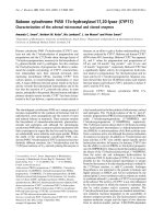
Glycoaldehyde-induced modifications of BSA were studied by
UV-visible absorption spectroscopy, gel electrophoresis, and
AGE-specific fluorescence. The absorption spectra of AGE-
BSA (AGEs) revealed a marked UV hyperchromicity (74.4%)
at 280 nm (Figure 1a). For further characterization of in vitro-
produced AGEs, samples were analyzed by SDS-PAGE
under reducing conditions. An accompanying decrease in the
electrophoretic mobility and smear towards higher molecular
size ranges from 65 to 98 kDa was noted, indicating a higher
level of glycoaldehyde-mediated modifications (Figure 1b).
AGE-specific absorbance was detected at 340 nm [21] and
was found to be significantly high in glycoaldehyde-treated
BSA as compared with native BSA (P < 0.001). AGE-specific
fluorescence was detected at the excitation/emission wave-
lengths described elsewhere [22,23] and were found to be
very high for glycoaldehyde-treated BSA compared with native
BSA, signifying the presence of a higher level of modifications.
Characterization of AGE-specific modifications on BSA result-
ing from incubation with glycoaldehyde is summarized in Table
1.
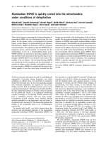
Human chondrocytes in monolayers maintain their
chondrogenic phenotype
We determined whether primary human OA chondrocytes and
chondrocytes passaged once used in these studies main-
tained their phenotype – by analyzing the expression of type-2
collagen, aggrecan, and SOX-9 mRNA, which are considered
to be signatures of the chondrogenic phenotype [44]. Our
results show that primary or passage-1 human OA chondro-
cytes in monolayer culture maintained their phenotype, when
they were plated (1 × 10
6
/ml) in 35-mm culture dishes in com-
plete Ham's F-12 medium with 10% FCS and were allowed to
grow at 37°C and 5% CO
2
in a tissue culture incubator, as
judged by the continued expression of COL2A1 (Figure 2a),
aggrecan, and SOX-9 mRNAs, whereas COL10A1 mRNA
was not expressed (Figure 2b). Based on these data, OA
chondrocytes were used within 72 hours after plating to avoid
possible dedifferentiation.
Induction of TNFα and MMP-13 expression by AGE-BSA
in human osteoarthritis chondrocytes
OA chondrocytes were treated with increasing doses of AGE-
BSA (20 to 600 μg/ml) and the mRNA expression of TNFα
and MMP-13 was determined using a highly sensitive and spe-
cific quantitative RT-PCR method. We found AGE-BSA dose-
dependently induced TNFα and MMP-13 mRNA expressions,
and the maximum stimulation of chondrocytes was found to be
at 600 μg/ml AGE-BSA (data not shown). Based on these
data, we used a concentration of 600 μg/ml AGE-BSA for
stimulation of chondrocytes in all of our experiments.
percentage inhibition
inhibited blank uninhibited
=− −1[( )/(AAA−−×A
blank
)] 100
Table 1
Characterization of native BSA and AGE-BSA under identical
experimental conditions
Parameter AGE-BSA Native BSA (control)
A
340
0.625 ± 0.13* 0.228 ± 0.05
Fluo [ex 360/em 430 nm] 749.4 ± 19.5** 80.4 ± 4.8
Fluo [ex 330/em 395 nm] 754.6 ± 17.2** 83.2 ± 5.7
Fluo [ex 365/em 440 nm] 741.4 ± 28.1** 84.8 ± 5.2
Fluo [ex 485/em 530 nm] 734.1 ± 20.4** 85.1 ± 5.3
Fluo [ex 280/em 350 nm] 733.2 ± 19.1** 84.0 ± 4.4
Data expressed as the mean ± standard deviation of 12 independent
assays. A
340
, absorbance at 340 nm; AGE, advanced glycation end
product; Fluo [ex/em], fluorescence [excitation/emission], Band
width was set at 40/40 for fluorescence measurement. *P < 0.01
versus control, **P < 0.0001 versus control.
Arthritis Research & Therapy Vol 11 No 3 Rasheed et al.
Page 6 of 13
(page number not for citation purposes)
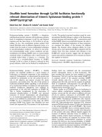
Effect of EGCG on AGE-BSA-induced expression and
production of TNFα in osteoarthritis chondrocytes
OA chondrocytes (80% confluent) were pretreated with
EGCG (25 to 150 μM) for 2 hours, and were then stimulated
with in vitro-generated AGE-BSA (600 μg/ml) for 8 hours. No
cytotoxic effect of EGCG was noted at the dose used (data
not shown). The level of TNFα mRNA was quantified by a
highly sensitive and specific quantitative RT-PCR method, and
values were compared with control. Our results showed that
OA chondrocytes treated with AGE-BSA had a higher level of
TNFα mRNA compared with unstimulated OA chondrocytes.
TNFα mRNA levels showed a marked decline, however, in the
samples pretreated with EGCG and then stimulated with
AGE-BSA (Figure 3a). To determine whether inhibition of
gene expression also affected the protein level, culture super-
natants were assayed for TNFα protein using TNFα-specific
ELISA. As shown in Figure 3b, pretreatment with 25 to 150
μM EGCG significantly decreased the AGE-BSA-induced
TNFα production in the culture supernatant of AGE-BSA-stim-
ulated OA chondrocytes. It is also to be noted that maximum
suppression was observed in cultures treated with 75 μM
EGCG, after which no further decline was found (Figure 3b).
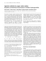
EGCG downregulated the expression and production of
MMP-13 in AGE-BSA-stimulated osteoarthritis
chondrocytes
Pretreatment of OA chondrocytes with EGCG inhibited AGE-
BSA-induced gene expression of MMP-13 in a dose-depend-
ent manner as determined by quantitative RT-PCR (Figure 4a).
Again the maximum inhibitory effect of EGCG was found at 75
μM concentration. We also determined the effect of EGCG on
the production of MMP-13 in the culture supernatant by west-
ern immunoblotting using anti-MMP-13 antibody (Figure 4b).
Analysis of the immunoblot revealed that the levels of MMP-13
were high in the supernatant of chondrocytes treated with
AGE-BSA when compared with the levels detected in
untreated chondrocytes culture, where MMP-13 was barely
detectable. Importantly, the AGE-BSA-stimulated increase in
MMP-13 expression was inhibited by EGCG to less than the
basal level (Figure 4b). These results were further verified
using gelatin zymography (Figure 4c). Zymographic analysis
showed that OA chondrocytes stimulated with AGE-BSA had
enhanced levels of active MMP-13 compared with the levels
detected in control OA chondrocytes. Importantly, chondro-
cytes pretreated with different doses of EGCG and then stim-
ulated with AGE-BSA showed reduced levels of active MMP-
13 in the culture supernatant. Taken together these data indi-
cate that the AGE-BSA-induced production of active MMP-13
was inhibited by EGCG in human OA chondrocytes (Figure
4c).
Effect of EGCG on the activation of MAPKs in AGE-BSA-
stimulated osteoarthritis chondrocytes
Activation of MAPKs is intimately associated with the expres-
sion of proinflammatory mediators [26]. To determine whether
the inhibition of TNFα and MMP-13 expressions was due to
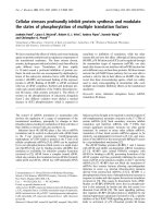
Figure 1
Absorbance spectra and electrophoresis of native BSA and advanced glycation end product-BSAAbsorbance spectra and electrophoresis of native BSA and advanced glycation end product-BSA. (a) Absorbance spectra of native BSA and
advanced glycation end product (AGE)-BSA. Samples were incubated with or without glycoaldehyde in PBS, pH 7.2, with equal protein concentra-
tions. (b) Electrophoresis of native BSA and AGE-BSA. Samples were electrophoresed using 10% SDS-PAGE with 4% stacking gel. The gel was
run for 1.5 hours at 125 V. The precision plus protein standard (Bio-Rad) served as the molecular size marker. AGE-BSA was derived from the reac-
tion between endotoxin-free BSA (2 mg/ml) and glycoaldehyde (70 mM).
Available online />Page 7 of 13
(page number not for citation purposes)
EGCG-mediated inhibition of the MAPK pathway, we exam-
ined the effect of EGCG on the activation of MAPKs in AGE-
BSA-stimulated human OA chondrocytes. OA chondrocytes
were pretreated with EGCG (25 to 150 μM) for 1 hour and
then stimulated with AGE-BSA for 45 minutes, and the cell
lysate was analyzed by western immunoblotting. Pretreatment
of chondrocytes with EGCG attenuated the AGE-BSA-
induced phosphorylation of p38-MAPK, JNK-MAPK and to a
lesser extent of ERK-MAPK (Figure 5a,b). To further
strengthen the relation of p38, JNK and ERK inhibition by
EGCG and proinflammatory cytokine TNFα and MMP-13
expressions in AGE-BSA-stimulated human OA chondro-
cytes, we used pharmacological agents that inhibit p38-, JNK-
and ERK-MAPKs.
Treatment of OA chondrocytes with the selective p38 inhibitor
SB202190 (100 μM), the JNK inhibitor SP600125 (10 μM),
and the ERK inhibitor PD98059 (50 μM) blocked the AGE-
BSA-induced TNFα mRNA expression as determined by
quantitative RT-PCR, but the effect was more pronounced in
the case of p38-MAPK and JNK (Figure 3a). AGE-BSA-
induced MMP-13 mRNA expression was significantly inhibited
by SB202190 and SP600125 (P < 0.05), but the affect was
more pronounced when SB202190 was used. Pretreatment
of OA chondrocytes with PD98059 had no effect on MMP-13
mRNA expression in AGE-BSA-stimulated OA chondrocytes
(Figure 4a). These data support the contention that inhibition
of AGE-BSA-induced TNFα (Figure 3a) and MMP-13 (Figure
4a) expressions by EGCG in OA chondrocytes was mediated,
at least in part, by the inhibition of AGE-induced activation of
the p38-MAPK and JNK-MAPK pathways.
Effect of EGCG on NF-κB activation in AGE-BSA-
stimulated osteoarthritis chondrocytes
NF-κB is an important transcriptional regulator of inflammatory
cytokine gene expression and plays a crucial role in immune
and inflammatory response. After the ubiquitination and phos-
phorylation of IκBα, the inhibitor is degraded and the NF-κB is
translocated to the nucleus, where it binds and activates the
promoter of target genes. To investigate the mechanism
responsible for the inhibitory effect of EGCG on proinflamma-
tory cytokine gene expression, we examined the effect of
EGCG on NF-κB activation and translocation to the nucleus
using western blotting. Stimulation of OA chondrocytes with
AGE-BSA induced the degradation of IκBα and nuclear trans-
location of NF-κB p65 (Figure 6a,b). Pretreatment with EGCG
(25 and 75 μM) inhibited the AGE-BSA-induced degradation
of IκBα and nuclear translocation of NF-κB p65 (Figure 6a,b).
To determine whether EGCG also inhibits AGE-BSA-induced
DNA binding activity of NF-κB, we used the Transcription Fac-
tor ELISA kit. Exposure of OA chondrocytes to AGE-BSA sig-
nificantly enhanced the DNA binding activity of NF-κB p65
compared with controls (P < 0.0001), and increasing doses
of EGCG (25 to 75 μM) significantly reduced the AGE-BSA-
induced DNA binding activity of NF-κB p65 (P < 0.05) (Figure
6c).
To further strengthen the relation of inhibition of the NF-κB
pathway and the expression of TNFα in our studies, we next
investigated the effect of a pharmacological agent (MG-132,
a known inhibitor of NF-κB) on the expression of TNFα and
MMP-13. Treatment of chondrocytes with the proteasome
inhibitor MG-132 (100 μM) significantly blocked the AGE-
BSA-induced TNFα expression (P < 0.05) (Figure 3a),
whereas MMP-13 mRNA expression was also inhibited by
MG-132 but the inhibition was not statistically significant (P >
0.05) (Figure 4a). Together these results suggest that EGCG
exerts its inhibitory effect on TNFα expression in AGE-BSA-
stimulated OA chondrocytes via modulation of the activation
and DNA binding activity of NF-κB.
Inhibition of IKKβ kinase activity by EGCG
The effect of EGCG on the phosphorylating activity of IKKβ
kinase was determined using an HTScan
®
IKKβ Kinase Assay
Kit (Cell Signaling Technology). Purified IKKβ kinase was pre-
treated with different doses of EGCG (5 to 200 μM) 5 minutes
prior to incubation with the substrate peptide. Figure 6d
shows that IKKβ kinase activity was significantly inhibited by
EGCG treatment (P < 0.001). A maximum of 85% inhibition of
enzymatic activity was observed with 5 μM EGCG, after which
no significant inhibition (P > 0.05) of enzyme activity was
Figure 2
Human osteoarthritis chondrocytes in monolayer culture maintain their phenotypeHuman osteoarthritis chondrocytes in monolayer culture maintain their
phenotype. Primary chondrocytes from osteoarthritis patients were cul-
tured for 72 hours and were then split and cultured for an additional 3
days (passage 1). Expression of (a) type-2 collagen (COL2A1) and (b)
aggrecan (ACAN), type-10 collagen (COL10A1) and SRY-box contain-
ing gene 9 (SOX-9) was determined by RT-PCR. M, 100 bp DNA lad-
der; P, positive control cDNA; P1, primary chondrocytes; P2, passage
1 chondrocytes.
Arthritis Research & Therapy Vol 11 No 3 Rasheed et al.
Page 8 of 13
(page number not for citation purposes)
observed (Figure 6d). These data suggest that EGCG sup-
presses the activation of NF-κB by inhibiting the enzyme activ-
ity of IKK complex.
Discussion
Chondrocytes are the only cellular components of cartilage.
Under normal physiologic conditions, chondrocytes maintain
an equilibrium between anabolic and catabolic activities that is
necessary for preservation of the structural and functional
integrity of the tissue. Chondrocytes express inflammatory
mediators such as TNFα and proteolytic enzymes – aggreca-
nases and MMPs – which under normal conditions, mediate a
very low matrix turnover required for cartilage remodeling [45].
However, in pathologic conditions such as OA, however,
chondrocyte production of these inflammatory mediators and
enzymes increases considerably, resulting in cartilage
destruction [46].
Age is by far the most important risk factor for the development
of OA [45]. By which mechanism aging is involved in the
development of this debilitating disease remains largely
unknown. Fatigue failure of the cartilage collagen network due
to repetitive loading has long been recognized as one of the
mechanisms involved in the development of OA [46]. With
increasing age, the strength of the collagen matrix to withstand
loading diminishes. Age-related changes in articular cartilage
that influence the composition and strength of the cartilage
matrix are therefore very probably involved in the development
of OA [47,48]. One such change, the age-related accumula-
tion of AGEs, has previously been shown to increase tissue
stiffness, to decrease extracellular matrix turnover (synthesis
and degradation), and to affect many cellular processes [17].
It is well documented that human chondrocytes are highly
responsive to AGEs [18-20], and the most striking effect of
AGEs or AGE-BSA on chondrocytes is to induce the produc-
tion of TNFα [18] and MMP-13 [18,19], which are important
sources of inflammation and cartilage degradation within the
arthritic joints.
Although arthritis is present in every population and OA is the
most common joint disorder, treatment is still limited to a few
classes of drugs, primarily nonsteroidal anti-inflammatory
drugs and corticosteroids [49,50]. While providing relief from
pain, however, none of these agents has been shown to inhibit
cartilage breakdown or to inhibit disease progress; they also
have varying degrees of gastrointestinal toxicity [51]. Previous
studies from our laboratory have shown that green tea inhib-
ited the development of arthritis in a mouse model and also
inhibited the production of various inflammatory mediators by
human chondrocytes stimulated with IL-1β [30-34]. Studies
from other investigators have shown that EGCG inhibits the
degradation of human cartilage proteoglycan and type-2 colla-
gen [52] and selectively inhibits the ADAMTS-1, ADAMTS-4,
and ADAMTS-5 [53]. In the present study, we determined the
effect of EGCG on the induction of the major proinflammatory
mediators TNFα and MMP-13 in AGE-BSA-stimulated human
Figure 3
Gene expression and production of TNFα in advanced glycation end product-BSA-stimulated osteoarthritis chondrocytesGene expression and production of TNFα in advanced glycation end product-BSA-stimulated osteoarthritis chondrocytes. (a) Effect of epigallocate-
chin-3-gallate (EGCG), specific inhibitors for mitogen-activated protein kinases and NF-κB on the gene expression of TNFα in advanced glycation
end product (AGE)-BSA-stimulated osteoarthritis (OA) chondrocytes. Primary chondrocytes were pretreated with EGCG (25 to 150 μM) for 2
hours and were stimulated by AGE-BSA (600 μg/ml) for 8 hours. Folds of TNFα mRNA expression, as compared with control and normalized to
GAPDH, were determined by quantitative RT-PCR. Concentrations of specific inhibitors of JNK (SP600125), ERK (PD98059), p38 (SB202190)
and NF-κB (MG-132) used in these studies were 10 μM, 50 μM, 100 μM and 100 μM, respectively. Native BSA (600 μg/ml) was used as a nega-
tive control. (b) Effect of EGCG on the production of TNFα in AGE-BSA-stimulated OA chondrocytes. Primary chondrocytes were pretreated with
EGCG (25 to 150 μM) for 2 hours and were stimulated by AGE-BSA (600 μg/ml) for 24 hours. The production level of TNFα was determined by
sandwich ELISA. Results are representative (mean ± standard error of the mean) of duplicate experiments with OA chondrocytes obtained from five
age-matched and sex-matched OA donors; data without a common letter differ, P < 0.05.
Available online />Page 9 of 13
(page number not for citation purposes)
OA chondrocytes. Almost complete inhibition of both TNFα
and MMP-13 expression and production was observed at a
concentration of 75 μM EGCG (P < 0.01) – although these
concentrations of EGCG may not be achieved physiologically
through oral consumption but may readily be achieved through
local administration. Our results presented here demonstrate
that EGCG is a potent inhibitor of AGE-BSA-induced expres-
sion and production of these inflammatory mediators in human
chondrocytes.
The signaling pathways characterized by the MAPKs p38,
ERK, and JNK are known to play a potential role in the regula-
tion of inflammatory response [26,54]. These are the key play-
ers in the molecular and cellular events associated with the
pathogenesis of inflammatory arthritis and are being studied
as a rational target for arthritis therapy [54]. The activation of
RAGE stimulates critical signaling pathways linked to inflam-
mation, resulting in the activation of various inflammatory
genes [55]. The interaction of MAPKs and RAGE has been
well reported [17]. In the present study we found that EGCG
specifically inhibited the AGE-BSA-induced activation of
MAPKs and inhibited the expression of TNFα and MMP-13 by
human OA chondrocytes. In addition, the p38-specific, JNK-
specific and ERK-specific inhibitors SB202190, SP600125
and PD98059 also reduced TNFα gene and MMP-13 expres-
sion in human chondrocytes. These data suggest that EGCG
has the potential to inhibit the inflammatory stimuli-induced
MAPK activation and the downstream TNFα and MMP-13
gene and protein expression.
Activation of the master transcription factor NF-κB leads to the
coordinated expression of many genes that encode cytokines,
chemokines, enzymes, and adhesion molecules involved in
mediator synthesis, and the further amplification and perpetu-
ation of the inflammatory reaction [42,43]. The NF-κB tran-
scription factors are present in the cytosol in an inactive state,
complexed with the inhibitory IκB proteins [56]. Activation of
NF-κB is a common pathway based on the induction of phos-
Figure 4
Gene expression and production of matrix metalloproteinase-13 in advanced glycation end product-BSA-stimulated osteoarthritis chondrocytesGene expression and production of matrix metalloproteinase-13 in advanced glycation end product-BSA-stimulated osteoarthritis chondrocytes. (a)
Effect of epigallocatechin-3-gallate (EGCG), specific inhibitors for mitogen-activated protein kinases and NF-κB on the gene expression of matrix
metalloproteinase (MMP)-13 in advanced glycation end product (AGE)-BSA-stimulated osteoarthritis (OA) chondrocytes. Primary human chondro-
cytes were pretreated with EGCG (25 to 150 μM) for 2 hours and were stimulated with AGE-BSA (600 μg/ml) for 8 hours. Expression of MMP-13
mRNA was normalized to GAPDH and compared with the levels present in control. Concentrations of specific inhibitors of JNK (SP600125), ERK
(PD98059), p38 (SB202190) and NF-κB (MG-132) used in these studies were 10 μM, 50 μM, 100 μM and 100 μM, respectively. Native BSA
(600 μg/ml) was used as negative control. Results are representative (mean ± standard error of the mean) of duplicate experiments with chondro-
cytes obtained from five age-matched and sex-matched OA donors; data without a common letter differ, P < 0.01. (b), (c) Effect of EGCG on the
production of MMP-13 in AGE-BSA-stimulated OA chondrocyte culture medium. Primary chondrocytes were pretreated with EGCG (25 to 150 μM)
for 2 hours and were stimulated with AGE-BSA (600 μg/ml) for 24 hours. MMP-13 production was analyzed in cell culture supernatant by (b) west-
ern blotting and (c) gelatin zymography. Equal volumes of culture supernatant were loaded on polyacrylamide gel. The MMP-13 positive control
(EMD Chemicals) was also used. Band images were digitally captured and the band intensities (pixels/band) were obtained using the Un-Scan-It
software and are expressed in arbitrary optical density units. Data shown are cumulative of two experiments. OD values presented as mean ± stand-
ard deviation; data without a common letter differ, P < 0.05.
Arthritis Research & Therapy Vol 11 No 3 Rasheed et al.
Page 10 of 13
(page number not for citation purposes)
phorylation, which mediates proteasomal degradation of IκB
[56]. The key regulatory step in this pathway involves activa-
tion of a high-molecular-weight IKK complex, whose catalysis
is generally carried out by three tightly associated IKK subu-
nits. IKKα and IKKβ serve as the catalytic subunits of the
kinase. IKKγ serves as the regulatory subunit [57]. Activation
of IKK depends on phosphorylation; serines 177 and 181 in
the activation loop of IKKβ (176 and 180 in IKKα) are the spe-
cific sites whose phosphorylation causes conformational
changes resulting in kinase activation [58]. It is also well doc-
umented that NF-κB is known to be involved in AGE-mediated
effects of RAGE signaling [17], and that expression of TNFα
and MMP-13 gene is dependent on the activation of transcrip-
tion factor NF-κB [17,59].
Suppression of NF-κB activation has been linked with anti-
inflammatory activity; we therefore postulated that EGCG
mediates its inhibitory effects on TNFα and MMP-13 expres-
sions, at least in part, through the suppression of NF-κB activ-
ity. In AGE-BSA-stimulated human OA chondrocytes, EGCG
inhibited the degradation of IκBα and nuclear translocation of
the NF-κB p65 (Figure 6a,b). In addition, DNA binding activity
of nuclear NF-κB p65, as demonstrated by highly sensitive
and specific ELISA, was also inhibited in OA chondrocytes.
These data further confirm that EGCG attenuates the inflam-
matory stimuli-induced activation and DNA binding activity of
NF-κB in human chondrocytes. In order to gain further insight
into the mechanism, we used an in vitro kinase activity assay.
Our results showed that EGCG inhibited the phosphorylating
activity of IKKβ kinase, indicating that the observed inhibition
of NF-κB in the above studies may have been achieved by inhi-
bition of the IKK activity in OA chondrocytes, causing IκBα to
accumulate in the nucleus.
In the present article we report for the first time that EGCG,
the most abundant and biologically active catechin of green
tea, inhibits the inflammatory activity of AGE-BSA-stimulated
human OA chondrocytes. This is achieved by blocking MAPKs
Figure 5
Mitogen-activated protein kinase phosphorylation in advanced glycation end product-BSA-stimulated osteoarthritis chondrocytesMitogen-activated protein kinase phosphorylation in advanced glycation end product-BSA-stimulated osteoarthritis chondrocytes. (a) Effect of epi-
gallocatechin-3-gallate (EGCG) on mitogen-activated protein kinase phosphorylation in advanced glycation end product (AGE)-BSA-stimulated
osteoarthritis (OA) chondrocytes. After pretreatment with EGCG (25 to 150 μM) for 1 hour at 37°C, primary human chondrocytes (70 to 80% con-
fluent) were incubated with AGE-BSA (400 μg/ml) for 45 minutes, and then the phosphorylation of p38, JNK, and ERK was determined by western
blot analysis. (b) Band images were digitally captured and the band intensities (pixels/band) were obtained using the Un-Scan-It software and are
expressed in arbitrary optical density units. Data shown are cumulative of two experiments. OD values presented as mean ± standard deviation; data
without a common letter differ, P < 0.05.
Available online />Page 11 of 13
(page number not for citation purposes)
and NF-κB activation in human chondrocytes. Our results also
point out that inhibition of NF-κB was achieved by inhibiting
the degradation of the inhibitor IκBα in the cytoplasm of
human OA chondrocytes as previously reported [31]. As
TNFα and MMP-13 genes are NF-κB dependent, inhibition of
NF-κB also inhibits their expression and production in AGE-
BSA-stimulated chondrocytes.
There are a number of studies documenting the beneficial
health effects of green tea consumption. Most of these studies
place emphasis on the anticancer properties of green tea [27-
29], which have now been attributed, at least in part, to the
ability of green tea polyphenols to inhibit the inflammatory
processes [30]. To this, based on our results, we can add that
EGCG is a potent inhibitor of AGE-BSA-induced induction of
TNFα and MMP-13 at a physiologically achievable concentra-
tion (25 μM; see Figures 3a and 4a) – but for more complete
inhibition higher dose is needed. We therefore conclude that
inhibition of arthritis following green tea consumption in an ani-
mal model [30] and inhibition of cartilage degradation and pro-
duction of inflammatory mediators by EGCG may be the result
of inhibition of some of the matrix-degrading enzymes/factors
at the mRNA level through inhibition of NF-κB. It is therefore
tempting to suggest that green tea polyphenol EGCG or com-
pounds derived from it may serve as lead agents in the design
of more potent and effective inhibitors of proinflammatory
Figure 6
Epigallocatechin-3-gallate inhibits activation and DNA binding of NF-κB in advanced glycation end product-BSA-stimulated osteoarthritis chondro-cytesEpigallocatechin-3-gallate inhibits activation and DNA binding of NF-κB in advanced glycation end product-BSA-stimulated osteoarthritis chondro-
cytes. Primary chondrocytes (70 to 80% confluent) were pretreated with epigallocatechin-3-gallate (EGCG) (25 and 75 μM) for 2 hours and were
stimulated by advanced glycation end product (AGE)-BSA (600 μg/ml). (a) IκBα degradation and NF-κB translocation were analyzed by western
immunoblotting using antibodies specific for the NF-κB p65 (Santa Cruz Biotech) (C-NF-κB, cytoplasmic NF-κB; N-NF-κB, nuclear NF-κB) and for
IκBα (Santa Cruz Biotech). (b) Band intensities were obtained as described above. Data shown are cumulative of three experiments, and the optical
density values (pixels/band) presented as mean ± standard deviation. (c) Activated NF-κB p65 in the nucleus was determined by the highly specific
Transcription Factor ELISA kit (Panomics). The positive control nuclear extract supplied with the kit was used. Data are representative of two experi-
ments and presented as mean ± standard deviation; data without a common letter differ, P < 0.05. (d) EGCG inhibited the IKKβ kinase activity in
vitro. IKKβ kinase activity was determined in the absence or presence of EGCG (5 to 200 μM) using the HTScan
®
IKKβ Kinase Assay Kit (Cell Sig-
naling Technology). Each point is representative of three individual kinase assays and presented as mean ± standard deviation.
Arthritis Research & Therapy Vol 11 No 3 Rasheed et al.
Page 12 of 13
(page number not for citation purposes)
cytokines and collagenases for use therapeutically to block
cartilage degradation in OA.
Conclusions
The present article is the first report that shows green tea cat-
echin EGCG inhibits the inflammatory activity against AGE-
induced activation of human OA chondrocytes. The results of
the present study indicate that EGCG inhibits AGE-BSA-
induced upregulation of TNFα and MMP-13 viainhibiting the
MAPK and NF-κB activation in human OA chondrocytes.
EGCG or EGCG-derived compounds may be of value for the
treatment of inflammatory arthritis in which AGEs play an
active role.
Competing interests
The authors declare that they have no competing interests.
Authors' contributions
ZR carried out the experimental work, data collection and inter-
pretation, and manuscript preparation. ANA, NA, SR, and FRV
carried out the experimental work and collection of data. TMH
conceived of the study design, coordinated the studies, data
interpretation and manuscript preparation. All authors have
read and approved the final manuscript.
Acknowledgements
The present work was supported in part by NIH/NCCAM grant AT-
003267 and by funds from the University of South Carolina, Columbia.
References
1. Felson DT, Lawrence RC, Dieppe PA, Hirsch R, Helmick CG, Jor-
dan JM, Kington RS, Lane NE, Nevitt MC, Zhang Y, Sowers M,
McAlindon T, Spector TD, Poole AR, Yanovski SZ, Ateshian G,
Sharma L, Buckwalter JA, Brandt KD, Fries JF: Osteoarthritis:
new insights. I. The disease and its risk factors. Ann Intern Med
2000, 133:635-646.
2. Felson DT, Zhang Y: An update on the epidemiology of knee
and hip osteoarthritis with a view to prevention [review].
Arthritis Rheum 1998, 41:1343-1355.
3. Verzijl N, Bank RA, TeKoppele JM, DeGroot J: Ageing and oste-
oarthritis: a different perspective. Curr Opin Rheumatol 2003,
15:616-622.
4. Verzijl N, DeGroot J, Thorpe SR, Bank RA, Shaw JN, Lyons TJ,
Bijlsma JW, Lafeber FP, Baynes JW, TeKoppele JM: Effect of col-
lagen turnover on the accumulation of advanced glycation end
products. J Biol Chem 2000, 275:39027-39031.
5. Bank RA, Bayliss MT, Lafeber FP, Maroudas A, TeKoppele JM:
Ageing and zonal variation in post-translational modification
of collagen in normal human articular cartilage: the age-
related increase in non-enzymatic glycation affects biome-
chanical properties of cartilage. Biochem J 1998,
330:345-351.
6. Verzijl N, DeGroot J, Oldehinkel E, Bank RA, Thorpe SR, Baynes
JW, Bayliss MT, Bijlsma JW, Lafeber FP, Tekoppele JM: Age-
related accumulation of Maillard reaction products in human
articular cartilage collagen. Biochem J 2000, 350:381-387.
7. Verzijl N, DeGroot J, Ben ZC, Brau-Benjamin O, Maroudas A, Bank
RA, Mizrahi J, Schalkwijk CG, Thorpe SR, Baynes JW, Bijlsma JW,
Lafeber FP, TeKoppele JM: Crosslinking by advanced glycation
end products increases the stiffness of the collagen network
in human articular cartilage: a possible mechanism through
which age is a risk factor for osteoarthritis. Arthritis Rheum
2002, 46:114-123.
8. DeGroot J, Bank RA, Tchetverikov I, Verzijl N, TeKoppele JM:
Molecular markers for osteoarthritis: the road ahead. Curr
Opin Rheumatol 2002, 14:585-589.
9. DeGroot J, Verzijl N, Bank RA, Lafeber FP, Bijlsma JW, TeKoppele
JM: Age-related decrease in proteoglycan synthesis of human
articular chondrocytes: the role of nonenzymatic glycation.
Arthritis Rheum 1999, 42:1003-1009.
10. DeGroot J, Verzijl N, Wenting-van Wijk MJ, Jacobs KM, van El B,
van Roermund PM, Bank RA, Bijlsma JW, TeKoppele JM, Lafeber
FP: Accumulation of advanced glycation end products as a
molecular mechanism for aging as a risk factor in osteoarthri-
tis. Arthritis Rheum 2004, 50:1207-1215.
11. Steenvoorden MM, Huizinga TW, Verzijl N, Bank RA, Ronday HK,
Luning HA, Lafeber FP, Toes RE, DeGroot J: Activation of recep-
tor for advanced glycation end products in osteoarthritis leads
to increased stimulation of chondrocytes and synoviocytes.
Arthritis Rheum 2006, 54:253-263.
12. McCance DR, Dyer DG, Dunn JA, Bailie KE, Thorpe SR, Baynes
JW, Lyons TJ: Maillard reaction products and their relation to
complications in insulin-dependent diabetes mellitus. J Clin
Invest 1993, 91:2470-2478.
13. Waine H, Nevinny D, Rosenthal J, Joffe IB: Association of oste-
oarthritis and diabetes mellitus. Tufts Folia Med 1961, 7:13-19.
14. Campbell WL, Feldman F: Bone and soft tissue abnormalities of
the upper extremity in diabetes mellitus. Am J Roentgenol
Radium Ther Nucl Med 1975, 124:7-16.
15. Bagge E, Bjelle A, Eden S, Svanborg A: Factors associated with
radiographic osteoarthritis: results from the population study
70-year-old people in Göteborg. J Rheumatol 1991,
18:1218-1222.
16. Sunahori K, Yamamura M, Yamana J, Takasugi K, Kawashima M,
Makino H: Increased expression of receptor for advanced gly-
cation end products by synovial tissue macrophages in rheu-
matoid arthritis. Arthritis Rheum 2006, 54:97-104.
17. Loeser RF, Yammani RR, Carlson CS, Chen H, Cole A, Im HJ, Bur-
sch LS, Yan SD: Articular chondrocytes express the receptor
for advanced glycation end products: potential role in osteoar-
thritis. Arthritis Rheum 2005, 52:2376-2385.
18. Nah SS, Choi IY, Yoo B, Kim YG, Moon H, Lee C: Advanced gly-
cation end products increases matrix metalloproteinase-1, -3,
and -13, and TNF-α in human osteoarthritic chondrocytes.
FEBS Lett 2007, 581:1928-1932.
19. Steenvoorden MM, Huizinga TW, Verzijl N, Bank RA, Ronday HK,
Luning HA, Lafeber FP, Toes RE, DeGroot J: Activation of recep-
tor for advanced glycation end products in osteoarthritis leads
to increased stimulation of chondrocytes and synoviocytes.
Arthritis Rheum 2006, 54:253-263.
20. Nah SS, Choi IY, Lee CK, Oh JS, Kim YG, Moon HB, Yoo B:
Effects of advanced glycation end products on the expression
of COX-2, PGE
2
and NO in human osteoarthritic chondrocytes.
Rheumatology (Oxford) 2008, 47:425-431.
21. Valencia JV, Weldon SC, Quinn D, Kiers GH, DeGroot J, TeKop-
pele JM, Hughes TE: Advanced glycation end product ligands
for the receptor for advanced glycation end products: bio-
chemical characterization and formation kinetics. Anal Bio-
chem 2004, 324:68-78.
22. Schmitt A, Schmitt J, Münch G, Gasic-Milencovic J: Characteriza-
tion of advanced glycation end products for biochemical stud-
ies: side chain modifications and fluorescence characteristics.
Anal Biochem 2005, 338:201-215.
23. Schmitt A, Nöller J, Schmitt J: The binding of advanced glycation
end products to cell surfaces can be measured using bead-
reconstituted cellular membrane proteins. Biochim Biophys
Acta 2007, 1768:1389-1399.
24. Farboud B, Aotaki-Keen A, Miyata T, Hjelmeland LM, Handa JT:
Development of a polyclonal antibody with broad epitope spe-
cificity for advanced glycation end products and localization of
these epitopes in Bruch's membrane of the aging eye. Mol Vis
1999, 5:11.
25. Sternlicht MD, Werb Z: How matrix metalloproteinases regu-
late cell behavior [review]. Annu Rev Cell Dev Biol 2001,
17:463-516.
26. Rasheed Z, Haqqi TM: Update on targets of biologic therapies
for rheumatoid arthritis. Curr Rheum Rev 2008, 4:246-253.
27. Khan N, Mukhtar H: Tea polyphenols for health promotion. Life
Sci 2007, 81:519-533.
28. Siddiqui IA, Malik A, Adhami VM, Asim M, Hafeez BB, Sarfaraz S,
Mukhtar H: Green tea polyphenol EGCG sensitizes human
prostate carcinoma LNCaP cells to TRAIL-mediated apoptosis
Available online />Page 13 of 13
(page number not for citation purposes)
and synergistically inhibits biomarkers associated with angio-
genesis and metastasis. Oncogene 2008, 27:2055-2063.
29. Siddiqui IA, Shukla Y, Adhami VM, Sarfaraz S, Asim M, Hafeez BB,
Mukhtar H: Suppression of NF κB and its regulated gene prod-
ucts by oral administration of green tea polyphenols in an
autochthonous mouse prostate cancer model. Pharm Res
2008, 25:2135-2142.
30. Haqqi TM, Anthony DD, Gupta S, Ahmed N, Lee MS, Kumar GK,
Mukhtar H: Prevention of collagen-induced arthritis in mice by
a polyphenolic fraction from green tea. Proc Natl Acad Sci USA
1999, 96:4524-4529.
31. Singh R, Ahmed S, Islam N, Goldberg VM, Haqqi TM: Epigallo-
catechin-3-gallate inhibits interleukin-1β-induced expression
of nitric oxide synthase and production of nitric oxide in
human chondrocytes: suppression of nuclear factor kappa B
activation by degradation of the inhibitor of nuclear factor
kappa B. Arthritis Rheum 2002, 46:2079-2086.
32. Ahmed S, Wang N, Lalonde M, Goldberg VM, Haqqi TM: Green
tea polyphenol epigallocatechin-3-gallate (EGCG) differen-
tially inhibits interleukin-1β-induced expression of matrix met-
alloproteinase-1 and -13 in human chondrocytes. J Pharmacol
Exp Ther 2004, 308:767-773.
33. Singh R, Ahmad S, Malemud CJ, Goldberg VM, Haqqi TM: Epigal-
locatechin-3-gallate selectively inhibits interleukin-1-induced
activation of mitogen activated protein kinase subgroup c-Jun
N-terminal kinase in human osteoarthritis chondrocytes. J
Orthop Res 2003, 21:102-109.
34. Ahmed S, Rahman A, Hasnain A, Lalonde M, Goldberg VM, Haqqi
TM: Green tea polyphenol epigallocatechin-3-gallate inhibits
the IL-1β-induced activity and expression of cyclooxygenase-
2 and nitric oxide synthase-2 in human chondrocytes. Free
Radic Biol Med 2002, 33:1097-1105.
35. Mandel S, Weinreb O, Amit T, Youdim MB: Cell signaling path-
ways in the neuroprotective actions of the green tea polyphe-
nol (-)-epigallocatechin-3-gallate: implications for
neurodegenerative diseases. J Neurochem 2004,
88:1555-1569.
36. Armstrong CG, Mow VC: Variations in the intrinsic mechanical
properties of human articular cartilage with age, degeneration,
and water content. J Bone Joint Surg Am 1982, 64:88-94.
37. Shukla M, Gupta K, Rasheed Z, Khan KA, Haqqi TM:
Bioavailable
constituents/metabolites of pomegranate (Punica granatum
L) preferentially inhibit COX2 activity ex vivo and IL-1β-induced
PGE
2
production in human chondrocytes in vitro. J Inflamm
(Lond) 2008, 5:9.
38. Mankin HJ, Dorfman H, Lippiello L, Zarins A: Biochemical and
metabolic abnormalities in articular cartilage from osteo-
arthritic human hips. II. Correlation of morphology with bio-
chemical and metabolic data. J Bone Joint Surg Am 1971,
53:523-537.
39. Laemmli UK: Cleavage of structural proteins during the assem-
bly of the head of bacteriophage T4. Nature 1970,
227:680-685.
40. Pfaffl MW: A new mathematical model for relative quantifica-
tion in real-time RT-PCR. Nucleic Acid Res 2001, 29:e45.
41. Gupta K, Shukla M, Cowland JB, Malemud CJ, Haqqi TM: Neu-
trophil gelatinase-associated lipocalin is expressed in oste-
oarthritis and forms a complex with matrix metalloproteinase
9. Arthritis Rheum 2007, 56:3326-3335.
42. Rasheed Z, Akhtar N, Anbazhagan AN, Ramamurthy S, Shukla M,
Haqqi TM: Polyphenol-rich pomegranate fruit extract (POMx)
suppresses PMACI-induced expression of pro-inflammatory
cytokines by inhibiting the activation of MAP Kinases and NF-
κB in human KU812 cells. J Inflamm (Lond) 2009, 6:1.
43. Hafeez BB, Ahmed S, Wang N, Gupta S, Zhang A, Haqqi TM:
Green tea polyphenols-induced apoptosis in human osteosa-
rcoma SAOS-2 cells involves a caspase-dependent mecha-
nism with downregulation of nuclear factor-kappaB. Toxicol
Appl Pharmacol 2006, 216:11-19.
44. de Crombrugghe B, Lefebvre V, Behringer RR, Bi W, Murakami S,
Huang W: Transcriptional mechanisms of chondrocyte differ-
entiation. Matrix Biol 2000, 19:389-394.
45. Poole AR: Cartilage in health and disease. In Arthritis and Allied
Conditions: A Textbook of Rheumatology 14th edition. Edited by:
Koopman W. Philadelphia: Lippincott Williams & Wilkins;
2001:226-284.
46. Pelletier JP, Martel-Pelletier J, Abramson SB: Osteoarthritis, an
inflammatory disease: potential implication for the selection of
new therapeutic targets [review]. Arthritis Rheum
2001,
44:1237-1247.
47. Weightman BO, Freeman MA, Swanson SA: Fatigue of articular
cartilage. Nature 1973, 244:303-304.
48. Kempson GE: The mechanical properties of articular cartilage.
In The Joints and Synovial Fluid Edited by: Sokoloff L. New York:
Academic Press; 1980:177-238.
49. Fendrick AM, Greenberg BP: A review of the benefits and risks
of nonsteroidal anti-inflammatory drugs in the management of
mild-to-moderate osteoarthritis. Osteopath Med Prim Care
2009, 3:1.
50. Bellamy N, Campbell J, Robinson V, Gee T, Bourne R, Wells G:
Intraarticular corticosteroid for treatment of osteoarthritis of
the knee. Cochrane Database Syst Rev 2006, 2:CD005328.
51. Fries JF, Bruce B: Rates of serious gastrointestinal events from
low dose use of acetylsalicylic acid, acetaminophen, and ibu-
profen in patients with osteoarthritis and rheumatoid arthritis.
J Rheumatol 2003, 30:2226-2233.
52. Adcocks C, Collin P, Buttle DJ: Catechins from green tea
(Camellia sinensis) inhibit bovine and human cartilage prote-
oglycan and type-II collagen degradation in vitro. J Nutr 2002,
132:341-346.
53. Vankemmelbeke MN, Jones GC, Fowles C, Ilic MZ, Handley CJ,
Day AJ, Knight CG, Mort JS, Buttle DJ: Selective inhibition of
ADAMTS-1, -4 and -5 by catechin gallate esters. Eur J Biochem
2003, 270:2394-2403.
54. Thalhamer T, McGrath MA, Harnett MM: MAPKs and their rele-
vance to arthritis and inflammation. Rheumatology 2008,
47:409-414.
55. Hofmann MA, Drury S, Fu C, Qu W, Taguchi A, Lu Y, Avila C, Kam-
bham N, Bierhaus A, Nawroth P, Neurath MF, Slattery T, Beach D,
McClary J, Nagashima M, Morser J, Stern D, Schmidt AM: RAGE
mediates a novel proinflammatory axis: a central cell surface
receptor for S100/calgranulin polypeptides. Cell 1999,
97:889-901.
56. Finco TS, Beg AA, Baldwin AS Jr: Inducible phosphorylation of
IκBα is not sufficient for its dissociation from NF-κB and is
inhibited by protease inhibitors. Proc Natl Acad Sci USA 1994,
91:11884-11888.
57. Zandi E, Rothwarf DM, Delhase M, Hayakawa M, Karin M: The IκB
kinase complex (IKK) contains two kinase subunits, IKKα and
IKKβ, necessary for IκB phosphorylation and NF-κB activation.
Cell 1997, 91:243-252.
58. DiDonato JA, Hayakawa M, Rothwarf DM, Zandi E, Karin M: A
cytokine-responsive IκB kinase that activates the transcription
factor NF-κB. Nature 1997, 388:548-554.
59. Azzolina A, Bongiovanni A, Lampiasi N: Substance P induces
TNF-α and IL-6 production through NF κB in peritoneal mast
cells. Biochim Biophys Acta 2003, 1643:75-83.