Báo cáo y học: "Neutrophils exhibit distinct phenotypes toward chitosans with different degrees of deacetylation: implications for cartilage repair" pdf
Bạn đang xem bản rút gọn của tài liệu. Xem và tải ngay bản đầy đủ của tài liệu tại đây (700.11 KB, 10 trang )
Open Access
Available online />Page 1 of 10
(page number not for citation purposes)
Vol 11 No 3
Research article
Neutrophils exhibit distinct phenotypes toward chitosans with
different degrees of deacetylation: implications for cartilage
repair
Pascale Simard
1,2
*, Hugo Galarneau
1
*, Sébastien Marois
1
, Daniel Rusu
1,3
, Caroline D Hoemann
4
,
Patrice E Poubelle
1,3
, Hani El-Gabalawy
5
and Maria JG Fernandes
1,2
1
Centre de Recherche en Rhumatologie et Immunologie, Centre de Recherche du CHUQ-CHUL, boul. Laurier, Québec, G1V 4G2, Canada
2
Department of Anatomy and Physiology, Université Laval, avenue de la Médecine, Québec, G1V 0A6, Canada
3
Department of Medicine, Université Laval, avenue de la Médecine, Québec, G1V 0A6, Canada
4
Department of Chemical Engineering, Institute of Biomedical Engineering, Ecole Polytechnique, boul. Édouard-Montpetit, Montréal, H3C 3A7,
Canada
5
Arthritis Centre, University of Manitoba, Sherbrook Street, Winnipeg, R3A 1M4, Canada
* Contributed equally
Corresponding author: Maria JG Fernandes,
Received: 22 Sep 2008 Revisions requested: 17 Nov 2008 Revisions received: 23 Feb 2009 Accepted: 21 May 2009 Published: 21 May 2009
Arthritis Research & Therapy 2009, 11:R74 (doi:10.1186/ar2703)
This article is online at: />© 2009 Simard et al.; licensee BioMed Central Ltd.
This is an open access article distributed under the terms of the Creative Commons Attribution License ( />),
which permits unrestricted use, distribution, and reproduction in any medium, provided the original work is properly cited.
Abstract
Introduction Osteoarthritis is characterized by the progressive
destruction of cartilage in the articular joints. Novel therapies
that promote resurfacing of exposed bone in focal areas are of
interest in osteoarthritis because they may delay the progression
of this disabling disease in patients who develop focal lesions.
Recently, the addition of 80% deacetylated chitosan to cartilage
microfractures was shown to promote the regeneration of
hyaline cartilage. The molecular mechanisms by which chitosan
promotes cartilage regeneration remain unknown. Because
neutrophils are transiently recruited to the microfracture site, the
effect of 80% deacetylated chitosan on the function of
neutrophils was investigated. Most studies on neutrophils use
preparations of chitosan with an uncertain degree of
deacetylation. For therapeutic purposes, it is of interest to
determine whether the degree of deacetylation influences the
response of neutrophils to chitosan. The effect of 95%
deacetylated chitosan on the function of neutrophils was
therefore also investigated and compared with that of 80%
deacetylated chitosan.
Methods Human blood neutrophils from healthy donors were
isolated by centrifugation on Ficoll-Paque. Chemotaxis was
performed using the chemoTX system. Production of
superoxide anions was evaluated using the cytochrome c
reduction assay. Degranulation was determined by evaluating
the release of myeloperoxidase and lactoferrin. The
internalization of fluorescently labelled 80% deacetylated
chitosan by neutrophils was studied by confocal microscopy.
Results Neutrophils were dose dependently attracted to 80%
deacetylated chitosan. In contrast, 95% deacetylated chitosan
was not chemotactic for neutrophils. Moreover, the majority of
the chemotactic effect of 80% deacetylated chitosan was
mediated by phospholipase-A
2
-derived bioactive lipids.
Contrary to the induction of chemotaxis, neither 80% nor 95%
deacetylated chitosan activated the release of granule enzymes
or the generation of active oxygen species. Despite the distinct
response of neutrophils toward 80% and 95% deacetylated
chitosan, both chitosans were internalized by neutrophils.
Conclusions Eighty per cent deacetylated chitosan induces a
phenotype in neutrophils that is distinct from the classical
phenotype induced by pro-inflammatory agents. Our
observations also indicate that the degree of deacetylation is an
important factor to consider in the use of chitosan as an
accelerator of repair because neutrophils do not respond to
95% deacetylated chitosan.
80 M: 80% deacetylated chitosan of medium viscosity; 95 M: 95% deacetylated chitosan of medium viscosity; cPLA2-α: cytosolic phospholipase
A
2
-α; DDA: degree of deacetylation; FBS: fetal bovine serum; fMLP: N-formyl-methionyl-leucyl-phenylalanine; HTAB: hexadecyltrimethylammonium
bromide; LTB
4
: leukotriene B
4
; MPO: myeloperoxidase; OA: osteoarthritis; PAF: platelet-activating factor; PDI: polydispersity index; PMN: polymor-
phonuclear neutrophil; RITC: rhodamine B isothiocyanate.
Arthritis Research & Therapy Vol 11 No 3 Simard et al.
Page 2 of 10
(page number not for citation purposes)
Introduction
Osteoarthritis (OA) is characterized by progressive destruc-
tion of cartilage in the articular joints [1]. Because it is one of
the main causes of disability, this form of arthritis is a burden
to both society and the patient. The incidence of OA increases
with age. Over 80% of the elderly population exhibits radio-
graphic evidence of OA.
Focal cartilage lesions in humans can be treated by microfrac-
ture. This resurfacing procedure, when successful, can re-sta-
bilize the joint and slow the progression of OA. Chitosan was
recently shown to promote the regeneration of articular carti-
lage through the application of an in situ solidifying chitosan-
glycerol phosphate/blood clot over lesions treated with micro-
fracture [2,3]. Chitosan-glycerol phosphate/blood clots repre-
sent a novel articular cartilage repair approach, which has
yielded promising results in the clinic [4].
Chitosan is a linear polymer of β (1→4)-linked glucosamine
and N-acetyl-
D-glucosamine residues obtained by the N-
deacetylation of chitin. Chitosan is biodegradable, non-toxic,
and nonimmunogenic [5-7]. The degree of deacetylation
(DDA) influences the physical properties of chitosan. As the
degree of deacetylation increases, the degree of solubility of
chitosan in different solvents decreases and susceptibility to
lysosomal biodegradation decreases [8,9]. The chitosan used
in the cartilage repair model is of medium viscosity and is 80%
deacetylated (80 M). In vivo, 50% to 80% deacetylated chi-
tosan is slowly degraded and eventually cleared by enzymatic
and cell-based mechanisms [5,10].
The presence of 80 M chitosan over repairing microfracture or
microdrill holes is associated with the recruitment of polymor-
phonuclear neutrophils (PMNs) to the granulation tissue as
well as remodeling and revascularization of the damaged
trabecular bone, and subsequent formation of more hyaline
repair tissue in both rabbit and sheep repair models [2,3,11].
In contrast, few PMNs home to microdrills in the absence of
chitosan [11]. Remarkably, PMNs persist in repairing defects
for several weeks, in parallel with clearance of the 80 M chi-
tosan particles. Therefore, in contrast to traditional notions that
persistence of PMNs in repairing wounds is detrimental to
repair, in this cartilage repair model the persistence of PMNs
during the first few weeks of repair is related to a more favora-
ble cartilage repair outcome.
To identify the mechanisms through which 80 M chitosan pro-
motes cartilage regeneration in this repair model, the first
objective of the present study was to investigate the effect of
80 M chitosan on the function of PMNs. Even though the
response of PMNs toward chitosan has been characterized in
vitro to some extent [12-16], it remains difficult to compare the
results between studies and to draw clear conclusions
because the chitosan preparations in most studies vary and
details on the quality of the chitosan preparations are rarely
provided. With regard to the latter, the presence of endotoxins
is an important consideration when investigating PMN
responses. In the present study, chitosan preparations of med-
ical grade were used. Regarding the former, some studies use
chitosan preparations of unspecified DDA whereas other
studies use water-soluble chitosan, which does not form a
solid implant [16] or semi-crystalline scaffolds [10]. This com-
plicates the interpretation of the results because the degree of
DDA is a determining factor for the physical properties of chi-
tosan, and it is not yet established to what extent PMNs
respond differently to chitosans of different DDA. Also, it
remains to be determined whether the PMN response varies
toward chitosan presented as a particulate or a cross-linked
scaffold. To optimize the use of chitosan in clinical applica-
tions, it is therefore critical to address the effect of DDA on the
ability of chitosan to activate PMNs and to compare different
preparations of chitosan (for instance, chitosan suspensions
versus scaffolds). The second objective of this study was
therefore to compare the response of PMNs to two chitosan
preparations of a defined DDA, namely 80 M chitosan and
95% deacetylated (95 M) chitosan. The 95 M chitosan was
investigated because our preliminary results indicate that
PMNs respond differently to chitosan of this DDA in vivo.
Materials and methods
All preparation and incubation procedures were performed
under sterile conditions.
Materials
The two medical grade chitosan preparations (80.6% or
94.6% DDA) used in this study are certified to contain under
0.2% weight/weight protein, <500 EU/g endotoxin, and <10
parts/million heavy metals. To prevent contamination by endo-
toxin of chitosan solutions, chitosan powder was dissolved in
double-distilled water filtered by MilliQ (Millipore, Billerica, MA,
USA), at a resistance below 18.2 MΩcm and levels of trace
organic compounds below 30 parts/billion with certified 1.0 N
HCl, using heat-treated endotoxin-free glassware and stir
bars. Chitosan solutions were manipulated under aseptic con-
ditions with laminar flow hoods and dispensed in sterile cryo-
vials with cryo-resistance silicone gaskets for storage at -
80°C. The chitosan solutions are of medium viscosity of 1,422
mPa.S for 80% DDA chitosan, termed 80 M (Mn = 176 kDa,
polydispersity index [PDI] = 1.4), or 2,964 mPa.S for 95%
DDA chitosan, termed 95 M (Mn = 147 kDa, PDI = 1.6), as
previously described by Ma and coworkers [17]. Chitosan
solutions were further diluted with sterile double-distilled
water to 5 mg/ml or 0.5 mg/ml stock solutions. The DDA of
chitosan was provided by the certificates of analysis from the
supplier (Bio Syntech Canada, Inc., Laval, QC, Canada).
Rhodamine B isothiocyanate (RITC) was covalently conju-
gated with each chitosan (RITC-chitosan) to generate either
RITC-80 M (80% DDA, number average molecular weight Mn
= 144 kDa, PDI = 1.3, 0.5% mol/mol RITC/chitosan) or RITC-
Available online />Page 3 of 10
(page number not for citation purposes)
95 M (95% DDA, Mn = 177 kDa, PDI = 1.1, 0.6% mol/mol
RITC/chitosan). The DDA of RITC-chitosan derivatives is
reported as unchanged, given that the derivatization level was
determined as only 0.5% mol RITC/mol chitosan (as reported
by Ma and coworkers [17]). Ficoll-Paque and dextran used for
the isolation of PMN were obtained from Pharmacia (Kirkland,
Québec, Canada) and fetal bovine serum (FBS) as well as
RPMI 1640 were purchased from Wisent (St-Bruno, Québec,
Canada). Calcein/AM was obtained from Invitrogen (Burling-
ton, Ontario, Canada). Migration was assessed using the
ChemoTx system from Neuroprobe (Gaithersburg, MD, USA)
and the purified myeloperoxidase (MPO), O-dianisidine dihy-
drochloride, hydrogen peroxide, and cytochalasin B used in
the MPO assay were obtained from Sigma-Aldrich (Oakville,
Ontario, Canada). Hexadecyltrimethylammonium bromide
(HTAB) was purchased from Fluka Chemie GmbH (Buchs,
Switzerland). Cytochrome c equine heart and pyrrolidine-1
were purchased from Calbiochem (Gibbstown, New Jersey,
USA).
Isolation of polymophonuclear neutrophils
The Institutional Review Board of the Université Laval
(Québec, QC, Canada) approved the study, and volunteers
signed a consent form. PMNs were isolated as previously
described [18]. Briefly, venous blood was obtained from
healthy adult volunteers, in accordance with institute-
approved protocols, in tubes containing heparin or isocitrate.
No difference was observed between the results obtained
with these two anticoagulants. After sedimentation of red
blood cells in 2% dextran, PMNs were aseptically purified by
centrifugation on Ficoll-Paque cushions. Contaminating eryth-
rocytes were removed by hypotonic lysis and PMNs were
resuspended in RPMI 1640 supplemented with 0.1% FBS
previously decomplemented at 56°C for 30 minutes, except
for the chemotaxis experiment (10% decomplemented FBS).
Chemotaxis
Chemotaxis was measured as described previously [13].
Briefly, PMNs were resuspended in RPMI-1640 and 10% FBS
at a concentration of 10
7
cells/ml and were pre-incubated with
1 μg/ml calcein/AM at 37°C for 30 minutes in the dark with
constant agitation. Cells were washed twice and resuspended
in RPMI/10% FBS at 5 × 10
7
cells/ml at 37°C. PMN migration
was monitored using a 96-well ChemoTx disposable chemo-
taxis system. The fluorescence of cells in the filters was meas-
ured using a microplate fluorescence reader (FL600; Bio-Tek
Instruments, Winooski, VT, USA; excitation wavelength 485
nm, emission wavelength 530 nm). Fluorescence was con-
verted to numbers of PMNs based on a standard curve gener-
ated by seeding known numbers of PMNs in the bottom of the
chamber. The results are expressed as percentage of
migrated cells, calculated as the fluorescence of migrated
PMNs/fluorescence of 20,000 PMNs/ml × 100, obtained from
the standard curve. In some experiments, PMNs were incu-
bated with a final concentration of 0.5 μg/ml pertussis toxin for
90 minutes or with 10
-7
mol/l pyrrolidine-1 for 10 minutes at
37°C before the chemotaxis assay.
Production of superoxide anions by polymorphonuclear
neutrophils
Superoxide anion production in response to 80 M and 95 M
chitosan was determined using the cytochrome c reduction
assay, as previously described [19]. Briefly, freshly isolated
PMNs were resuspended at a concentration of 1 × 10
6
cells/
ml in RPMI supplemented with 0.1% decomplemented FBS
and cytochrome c at a final concentration of 125 μg/ml. The
cells were incubated at 37°C for 5 minutes before the addition
of the indicated concentrations of 80 M or 95 M chitosan.
Cells were incubated for an additional 10 minutes at 37°C and
the reaction stopped on ice for 10 minutes. The samples were
then centrifuged at 12,000 g for 2 minutes at 4°C and the opti-
cal density of the supernatants was read at 540, 550, and 560
nm in a spectrophotometer (Milton Roy Spectronic 1001 Plus
spectrophotometer, Milton Roy, Rochester, NY, USA). The
amount of superoxide produced was calculated using the fol-
lowing formula: A
550
- ([A
540
+ A
560
]/2). The absorbance was
transformed into the amount of superoxide produced (nmol/
10
6
neutrophils) by using a conversion factor of 47.4, derived
from the molar extinction coefficient of cytochrome c.
Release of myeloperoxidase and lactoferrin by
polymorphonuclear neutrophils
Degranulation was determined using the MPO and lactoferrin
assay, as described by Bradley and coworkers [20] and Moc-
sai and colleagues [21], respectively.
For the MPO assay, PMNs (10
7
cells/ml) were incubated with
10 μmol/l cytochalasin B, an actin depolymerizing agent that
is known to amplify granule exocytosis, for 2 minutes at 37°C
and then with the indicated concentrations of 80 M or 95 M
chitosan for 30 minutes at 37°C. A negative control with cyto-
chalasin B and a positive control with cytochalasin B + N-
formyl-methionyl-leucyl-phenylalanine (fMLP; 5 minutes incu-
bation) were also performed. PMNs were then centrifuged for
1 minute at 12,000 g and lysed in HTAB lysis buffer (0.5%
HTAB, 50 mmol/l K
2
HPO
4
buffer [pH 6.0]). One hundred
microliters of the lysate and cell pellet was mixed with 2.4 ml
of a K
2
HPO
4
buffer (50 mmol/l phosphate buffer [pH 6.0], 0.2
mg/ml o-dianisidine dihydrochloride, and 0.003% hydrogen
peroxide) before reading the optical density at 460 nm in a
spectrophotometer (Milton Roy Spectronic 1001 Plus spec-
trophotometer). Purified human MPO (from 0.0625 to 1 U)
was used to generate a standard curve. The value '% release
of MPO' corresponds to the ratio of the amount of MPO
released/the total amount of released and cellular MPO.
For the lactoferrin assay, PMNs (5 × 10
5
cells/ml) were incu-
bated as described above for the MPO experiments. The
release of lactoferrin was determined by enzyme-linked immu-
nosorbent assay [21]. Supernatants were diluted 10-fold and
Arthritis Research & Therapy Vol 11 No 3 Simard et al.
Page 4 of 10
(page number not for citation purposes)
100-fold in 50 mmol/l CO
2
/HCO
3
buffer (pH 9.6) and 100 μl
of the diluted supernatants or of known concentrations of
human lactoferrin were added to 96-microwell plates (Nalge
Nunc International, Rochester, NY, USA) and incubated over-
night at 4°C. Nonspecific binding sites were blocked with
phosphate-buffered saline supplemented with 0.5% bovine
serum albumin and 0.5% Tween-20 overnight at room temper-
ature. One hundred microliters of rabbit anti-human lactoferrin
antibody (1:500 dilution of 17.05 mg/ml stock) was then
added to each well and incubated for 2 hours. One hundred
microliters of the secondary antibody, peroxidase-conjugated
anti-rabbit antibody (1:40,000 dilution), was then added and
incubated for 30 minutes. Each of the above steps was per-
formed at room temperature and between each step the plates
were repeatedly washed with 1× Tris-buffered saline/0.1%
Tween-20. Tetra-methyl-benzidene was added before stop-
ping the reaction with 50 μl of 1 mol/l sulfuric acid. Absorb-
ance was read at 450 nm with a microplate reader, and the
lactoferrin concentration was calculated using the standard
curve.
Internalization of 80 M and 95 M chitosan by
polymorphonuclear neutrophils
Freshly isolated PMNs were resuspended in RPMI supple-
mented with 0.1% decomplemented autologous serum, pre-
stained with 1 μg/ml calcein/AM for 30 minutes at 37°C, and
then incubated with 30 μg/ml of RITC-80 M or RITC-95 M for
3 hours at 37°C. PMNs were then centrifuged for 2 minutes at
1,500 g at room temperature and plated on a glass slide
coated with 100% decomplemented autologous serum.
Slides were coated with autologous serum that was prepared
by centrifuging clotted blood for 15 minutes at 700 g at room
temperature and decomplemented for 30 minutes at 56°C to
avoid activation of PMNs by the glass surface of the slide.
PMNs were visualized live at 37°C in an environment chamber
with 5% CO
2
through a spinning disc confocal microscope
equipped with a 63× objective (Quorum Spinning Disc Wave
FX, ON, Canada). The index of internalization of chitosan was
calculated as the percentage age of PMNs that internalized
any visually detectable quantity of RITC-chitosan. One hun-
dred cells were observed for each experimental condition.
Interaction of 80 M and 95 M chitosan with monocytes,
granulocytes, and lymphocytes in whole blood
The interaction of RITC-80 M and RITC-95 M chitosan with
blood cells was determined by flow cytometry. Blood samples
were first treated to eliminate erythrocytes by lysis, as
described by Desmeules and coworkers [22], and were then
stimulated for 30 minutes at 37°C with 5 μg/ml RITC-80 M.
Chitosan binding to cells was visualized using FACScan flow
cytometry. The binding index was calculated as fluorescence
units of a cellular population incubated with RITC-80 M chi-
tosan/fluorescence units of a cellular population incubated in
the same volume of the diluent (double-distilled water).
Statistical analysis
Results are expressed as means ± standard error. Statistical
analyses were performed using GraphPad Instat 3.0 (Graph-
Pad Software, Inc., San Diego, CA, USA). Comparisons made
between two groups were analyzed with the unpaired Stu-
dent's t-test. Comparisons made between two or more groups
were analyzed by one-way analysis of variance and the Tukey-
Kramer post hoc test. P < 0.05 was regarded to indicate sta-
tistical significance.
Results
The chemotactic effect of 80 M and 95 M chitosan on
polymorphonuclear neutrophils
Before we conducted the study, we verified that we could
reproduce the chemotactic activity of 80 M chitosan toward
PMNs observed in vivo, in the cartilage repair model, with an
in vitro chemotaxis assay. Briefly, isolated PMNs were labeled
with calcein/AM and the chemotactic activity of 80 M chitosan
was determined using the ChemoTx chemotaxis system – a
transwell migration assay. We provide direct evidence that
under our experimental conditions the 80 M chitosan prepara-
tion was chemotactic for PMNs (Figure 1).
Chitosan preparations composed of chitosan of varying
degrees of DDA, greater than 80%, have been reported to be
chemotactic for PMNs in vitro and in vivo [12]. It is not clear
from these studies, however, whether the chemotactic activity
of chitosan is dependent on the degree of DDA. To determine
whether the chemotaxis of PMNs toward chitosan is depend-
ent on the degree of DDA, a similar chemotaxis experiment
was performed with 95 M chitosan. In contrast to 80 M chi-
tosan, 95 M chitosan was not chemotactic for PMNs under the
same experimental conditions (Figure 1). These results not
only confirmed the potential of 80 M chitosan to attract PMNs
but also indicated that the degree of acetylation of chitosan
affected its chemotactic activity toward PMNs.
Mediator(s) of the chemotactic effect of 80 M and 95 M
chitosan on polymorphonuclear neutrophils
We then investigated the molecular mechanism through which
80 M chitosan induces chemotaxis of PMNs. Because neu-
trophils are a major source of the strong chemotactic media-
tors leukotriene B
4
(LTB
4
) and platelet-activating factor (PAF),
we studied the activation of this metabolic pathway in
response to 80 M chitosan. LTB
4
is generated by the oxygen-
ation of arachidonic acid by a 5-lipoxygenase. Arachidonic
acid becomes available to 5-lipoxygenase once it is released
from 1-O-alkyl-2-acyl-glycerophosphocholine by cytosolic
phospholipase A
2
-α (cPLA
2
-α) that also releases lyso-PAF
simultaneously [23]. To determine whether these phospholi-
pid-derived metabolites are responsible for the chemotactic
activity of 80 M chitosan toward human PMNs, the effect of
pyrrolidine-1, an inhibitor of cPLA
2
-α, on the chemotaxis of
PMNs induced by 80 M chitosan was determined. Briefly,
PMNs were pre-incubated with pyrrolidine-1 and then allowed
Available online />Page 5 of 10
(page number not for citation purposes)
to migrate toward 80 M chitosan. Pyrrolidine-1 decreased the
chemotaxis of PMNs toward 80 M chitosan by 50% (Figure 2).
These findings indicate that arachidonic acid metabolites are
responsible, at least in part, for the chemotactic activity of 80
M chitosan toward PMNs. As a general rule, PMN chemotactic
factors (for example, fMLP, interleukin-8, PAF, and LTB
4
) bind
G-protein-coupled receptors. The activation of these G-pro-
tein-coupled receptors can be inhibited by pertussis toxin. We
provide direct evidence that pertussis toxin significantly inhib-
ited the chemotaxis of PMNs toward 80 M chitosan by 80%
(Figure 2).
Production of superoxide anions and degranulation by
polymorphonuclear neutrophils in response to 80 M and
95 M chitosan
The mechanisms by which PMNs are thought to impair healing
include the production of reactive oxygen species and the
release of granule contents. Because PMNs promote wound
healing and cartilage regeneration in the presence of 80 M chi-
tosan, it is of interest to determine whether – in the presence
of 80 M chitosan – PMNs produce these microbicidal sub-
stances. PMNs produce superoxide in response to fMLP, a
bacterial-derived antigen (Figure 3). In contrast to the large
superoxide burst observed in response to fMLP, neither 80 M
(Figure 3a) nor 95 M (Figure 3b) chitosan induced the release
of superoxide by PMNs at all of the concentrations of chitosan
tested (from 10 to 100 μg/ml). The amounts of superoxide
released by PMNs incubated with 80 M or 95 M chitosan were
comparable to those of the negative control.
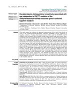
Figure 1
The chemotactic effect of chitosan on PMNsThe chemotactic effect of chitosan on PMNs. Freshly isolated polymor-
phonuclear neutrophils (PMNs) were pre-stained with 1 μg/ml calcein-
AM and seeded on a polycarbonate filter placed above a well contain-
ing (a) N-formyl-methionyl-leucyl-phenylalanine (fMLP; positive control)
or (b) 80% deacetylated (80 M; black line) or 95% deacetylated (95 M;
gray line) chitosan. PMNs were allowed to migrate for 1 hour before
assessing migration, as described in 'Materials and Methods'. Percent-
age migration of PMNs = the fluorescence of migrated PMNs/fluores-
cence of 20,000 PMNs/ml × 100, obtained from the standard curve.
Results are presented as mean ± standard error. P values from Stu-
dent's two-tailed unpaired t-test: *P = 0.01, fluorescence of PMNs
migrated toward 25 μg/ml 80 M chitosan versus fluorescence of PMNs
migrated toward RPMI (Ctrl-); **P = 0.004, fluorescence of PMNs
migrated toward 50 μg/ml 80 M chitosan versus fluorescence of PMNs
migrated toward RPMI. The positive control is the fMLP curve. This fig-
ure represents the results of three independent experiments.
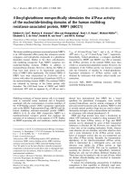
Figure 2
The effect of inhibitors on PMN chemotaxis towards chitosanThe effect of inhibitors on PMN chemotaxis towards chitosan. Freshly
isolated polymorphonuclear neutrophils (PMNs) were resuspended in
RPMI 1640 supplemented with 0.1% decomplemented fetal bovine
serum, pre-stained with 1 μg/ml calcein-AM for 30 minutes at 37°C and
incubated with 0.5 μg/ml pertussis toxin for 90 minutes and seeded on
a polycarbonate filter above a well containing 50 μg/ml 80%
deacetylated (80 M) chitosan. Alternatively, PMNs were incubated with
10
-7
mol/l pyrrolidine for the last 10 minutes of the incubation with cal-
cein-AM. Chemotaxis was performed as described in Figure 1. The per-
centage inhibition of migration corresponds to the fluorescence of
PMNs incubated with the inhibitors that migrated toward 80 M chitosan
versus fluorescence of PMN incubated in media that migrated toward
80 M chitosan. This figure represents the results of at least three inde-
pendent experiments. *P = 0.02 and ***P = 0.0001.
Arthritis Research & Therapy Vol 11 No 3 Simard et al.
Page 6 of 10
(page number not for citation purposes)
To determine whether 80 M and 95 M chitosan induce PMNs
to degranulate, the release of the contents of the primary and
secondary granules was determined by measuring the amount
of MPO and lactoferrin released. When incubated with 10 to
100 μg/ml 80 M or 95 M chitosan, the amounts of MPO and
lactoferrin released into the media by PMNs were negligible
(Figure 4). Together, the above observations indicate that the
effects of 80 M chitosan on PMNs are not associated with the
release of granule substances from PMNs.
The interaction of 80 M and 95 M chitosan with
polymorphonuclear neutrophils
The difference in chemotactic activity between 80 M and 95
M chitosan toward PMNs may be due to the inability of PMNs
to bind and/or internalize 95 M chitosan. The binding and inter-
nalization of RITC-80 M and RITC-95 M chitosan by PMNs
was investigated by live cell confocal microscopy. Live cell
imaging revealed that PMNs internalized both RITC-80 M and
RITC-95 M chitosan in the presence of decomplemented
serum, although internalization was much greater for fluores-
cent zymosan under similar conditions (Figure 5).
Because all of the white blood cells are present at the microf-
racture sites and could be involved in the effects of chitosan
on cartilage repair and wound healing, we also investigated
the ability of 80 M chitosan to interact with other leukocytes.
To observe the interaction of 80 M chitosan with leukocytes
with the same differential ratio in which these cells normally co-
exist, this analysis was performed in whole blood devoid of
erythrocytes. Flow cytometry analysis of leukocytes in whole
blood revealed that a greater amount of RITC-80 M chitosan
associates with monocytes than granulocytes and lym-
phocytes (Figure 6a). Confocal microscopy revealed that
monocytes readily internalize large amounts of RITC-80 M and
RITC-95 M chitosan (Figure 6b).
Discussion
Novel therapeutic modalities that promote cartilage regenera-
tion have the potential to delay significantly the progression of
OA in patients who develop focal lesions. We therefore inves-
tigated some of the molecular mechanisms involved in the clin-
ically beneficial effects of 80 M chitosan, which was recently
shown to promote cartilage regeneration in both large and
small animal cartilage repair models [2,3]. In recent years evi-
dence has accumulated that the PMN is more than just a leu-
kocyte that phagocytoses foreign antigens. PMNs differentiate
into dendritic cells [24] and have the capacity to modulate the
adaptive immune response. The PMN therefore adopts differ-
ent phenotypes that are determined by its environment. In this
light, the response of PMNs to 80 M chitosan was investigated
to identify the characteristics of the phenotype of PMNs that
promotes cartilage regeneration. We report that 80 M chi-
tosan selectively activates a subset of PMN functional
responses.
The distinct phenotype of PMNs in response to 80 M chitosan
is characterized by PMN chemotaxis and the absence of the
production of superoxide and degranulation. In comparison,
fMLP, a bacterial-derived peptide, is not only chemotactic for
PMNs but also stimulates them to produce superoxide and
degranulate. These observations strongly suggest that the
PMN phenotype in the presence of 80 M chitosan promotes
repair due, at least in part, to the lack of superoxide production
and PMN degranulation. Our data agree with a recent report
indicating that water-soluble chitosan oligomers suppress the
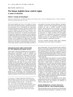
Figure 3
Production of superoxide anions by PMN in response to chitosanProduction of superoxide anions by PMN in response to chitosan.
Superoxide anion production was determined using the cytochrome c
reduction assay. Freshly isolated polymorphonuclear neutrophils
(PMNs) resuspended in RPMI 1640 supplemented with 0.1% decom-
plemented fetal bovine serum were incubated with the indicated con-
centrations of 80% deacetylated (80 M) or 95% deacetylated (95 M)
chitosan for 10 minutes at 37°C. Results are presented as mean ±
standard error. The difference from the negative control is statistically
significant: *P < 0.001 (Tukey-Kramer test). The negative control =
PMNs incubated in Hanks' Balanced Salt Solution supplemented with
0.1% decomplemented fetal bovine serum and incubated with diluent
(dimethyl sulfoxide [DMSO]). This figure represents the results of at
least three independent experiments. fMLP, N-formyl-methionyl-leucyl-
phenylalanine.
Available online />Page 7 of 10
(page number not for citation purposes)
capacity for PMNs to respond to phorbol myristate acetate
[16].
Because 80 M chitosan is chemotactic for PMNs, it must inter-
act at the surface of PMNs to elicit a chemotactic response.
The majority of chemotactic factors mediate their effect
through G-protein-coupled receptors. To determine whether
this applies to 80 M chitosan, we assessed the effect of per-
tussis toxin on 80 M chitosan-induced chemotaxis of PMN.
Pertussis-toxin inhibited PMN chemotaxis by 80%, implicating
a G-protein-coupled receptor. The mechanism through which
80 M chitosan activates a G-protein-coupled receptor remains
to be determined. It was previously found that conditioned
media from canine PMNs stimulated with >80% DDA chitosan
particles promoted chemotaxis of neutrophils [14]. We pro-
vide evidence, using a specific cPLA
2
-α inhibitor, that phos-
pholipid-derived mediators, possibly the chemotactic factors
LTB
4
and PAF, are involved in the direct chemotactic activity
Figure 4
Release of myeloperoxidase and lactoferrin by PMNs in response of chitosanRelease of myeloperoxidase and lactoferrin by PMNs in response of chitosan. Degranulation was determined using the (a, b) myeloperoxidase
(MPO) and (c, d) lactoferrin assay, as described in 'Materials and Methods'. Freshly isolated polymorphonuclear neutrophils (PMNs) resuspended in
RPMI 1640 supplemented with 0.1% decomplemented fetal bovine serum were treated with cytochalasin B and further incubated with the indicated
concentrations of 80% deacetylated (80 M; panels a and c) or 95% deacetylated (95 M; panels b and d) chitosan for 30 minutes at 37°C. The
quantity of MPO released is expressed as '% MPO', which corresponds to the ratio of the amount of MPO released/total amount of cellular MPO.
The amount of lactoferrin released is expressed in ng/ml. Results are presented as mean ± standard error. The difference from the negative control
is statistically significant: *P < 0.001 (Tukey-Kramer test). The negative control = PMNs incubated in Hanks' Balanced Salt Solution supplemented
with 0.1% decomplemented fetal bovine serum and incubated with cytochalasin B. This figure represents the results of at least three independent
experiments. fMLP, N-formyl-methionyl-leucyl-phenylalanine.
Arthritis Research & Therapy Vol 11 No 3 Simard et al.
Page 8 of 10
(page number not for citation purposes)
of human PMNs toward a pure and sterile 80 M chitosan prep-
aration. This is the first study to demonstrate that such lipid
mediators contribute to half of the chemotactic activity of
human PMNs toward chitosan. The cPLA
2
-α inhibitor pyrrolid-
ine-1 inhibited the chemotaxis of PMNs by 50%. Moreover, the
inhibition of chemotaxis by 80% in the presence of pertussis
toxin suggests that additional chemotactic agents acting
through G-protein-coupled receptors participate in the chem-
otaxis of PMNs toward chitosan. Further investigation is
required to characterize fully the molecular mechanisms that
are involved in 80 M chitosan-induced chemotaxis of human
PMNs.
Having characterized the response of PMNs toward 80 M chi-
tosan, we conducted similar experiments with 95 M chitosan
because we had observed a distinct response of PMNs
toward 95 M chitosan in vivo. This is the first report on the
effect of 95 M chitosan on PMN effector functions. Chitosan
95 M (95% glucosamine, 5% N-acetyl glucosamine) was una-
ble to induce chemotactic activity, superoxide production, or
the release of granule contents by PMNs. The lack of chemo-
tactic activity of 95 M chitosan toward PMNs was not due to
an effect on the viability of PMNs (data not shown). The per-
centage DDA of chitosan is therefore a determining factor for
Figure 5
The internalization of chitosan by PMNsThe internalization of chitosan by PMNs. Freshly isolated polymorpho-
nuclear neutrophils (PMNs) were resuspended in RPMI 1640 supple-
mented with 0.1% decomplemented fetal bovine serum, pre-stained
with 1 μg/ml calcein-AM for 30 minutes at 37°C and incubated with
100 μg/1 × 10
6
cells rhodamine B isothiocyanate (RITC)-zymosan for
1.5 hours (a positive control), 15 μg/ml RITC-80% deacetylated (80 M)
or RITC-95% deacetylated (95 M) chitosan for 3 hours at 37°C. PMNs
were then centrifuged and plated on a slide coated with 100% decom-
plemented autologous serum and visualized by live confocal micros-
copy. The index of internalization of chitosan by PMNs was calculated
as the percentage of cells that internalized RITC-chitosan. Results are
presented as mean ± standard error. This figure represents the results
of three independent experiments.
Figure 6
The interaction of chitosan with monocytes, granulocytes, and lym-phocytes in whole bloodThe interaction of chitosan with monocytes, granulocytes, and lym-
phocytes in whole blood. (a) Whole blood was incubated for 30 min-
utes with 5 μg/ml rhodamine B isothiocyanate (RITC)-80%
deacetylated (80 M) chitosan at 37°C for 30 minutes before analysis by
flow cytometry. The binding index was calculated as fluorescence units
of each leukocyte population incubated with RITC-80 M chitosan/fluo-
rescence units of leukocyte population in the absence of RITC-chi-
tosan. Results are presented as mean ± standard error. P values from
Student's two-tailed unpaired t-test: **P < 0.005 versus autofluores-
cence for each leukocyte population. (b) Macrophages were seeded
on glass slides (2 × 10
6
cells/ml) in RPMI 1640 supplemented with
0.1% decomplemented fetal bovine serum, pre-stained with 1 μg/ml
calcein AM for 30 minutes at 37°C, and incubated with 100 μg/1 ×
10
6
cells RITC-zymosan for 1.5 hours (a positive control), 15 μg/ml
RITC-80 M, or RITC-95% deacetylated (95 M) chitosan for 3 hours at
37°C. Macrophages were then visualized live through a spinning disc
confocal microscope with a 63× objective. The top panels are images
taken in the X-Y plane and the lower panels are images taken in the X-Z
plane. This figure represents the results of three independent experi-
ments.
Available online />Page 9 of 10
(page number not for citation purposes)
the activation of PMNs by chitosan and potentially for the ther-
apeutic use of chitosan. Our findings indicate that chitosans in
the range from 80% to 95% DDA can elicit quite different bio-
logic responses, and highlight the importance of defining the
DDA level when conducting biologic assays. Some of the dif-
ferential responses could be related to the very low solubility
of 95% DDA chitosan at neutral pH [8]. In the light of these
findings, it is of interest to investigate fully the effect of chi-
tosan with other percentages of DDA on PMNs to determine
whether there is a percentage DDA that induces maximal
chemotactic activity in PMNs and consequently an optimal
therapeutic effect.
Another parameter that may modify the response of PMNs to
chitosan is the form in which the chitosan is used. Vandevord
and coworkers [6] reported that a chitosan scaffold made with
chitosan of 92% DDA is chemotactic for PMN in vivo.
Because 92% DDA chitosan is structurally more similar to 95
M than to 80 M chitosan, our data indicate that PMN migration
to 92% DDA chitosan should be quite modest. The discrep-
ancy between this previous observation and our findings could
potentially be explained by the fact that the scaffold used in the
in vivo study was prepared by coating polytetrafluoroethylene
tubes with 92% DDA chitosan, and did not employ pure chi-
tosan. It will be of therapeutic interest to determine how differ-
ently PMNs react to chitosans of the same percentage DDA of
different structural forms – suspension versus scaffold.
It is generally accepted that PMN phagocytose chitosan, but
no microscopy studies have been performed to demonstrate
that PMNs indeed internalize chitosan. This is a relevant ques-
tion because PMNs can respond to foreign material without
necessarily internalizing it. We provide direct evidence that
PMNs can internalize 80 M chitosan without stimulating
degranulation. Around 10% of PMNs internalized 80 M chi-
tosan, in the presence of 0.5% heat-inactivated serum. These
observations are quite different to those in PMN and monoso-
dium urate crystals, which have a poor capacity for internaliza-
tion while strongly activating PMNs, probably because of an
autocrine effect [25]. It is highly likely that lipid mediators are
involved in this autocrine effect. In our internalization assay,
PMNs readily internalized zymosan, a yeast cell wall prepara-
tion that activates neutrophils. Because both 95 M and 80 M
were internalized without activating neutrophils, our data dem-
onstrate that internalization of a polysaccharide biomaterial
does not automatically trigger degranulation.
Recently, PMNs were reported to express the mannose recep-
tor [26], a receptor that is implicated in the internalization of
chitosan by macrophages [27,28]. We provide evidence that
monocytes internalize 80 M chitosan more readily than PMNs,
suggesting that the molecular mechanisms involved in the
internalization of 80 M chitosan by PMNs differ from those of
macrophages. This does not imply a less important role of
PMNs in chitosan-based wound healing. PMNs usually out-
number macrophages in certain phases of wound healing and
can collectively synthesize large quantities of soluble media-
tors.
Conclusions
In summary, 80 M chitosan is chemotactic for human PMNs
but does not activate additional PMN effector functions such
as degranulation and superoxide production. Because the
beneficial therapeutic effects of 80 M chitosan are preceded
by the recruitment of a significant number of PMNs, this chi-
tosan-induced PMN phenotype could be associated with pro-
motion of repair. Our observations also indicate that the
degree of deacetylation is an important factor to consider in
the use of chitosan as an accelerator of repair because PMNs
exhibit a differential capacity to migrate towards 80 M and 95
M chitosan.
Competing interests
The authors declare that they have no competing interests.
Authors' contributions
PS and HG made equal contributions to the experimental
aspects of this study. They performed the majority of the exper-
iments. SM and DR made important contributions to the con-
duct of certain experiments as well as the interpretation of the
data. MF, PP, and CH contributed to the design of the experi-
ments and the interpretation of the data. MF wrote the manu-
script, and PP and CH revised it. All the experiments were
performed and supervised in MF's laboratory. HEG contrib-
uted to the interpretation of the data.
Acknowledgements
This research was funded by a Discovery Advancement Program grant
awarded by the Canadian Arthritis Network to CDH, MJGF, PEP, and
HEG. PS is a recipient of a scholarship awarded by the Canadian Arthri-
tis Network, MJGF received a salary award from The Arthritis Society,
and CDH received a salary award from the Fonds de la Recherche
Santé Québec. We should like to thank Dr Matthew Shive for insightful
comments and critical reading of the manuscript.
References
1. Felson DT: An update on the pathogenesis and epidemiology
of osteoarthritis. Radiol Clin North Am 2004, 42:1-9.
2. Hoemann CD, Hurtig M, Rossomacha E, Sun J, Chevrier A, Shive
MS, Buschmann MD: Chitosan-glycerol phosphate/blood
implants improve hyaline cartilage repair in ovine microfrac-
ture defects. J Bone Joint Surg Am 2005, 87:2671-2686.
3. Hoemann CD, Sun J, Mckee MD, Chevrier A, Rossomacha E,
Rivard GE, Hurtig M, Buschmann MD: Chitosan-glycerol phos-
phate/blood implants elicit hyaline cartilage repair integrated
with porous subchondral bone in microdrilled rabbit defects.
Osteoarthritis Cartilage 2007, 15:78-89.
4. Shive MS, Hoemann CD, Restrepo A, Hurtig MB, Duval N, Ranger
P, Stannish W, Buschmann MD: BST-CarGel: in situ chondroin-
duction for cartilage repair. Operative techniques in orthopae-
dics: articular cartilage surgery. Operative Techn Orthopaedics
2006, 16:271-278.
5. Onishi H, Machida Y: Biodegradation and distribution of water-
soluble chitosan in mice. Biomaterials 1999, 20:175-182.
6. VandeVord PJ, Matthew HWT, DeSilva SP, Mayton L, Wu B, Woo-
ley PH: Evaluation of the biocompatibility of a chitosan scaffold
in mice. J Biomed Mater Res 2002, 59:585-590.
Arthritis Research & Therapy Vol 11 No 3 Simard et al.
Page 10 of 10
(page number not for citation purposes)
7. Malette WG, Quigley HJ, Gaines RD, Johnson ND, Rainer WG:
Chitosan – a new hemostatic. Ann Thorac Surg 1983,
36:55-58.
8. Varum KM, Holme HK, Izume M, Stokke BT, Smidsrod O: Deter-
mination of enzymatic hydrolysis specificity of partially N-
acetylated chitosans. Biochim Biophys Acta 1996, 1291:5-15.
9. Sashiwa H, Saimoto H, Shigemasa Y, Ogawa R, Tokura S: Lys-
ozyme susceptibility of partially deacetylated chitin. Int J Biol
Macromol 1990, 12:295-296.
10. Tomihata K, Ikada Y: In vitro and in vivo degradation of films of
chitin and its deacetylated derivatives. Biomaterials 1997,
18:567-575.
11. Chevrier A, Hoemann CD, Sun J, Buschmann MD: Chitosan-glyc-
erol phosphate/blood implants increase cell recruitment, tran-
sient vascularization and subchondral bone remodeling in
drilled cartilage defects. Osteoarthritis Cartilage 2007,
15:316-327.
12. Usami Y, Okamoto Y, Minami S, Matsuhashi A, Kumazawa NH,
Tanioka S, Shigemasa Y: Migration of canine neutrophils to chi-
tin and chitosan. J Vet Med Sci 1994, 56:1215-1216.
13. Usami Y, Okamoto Y, Minami S, Matsuhashi A, Kumazawa NH,
Tanioka S, Shigemasa Y: Chitin and chitosan induce migration
of bovine polymorphonuclear cells. J Vet Med Sci 1994,
56:761-762.
14. Usami Y, Okamoto Y, Takayama T, Shigemasa Y, Minami S: Chitin
and chitosan stimulate canine polymorphonuclear cells to
release leukotriene B-4 and prostaglandin E-2. J Biomed
Mater Res 1998, 42:517-522.
15. Ueno H, Yamada H, Tanaka I, Kaba N, Matsuura M, Okumura M,
Kadosawa T, Fujinaga T: Accelerating effects of chitosan for
healing at early phase of experimental open wound in dogs.
Biomaterials 1999, 20:1407-1414.
16. Dou JL, Tan CY, Du YG, Bai XF, Wang KY, Ma XJ: Effects of chi-
tooligosaccharides on rabbit neutrophils in vitro. Carbohyd
Polym 2007, 69:209-213.
17. Ma O, Lavertu M, Sun J, Nguyen S, Buschmann MD, Winnik FM,
Hoemann CD: Precise derivatization of structurally distinct chi-
tosans with rhodamine B isothiocyanate. Carbohyd Polym
2008, 72:
616-624.
18. Fernandes MJ, Rollet-Labelle E, Pare G, Marois S, Tremblay ML,
Teillaud JL, Naccache PH: CD16b associates with high-density,
detergent-resistant membranes in human neutrophils. Bio-
chem J 2006, 393:351-359.
19. Gilbert C, Levasseur S, Desaulniers P, Dusseault AA, Thibault N,
Bourgoin SG, Naccache PH: Chemotactic factor-induced
recruitment and activation of Tec family kinases in human
neutrophils. II. Effects of LFM-A13, a specific Btk inhibitor. J
Immunol 2003, 170:5235-5243.
20. Bradley PP, Priebat DA, Christensen RD, Rothstein G: Measure-
ment of cutaneous inflammation: estimation of neutrophil
content with an enzyme marker. J Invest Dermatol 1982,
78:206-209.
21. Mocsai A, Ligeti E, Lowell CA, Berton G: Adhesion-dependent
degranulation of neutrophils requires the Src family kinases
Fgr and Hck. J Immunol 1999, 162:1120-1126.
22. Desmeules P, Dufour M, Fernandes MJ: A rapid flow cytometry
assay for the assessment of calcium mobilization in human
neutrophils in a small volume of lysed whole-blood. J Immunol
Methods 2009, 340:154-157.
23. Bauldry SA, Wooten RE: Leukotriene B4 and platelet activating
factor production in permeabilized human neutrophils: role of
cytosolic PLA2 in LTB4 and PAF generation. Biochim Biophys
Acta 1996, 1303:63-73.
24. Iking-Konert C, Cseko C, Wagner C, Stegmaier S, Andrassy K,
Hansch GM: Transdifferentiation of polymorphonuclear neu-
trophils: acquisition of CD83 and other functional characteris-
tics of dendritic cells. J Mol Med 2001, 79:464-474.
25. Desaulniers P, Fernandes M, Gilbert C, Bourgoin SG, Naccache
PH: Crystal-induced neutrophil activation. VII. Involvement of
Syk in the responses to monosodium urate crystals. J Leukoc
Biol 2001, 70:659-668.
26. Valera I, Vigo AG, Alonso S, Barbolla L, Crespo MS, Fernandez N:
Peptidoglycan and mannose-based molecular patterns trigger
the arachidonic acid cascade in human polymorphonuclear
leukocytes. J Leukocyte Biol 2007, 81:925-933.
27. Mori T, Murakami M, Okumura M, Kadosawa T, Uede T, Fujinaga T:
Mechanism of macrophage activation by chitin derivatives. J
Vet Med Sci 2005, 67:51-56.
28. Feng J, Zhao L, Yu Q: Receptor-mediated stimulatory effect of
oligochitosan in macrophages. Biochem Biophys Res Commun
2004, 317:414-420.