Báo cáo y học: "Expression of 5-lipoxygenase and 15-lipoxygenase in rheumatoid arthritis synovium and effects of intraarticular glucocorticoids" docx
Bạn đang xem bản rút gọn của tài liệu. Xem và tải ngay bản đầy đủ của tài liệu tại đây (2.89 MB, 11 trang )
Open Access
Available online />Page 1 of 11
(page number not for citation purposes)
Vol 11 No 3
Research article
Expression of 5-lipoxygenase and 15-lipoxygenase in rheumatoid
arthritis synovium and effects of intraarticular glucocorticoids
Karina Roxana Gheorghe
1,2
, Marina Korotkova
2
, Anca Irinel Catrina
2
, Linda Backman
3
, Erik af Klint
2
,
Hans-Erik Claesson
3,4
, Olof Rådmark
4
and Per-Johan Jakobsson
2,5
1
Department of Biosciences and Nutrition, Novum, Karolinska Institute, SE-141 57 Huddinge, Sweden
2
Department of Medicine, Rheumatology Unit, Karolinska University Hospital and Karolinska Institute, S-171 76 Stockholm, Sweden
3
Orexo AB, Virdings allé 32 A, SE-751 05 Uppsala, Sweden
4
Department of Medical Biochemistry and Biophysics, Karolinska Institutet, SE-171 77 Stockholm, Sweden
5
Karolinska Biomic Center, Karolinska University Hospital and Karolinska Institute, S-171 76 Stockholm, Sweden
Corresponding author: Per-Johan Jakobsson,
Received: 6 Jan 2009 Revisions requested: 24 Feb 2009 Revisions received: 6 May 2009 Accepted: 4 Jun 2009 Published: 4 Jun 2009
Arthritis Research & Therapy 2009, 11:R83 (doi:10.1186/ar2717)
This article is online at: />© 2009 Gheorghe et al.; licensee BioMed Central Ltd.
This is an open access article distributed under the terms of the Creative Commons Attribution License ( />),
which permits unrestricted use, distribution, and reproduction in any medium, provided the original work is properly cited.
Abstract
Introduction It was previously shown that lipoxygenase (LO)
pathways are important in the rheumatoid arthritis (RA)
inflammatory process and that synovial fluid from RA patients
contains high amounts of leukotrienes. We therefore aimed to
investigate the 5-LO and 15-LO-1 expression pattern in RA and
ostheoarthritis (OA) synovial tissue and to study the effect of
intraarticular glucocorticoid (GC) therapy on enzyme
expression.
Methods Expression of LOs was evaluated by
immunohistochemistry in RA and OA synovial biopsies. Cellular
localization of these enzymes was analyzed by double
immunofluorescence. In synovial biopsies from 11 RA patients,
5-LO and 15-LO-1 expression was evaluated before and after
triamcinolone hexacetonide knee injection and assessed by
image analysis to quantify their expression. We also investigated
the presence of 15-LO-1 by immunohistochemistry in synovial
fluid (SF) cells as well as their ability to form 15-
hydroxyeicosatetraenoic acid (15-HETE) following treatment
with arachidonic acid (AA).
Results 5-LO and 15-LO-1 are present in RA and OA synovium,
with 5-LO being mostly expressed in lining and sublining
macrophages, neutrophils and mast cells and 15-LO-1 mainly in
lining macrophages, fibroblasts and sublining endothelial cells.
Intraarticular GC treatment resulted in a significant suppression
of 5-LO expression, but did not influence the 15-LO-1 enzyme
significantly. Also, SF cells express a functional 15-LO-1 and
produce 15-HETE when challenged with AA.
Conclusions These data demonstrate that local therapy with
GC decreases 5-LO expression in RA synovium and offer an
additional possible mechanism for the efficiency of intraarticular
adjuvant therapy in RA.
Introduction
Rheumatoid arthritis (RA) is a chronic inflammatory disease
characterized by polyarticular joint inflammation, synovial
hyperplasia, and cartilage and bone destruction, with subse-
quent joint deformities. The inflammatory synovial fluid in RA
patients contains–in addition to various cytokines and growth
factors–high levels of leukotrienes, with leukotriene B
4
(LTB
4
)
being predominant [1].
LTB
4
is a powerful proinflammatory lipid mediator and one of
the most potent chemotactic agents known to date [2]. This
leukotriene is produced mainly by neutrophils, macrophages
and mast cells, and promotes neutrophil recruitment and acti-
vation [3]. Neutrophils are the most abundant leukocytes in
rheumatoid joints [4], and have destructive potential by secret-
ing proteases and reactive oxygen species and by promoting
synthesis of matrix metalloproteinases [5,6]. Several lines of
evidence have implicated LTB
4
as an important mediator of
joint inflammation in RA. LTB
4
is present at higher levels in
15-HETE: 15-hydroxyeicosatetraenoic acid; AA: arachidonic acid; GC: glucocorticoid; IL: interleukin; LO: lipoxygenase; LTB
4
: leukotriene B
4
; OA:
osteoarthritis; PBS: phosphate-buffered saline; RA: rheumatoid arthritis; RANKL: receptor activator of NF-κB ligand; TNF: tumor necrosis factor.
Arthritis Research & Therapy Vol 11 No 3 Gheorghe et al.
Page 2 of 11
(page number not for citation purposes)
serum of patients with active RA compared with patients with
inactive arthritis or normal subjects [7], and its levels correlate
with the disease severity [8].
A critical contribution of neutrophil-derived LTB
4
to arthritis
induction and severity has recently been revealed in a mouse
serum transfer model of inflammatory arthritis [9]. In this study
it was shown that mice lacking 5-lipoxygenase (5-LO) or leu-
kotriene A
4
hydrolase enzymes are protected from developing
the disease and that there is a specific requirement for LTB
4
and not other leukotrienes for the pathogenesis in this model.
5-LO and 5-LO activating protein (FLAP), followed by leukot-
riene A
4
hydrolase, are the enzymes responsible for the
sequential formation of LTB
4
from arachidonic acid (AA).
15-Lipoxygenase (15-LO) is a lipid-peroxidizing enzyme mainly
expressed in airway epithelial cells, eosinophils, reticulocytes
and macrophages. In humans, 15-LO exists as two different
enzymes with different cell localizations and product profiles
[10]. 15-LO-1 converts AA to an unstable intermediate, 15-
hydroperoxyeicosatetraenoic acid, which can be further con-
verted to 15-hydroxyeicosatetraenoic acid (15-HETE). The 15-
LO-1 enzyme has proinflammatory actions, with high levels of
15-HETE reported in sputum of asthmatic patients along with
increased macrophage 15-LO-1 mRNA expression [11]. 15-
LO-1 expression is induced by IL-13 in human blood mono-
cytes [12] and by IL-4 in monocytes, alveolar macrophages,
dendritic cells, mast cells and rheumatoid arthritis synovial
cells [12-18]. Only recently was it reported that 15-LO-1 can
catalyze the metabolism of AA to the proinflammatory eoxins
that can increase permeability of the endothelial cell monol-
ayer in vitro, indicating that they can enhance vascular perme-
ability [19]. 15-LO-1 products, however, were also
demonstrated to have protective roles in inflammatory disor-
ders due to formation of anti-inflammatory lipoxins [20-22].
The 15-LO-1 mRNA was demonstrated to be present in RA
synovial membranes [23] and its expression was stronger in
RA compared with osteoarthritis (OA) biopsies [24].
The 5-LO cascade and the role of LTB
4
in RA are well docu-
mented. Although the presence of 5-LO enzyme in the synovial
lining of rheumatoid tissue has recently been reported [24], a
detailed characterization of cells expressing 5-LO in human
synovial tissue is lacking. Evidence is also limited regarding
the influence of current therapy for RA on this pathway.
Glucocorticoids (GCs) are used in RA as an efficient adjuvant
therapy and their efficacy is related to their broad anti-inflam-
matory profile, with inhibition of inflammatory cells functions
[25]. Controversial results have been reported about the
effects of GCs on 5-LO expression and LTB
4
formation. Some
studies reported that 5-LO pathway activity is decreased in
the presence of GCs [26,27], while other investigators have
shown that in vivo GC administration had no influence on
LTB
4
formation [28,29]. In contrast, leukotriene synthesis and
5-LO expression were increased in human blood monocytes
[30] and mast cells [31] by dexamethasone. In addition, blood
polymorphonuclear neutrophils from RA patients released
higher amounts of LTB
4
after GC pulse therapy [32] while
intraarticular corticosteroids reduced the LTB
4
level in synovial
fluid of RA patients [33].
In comparison, there are few studies to date investigating the
effects of corticosteroids on 15-LO-1 expression. In a rabbit
model for atherosclerosis, corticosteroid treatment was shown
to decrease atherosclerotic plaque formation along with
increasing 15-LO-1 expression in the arterial wall [34]. GC
treatment of asthma patients, however, has been reported to
decrease the expression of 15-LO-1 in the lung [35].
In the present study we characterized the expression pattern
of 5-LO and 15-LO-1 enzymes in synovial tissue of RA and OA
patients and phenotyped the positive cells. In addition, we
determined the effects of intraarticular glucocorticoids on the
expression of these enzymes in RA synovium.
Materials and methods
Patients
In the first study group, we analyzed synovial biopsies from six
RA patients and from five OA patients collected at the time of
orthopedic surgery. In a second group, 11 RA patients were
recruited into the study. The demographical and clinical data
of the second patients group are presented in Table 1.
All patients in the second group received an intraarticular knee
injection of 40 mg triamcinolone hexacetonide, and synovial
biopsies were collected by arthroscopy immediately prior to
treatment and a median of 10 days after treatment. The treat-
ment regimen remained unchanged from at least 2 weeks prior
to and during the entire study period.
Table 1
Demographical and clinical data of the second patient group (n
= 11)
Characteristic Value
Age (years) 68 (35 to 83)
Gender (male/female) 3/8
Disease duration (months) 24 (3 to 240)
Current knee arthritis episode duration (months) 2 (0.5 to 6)
Taking disease-modifying antirheumatic drugs 6
Taking oral corticosteroids 2
Taking nonsteroidal anti-inflammatory drugs 4
Time between biopsies (days) 10 (7 to 12)
Data presented as median (range) or number of patients.
Available online />Page 3 of 11
(page number not for citation purposes)
All RA patients fulfilled the 1987 American College of Rheu-
matology diagnostic criteria for RA [36]. The ethics committee
at the Karolinska Hospital approved all experiments on human
cells and tissues. Informed consent was obtained from all
study subjects.
Tissue preparation and immunohistochemical analysis
Serial cryostat sections (7 μm) were fixed for 20 minutes in 2%
formaldehyde (v/v), air-dried and then stored at -70°C. Immu-
nohistochemical staining was performed as described previ-
ously [37]. The inhouse antibodies used were affinity-purified
rabbit polyclonal antibody against human 5-LO and rabbit pol-
yclonal anti-human 15-LO-1 antibody. Rabbit IgG served as
the negative control. Stained synovial biopsies were evaluated
using a Polyvar II microscope (Reichert-Jung, Vienna, Austria)
and photographs were taken with a digital camera (300F;
Leica, Cambridge, UK). Synovial expression of 5-LO and 15-
LO-1 was quantified by computer-assisted image analysis and
was expressed as the percentage of positive stained area ver-
sus total tissue area.
Synovial fluid cells from RA patients were collected on slides
by cytospin centrifugation. The slides were then fixed and
processed for immunhistochemical detection of 15-LO-1 as
described above.
Immunofluorescence staining
Double immunofluorescence staining was performed using
rabbit anti-human 5-LO or 15-LO-1, mouse anti-human
CD163 (Ber-MAC3; DakoCytomation, Glostrup, Denmark),
mouse anti-human CD68 (KP1; DakoCytomation), mouse anti-
human prolyl 4-hydrolase (DakoCytomation), mouse anti-
human CD66b (80H3; Beckman Coulter, France), mouse anti-
human CD3 (SK7; BD Biosciences, San Jose, CA, USA),
mouse anti-human CD20 (DakoCytomation), mouse anti-
human CD31 (EN4; Novakemi AB, Handen, Sweden), and
mouse anti-human mast cell tryptase (Chemicon International,
Temecula, CA, USA) antibodies.
The staining procedure has been published previously [38].
Briefly, after blocking with an avidin–biotin kit (Vector Labora-
tories, Peterborough, UK), sections were incubated overnight
with primary antibodies. Subsequently, slides were incubated
with secondary biotinylated goat anti-rabbit antibody (heavy
and light chain; Vector Laboratories) and streptavidin-conju-
gated fluorochrome Alexa 488 (Molecular Probes, Leiden, the
Netherlands). The slides were blocked again with the avidin–
biotin kit and were incubated with the next secondary bioti-
nylated horse anti-mouse antibody (IgG heavy and light chain;
Vector Laboratories), followed by streptavidin-conjugated
fluorochrome Alexa 546 (Molecular Probes). Matched IgG iso-
type controls were included for all markers.
15-LO-1 product measurement in RA synovial fluid cells
Synovial fluid from RA patients was centrifuged and the pel-
leted cells were resuspended in PBS and washed twice. The
cellular composition of synovial fluid cells was analyzed using
flow cytometry. Monocyte, neutrophil and lymphocyte popula-
tions were identified using a FACSCalibur (Becton Dickinson,
San Jose, CA, USA) and Cell Quest software (Becton Dickin-
son). AA was added to a final concentration of 40 μM and the
cells were incubated for 5 minutes at 37°C. Buffer control
without cells was used to assess for spontaneous degradation
of AA. Subsequently, the samples were centrifuged and the
supernatant collected and stored at -70°C until analysis by
enzyme immunoassay according to the manufacturer's instruc-
tions (Cayman Chemicals, Ann Arbor, MI, USA).
Statistical analysis
Statistical analysis was performed using the Wilcoxon test and
Bonferroni correction for multiple comparisons for paired sam-
ples for the synovial biopsy data, and using the Mann–Whitney
test for 15-HETE production.
Results
RA synovial tissue displays a higher expression of 5-LO
and 15-LO-1 enzymes compared with OA samples
We detected intracellular 5-LO staining in all RA samples
studied. Sections incubated with the preadsorbed 5-LO anti-
body showed no significant staining, confirming the specificity
of the antibody for the 5-LO enzyme (Figure 1a, inset). Strong
5-LO staining was shown in macrophage-like cells within the
synovial lining layer and in sublining tissue (Figure 1a,b). 5-LO
positivity was scarce in the follicular mononuclear infiltrates,
with the majority of patients not having detectable staining in
these areas. By contrast, 15-LO-1 showed a very strong stain-
ing pattern mainly in the synovial lining cells and in vessels,
with lower expression in scattered sublining macrophage-like
and fibroblast-like cells (Figure 1e). The specificity of 15-LO-1
antibody was tested in bronchial tissue, and the airway epithe-
lium was strongly stained (Figure 1d). In contrast, there was no
staining after preincubation with the 15-LO-1-specific peptide
against which the antibody was raised (Figure 1d, inset).
The OA synovial samples displayed mostly large areas of fibro-
sis and cartilage, with limited synovial membranes. Positive
staining for 5-LO and 15-LO was detected almost exclusively
in the synovial membrane areas. OA tissue showed low
expression of both 5-LO and 15-LO-1 enzymes, with few
stained cells scattered in the synovial membrane areas (Figure
1c,f). Strong staining for 15-LO-1, however, was detected in
blood vessel cells.
We then quantitatively analyzed the expression of LO enzymes
in RA and OA synovial tissue sections. Both 5-LO and 15-LO-
1 showed a lower expression in OA synovial tissue compared
with RA samples (Figure 2).
Arthritis Research & Therapy Vol 11 No 3 Gheorghe et al.
Page 4 of 11
(page number not for citation purposes)
Phenotype of cells expressing 5-LO and 15-LO in RA
synovium
We characterized the cellular distribution of the respective
enzymes in RA synovial tissue. As shown by double immun-
ofluorescence, 5-LO was mainly detected in synovial CD163
+
macrophages (Figure 3a) and in CD68
+
macrophages (data
not shown), but not in fibroblasts. 5-LO expression was also
detected in scattered CD66b
+
neutrophils and tryptase-posi-
tive mast cells (Figure 3b,c). 15-LO-1-positive staining was
identified in macrophages, fibroblasts and CD31
+
endothelial
cells (Figure 4). No staining was observed for either enzyme in
CD3
+
T cells or in CD20
+
B cells (data not shown).
The clinical response after intraarticular GC
administration is associated with a decrease in 5-LO
expression but not in 15-LO-1 expression in rheumatoid
synovium
All patients included in the study were clinical responders as
assessed by the arthroscopy-performing physician. Figure 5
demonstrates that intraarticular GCs significantly reduced the
expression of 5-LO enzyme in the synovium (P = 0.002). By
contrast, the 15-LO-1 enzyme displayed a reduced expression
after therapy in nine out of 11 patients, while two patients had
a higher expression. Overall in this analysis, however, the 15-
LO-1 pattern did not significantly change following intraarticu-
lar corticosteroid therapy (Figure 5d to 5f).
Synovial fluid cells express a functional 15-LO-1 enzyme
and form 15-HETE
RA synovial fluid cells demonstrated strong positive staining
for 15-LO-1 in mononuclear cells and possibly in neutrophils
(Figure 6a,b). Direct measurement of the 15-HETE content in
synovial fluid obtained from RA patients was not possible,
however, since the concentrations were below the limits of
detection (data not shown). We therefore analyzed the func-
tional ability of 15-LO-1 in cells isolated from RA synovial fluid.
Flow cytometry analysis has shown that synovial fluid cells are
composed mainly of neutrophils (~70%), monocytes and lym-
phocytes. The cellular composition of the synovial fluid sam-
ples is shown in Figure 6d.
After incubation of synovial fluid cells with or without AA at
37°C for 5 minutes, 15-HETE could be measured (mean ±
standard error of the mean (n = 5), 1.5 ± 0.03 pmol/10
6
cells
compared with 0.08 ± 0.02 pmol/10
6
cells in controls) (Figure
6c). Any nonenzymatic 15-HETE present in AA or formed dur-
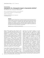
Figure 1
Lipoxygenase enzymes in rheumatoid arthritis and osteoarthritis synovial tissueLipoxygenase enzymes in rheumatoid arthritis and osteoarthritis synovial tissue. 5-Lipoxygenase (5-LO) and 15-LO-1 enzymes are present in both
rheumatoid arthritis (RA) and osteoarthritis (OA) synovial tissue. Photographs illustrating brown (diaminobenzidine) immunoperoxidase staining for
(a, b, c) 5-LO and (d, e, f) 15-LO-1 in sections from frozen synovial biopsies of (a, b, e) RA and (c, f) OA patients (hematoxylin counterstained). (d)
Bronchial epithelium staining positive for 15-LO-1. Insets: (a) RA synovium stained with 5-LO antibody and (d) bronchial epithelium stained with 15-
LO-1 antibody, preabsorbed with purified 5-LO and 15-LO-1 protein, respectively. Original magnification: ×100 (a, d and insets) and ×200 (b, c, e,
f).
Available online />Page 5 of 11
(page number not for citation purposes)
ing the incubation period in corresponding buffer controls was
subtracted from these results.
Discussion
The leukotriene pathway, and in particular LTB
4
, has long been
recognized to have deleterious effects in arthritis. Neverthe-
less, the enzymes responsible for arthritis formation have not
been well characterized in synovial tissues, and nor is it known
whether they are targeted by current RA therapy.
In the present study we showed that 5-LO is expressed in syn-
ovial tissue from patients with RA, mainly in macrophage-like
cells and to a lesser extent in neutrophils and mast cells. We
did not, however, detect 5-LO enzyme in T cells or B cells in
RA biopsies. Although previous studies indicate that tonsillar
B lymphocytes and B-cell lines are abundant in 5-LO protein
[39,40], recent data reveal that, within the tonsils, it is the man-
tle-zone B cells that are 5-LO-positive and not the germinal-
centre B cells or plasma cells [41]. In fact, it has been sug-
gested that RA synovial B cells mainly represent mature acti-
vated memory B cells and plasma cells [42]. Our findings that
RA CD20
+
B cells display no detectable 5-LO staining may
therefore be explained in part by the specific B-cell subsets
present in RA synovium. The wide expression of 5-LO in the
synovial tissue of RA patients is in agreement with studies
describing the LTB
4
presence in RA synovial fluid [1] and the
5-LO-positive immunostaining in areas coinciding with macro-
phage localization [24].
We also observed a low number of cells expressing 5-LO in
OA tissue, scattered in areas with more abundant synovial
membranes. By quantifying the positive staining areas, we
showed that OA synovium expresses significantly less 5-LO
than RA tissue. Indeed, OA synovial fluid has been shown to
contain less LTB
4
than RA fluid [8] and OA synovium is known
to contain a low degree of infiltrating inflammatory cells, which
is in line with our observations.
There are a limited number of studies investigating the 15-LO-
1 pathway in chronic inflammatory disorders, although the
products of this pathway have long been recognized to play
important roles in immune regulation and inflammation [43].
We underwent a detailed study characterizing the expression
of 15-LO-1 enzyme in RA synovium, showing that it is highly
expressed in synovial lining and scattered sublining fibroblast
and macrophages and also in vessels of different sizes. In
addition, we showed here that endothelial cells from both RA
and OA biopsies express 15-LO-1. In OA, however, few syno-
vial lining cells stained positively for 15-LO-1 while 15-LO-1
was abundantly present in vessels. The overall 15-LO expres-
sion was significantly lower in OA synovium compared with RA
synovium.
The expression of functional 15-LO-1 in endothelial cells has
been somewhat controversial, although some studies have
demonstrated expression of 15-LO-1 in these cells [44].
Human and rabbit aortic endothelial cells, however, were more
recently revealed to express 15-LO-1 mRNA and protein [45].
In addition, the presence of 15-LO-1 in endothelial cells was
correlated with an induction of NF-κB activity and a subse-
quent increase in intracellular adhesion molecule expression
[46], which may augment the local influx of cells. Our finding
Figure 2
Osteoarthritis versus rheumatoid arthritis synovial expression of lipoxygenase enzymesOsteoarthritis versus rheumatoid arthritis synovial expression of lipoxygenase enzymes. Ostheoarthritis (OA) synovial tissue displays a lower expres-
sion of 5-lipoxygenase (5-LO) and 15-LO-1 compared with rheumatoid arthritis (RA) synovium. Graphs show computer assisted-image analysis
results for (a) 5-LO and (b) 15-LO-1 expression in RA tissue (n = 6) and OA tissue (n = 5). Results expressed as percentages of the total area of
counterstained tissue. Horizontal lines, median values; whiskers, range values. **P < 0.01.
Arthritis Research & Therapy Vol 11 No 3 Gheorghe et al.
Page 6 of 11
(page number not for citation purposes)
that 15-LO-1 is localized in endothelial cells from RA synovium
may therefore be related to its ability to form mediators that
locally attract immune cells and promote inflammation.
Although 15-LO-1 is largely present in the synovial tissue, its
main product (15-HETE) was not detectable in synovial fluid in
the present study. Synovial fluid cells incubated with AA form
only small amounts of this eicosanoid product. One explana-
tion for this may reside in the methodology we used, such as
a short incubation time. Furthermore, the synovial fluid was iso-
lated from patients treated with various regimens. Cells incu-
bated with AA still form significantly higher amounts of 15-
HETE compared with cells without AA, demonstrating the
capacity of these cells to produce 15-HETE.
We further demonstrated that 5-LO expression in synovial tis-
sue was significantly decreased following intraarticular admin-
istration of GCs. This finding is consistent with previous work
documenting reduced synthesis of LTB
4
in neutrophils of
patients with RA after intraarticular GC injection [33]. It has
been demonstrated previously that the number of macro-
phages in RA synovial tissue is not influenced by therapy with
Figure 3
Synovial distribution pattern of 5-LipoxygenaseSynovial distribution pattern of 5-Lipoxygenase. CD163
+
macrophages, CD66b
+
neutrophils and tryptase-positive mast cells express 5-lipoxygenase
(5-LO) in rheumatoid arthritis synovium. Photomicrographs showing fluorescent staining of (a) CD163
+
cells, (b) CD66b
+
cells and (c) tryptase-pos-
itive cells (Alexa 546, red), 5-LO-positive cells (Alexa 488, green), and superimposed staining. White arrows, double-positive mast cells expressing
5-LO. Original magnification: ×400.
Available online />Page 7 of 11
(page number not for citation purposes)
local GCs [47]. This suggests that the decrease in 5-LO
expression we observe here most probably reflects a decrease
in cellular expression and not a lower number of cells locally
present. Other investigators, however, have found that sys-
temic treatment with GCs is followed by reduced macrophage
infiltration in RA synovium [48]. Different biological mecha-
nisms may operate when administrating GCs intraarticularly or
systemically. Further investigation is therefore needed to eluci-
date the mechanism for the reduction in 5-LO expression.
GCs are very efficient in achieving important clinical and radi-
ographic outcomes in RA [49]. Intraarticular GC may also con-
fer a bone-protecting effect in RA by decreasing the RANKL/
osteoprotegerin ratio [50]. Previous studies have indicated
LTB
4
to be a negative regulator of bone metabolism by activat-
ing osteoclasts and inhibiting osteoblasts, thus promoting
bone degradation and inhibiting bone formation [51,52]. In
this context, the decrease in 5-LO expression after intraarticu-
lar GC therapy may indicate a potential role for 5-LO in bone
degradation associated with inflammatory arthritis and sug-
Figure 4
Synovial distribution pattern of 15-Lipoxygenase-1Synovial distribution pattern of 15-Lipoxygenase-1. CD163
+
macrophages, CD31
+
endothelial cells and prolyl 4-hydrolase-positive fibroblast cells
express 15-lipoxygenase-1 (15-LO-1) in rheumatoid arthritis synovium. Photomicrographs showing fluorescent staining of (a) CD163
+
cells, (b)
CD31
+
cells and (c) prolyl 4-hydrolase-positive cells (Alexa 546, red), 15-LO-positive cells (Alexa 488, green), and superimposed stainings. Original
magnification: (a, b) ×200 and (c) ×400.
Arthritis Research & Therapy Vol 11 No 3 Gheorghe et al.
Page 8 of 11
(page number not for citation purposes)
gests a new mechanism for the bone-protecting effects of
intraarticular GCs.
Since LTB4 has been demonstrated to be a key regulator in
the pathogenesis of murine arthritis [9], it may be conceivable
that targeting the 5-LO pathway could provide additional ben-
efits in the treatment of RA, by reducing the formation of LTB
4
and, by this means, decreasing the chemotaxis of inflammatory
cells. Few studies have investigated the effects of 5-LO path-
way inhibition in RA patients. In a 4-week clinical trial, treat-
ment with zileuton showed a trend towards clinical
improvement, but the duration of the study was not adequate
to assess efficacy [53]. Novel 5-LO inhibitors may possibly
offer better treatment effects.
There are few studies to date on 15-LO-1 in RA, and the role
of its products in inflammation is not clearly defined. We dem-
onstrate here that locally administered corticosteroids do not
significantly change the expression of 15-LO-1 in RA syn-
ovium. Previously, it was shown that 15-LO-1 metabolites con-
fer proinflammatory actions by increasing vascular
permeability in vitro [19], enhancing expression of monocyte
chemotactic protein-1 and TNFα in vascular smooth muscle
cells via activation of NF-κB [54]. On the other hand, 15-LO-1
products may also have anti-inflammatory properties, by
reducing synovitis through decreased LTB
4
in experimental
arthritis [55], inhibiting chemotaxis of neutrophils to LTB
4
[56]
or through transcellular formation of lipoxins [57]. In this sense,
it is noteworthy that IL-13, known to increase 15-LO-1 expres-
sion in several cell systems, is constantly present in synovial
fluid of RA patients and has the ability to decrease proinflam-
matory cytokine production by synovial fluid mononuclear cells
[58]. 15-LO-1 and its metabolites may therefore have dual
roles in inflammation, and the net effect in RA needs further
investigation.
Conclusions
In the present study we have shown that RA synovium
expresses 5-LO and 15-LO-1, and that administration of
intraarticular corticosteroids is followed by a significant reduc-
Figure 5
Intraarticular glucocorticoids effects on lipoxygenase expression in rheumatoid arthritis synoviumIntraarticular glucocorticoids effects on lipoxygenase expression in rheumatoid arthritis synovium. Intraarticular glucocorticoids decrease 5-lipoxyge-
nase (5-LO) expression in rheumatoid arthritis (RA) synovium but leave unaltered the expression of 15-LO-1 enzyme. RA synovial tissue (n = 11)
showing diaminobenzidine (brown) staining for 5-LO (a) before and (b) after treatment, and for 15-LO-1 (d) before and (e) after therapy (hematoxy-
lin counterstained). Graphs show image analysis results for (c) 5-LO and (f) 15-LO-1 expression in synovial biopsy sections taken before and after
intraarticular corticosteroid injection. Results expressed as percentage of the total area of counterstained tissue. Horizontal lines, median values;
whiskers, range values. **P < 0.01. Original magnification: (a, b) ×125 and (c, d) ×160.
Available online />Page 9 of 11
(page number not for citation purposes)
tion in 5-LO expression while leaving the 15-LO-1 enzyme
unaffected. Our results provide an additional explanation for
the beneficial effects of local corticosteroids in RA, through
inhibition of 5-LO enzyme and reduced formation of its proin-
flammatory products. Together with previous studies incrimi-
nating LTB
4
as a potent mediator of joint inflammation and
destruction in RA, the present study suggests the use of 5-LO
inhibitors as add-on therapy.
Competing interests
The authors declare that they have no competing interests.
Figure 6
15-Lipoxygenase-1 expression in rheumatoid arthritis synovial fluid cells and 15-hydroxyeicosatetraenoic acid production15-Lipoxygenase-1 expression in rheumatoid arthritis synovial fluid cells and 15-hydroxyeicosatetraenoic acid production. Rheumatoid arthritis (RA)
synovial fluid cells express 15-lipoxygenase-1 (15-LO-1) and produce 15-hydroxyeicosatetraenoic acid (15-HETE) upon stimulation with arachidonic
acid (AA). (a, b) Cytospin preparation of synovial fluid cells shows brown (diaminobenzidine) staining for 15-LO-1. Inset: isotype control. (c) 15-
HETE formation in control synovial fluid cells and synovial fluid cells incubated with AA. (d) Cellular composition of the RA synovial fluid showing the
percentage of neutrophils, monocytes and lymphocytes in samples from five patients. Horizontal lines, median values; whiskers, range values. Origi-
nal magnification: (a) ×500 and (b) ×800.
Arthritis Research & Therapy Vol 11 No 3 Gheorghe et al.
Page 10 of 11
(page number not for citation purposes)
Authors' contributions
KRG performed acquisition and interpretation of data, per-
formed statistical analysis and wrote the manuscript. MK par-
ticipated in acquisition and interpretation of data, and in
writing the manuscript. AIC provided the patient biopsies and
their clinical data and participated in writing the manuscript.
LB participated in the collection of data. EaK provided patient
biopsies and participated in writing the manuscript. H-EC par-
ticipated in the study design and preparation of the manu-
script. OR participated in writing the manuscript. P-JJ was
responsible for study design, interpretation of data and partic-
ipated in writing the manuscript.
Acknowledgements
The authors thank Professor Lars Klareskog for valuable scientific advice
and suggestions regarding planning of the study and writing the manu-
script. The present work was supported by funds from Karolinska Insti-
tutet, the Swedish Research Council, The Swedish County Council, The
Swedish Rheumatism Association, The Swedish Medical Society and
the King Gustaf V 80-year fund.
References
1. Davidson EM, Rae SA, Smith MJ: Leukotriene B4, a mediator of
inflammation present in synovial fluid in rheumatoid arthritis.
Ann Rheum Dis 1983, 42:677-679.
2. Henderson WR Jr: The role of leukotrienes in inflammation.
Ann Intern Med 1994, 121:684-697.
3. Lewis RA, Austen KF, Soberman RJ: Leukotrienes and other
products of the 5-lipoxygenase pathway. Biochemistry and
relation to pathobiology in human diseases. N Engl J Med
1990, 323:645-655.
4. Pillinger MH, Abramson SB: The neutrophil in rheumatoid
arthritis. Rheum Dis Clin North Am 1995, 21:691-714.
5. Ahluwalia N, Lin AY, Tager AM, Pruitt IE, Anderson TJ, Kristo F,
Shen D, Cruz AR, Aikawa M, Luster AD, Gerszten RE: Inhibited
aortic aneurysm formation in BLT1-deficient mice. J Immunol
2007, 179:691-697.
6. Leppert D, Hauser SL, Kishiyama JL, An S, Zeng L, Goetzl EJ:
Stimulation of matrix metalloproteinase-dependent migration
of T cells by eicosanoids. Faseb J 1995, 9:1473-1481.
7. Gursel T, Firat S, Ercan ZS: Increased serum leukotriene B4
level in the active stage of rheumatoid arthritis in children.
Prostaglandins Leukot Essent Fatty Acids 1997, 56:205-207.
8. Ahmadzadeh N, Shingu M, Nobunaga M, Tawara T: Relationship
between leukotriene B4 and immunological parameters in
rheumatoid synovial fluids. Inflammation 1991, 15:497-503.
9. Chen M, Lam BK, Kanaoka Y, Nigrovic PA, Audoly LP, Austen KF,
Lee DM: Neutrophil-derived leukotriene B4 is required for
inflammatory arthritis. J Exp Med 2006, 203:837-842.
10. Andersson E, Schain F, Svedling M, Claesson HE, Forsell PK:
Interaction of human 15-lipoxygenase-1 with phosphatidyli-
nositol bisphosphates results in increased enzyme activity.
Biochim Biophys Acta 2006, 1761:1498-1505.
11. Profita M, Sala A, Riccobono L, Paterno A, Mirabella A, Bonanno
A, Guerrera D, Pace E, Bonsignore G, Bousquet J, Vignola AM:
15-Lipoxygenase expression and 15(S)-hydroxyeicoisatetrae-
noic acid release and reincorporation in induced sputum of
asthmatic subjects. J Allergy Clin Immunol 2000, 105:711-716.
12. Deleuran B, Iversen L, Deleuran M, Yssel H, Kragballe K, Sten-
gaard-Pedersen K, Thestrup-Pedersen K: Interleukin 13 sup-
presses cytokine production and stimulates the production of
15-HETE in PBMC. A comparison between IL-4 and IL-13.
Cytokine 1995, 7:319-324.
13. Nassar GM, Morrow JD, Roberts LJ 2nd, Lakkis FG, Badr KF:
Induction of 15-lipoxygenase by interleukin-13 in human
blood monocytes. J Biol Chem 1994, 269:27631-27634.
14. Conrad DJ, Kuhn H, Mulkins M, Highland E, Sigal E: Specific
inflammatory cytokines regulate the expression of human
monocyte 15-lipoxygenase. Proc Natl Acad Sci USA 1992,
89:217-221.
15. Spanbroek R, Hildner M, Kohler A, Muller A, Zintl F, Kuhn H, Rad-
mark O, Samuelsson B, Habenicht AJ: IL-4 determines eicosa-
noid formation in dendritic cells by down-regulation of 5-
lipoxygenase and up-regulation of 15-lipoxygenase 1 expres-
sion. Proc Natl Acad Sci USA 2001, 98:5152-5157.
16. Levy BD, Romano M, Chapman HA, Reilly JJ, Drazen J, Serhan CN:
Human alveolar macrophages have 15-lipoxygenase and gen-
erate 15(S)-hydroxy-5,8,11-cis-13-trans-eicosatetraenoic acid
and lipoxins. J Clin Invest 1993, 92:1572-1579.
17. Gulliksson M, Brunnstrom A, Johannesson M, Backman L, Nilsson
G, Harvima I, Dahlen B, Kumlin M, Claesson HE: Expression of
15-lipoxygenase type-1 in human mast cells. Biochim Biophys
Acta 2007, 1771:1156-1165.
18. Harada S, Sugiyama E, Takebe S, Taki H, Shinoda K, Mohamed
SG, Maruyama M, Hamazaki T, Kobayashi M: Cooperative induc-
tion of 15-lipoxygenase in rheumatoid synovial cells by IL-4
and proinflammatory cytokines. Clin Exp Rheumatol 2003,
21:753-758.
19. Feltenmark S, Gautam N, Brunnstrom A, Griffiths W, Backman L,
Edenius C, Lindbom L, Bjorkholm M, Claesson HE: Eoxins are
proinflammatory arachidonic acid metabolites produced via
the 15-lipoxygenase-1 pathway in human eosinophils and
mast cells. Proc Natl Acad Sci USA 2008, 105:680-685.
20. Serhan CN: Lipoxins and aspirin-triggered 15-epi-lipoxins are
the first lipid mediators of endogenous anti-inflammation and
resolution. Prostaglandins Leukot Essent Fatty Acids 2005,
73:141-162.
21. Serhan CN, Jain A, Marleau S, Clish C, Kantarci A, Behbehani B,
Colgan SP, Stahl GL, Merched A, Petasis NA, Chan L, Van Dyke
TE: Reduced inflammation and tissue damage in transgenic
rabbits overexpressing 15-lipoxygenase and endogenous
anti-inflammatory lipid mediators.
J Immunol 2003,
171:6856-6865.
22. Wittwer J, Hersberger M: The two faces of the 15-lipoxygenase
in atherosclerosis. Prostaglandins Leukot Essent Fatty Acids
2007, 77:67-77.
23. Liagre B, Vergne P, Rigaud M, Beneytout JL: Arachidonate 15-
lipoxygenase of reticulocyte-type in human rheumatoid arthri-
tis type B synoviocytes and modulation of its activity by proin-
flammatory cytokines. J Rheumatol 1999, 26:1044-1051.
24. Hashimoto A, Hayashi I, Murakami Y, Sato Y, Kitasato H, Matsus-
hita R, Iizuka N, Urabe K, Itoman M, Hirohata S, Endo H: Antiin-
flammatory mediator lipoxin A4 and its receptor in synovitis of
patients with rheumatoid arthritis. J Rheumatol 2007,
34:2144-2153.
25. Barnes PJ: Corticosteroids: the drugs to beat. Eur J Pharmacol
2006, 533:2-14.
26. Peters-Golden M, Thebert P: Inhibition by methylprednisolone
of zymosan-induced leukotriene synthesis in alveolar macro-
phages. Am Rev Respir Dis 1987, 135:1020-1026.
27. Fuller RW, Kelsey CR, Cole PJ, Dollery CT, MacDermot J: Dexam-
ethasone inhibits the production of thromboxane B2 and leu-
kotriene B4 by human alveolar and peritoneal macrophages in
culture. Clin Sci (Lond) 1984, 67:653-656.
28. Sebaldt RJ, Sheller JR, Oates JA, Roberts LJ 2nd, FitzGerald GA:
Inhibition of eicosanoid biosynthesis by glucocorticoids in
humans. Proc Natl Acad Sci USA 1990, 87:6974-6978.
29. Freeland HS, Pipkorn U, Schleimer RP, Bascom R, Lichtenstein
LM, Naclerio RM, Peters SP: Leukotriene B4 as a mediator of
early and late reactions to antigen in humans: the effect of sys-
temic glucocorticoid treatment in vivo. J Allergy Clin Immunol
1989, 83:634-642.
30. Riddick CA, Ring WL, Baker JR, Hodulik CR, Bigby TD: Dexame-
thasone increases expression of 5-lipoxygenase and its acti-
vating protein in human monocytes and THP-1 cells. Eur J
Biochem 1997, 246:112-118.
31. Colamorea T, Di Paola R, Macchia F, Guerrese MC, Tursi A, But-
terfield JH, Caiaffa MF, Haeggstrom JZ, Macchia L: 5-Lipoxygen-
ase upregulation by dexamethasone in human mast cells.
Biochem Biophys Res Commun 1999, 265:617-624.
32. Thomas E, Leroux JL, Blotman F, Descomps B, Chavis C:
Enhancement of leukotriene A4 biosynthesis in neutrophils
from patients with rheumatoid arthritis after a single glucocor-
ticoid dose.
Biochem Pharmacol 1995, 49:243-248.
Available online />Page 11 of 11
(page number not for citation purposes)
33. Klickstein LB, Shapleigh C, Goetzl EJ: Lipoxygenation of arachi-
donic acid as a source of polymorphonuclear leukocyte chem-
otactic factors in synovial fluid and tissue in rheumatoid
arthritis and spondyloarthritis. J Clin Invest 1980,
66:1166-1170.
34. Makheja AN, Bloom S, Muesing R, Simon T, Bailey JM: Anti-
inflammatory drugs in experimental atherosclerosis. 7. Spon-
taneous atherosclerosis in WHHL rabbits and inhibition by cor-
tisone acetate. Atherosclerosis 1989, 76:155-161.
35. Laprise C, Sladek R, Ponton A, Bernier MC, Hudson TJ, Laviolette
M: Functional classes of bronchial mucosa genes that are dif-
ferentially expressed in asthma. BMC Genomics 2004, 5:21.
36. Arnett FC, Edworthy SM, Bloch DA, McShane DJ, Fries JF, Cooper
NS, Healey LA, Kaplan SR, Liang MH, Luthra HS, Medsger TA Jr,
Mitchell DM, Neustadt DH, Pinals RS, Schaller JG, Sharp JT,
Wilder RL, Hunder GG: The American Rheumatism Association
1987 revised criteria for the classification of rheumatoid arthri-
tis. Arthritis Rheum 1988, 31:315-324.
37. Ulfgren AK, Lindblad S, Klareskog L, Andersson J, Andersson U:
Detection of cytokine producing cells in the synovial mem-
brane from patients with rheumatoid arthritis. Ann Rheum Dis
1995, 54:654-661.
38. Westman M, Korotkova M, af Klint E, Stark A, Audoly LP, Klareskog
L, Ulfgren AK, Jakobsson PJ: Expression of microsomal prostag-
landin E synthase 1 in rheumatoid arthritis synovium. Arthritis
Rheum 2004, 50:1774-1780.
39. Werz O, Tretiakova I, Michel A, Ulke-Lemee A, Hornig M, Franke L,
Schneider G, Samuelsson B, Radmark O, Steinhilber D: Caspase-
mediated degradation of human 5-lipoxygenase in B lym-
phocytic cells. Proc Natl Acad Sci USA 2005,
102:13164-13169.
40. Jakobsson PJ, Odlander B, Steinhilber D, Rosen A, Claesson HE:
Human B lymphocytes possess 5-lipoxygenase activity and
convert arachidonic acid to leukotriene B4. Biochem Biophys
Res Commun 1991, 178:302-308.
41. Mahshid Y, Lisy MR, Wang X, Spanbroek R, Flygare J, Christens-
son B, Bjorkholm M, Sander B, Habenicht AJ, Claesson HE: High
expression of 5-lipoxygenase in normal and malignant mantle
zone B lymphocytes. BMC Immunol 2009, 10:2.
42. Cantaert T, Kolln J, Timmer T, Pouw Kraan TC van der, Vandooren
B, Thurlings RM, Canete JD, Catrina AI, Out T, Verweij CL, Zhang
Y, Tak PP, Baeten D: B lymphocyte autoimmunity in rheuma-
toid synovitis is independent of ectopic lymphoid neogenesis.
J Immunol 2008, 181:785-794.
43. Kuhn H, O'Donnell VB: Inflammation and immune regulation by
12/15-lipoxygenases. Prog Lipid Res 2006, 45:334-356.
44. Lee YW, Kuhn H, Kaiser S, Hennig B, Daugherty A, Toborek M:
Interleukin 4 induces transcription of the 15-lipoxygenase I
gene in human endothelial cells. J Lipid Res 2001, 42:783-791.
45. Tang X, Holmes BB, Nithipatikom K, Hillard CJ, Kuhn H, Campbell
WB: Reticulocyte 15-lipoxygenase-I is important in acetylcho-
line-induced endothelium-dependent vasorelaxation in rabbit
aorta. Arterioscler Thromb Vasc Biol 2006, 26:78-84.
46. Bolick DT, Orr AW, Whetzel A, Srinivasan S, Hatley ME, Schwartz
MA, Hedrick CC: 12/15-Lipoxygenase regulates intercellular
adhesion molecule-1 expression and monocyte adhesion to
endothelium through activation of RhoA and nuclear factor-
κB. Arterioscler Thromb Vasc Biol 2005, 25:2301-2307.
47. af Klint E, Grundtman C, Engstrom M, Catrina AI, Makrygiannakis
D, Klareskog L, Andersson U, Ulfgren AK: Intraarticular glucocor-
ticoid treatment reduces inflammation in synovial cell infiltra-
tions more efficiently than in synovial blood vessels. Arthritis
Rheum 2005, 52:3880-3889.
48. Gerlag DM, Haringman JJ, Smeets TJ, Zwinderman AH, Kraan MC,
Laud PJ, Morgan S, Nash AF, Tak PP: Effects of oral pred-
nisolone on biomarkers in synovial tissue and clinical
improvement in rheumatoid arthritis. Arthritis Rheum 2004,
50:3783-3791.
49. Harris ED Jr: Prednisolone in early rheumatoid arthritis: an anti-
invasive effect. Arthritis Rheum 2005, 52:3324-3325.
50. Makrygiannakis D, af Klint E, Catrina SB, Botusan IR, Klareskog E,
Klareskog L, Ulfgren AK, Catrina AI: Intraarticular corticosteroids
decrease synovial RANKL expression in inflammatory arthritis.
Arthritis Rheum 2006, 54:1463-1472.
51. Garcia C, Boyce BF, Gilles J, Dallas M, Qiao M, Mundy GR, Bone-
wald LF: Leukotriene B4 stimulates osteoclastic bone resorp-
tion both in vitro and in vivo. J Bone Miner Res 1996,
11:1619-1627.
52. Traianedes K, Dallas MR, Garrett IR, Mundy GR, Bonewald LF: 5-
Lipoxygenase metabolites inhibit bone formation in vitro.
Endocrinology 1998, 139:
3178-3184.
53. Weinblatt ME, Kremer JM, Coblyn JS, Helfgott S, Maier AL, Petrillo
G, Henson B, Rubin P, Sperling R: Zileuton, a 5-lipoxygenase
inhibitor in rheumatoid arthritis. J Rheumatol 1992,
19:1537-1541.
54. Dwarakanath RS, Sahar S, Reddy MA, Castanotto D, Rossi JJ,
Natarajan R: Regulation of monocyte chemoattractant protein-
1 by the oxidized lipid, 13-hydroperoxyoctadecadienoic acid,
in vascular smooth muscle cells via nuclear factor-kappa B
(NF-κB). J Mol Cell Cardiol 2004, 36:585-595.
55. Hansen ES, Fogh K, Hjortdal VE, Henriksen TB, Noer I, Ewald H,
Herlin T, Kragballe K, Bunger C: Synovitis reduced by inhibition
of leukotriene B4. Carrageenan-induced gonarthritis studied
in dogs. Acta Orthop Scand 1990, 61:207-212.
56. Fogh K, Hansen ES, Herlin T, Knudsen V, Henriksen TB, Ewald H,
Bunger C, Kragballe K: 15-Hydroxy-eicosatetraenoic acid (15-
HETE) inhibits carrageenan-induced experimental arthritis
and reduces synovial fluid leukotriene B4 (LTB4). Prostaglan-
dins 1989, 37:213-228.
57. Yacoubian S, Serhan CN: New endogenous anti-inflammatory
and proresolving lipid mediators: implications for rheumatic
diseases. Nat Clin Pract Rheumatol 2007, 3:570-579.
58. Isomaki P, Luukkainen R, Toivanen P, Punnonen J: The presence
of interleukin-13 in rheumatoid synovium and its antiinflam-
matory effects on synovial fluid macrophages from patients
with rheumatoid arthritis. Arthritis Rheum 1996, 39:1693-1702.