Báo cáo y học: "Developments in the clinical understanding of lupus" pot
Bạn đang xem bản rút gọn của tài liệu. Xem và tải ngay bản đầy đủ của tài liệu tại đây (677.1 KB, 11 trang )
Available online />Page 1 of 11
(page number not for citation purposes)
Abstract
Advances in genetics and new understanding of the molecular
pathways that mediate innate and adaptive immune system
activation, along with renewed focus on the role of the complement
system as a mediator of inflammation, have stimulated elaboration
of a scheme that might explain key mechanisms in the patho-
genesis of systemic lupus erythematosus. Clinical observations
identifying important comorbidities in patients with lupus have been
a recent focus of research linking immune mechanisms with clinical
manifestations of disease. While these advances have identified
rational and promising targets for therapy, so far the therapeutic
trials of new biologic agents have not met their potential.
Nonetheless, progress in understanding the underlying immuno-
pathogenesis of lupus and its impact on clinical disease has
accelerated the pace of clinical research to improve the outcomes
of patients with systemic lupus erythematosus.
Introduction
Systemic lupus erythematosus (SLE) is often considered the
prototype systemic autoimmune disease, as virtually all
components of the immune system contribute to the
characteristic autoimmunity and tissue pathology. The utility
of lupus research extends beyond defining lupus-specific
mechanisms, as the disease can serve as a model system for
consideration of immune system responses to microbial
infection and control of hematologic malignancies. Especially
in recent years, as new concepts have evolved to explain
mechanisms that link the nucleic acid targets of lupus auto-
antibodies to immune system activation and inflammation, the
intellectual rewards of research on this most complex medical
syndrome have grown. Yet this is a disease with high impact
on patients, particularly women in the reproductive years. The
satisfaction derived from new understanding of disease
mechanisms will only be fully realized when those insights are
translated into new therapies. Despite some frustrations in
efforts to develop new lupus drugs, clinical care of lupus
patients continues to improve, and the scope of clinical
research in search of new lupus therapies has significantly
expanded to include both traditional and new biologic agents.
The etiopathogenesis of lupus comprises genetic contribu-
tions, environmental triggers, and stochastic events, as
demonstrated in murine models in the late 1980s [1]. These
factors play out at the level of the immune system, with
multiple genetic hits and an undefined complement of
exogenous or endogenous triggers required for initiation of
autoimmunity. When the genetic load is sufficient, immune
triggers are available and chance favors effective immune
system activation, the disease process can move forward [2]
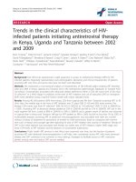
(Figure 1). A concept that has been developed in recent
years considers the kinetics of the disease, with lupus
autoantibodies present in serum of lupus patients up to
5 years prior to the development of clinical manifestations of
disease [3]. It is notable that autoimmunity, when considered
in a population of lupus patients, develops in a stereotypical
manner, with anti-Ro and anti-La antibodies, common to
several systemic autoimmune diseases, developing early in
the pre-clinical stage of disease, while anti-Sm and anti-RNP
antibodies, those that are more specific for SLE, developing
very close to the time that disease becomes clinically
apparent.
It is now recognized that autoantibodies and their associated
nucleic acids can play an amplifying role in immune system
activation, most likely through stimulation of innate immune
pathways. Insights into the genetic variations that are
associated with lupus, along with this new awareness of how
autoimmunity, immune dysfunction, and tissue damage
develop over time, are providing a more complete picture of
disease risk, the steps in pathogenesis, and most signifi-
cantly, new therapeutic targets.
Review
Developments in the clinical understanding of lupus
Mary K Crow
Autoimmunity and Inflammation Program, Hospital for Special Surgery, 535 East 70th Street, New York, NY 10021, USA
Corresponding author: Mary K Crow,
Published: 14 October 2009 Arthritis Research & Therapy 2009, 11:245 (doi:10.1186/ar2762)
This article is online at />© 2009 BioMed Central Ltd
ACR = American College of Rheumatology; BLyS = B lymphocyte stimulator; dsDNA = double-stranded DNA; FDA = Food and Drug Administra-
tion; GWAS = genome-wide association study; HMGB1 = high mobility group box 1; ICOS = inducible costimulator; IFN = interferon; IL = inter-
leukin; MMF = mycophenolate mofetil; RA = rheumatoid arthritis; SLE = systemic lupus erythematosus; TLR = Toll-like receptor; TNF = tumor
necrosis factor.
Arthritis Research & Therapy Vol 11 No 5 Crow
Page 2 of 11
(page number not for citation purposes)
New concepts in lupus pathogenesis
Genetics
Two types of genetic variants associated with a diagnosis of
SLE, common single nucleotide variants and rare genetic
mutations, are stimulating studies of functional alterations in
molecular pathways important in lupus pathogenesis. A third
type of genetic variant, copy number variation, has been
observed in a murine model of lupus, the BXSB mouse,
where a duplication of a region of the X chromosome contain-
ing the Toll-like receptor (TLR) 7 gene (TLR7) is associated
with increased type I IFN production, macrophage activation,
autoantibody production and poor survival [4-6].
Establishment of large collections of DNA samples from
lupus patients and controls, along with advances in tech-
nology that have made large scale studies of genetic variants
more affordable, have led to successful genome-wide asso-
ciation studies (GWAS) supported by government agencies,
foundations, industry and academic centers [7-10]. Data from
these studies have confirmed several candidate genes
previously associated with lupus, identified some new lupus-
associated genes and gene loci, and identified variants in a
gene (ITGAM) whose protein product had been studied in
SLE but was not previously known to have a genetic
association with lupus [11]. An earlier publication in this
series, ‘Developments in the Scientific Understanding of
Lupus’, has listed some of the genes showing a statistical
association with a diagnosis of lupus in GWAS [12]. Several,
including PTPN22, IRF5, STAT4, FCGRIIA, and of course
the HLA region, have been previously described prior to the
publication of the GWAS data. Some recently identified
lupus-associated genetic variants, including BLK, PXK, and
BANK1, may modify lymphocyte signaling and provide new
insights into molecular pathways relevant to lupus patho-
genesis. The protein product of ITGAM, also identified as a
lupus-associated gene and known as CD11b, Mac1 and
complement receptor 3, had not been previously linked to
lupus at the genetic level but its expression was known to be
increased on neutrophils of active lupus patients and it can
mediate adhesion to endothelial cells [11]. In recent months
additional lupus associated genes have been described,
including LYN, a src-tyrosine kinase, IRAK1, an IL-1 receptor
associated kinase, TNFAIP3, which encodes A20, and
OX40L, a costimulatory molecule [13-16]. KLK1 and KLK3,
encoding kallikreins, have been associated with altered
protection from anti-glomerular basement membrane disease
and lupus nephritis [17].
What is striking about most of these lupus-associated genes
is that their function is most likely associated with activation
or regulation of the immune response. Based on identification
of these genes and their known functions, we can hypo-
thesize a role for activation of the innate immune response
through TLRs (IRF5, FCGRIIA, TNFAIP3), response to
cytokines (STAT4, IRAK1), or lymphocyte activation and
regulation (PTPN22, PLK, BANK1, LYN, OX40L, SPP1)
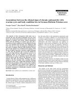
[18-22] (Figure 2). In addition, some of these genetic variants
might contribute to directing the immune response to target
organs and contribute to tissue inflammation and damage
(ITGAM).
In addition to the GWAS, which identify common genetic
variants, old observations of the high risk of SLE in rare
patients with C2, C4 and C1q deficiencies have now been
supplemented with data from several groups identifying lupus
in patients with mutations in a DNase encoded by TREX1
[23]. Rare mutations in that gene are associated with a lupus-
like syndrome characterized by anti-DNA antibodies, high
levels of IFN-alpha and neurologic disease and have led to
studies of lupus cohorts and detection of occasional TREX1
mutations. It appears that altered structure or function of the
TREX1-encoded DNase results in inefficient clearance of
intracellular DNA rich in endogenous genomic repeat element
sequences and induction of type I IFN [24].
To some extent, data from genetic studies are confirming
what we have known - that the immune response underlies
lupus pathogenesis [7]. But those studies are also providing
some surprises, such as the TREX1 observation, that will lead
to research on previously unsuspected pathways. Clinical
insights from genetic data are just beginning to emerge. For
example, recent data identify variations in LYN that confer
protection from hematologic manifestations in a lupus
subgroup defined by the presence of certain autoantibodies
[13], and the association of IFN-alpha and neurologic
manifestations in patients with TREX1 mutations may lead to
greater understanding of the molecular basis of neurologic
involvement in patients with SLE. Analysis of the function of
Figure 1
Stages of lupus pathogenesis. Genetic factors and environmental
triggers, whether exogenous or endogenous, along with stochastic
events, act on the immune system to initiate autoimmunity.
Autoantibodies and their antigens, cytokines and chemokines amplify
immune system activation and generate tissue damage. Autoantibody
production occurs years prior to the development of clinical signs and
symptoms of systemic lupus erythematosus (SLE). Organ damage has
likely occurred by the time lupus is diagnosed. Sx, symptoms; Dx,
diagnosis.
the lupus-associated genetic variants should provide impor-
tant insights into pathogenic mechanisms that can be applied
to development of highly targeted therapeutics.
Apoptotic cells
Apoptotic cells remain attractive candidates as a source of
self-antigens that may initiate and direct the autoimmune
response. Longstanding observations have documented the
concentration of lupus autoantigens in apoptotic cell blebs
[25], and in vitro studies have demonstrated stimulation of
autoreactive T cells by dendritic cells that have processed
autologous apoptotic cell components [26]. Some lupus
patients demonstrate increased spontaneous apoptosis or
impaired clearance of apoptotic peripheral blood cells
[27,28]. Recent data have supported the hypothesis that
components of the classical complement pathway are
required for phagocytic clearance of apoptotic cells,
providing a possible explanation for the high frequency of
SLE among the rare individuals with genetic deficiencies of
those components, particularly C1q [29]. In addition to C1q,
similar molecules with collagen-like structural features,
including mannose-binding lectin and ficolin 3, can contribute
to uptake of late apoptotic cells by macrophages [30]. The
mechanisms that might account for induction of immune
dysregulation and autoimmunity by apoptotic cell compo-
nents are of great interest. Recent data support a role for
high mobility group box 1 (HMGB1)-nucleosome complexes
derived from apoptotic cells in the induction of pro-
inflammatory mediators, dendritic cell maturation, and anti-
double-stranded DNA (anti-dsDNA) autoantibodies [31,32].
Innate immune response
Among the autoimmune and rheumatic diseases, studies of
SLE have arguably provided the strongest evidence for an
essential role of TLRs and the innate immune response in
disease pathogenesis [33-35]. The immunomodulatory
effects of the HMGB1-nucleosome complexes are apparently
mediated by interactions with TLR2 [32]. In addition, several
lupus genes encode proteins that mediate or regulate TLR
signals and are associated with increased plasma IFN-alpha
among patients with particular autoantibodies. Those
antibodies could potentially deliver stimulatory nucleic acids
to TLR7 or TLR9 in their intracellular compartments [36-40].
Activation of the IFN pathway has been associated with the
presence of autoantibodies specific for RNA-associated
proteins, and the current literature supports RNA-mediated
activation of TLR as an important mechanism contributing to
production of IFN-alpha and other proinflammatory cytokines
[41]. Activation of the IFN pathway is associated with renal
disease and many measures of disease activity [42-45].
Ongoing studies are evaluating the temporal relationship
between expression of IFN-inducible genes in peripheral
Available online />Page 3 of 11
(page number not for citation purposes)
Figure 2
Genetic determinants of lupus pathogenesis. Genome-wide association studies are confirming previous data identifying genetic variants that are
statistically associated with systemic lupus erythematosus and are finding new lupus-associated genes. Most lupus-associated genes represent
common variants, but several (C2, C4, C1q and TREX1) are characterized by rare mutations. We suggest that lupus-associated genes contribute
to one or more essential mechanisms that must be implemented to generate lupus susceptibility. Some genetic variants will facilitate innate immune
system activation, particularly type I IFN production; other genetic variants will result in increased availability of self-antigen; and other genetic
variants will alter the threshold for activation or regulation of cells of the adaptive immune response, resulting in production of autoantibodies.
Additional genetic variants might promote inflammation and damage to target organs or fail to protect those organs from proinflammatory
mediators. The lupus-associated genetic variants prepare the immune system and target organs to be responsive to exogenous or endogenous
triggers. Lupus-associated genes are shown in red.
blood mononuclear cells of SLE patients and disease flares,
as measured by conventional tools such as the British Isles
Lupus Assessment Group (BILAG) index or the Systemic
Lupus Erythematosus Disease Activity Index (SLEDAI). In
some patients, increases in IFN-inducible gene expression
precede flares in disease activity by several months,
suggesting that the increased IFN activity might contribute to
increased immune system activity and tissue damage. In view
of the wide effects of type I IFN on immune system function,
including induction of macrophage differentiation toward a
dendritic cell phenotype, increased immunoglobulin class
switching and generalized priming of the immune system for
increased responsiveness to subsequent stimuli, IFN-alpha
represents a rational therapeutic target [35,46].
Adaptive immune response
Activated T and B cells are features of SLE, and many of the
genetic variants that are being studied in association with
SLE are likely to contribute to immune activation and clinical
disease by altering the threshold for lymphocyte activation or
modifying the capacity of inhibitors of signaling pathways to
appropriately function. Analysis of cell surface molecules on
lupus cells has led to descriptions of the phenotype of
lymphocytes from patients with increased disease activity.
Broad polyclonal activation of T cells is detected by
increased or prolonged expression of CD40 ligand, and
circulating B cells with a memory cell phenotype are
increased in patients [47,48]. The soluble TNF family member
B lymphocyte stimulator (BLyS) is increased in serum of
many lupus patients and promotes B cell survival and
differentiation [49], and interactions between co-stimulatory
ligands and receptors on T and B cells, including CD80 and
CD86 with CD28, inducible costimulator (ICOS) ligand with
ICOS, and CD40 ligand with CD40, contribute to B cell
differentiation to antibody producing plasma cells [48]. The
autoantibodies produced as a result of these T and B cell
interactions may directly contribute to inflammation and tissue
damage in target organs but also amplify immune system
activation and autoimmunity through their delivery of stimu-
latory nucleic acids to TLRs, as described above. The
contribution of T and B cells in lupus pathogenesis is not
restricted to their role in inducing autoantibodies, but likely
also includes their production of cytokines and chemokines
that shape the immune response and promote tissue damage.
The anecdotal reports of excellent therapeutic responses in
some patients treated with co-stimulatory molecule blockade
or anti-B cells agents, in spite of persistent autoantibody
titers, suggest that those additional mechanisms of lympho-
cyte function are likely contributing to clinical disease [50].
Target organ damage
Effector functions of the immune system, particularly those
induced by Fc receptor ligation and complement activation,
contribute to tissue damage through complex mechanisms
that include induction of reactive oxygen intermediates,
recruitment of inflammatory cells, induction of proinflam-
matory mediators such as TNF, and modulation of the clotting
cascade. In fact the complement system, for many years only
assessed as a measure of immune complex-mediated
activation, is increasingly recognized to play a prominent role
in many lupus-associated inflammatory states, including some
that do not involve a major role for immune complexes. Anti-
phospholipid antibodies binding to membranes of the
placenta can contribute to complement activation, placental
inflammation and fetal loss in a murine system [51,52]. The
presence of complement and complement regulatory proteins
in association with high density lipoprotein particles suggests
that one function of those particles might be to deliver
complement regulators to the vasculature where chronic
inflammation can take place, possibly modulating athero-
sclerotic mechanisms [53].
Autoantibody mediated tissue damage has been proposed
as a possible mechanism that contributes to central nervous
system manifestations of SLE, particularly cognitive dys-
function [54]. Antibodies that react with both DNA and
glutamate receptors on neurons are proposed to mediate
excitotoxic neuronal cell death. In addition to autoantibodies
or immune complexes, cytokines might contribute to central
nervous system dysfunction and clinical symptoms. As noted
above, high levels of IFN-alpha have been associated with
central nervous system disease in patients with TREX1
mutations [23]. In addition, administering recombinant IFN-
alpha to patients with hepatitis C infection can lead to
depression and cognitive dysfunction, perhaps similar to
those manifestations in SLE. In recent studies, immune
complexes present in cerebrospinal fluid were shown to
provide potent induction of type I IFN in target cells [55].
TNF is another cytokine that is likely to contribute to
inflammation and tissue damage. Small studies using TNF
antagonist therapy in patients with arthritis or nephritis
suggest some efficacy of that approach, although controlled
studies are needed [56]. Together, these observations
suggest that cytokines, particularly IFN-alpha, may contribute
to target organ damage.
While antibodies, immune complexes, cytokines, and
products generated by Fc receptor ligation and complement
activation likely represent important mediators of tissue
damage in SLE, the cells that produce some of those
products deserve further study. The properties of macro-
phages, dendritic cells and lymphocytes that infiltrate the
kidney and other target organs might suggest cell surface
molecules or components of signaling pathways that could
be therapeutically targeted to relieve some of the damage
mediated by those cells [57,58]. The strong association of a
polymorphism in the ITGAM gene raises the possibility that
leukocytes expressing the lupus-associated ITGAM variant
might demonstrate a propensity to adhere more avidly to the
local renal vasculature. In addition to augmented inflammatory
mechanisms, target organ damage, particularly in the kidney,
might be amplified by impaired protective mechanisms.
Arthritis Research & Therapy Vol 11 No 5 Crow
Page 4 of 11
(page number not for citation purposes)
Recent data demonstrating an association of variants of
KLK1 and KLK3 with lupus nephritis suggest a possible
defect in the protective function of kallikreins in some lupus
patients [17].
A summary of current concepts of lupus pathogenesis would
include an important role for genetic variants that prime both
innate and adaptive immune systems for increased respon-
siveness to cell activation, increased production of and
response to IFN-alpha, increased capacity to generate auto-
antibodies, and perhaps an increased targeting of inflam-
matory cells - or decreased protection from the products of
those cells - to target organs. As additional genetic data are
collected and analyzed, we will gain a better understanding of
how lupus susceptibility genes interact and the level of risk
conferred by each additional variant. Recent data suggest
that the risk of each disease-associated single nucleotide
polymorphism in IRF5 and STAT4 confers additive risk of
disease [59]. While the manner in which environmental
triggers interact with genetic risk remains to be understood
[60], we have already gained substantial insights into the
major pathways used by the immune system to initiate and
amplify immune system activation and inflammation. The new
information regarding candidate protective mechanisms in
target organs should stimulate new attention to the response
of tissue to the insults delivered by the immune system and
might suggest very novel and as yet unexplored approaches
to organ protection or repair.
Recent focus on comorbidties
The characteristic clinical features of SLE, including those
included in the American College of Rheumatology (ACR)
classification criteria, tend to be the focus of patient
management and therapy. But the past 10 years have
witnessed increased attention to comorbidities that have
substantial impact on patient outcomes and quality of life.
These comorbidities, beyond their impact on patients and
their medical management, have provided opportunities for
novel research observations with impact beyond SLE. Three
comorbidities that are associated with, but not exclusive to,
SLE will be briefly discussed: accelerated atherosclerosis,
antiphospholipid syndrome, and fetal loss.
Accelerated atherosclerosis
With the description of increased occurrence of myocardial
infarction by Urowitz in 1976 [61] and the ready availability of
tools, such as carotid ultrasound, to detect preclinical athero-
sclerotic lesions, the rheumatology community is now well
aware of the additional risk of accelerated atherosclerosis
conferred by lupus beyond that attributable to traditional
cardiovascular risk factors [61-64]. Studies from Manzi and
colleagues [63], Roman and colleagues [64], and others
have documented the high prevalence of premature athero-
sclerosis in SLE patients compared to control populations
without lupus, with Roman and colleagues’ study demon-
strating carotid plaque in 37% of SLE patients compared to
15% of age, race, gender and hypertension-matched control
subjects. In follow-up studies 28% of those SLE patients
developed new or more extensive plaque over approximately
3 years, with plaque progression associated with increased
homocysteine levels [65]. In addition to plaque, radial
applanation tonometry was used to show that SLE patients
also demonstrate increased vascular stiffness that was
associated with duration of disease, cholesterol, and serum
IL-6 and C-reactive protein levels [66].
In addition to the data pointing to pro-inflammatory cytokines
and homocysteine as possible mediators in the development
of cardiovascular disease, data from several groups have
linked IFN-alpha to decreased availability of endothelial
precursor cells and impaired endothelial function [67,68].
Even when SLE patients and controls have a similar degree
of atherosclerotic plaque, the SLE patients show increased
endothelial dysfunction, as measured by flow-mediated
dilatation [69]. In that study endothelial dysfunction was
associated with disease activity. A role for type I IFN in the
premature atherosclerosis of lupus patients is an attractive
concept in light of the growing literature implicating this
cytokine in many aspects of altered immune function in SLE.
But investigation of mechanisms that provide a functional link
between homocysteine and arterial stiffness might be another
fruitful research direction. At this time, it is advisable to be
vigilant in addressing traditional cardiovascular risk factors in
management of lupus patients. Additional translational and
clinical studies will be needed to better define the
mechanisms that account for the added risk experienced by
lupus patients beyond that in the general population.
Catastrophic antiphospholipid syndrome
The facilitated communication and collaboration presented by
the internet has been utilized by rheumatologists to gain new
knowledge about a significant cause of morbidity and
mortality among lupus patients: the catastrophic antiphos-
pholipid syndrome [70]. A website was established by the
European Forum on Antiphospholipid Antibodies that
provides a site for collection and analysis of clinical data on
those patients, whether associated with a diagnosis of SLE
or not [71-73]. This severe but rare clinical syndrome, seen in
perhaps 1% of patients with antiphospholipid syndrome, is
associated with SLE in approximately half the cases [74,75].
The clinical manifestations can appear suddenly, often
precipitated by an infection or tissue trauma such as surgery.
Occlusion of small or large vessels with thrombi can result in
renal disease, cerebrovascular thrombosis, gastrointestinal or
pancreas involvement, acute respiratory distress syndrome,
severe thrombocytopenia, peripheral gangrene, and other
manifestations. An analysis of 280 patients enrolled in the
registry documented a mortality rate of 44% [75]. Treatment
with anticoagulation, steroids, and plasma exchange or intra-
venous gamma globulin resulted in the best survival (63%).
Ongoing studies are investigating anti-B cell therapy in this
dramatic syndrome. While the mechanisms by which a
Available online />Page 5 of 11
(page number not for citation purposes)
precipitating event and antiphospholipid antibodies might
induce the multisystem failure seen in these patients are not
understood, the system established by this investigator group
provides novel opportunities to share observations, compare
results, and organize patient data to gain improved know-
ledge of a clinical syndrome with very high mortality.
Fetal loss
Antiphospholipid antibodies have also been implicated in
pregnancy complications in lupus patients, including fetal
loss. Data from studies of the effect of those antibodies in
murine models established a contribution of complement
activation to the placental inflammation, TNF production,
neutrophil accumulation and fetal death that mimics the
events that sometimes occur in lupus patients with anti-
phospholipid antibodies [51,52]. Those antibodies are rapidly
adsorbed onto the membranes of placental trophoblast cells
and trigger activation of the complement system. One of the
interesting observations from these studies that impacts our
understanding of current therapeutic approaches, while not
substantially changing them, is that heparin, commonly used
to prevent fetal loss in patients with previous losses, may be
beneficial by virtue of its inhibition of the complement system
rather than its anticoagulant effects [76].
Nephritis in systemic lupus erythematosus
Nephritis remains the most significant major organ system
manifestation of SLE and continues to be a therapeutic
challenge. In 2004 a revision of the pathologic classification
of lupus nephritis sponsored by the International Society of
Nephrology and the Renal Pathology Society was published,
and in 2009 a beautifully illustrated discussion of this
classification was presented [77]. The revised classification
devotes special attention to qualitative as well as quantitative
morphologic data and distinguishes segmental (involving less
than half of a glomerular tuft) from global disease. The classi-
fication also notes the presence of tubulointerstitial compo-
nents and vascular lesions. Tubulointerstitial inflammation
often accompanies proliferative glomerulonephritis, with T cells,
plasma cells and macrophages prominent in the infiltrate
[57,58]. Focal tubulitis can be present in active disease, and
tubular atrophy and interstitial fibrosis characterize chronic
renal disease, contributing to impaired renal function. The
degree of tubular atrophy and interstitial fibrosis can be useful
in predicting time to dialysis in lupus nephritis. A morpho-
metric measure of chronic renal damage, based on image
analysis and an index of chronic damage as a proportion of
cortical area, was developed and was a strong indicator of
risk of progression to renal failure [78]. The poor prognosis
associated with renal damage was also shown in data from
the LUMINA study, describing lupus patients of African-
American, Hispanic or Caucasian ethnicity [79]. The renal
domain of the Systemic Lupus International Collaborating
Clinics (SLICC) damage index was independently associated
with a shorter time to death when poverty was excluded from
a multivariate analysis.
Vascular lesions are another important component of lupus
nephritis that deserve more investigation. In addition to
immune complex-mediated vasculopathy, thrombotic micro-
angiopathy and occasionally necrotizing vasculitis of intra-
renal arterioles and small arteries can occur [77]. Endothelial
damage may be a common mechanism when vascular
damage is present, although diverse mediators can be
responsible for that damage, including antiphospholipid anti-
bodies. As renal thrombotic microangiopathy can occur even
in the absence of glomerular immune complexes and can be
associated with hypertension and renal fibrosis, its mecha-
nisms deserve further study. A recent report implicates
activation of the classical complement pathway in this setting,
with a strong relationship between glomerular deposition of
C4d and presence of microthrombi [80].
Old treatments for systemic lupus
erythematosus
The advances in basic science related to the TLR family
have stimulated new concepts of lupus pathogenesis. They
have also provided a possible mechanistic basis for the
broad and generally effective use of antimalarial therapy in
SLE. Choroquine and hydroxychloroquine are weak bases
and gain access to late endosomal vesicles where they can
raise the pH. In vitro studies have documented the capacity
of these agents to inhibit induction of type I IFN and other
proinflammatory mediators by lupus immune complexes.
While additional mechanisms relevant to lupus patho-
genesis may also come into play, the effect on TLR
signaling provides considerable rationale for use of hydroxy-
chloroquine to control disease activity and perhaps inhibit
the amplification of immune system activation mediated by
type I IFN.
A randomized placebo-controlled study of the withdrawal of
hydroxychloroquine treatment in clinically stable SLE patients
was published in 1991 by the Canadian Hydroxychloroquine
Study Group and showed a 2.5-fold increase in flare rate and
a shorter time to flare in those patients who received placebo
for 24 weeks [81]. After more than 3 years of follow-up, those
who had been randomized to continue hydroxychloroquine
had a relative risk of hospitalization for major flare of 0.58
compared to those who received placebo [82]. A subsequent
controlled trial of chloroquine supported its utility in reducing
steroid requirements and avoiding disease flare [83]. These
studies initiated a shift from the previous practice of using
hydroxychloroquine and related agents predominantly for
management of skin and joint symptoms toward a broader
and more consistent use in many lupus patients [84].
A recent review has summarized the available literature
addressing the impact of hydroxychloroquine on lupus activity
and its comorbidities [85]. While severe lupus requires
addition of more active therapeutic agents, the current
recommendation is for use of this drug throughout the course
of disease.
Arthritis Research & Therapy Vol 11 No 5 Crow
Page 6 of 11
(page number not for citation purposes)
Development of new therapies for systemic
lupus erythematosus
Aspirin, hydroxychloroquine and prednisone remain the only
US Food and Drug Administration (FDA)-approved drugs for
SLE, and in spite of the improved outcomes associated with
wider use of hydroxychloroquine, there is an urgent need for
improved therapies for active SLE, its significant organ
involvement and its comorbidities. One approach that has
been taken to identify more effective therapies is to extend
the use of drugs first studied for other diseases to treatment
of SLE. This approach is being used for both immuno-
suppressive agents as well as biologic therapies. Particularly
with the biologic therapies, the growing knowledge of lupus
pathogenesis is stimulating studies of therapeutic
approaches that appear rational and likely to target important
mechanisms of autoimmunity and inflammation. Unfortunately,
this latter approach has only recently begun to demonstrate
efficacy in randomized clinical trials of biologic agents. In
contrast to the success that has been met in rheumatoid
arthritis (RA), where TNF antagonists, CTLA4-Ig and anti-
CD20 therapies are significantly better than the placebo
comparators in clinical trials, leading to FDA approvals, only
one controlled clinical trial in SLE has met its primary
outcome measure. Nonetheless, introduction of mycopheno-
late mofetil (MMF) has increased therapeutic options for
lupus nephritis and off-label use of available biologics has
proved successful in select cases, with case studies and
anecdotal reports supportive of their use. Definition of the
clinical manifestations that are most responsive to biologic
agents is needed. Perhaps future clinical trials that focus on
defined clinical subgroups rather than ‘all comers’ will result in
more positive results.
Mycophenolate mofetil
The application of MMF, a drug approved for use in
prophylaxis of organ rejection, to treatment of lupus nephritis
has provided a new alternative to cyclophosphamide for this
severe manifestation of SLE [86]. Ginzler and colleagues
[87] initiated a 24-week randomized, open-label, non-
inferiority trial comparing oral mycophenolate mofetil (1 g per
day, increased to 3 g per day) with monthly intravenous
cyclophosphamide and reported that more patients receiving
MMF than those receiving cyclophosphamide achieved
complete remission, and a comparable number of patients in
the two groups achieved partial remission. There were fewer
infectious complications in the MMF group. The results from
an international randomized, controlled trial comparing MMF
to intravenous cyclophosphamide for induction therapy in
370 patients with lupus nephritis were recently published
[88]. The primary outcome - decrease in urine protein/
creatinine ratio and stabilization or improvement in serum
creatinine - was similar between the two groups. Adverse
events were also similar between the two groups, although
there were more deaths in the MMF group. While it was
hoped that MMF might prove superior to cyclophosphamide,
demonstration of equivalence provides additional support for
this approach as an appropriate therapeutic option for lupus
nephritis.
Biologic therapies
As described above, it is recognized that T and B lympho-
cytes collaborate to generate lupus autoantibodies. Inter-
ruption of interaction between these cell types or selective
inhibition of their activation or survival represents a promising
therapeutic strategy.
The soluble inhibitor of interaction between CD28 on T cells
and CD80/86 on antigen presenting cells, CTLA4-Ig
(abatacept), improves joint pain and swelling in RA. However,
the controlled trials of abatacept in SLE have not yet met their
defined endpoints. In data presented at the ACR Annual
Scientific Meeting in 2008, SLE patients selected for active
polyarthritis, serositis or discoid lesions received 10 mg/kg
abatacept or placebo over 1 year, along with 30 mg/day
prednisone that was tapered after the first month. The
outcomes for the abatacept and control subjects were
comparable, as measured by new flares. In spite of these
negative data, some hint of possible efficacy was suggested
by improved quality of life related to physical health and less
fatigue in the abatacept group. Inhibition of T cell activation
remains a rational therapeutic approach. Future studies of
abatacept, along with tests of biologics targeting CD40
ligand or the ICOS-ICOS ligand pathway, will provide
additional data related to T cell function in SLE.
B cells, the precursors of autoantibody-producing plasma
cells, are currently the most popular candidate therapeutic
target for clinical investigation in SLE. In addition to their role
in differentiating to antibody-producing cells, B cells can
potentially contribute to SLE pathophysiology through their
capacity to focus relevant antigens for presentation to T cells,
by production of cytokines, through their role in organizing the
anatomy of the germinal centers and other sites of productive
immune responses, and perhaps other functions. Recent
studies have defined a B cell phenotype that is associated
with lupus disease activity [47].
B cell depletion is an approach borrowed from the lymphoma
field, and anti-CD20 monoclonal antibody (rituximab) is
increasingly used for treatment of lupus patients refractory to
more traditional therapies [50,89-92]. As CD20 is expressed
on mature B cells but not on plasma cells, it is not surprising
that rituximab therapy does not deplete serum immuno-
globulin or autoantibodies, even in the context of effective
peripheral B cell depletion. Studies of B cell depletion in
target organs are limited in SLE, but in RA, several studies
have shown extensive variability in depletion of B cells in the
RA synovial membrane, perhaps a correlate of clinical
response. Case studies and anecdotal reports of rituximab
therapy in patients with active SLE have supported use of this
agent in clinical practice [50], but randomized, placebo-
controlled clinical trials of rituximab in generalized non-renal
Available online />Page 7 of 11
(page number not for citation purposes)
lupus, and more recently in lupus nephritis, have not met their
primary or secondary outcome measures. Results of the
phase II/III study of rituximab compared to placebo over one
year in patients with moderate to severe active lupus in 257
subjects on stable immunosuppressive therapy were
presented at the 2008 ACR meeting. Neither primary nor
secondary endpoints were achieved. Active debate in the
clinical research community has included the possibility that
prednisone, administered early in the trial and then tapered,
might have blunted differences in the responses of the
rituximab and placebo groups. It must also be acknowledged
that targeting the B cell, or the B cell depletion approach,
might not have the anticipated impact on the relevant
pathogenic mechanisms in the lupus patients studied. Future
studies might focus on defined clinical subgroups reported to
benefit from anti-B cell therapy in anecdotal reports, such as
those characterized by cytopenias. Review of protocol design
as well as careful comparison of data from responders and
non-responders will help to guide future trials.
Additional approaches to targeting of B cells in SLE may
provide support for the value of moving forward with a range
of B cell therapies. While abetimus (LJP394), a putative B
cell tolerogen, reduced anti-dsDNA antibody levels but did
not reduce time to lupus flare, other B cell targeted therapies
may be more promising [93]. Non-depleting anti-B cell mono-
clonal antibodies and inhibitors of BLyS and a proliferation-
inducing ligand (APRIL) pathway are being tested and will
provide informative data. BLyS and APRIL provide survival
and differentiation signals to B cells [94]. TACI-Ig (atacicept),
a soluble receptor that is predicted to block both of these
factors, may reduce serum IgG levels, as may anti-BLyS
monoclonal antibody (belimumab). Results from a 52-week
double-blind placebo-controlled trial of belimumab in 449
SLE patients showed sustained improvement in disease
activity through 3 years of therapy in seropositive patients
(antinuclear antibody (ANA) test >1:80 or anti-dsDNA >301
units), representing 72% of the original cohort, but not in the
total enrolled patient group. With the use of a new composite
outcome measure, a phase III trial of belimumab has recently
been reported to have met its primary endpoint. Clinical
studies continue to evaluate these agents, along with a
monoclonal antibody reactive with the IL-6 receptor, in SLE
[95]. Together, these studies and linked evaluation of immune
mechanisms affected by those interventions should allow a
fair assessment of the value of B cell targeted therapies in
SLE as well as new insights into the underlying disease
mechanisms.
With the recognition of the possibly central role of innate
immune system activation and nucleic acid-triggered TLRs in
the pathogenesis of SLE, increasing interest in inhibiting that
pathway has moved toward clinical trials of new biologic
agents. Several distinct anti-IFN-alpha monoclonal antibodies
are being tested in early phase clinical trials, with some
indication of blockade of IFN-inducible gene expression.
Additional approaches that are rational yet may encounter
challenges with delivery, stability or specificity include
oligonucleotide inhibitors of TLRs or inhibitors of downstream
signaling pathways.
Conclusion
Paradigm-changing advances in basic immunology have led
to significant progress in characterizing key pathogenic
mechanisms in SLE. New focus on the activation of the innate
immune response by nucleic acid-containing immune com-
plexes that signal production of IFN-alpha and other pro-
inflammatory mediators through TLRs has enriched our
understanding of initiation and amplification of autoimmunity
and inflammation. Lupus-associated genetic variants support
the important contributions of altered regulation of T and B
cell activation, along with the TLR pathways. The role of
complement activation in target organ damage has gained
renewed attention. All of these mechanisms are being applied
to improved understanding of the diverse clinical manifes-
tations of lupus disease. Clinical observations of co-
morbidities associated with lupus are stimulating more
comprehensive management of lupus patients as well as
research studies to determine lupus-related mechanisms
involved in premature atherosclerosis, catastrophic anti-
phospholipid syndrome, and fetal loss. Each of these
developments has contributed to accelerated efforts in drug
development for lupus patients. While more consistent use of
hydroxychloroquine and addition of MMF to the armamen-
tarium of therapeutic options for lupus patients have
improved patient management, the lupus community still
awaits the pay-off that should follow from the insights into
mechanisms and the development of biologic therapies.
Competing interests
MKC has served as a consultant for the following companies
related to development of lupus therapeutics or diagnostics:
Biogen-IDEC, Bristol-Myers Squibb, Genentech, Idera
Pharmaceuticals, MedImmune, Merck Serono, Novo-Nordisk,
Roche, Teva, XDx.
Arthritis Research & Therapy Vol 11 No 5 Crow
Page 8 of 11
(page number not for citation purposes)
This article is part of a special collection of reviews, The
Scientific Basis of Rheumatology: A Decade of
Progress, published to mark Arthritis Research &
Therapy’s 10th anniversary.
Other articles in this series can be found at:
/>The Scientific Basis
of Rheumatology:
A Decade of Progress
References
1. Eisenberg RA, Craven SY, Warren RW, Cohen PL: Stochastic
control of anti-Sm autoantibodies in MRL/Mp-lpr/lpr mice. J
Clin Invest 1987, 80:691-697.
2. Niewold TB, Hua J, Lehman TJA, Harley JB, Crow MK: High
serum interferon alpha activity is a heritable risk factor for
systemic lupus erythematosus. Genes Immun 2007, 8:492-
502.
3. Arbuckle MR, McClain MT, Rubertone MV, Scofield RH, Dennis
GJ, James JA, Harley JB: Development of autoantibodies
before the clinical onset of systemic lupus erythematosus. N
Engl J Med 2003, 349:1526-1533.
4. Schaschl H, Aitman TJ, Vyse TJ: Copy number variation in the
human genome and its implication in autoimmunity. Clin Exp
Immunol 2009, 156:12-16.
5. Subramanian S, Tus K, Li QZ, Wang A, Tian XH, Zhou J, Liang C,
Bartov G, McDaniel LD, Zhou XJ, Schultz RA, Wakeland EK: A
TLR7 translocation accelerates systemic autoimmunity in
murine lupus. Proc Natl Acad Sci USA 2006, 103:9970-9975.
6. Pisitkun P, Deane JA, Difilippantonio MJ, Tarasenko T, Satterth-
waite AB, Bolland S: Autoreactive B cell responses to RNA-
related antigens due to TLR7 gene duplication. Science 2006,
312:1606-1608.
7. Crow MK: Collaboration, genetic associations, and lupus ery-
thematosus. New Engl J Med 2008, 358:956-961.
8. Harley IT, Kaufman KM, Langefeld CD, Harley JB, Kelly JA:
Genetic susceptibility to SLE: new insights from fine mapping
and genome-wide association studies. Nat Rev Genet 2009,
10:285-290.
9. International Consortium for Systemic Lupus Erythematosus
Genetics (SLEGEN), Harley JB, Alarcón-Riquelme ME, Criswell
LA, Jacob CO, Kimberly RP, Moser KL, Tsao BP, Vyse TJ, Lange-
feld CD, Nath SK, Guthridge JM, Cobb BL, Mirel DB, Marion MC,
Williams AH, Divers J, Wang W, Frank SG, Namjou B, Gabriel
SB, Lee AT, Gregersen PK, Behrens TW, Taylor KE, Fernando M,
Zidovetzki R, Gaffney PM, Edberg JC, Rioux JD, et al.: Genome-
wide association scan in women with systemic lupus erythe-
matosus identifies susceptibility variants in ITGAM, PXK,
KIAA1542 and other loci. Nat Genet 2008, 40:204-210.
10. Hom G, Graham RR, Modrek B, Taylor KE, Ortmann W, Garnier
S, Lee AT, Chung SA, Ferreira RC, Pant PV, Ballinger DG, Kosoy
R, Demirci FY, Kamboh MI, Kao AH, Tian C, Gunnarsson I,
Bengtsson AA, Rantapää-Dahlqvist S, Petri M, Manzi S, Seldin
MF, Rönnblom L, Syvänen AC, Criswell LA, Gregersen PK,
Behrens TW: Association of systemic lupus erythematosus
with C8orf13-BLK and ITGAM-ITGAX. N Engl J Med 2008, 358:
900-909.
11. Buyon JP, Shadick N, Berkman R, Hopkins P, Dalton J, Weiss-
mann G, Winchester R, Abramson SB: Surface expression of
Gp 165/95, the complement receptor CR3, as a marker of
disease activity in systemic lupus erythematosus. Clin
Immunol Immunopathol 1988, 46:141-149.
12. Ardoin SP, Pisetsky DS: Developments in the scientific under-
standing of lupus. Arthritis Res Ther 2008, 10:218.
13. Lu R, Vidal GS, Kelly JA, Delgado-Vega AM, Howard XK,
Macwana SR, Dominguez N, Klein W, Burrell C, Harley IT,
Kaufman KM, Bruner GR, Moser KL, Gaffney PM, Gilkeson GS,
Wakeland EK, Li QZ, Langefeld CD, Marion MC, Divers J, Alarcón
GS, Brown EE, Kimberly RP, Edberg JC, Ramsey-Goldman R,
Reveille JD, McGwin G Jr, Vilá LM, Petri MA, Bae SC, et al.:
Genetic associations of LYN with systemic lupus erythemato-
sus. Genes Immun 2009, 10:397-403.
14. Jacob CO, Zhu J, Armstrong DL, Yan M, Han J, Zhou XJ, Thomas
JA, Reiff A, Myones BL, Ojwang JO, Kaufman KM, Klein-Gitelman
M, McCurdy D, Wagner-Weiner L, Silverman E, Ziegler J, Kelly JA,
Merrill JT, Harley JB, Ramsey-Goldman R, Vila LM, Bae SC, Vyse
TJ, Gilkeson GS, Gaffney PM, Moser KL, Langefeld CD, Zidovet-
zki R, Mohan C: Identification of IRAK1 as a risk gene with crit-
ical role in the pathogenesis of systemic lupus
erythematosus. Proc Natl Acad Sci USA 2009, 106:6256-6561.
15. Musone SL, Taylor KE, Lu TT, Nititham J, Ferreira RC, Ortmann W,
Shifrin N, Petri MA, Kamboh MI, Manzi S, Seldin MF, Gregersen
PK, Behrens TW, Ma A, Kwok PY, Criswell LA: Multiple polymor-
phisms in the TNFAIP3 region are independently associated
with systemic lupus erythematosus. Nat Genet 2008, 40:1062-
1064.
16. Manku H, Graham DS, Vyse TJ: Association of the co-stimulator
OX40L with systemic lupus erythematosus. J Mol Med 2009,
87:229-234.
17. Liu K, Li QZ, Delgado-Vega AM, Abelson AK, Sánchez E, Kelly JA,
Li L, Liu Y, Zhou J, Yan M, Ye Q, Liu S, Xie C, Zhou XJ, Chung SA,
Pons-Estel B, Witte T, de Ramón E, Bae SC, Barizzone N, Sebas-
tiani GD, Merrill JT, Gregersen PK, Gilkeson GG, Kimberly RP,
Vyse TJ, Kim I, D’Alfonso S, Martin J, Harley JB, et al.: Kallikrein
genes are associated with lupus and glomerular basement
membrane-specific antibody-induced nephritis in mice and
humans. J Clin Invest 2009, 119:911-923.
18. Niewold TB, Kelly JA, Flesch MH, Espinoza LR, Harley JB, Crow
MK: Association of the IRF5 risk haplotype with high serum
interferon-alpha activity in systemic lupus erythematosus
patients. Arthritis Rheum 2008, 58:2481-2487.
19. Kariuki SN, Kirou KA, MacDermott EJ, Barillas-Arias L, Crow MK,
Niewold TB: Cutting edge: Autoimmune disease risk variant of
STAT4 confers increased sensitivity to IFN-alpha in lupus
patients in vivo. J Immunol 2009, 182:34-38.
20. Kariuki SN, Crow MK, Niewold TB: The PTPN22 C1858T poly-
morphism is associated with skewing of cytokine profiles
toward high IFN-alpha activity and low tumor necrosis factor-
alpha levels in patients with lupus. Arthritis Rheum 2008, 58:
2818-2823.
21. Arechiga AF, Habib T, He Y, Zhang X, Zhang ZY, Funk A, Buckner
JH: Cutting edge: The PTPN22 allelic variant associated with
autoimmunity impairs B cell signaling. J Immunol 2009, 182:
3343-3347.
22. Kariuki SN, Moore KA, Kirou KA, Crow MK, Utset TO, Niewold
TB: Age- and gender-specific modulation of serum osteopon-
tin and interferon-
αα
by osteopontin genotype in systemic
lupus erythematosus. Genes Immun 2009, 10:487-494.
23. Rice G, Newman WG, Dean J, Patrick T, Parmar R, Flintoff K,
Robins P, Harvey S, Hollis T, O’Hara A, Herrick AL, Bowden AP,
Perrino FW, Lindahl T, Barnes DE, Crow YJ: Heterozygous
mutations in TREX1 cause familial chilblain lupus and domi-
nant Aicardi-Goutieres syndrome. Am J Hum Genet 2007, 80:
811-815.
24. Stetson DB, Ko JS, Heidmann T, Medzhitov R: Trex1 prevents
cell-intrinsic initiation of autoimmunity. Cell 2008, 134:569-
571.
25. Casciola-Rosen LA, Anhalt G, Rosen A: Autoantigens targeted
in systemic lupus erythematosus are clustered in two popula-
tions of surface structures on apoptotic keratinocytes. J Exp
Med 1994, 179:1317-1330.
26. Chernysheva AD, Kirou KA, Crow MK: T cell proliferation
induced by autologous non-T cells is a response to apoptotic
cells processed by dendritic cells. J Immunol 2002, 169:1241-
1250.
27. Georgescu L, Vakkalanka RK, Elkon KB, Crow MK: Interleukin-
10 promotes activation-induced cell death of SLE lympho-
cytes mediated by Fas ligand. J Clin Invest 1997, 100:
2622-2633.
28. Herrmann M, Voll RE, Zoller OM, Hagenhofer M, Ponner BB,
Kalden JR: Impaired phagocytosis of apoptotic cell material by
monocyte-derived macrophages from patients with systemic
lupus erythematosus. Arthritis Rheum 1998, 41:1241-1250.
29. Gullstrand B, Mårtensson U, Sturfelt G, Bengtsson AA, Trueds-
son L: Complement classical pathway components are all
important in clearance of apoptotic and secondary necrotic
cells. Clin Exp Immunol 2009, 156:303-311.
30. Honoré C, Hummelshoj T, Hansen BE, Madsen HO, Eggleton P,
Garred P: The innate immune component ficolin 3 (Hakata
antigen) mediates the clearance of late apoptotic cells. Arthri-
tis Rheum 2007, 56:1598-1607.
31. Ardoin SP, Pisetsky DS: The role of cell death in the pathogen-
esis of autoimmune disease: HMGB1 and microparticles as
intercellular mediators of inflammation. Mod Rheumatol 2008,
18:319-326.
32. Urbonaviciute V, Fürnrohr BG, Meister S, Munoz L, Heyder P, De
Marchis F, Bianchi ME, Kirschning C, Wagner H, Manfredi AA,
Kalden JR, Schett G, Rovere-Querini P, Herrmann M, Voll RE:
Induction of inflammatory and immune responses by HMGB1-
nucleosome complexes: implications for the pathogenesis of
SLE. J Exp Med 2008, 205:3007-3018.
33. Rönnblom L, Alm GV: A pivotal role for the natural interferon
alpha-producing cells (plasmacytoid dendritic cells) in the
pathogenesis of lupus. J Exp Med 2001, 194:F59-63.
Available online />Page 9 of 11
(page number not for citation purposes)
34. Crow MK: Interferon-
αα
: A new target for therapy in systemic
lupus erythematosus? Arthritis Rheum 2003, 48:2396-2401.
35. Crow MK: Type I interferon in systemic lupus erythematosus.
Curr Top Microbiol Immunol 2007, 316:359-386.
36. Vallin H, Perers A, Alm GV, Rönnblom L: Anti-double-stranded
DNA antibodies and immunostimulatory plasmid DNA in com-
bination mimic the endogenous IFN-alpha inducer in systemic
lupus erythematosus. J Immunol 1999, 163:6306-6313.
37. Lövgren T, Eloranta ML, Båve U, Alm GV, Rönnblom L: Induction
of interferon-alpha production in plasmacytoid dendritic cells
by immune complexes containing nucleic acid releases by
necrotic or late apoptotic cells and lupus IgG. Arthritis Rheum
2004, 50:1861-1872.
38. Vollmer J, Tluk S, Schmitz C, Hamm S, Jurk M, Forsbach A, Akira
S, Kelly KM, Reeves WH, Bauer S, Krieg AM: Immune stimula-
tion mediated by autoantigen binding sites within small
nuclear RNAs involves Toll-like receptors 7 and 8. J Exp Med
2005, 202:1575-1585.
39. Barrat FJ, Meeker T, Gregorio J, Chan JH, Uematsu S, Akira S,
Chang B, Duramad O, Coffman RL: Nucleic acids of mam-
malian origin can act as endogenous ligands for Toll-like
receptors and may promote systemic lupus erythematosus. J
Exp Med 2005, 202:1131-1139.
40. Kelly KM, Zhuang H, Nacionales DC, Scumpia PO, Lyons R,
Akaogi J, Lee P, Williams B, Yamamoto M, Akira S, Satoh M,
Reeves WH: ‘Endogenous adjuvant’ activity of the RNA com-
ponents of lupus autoantigens Sm/RNP and Ro60. Arthritis
Rheum 2006, 54:1557-1567.
41. Kirou KA, Lee C, George S, Louca K, Peterson, Crow MK: Inter-
feron-alpha pathway activation identifies a subgroup of sys-
temic lupus erythematosus patients with distinct serologic
features and active disease. Arthritis Rheum 2005, 52:1491-
1503.
42. Baechler EC, Batliwalla FM, Karypis G, Gaffney PM, Ortmann
WA, Espe KJ, Shark KB, Grande WJ, Hughes KM, Kapur V,
Gregersen PK, Behrens TW: Interferon-inducible gene expres-
sion signature in peripheral blood cells of patients with
severe lupus. Proc Natl Acad Sci USA 2003, 100:2610-2615.
43. Bennett L, Palucka AK, Arce E, Cantrell V, Borvak J, Banchereau
J, Pascual V: Interferon and granulopoiesis signatures in sys-
temic lupus erythematosus blood. J Exp Med 2003, 197:711-
723.
44. Crow MK, Kirou KA, Wohlgemuth J: Microarray analysis of inter-
feron-regulated genes in SLE. Autoimmunity 2003, 36:481-
490.
45. Kirou KA, Lee C, George S, Louca K, Papagiannis IG, Peterson
MG, Ly N, Woodward RN, Fry KE, Lau AY, Prentice JG, Wohlge-
muth JG, Crow MK: Coordinate overexpression of interferon-
alpha-induced genes in systemic lupus erythematosus.
Arthritis Rheum 2004, 50:3958-3967.
46. Blanco P, Palucka AK, Gill M, Pascual V, Banchereau J: Induction
of dendritic cell differentiation by IFN-alpha in systemic lupus
erythematosus. Science 2001,
294:1540-1543.
47. Jacobi AM, Mei H, Hoyer BF, Mumtaz IM, Thiele K, Radbruch AH,
Burmester GR, Hiepe F, Dörner T: HLA-DRhigh/CD27high plas-
mablasts indicate active disease in patients with SLE. Ann
Rheum Dis 2009 [Epub ahead of print].
48. Crow MK: Costimulatory molecules and T cell-B cell interac-
tions. Rheum Dis Clin N Amer 2003, 30:175-191.
49. Cheema GS, Roschke V, Hilbert DM, Stohl W: Elevated serum
B lymphocyte stimulator levels in patients with systemic
immune-based rheumatic diseases. Arthritis Rheum 2001, 44:
1313-1319.
50. Ramos-Casals M, Brito-Zerón P, Muñoz S, Soto MJ; BIOGEAS
STUDY Group: A systematic review of the off-label use of bio-
logical therapies in systemic autoimmune diseases. Medicine
(Baltimore) 2008, 87:345-364
51. Holers VM, Girardi G, Mo L, Guthridge JM, Molina H, Pierangeli
SS, Espinola R, Xiaowei LE, Mao D, Vialpando CG, Salmon JE:
Complement C3 activation is required for antiphospholipid
antibody-induced fetal loss. J Exp Med 2002, 195:211-220.
52. Girardi G, Berman J, Redecha P, Spruce L, Thurman JM, Kraus D,
Hollmann TJ, Casali P, Caroll MC, Wetsel RA, Lambris JD, Holers
VM, Salmon JE: Complement C5a receptors and neutrophils
mediate fetal injury in the antiphospholipid syndrome. J Clin
Invest 2003, 112:1644-1654.
53. Vaisar T, Pennathur S, Green PS, Gharib SA, Hoofnagle AN,
Cheung MC, Byun J, Vuletic S, Kassim S, Singh P, Chea H,
Knopp RH, Brunzell J, Geary R, Chait A, Zhao XQ, Elkon K, Mar-
covina S, Ridker P, Oram JF, Heinecke JW: Shotgun proteomics
implicates protease inhibition and complement activation in
the anti-inflammatory properties of HDL. J Clin Invest 2007,
117:595-598.
54. DeGiorgio LA, Konstantinov KN, Lee SC, Hardin JA, Volpe BT,
Diamond B: A subset of lupus anti-DNA antibodies cross-
reacts with the NR2 glutamate receptor in systemic lupus ery-
thematosus. Nat Med 2001, 7:1189-1193.
55. Santer DM, Yoshio T, Minota S, Möller T, Elkon KB: Potent induc-
tion of IFN-alpha and chemokines by autoantibodies in the
cerebrospinal fluid of patients with neuropsychiatric lupus. J
Immunol 2009, 182:1192-1201.
56. Aringer M, Graninger WB, Steiner G, Smolen JS: Safety and effi-
cacy of tumor necrosis factor alpha blockade in systemic
lupus erythematosus: an open-label study. Arthritis Rheum
2004, 50:3161-3169.
57. Enghard P, Humrich JY, Rudolph B, Rosenberger S, Biesen R,
Kuhn A, Manz R, Hiepe F, Radbruch A, Burmester GR, Riemekas-
ten G: CXCR3+CD4+ T cells are enriched in inflamed kidneys
and urine and provide a new biomarker for acute nephritis
flares in systemic lupus erythematosus patients. Arthritis
Rheum 2009, 60:199-206.
58. Eardley KS, Kubal C, Zehnder D, Quinkler M, Lepenies J, Savage
CO, Howie AJ, Kaur K, Cooper MS, Adu D, Cockwell P: The role
of capillary density, macrophage infiltrations and interstitial
scarring in the pathogenesis of human chronic kidney
disease. Kidney Int 2008, 74:495-504.
59. Abelson AK, Delgado-Vega AM, Kozyrev SV, Sánchez E,
Velázquez-Cruz R, Eriksson N, Wojcik J, Linga Reddy P, Lima G,
D’Alfonso S, Migliaresi S, Baca V, Orozco L, Witte T, Ortego-
Centeno N, Abderrahim H, Pons-Estel BA, Gutiérrez C, Suárez A,
González-Escribano MF, Martin J, Alarcón-Riquelme ME: STAT4
associates with SLE through two independent effects that
correlate with gene expression and act additively with IRF5 to
increase risk. Ann Rheum Dis
2008 [Epub ahead of print].
60. Poole BD, Templeton AK, Guthridge JM, Brown EJ, Harley JB,
James JA: Aberrant Epstein-Barr viral infection in systemic
lupus erythematosus. Autoimmun Rev 2009, 8:337-342.
61. Urowitz MB, Bookman AA, Koehler BE, Gordon DA, Smythe HA,
Ogryzlo MA: The bimodal mortality pattern of systemic lupus
erythematosus. Am J Med 1976, 60:221-225.
62. Bruce IN, Urowitz MB, Gladman DD, Ibañez D, Steiner G: Risk
factors for coronary heart disease in women with systemic
lupus erythematosus: the Toronto Risk Factor Study. Arthritis
Rheum 2003, 48:3159-3167.
63. Manzi S, Selzer F, Sutton-Tyrrell K, Fitzgerald SG, Rairie JE, Tracy
RP, Kuller LH: Prevalence and risk factors of carotid plaque in
women with systemic lupus erythematosus. Arthritis Rheum
1999, 42:51-60.
64. Roman MJ, Shanker B-A, Davis A, Lockshin MD, Sammaritano L,
Simantov R, Crow MK, Schwartz JE, Paget SA, Devereux RB,
Salmon JE: Prevalence and correlates of accelerated athero-
sclerosis in systemic lupus erythematosus. N Engl J Med
2003, 349:2399-2406.
65. Roman MJ, Crow MK, Lockshin MD, Devereux RB, Paget SA,
Sammaritano L, Levine DM, Davis A, Salmon JE: Rate and deter-
minants of progression of atherosclerosis in systemic lupus
erythematosus. Arthritis Rheum 2007, 56:3412-3419.
66. Roman MJ, Devereux RB, Schwartz JE, Lockshin MD, Paget SA,
Davis A, Crow MK, Sammaritano L, Levine DM, Shankar BA,
Moeller E, Salmon JE: Arterial stiffness in chronic inflammatory
diseases. Hypertension 2005, 46:194-199.
67. Denny MF, Thacker S, Mehta H, Somers EC, Dodick T, Barrat FJ,
McCune WJ, Kaplan MJ: Interferon-alpha promotes abnormal
vasculogenesis in lupus: a potential pathway for premature
atherosclerosis. Blood 2007, 110:2907-2915.
68. Lee PY, Li Y, Richards HB, Chan FS, Zhuang H, Narain S, But-
filoski EJ, Sobel ES, Reeves WH, Segal MS: Type I interferon as
a novel risk factor for endothelial progenitor cell depletion
and endothelial dysfunction in systemic lupus erythematosus.
Arthritis Rheum 2007, 56:3759-3769.
69. Valdivielso P, Gómez-Doblas JJ, Macias M, Haro-Liger M, Fernán-
dez-Nebro A, Sánchez-Chaparro MA, González-Santos P: Lupus-
associated endothelial dysfunction, disease activity and
arteriosclerosis. Clin Exp Rheumatol 2008, 26:827-833.
Arthritis Research & Therapy Vol 11 No 5 Crow
Page 10 of 11
(page number not for citation purposes)
70. Asherson RA: Multiorgan failure and antiphospholipid antibod-
ies: the catastrophic antiphospholipid (Asherson’s) syndrome.
Immunobiology 2005, 210:727-733.
71. European Forum on Antiphospholipid Antibodies: CAPS Reg-
istry [ />72. Cervera R, Font J, Gómez-Puerta JA, Espinosa G, Cucho M, Buc-
ciarelli S, Ramos-Casals M, Ingelmo M, Piette JC, Shoenfeld Y,
Asherson RA; Catastrophic Antiphospholipid Syndrome Registry
Project Group: Validation of the preliminary criteria for the
classification of catastrophic antiphospholipid syndrome. Ann
Rheum Dis 2005, 64:1205-1209.
73. Cervera R, Bucciarelli S, Plasín MA, Gómez-Puerta JA, Plaza J,
Pons-Estel G, Shoenfeld Y, Ingelmo M, Espinos G; Catastrophic
Antiphospholipid Syndrome (CAPS) Registry Project Group
(European Forum On Antiphospholipid Antibodies): Catastrophic
antiphospholipid syndrome (CAPS): Descriptive analysis of a
series of 280 patients from the ‘CAPS Registry’. J Autoimmun
2009, 32:240-245.
74. Bayraktar UD, Erkan D, Bucciarelli S, Espinosa G, Asherson R;
Catastrophic Antiphospholipid Syndrome Project Group: The
clinical spectrum of catastrophic antiphospholipid syndrome
in the absence and presence of lupus. J Rheumatol 2007, 34:
346-352.
75. Bucciarelli S, Espinosa G, Cervera R, Erkan D, Gómez-Puerta JA,
Ramos-Casals M, Font J, Asherson RA; European Forum on
Antiphospholipid Antibodies: Mortality in the catastrophic
antiphospholipid syndrome: causes of death and prognostic
factors. Autoimmun Rev 2006, 6:72-75.
76. Girardi G, Redecha P, Salmon JE: Heparin prevents antiphos-
pholipid antibody-induced fetal loss by inhibiting complement
activation. Nat Med 2004, 10:1222-1226.
77. Seshan SV, Jennette JC: Renal disease in systemic lupus ery-
thematosus with emphasis on classification of lupus
glomerulonephritis: advances and implications. Arch Pathol
Lab Med 2009, 133:233-248.
78. Howie AJ, Turhan N, Adu D: Powerful morphometric indicator
of prognosis in lupus nephritis. QJM 2003, 96:411-420.
79. Danila MI, Pons-Estel GJ, Zhang J, Vilá LM, Reveille JD, Alarcón
GS: Renal damage is the most important predictor of mortal-
ity within the damage index: data from LUMINA LXIV, a multi-
ethnic US cohort. Rheumatology (Oxford) 2009, 48:542-545.
80. Cohen D, Koopmans M, Kremer Hovinga IC, Berger SP, Roos van
Groningen M, Steup-Beekman GM, de Heer E, Bruijn JA, Bajema
IM: Potential for glomerular C4d as an indicator of thrombotic
microangiopathy in lupus nephritis. Arthritis Rheum 2008, 58:
2460-2469.
81. The Canadian Hydroxychloroquine Study Group: A randomized
study of the effect of withdrawing hydroxychloroquine sulfate
in systemic lupus erythematosus. N Engl J Med 1991, 324:
150-154.
82. Tsakonas E, Joseph L, Esdaile JM, Choquette D, Senecal JL,
Cividino A, Danoff D, Osterland CK, Yeadon C, Smith CD: A
long-term study of hydroxychloroquine withdrawal on exacer-
bations in systemic lupus erythematosus. The Canadian
Hydroxychloroquine Study Group. Lupus 1998, 7:80-85.
83. Meinao IM, Sata EI, Andrade LE, Ferraz MB, Atra E: Controlled
trial with chloroquine diphosphate in systemic lupus erythe-
matosus. Lupus 1996, 5:237-241.
84. Shinjo SK: Systemic lupus erythematosus in the elderly: anti-
malarials in disease remission. Rheumatol Int 2009, 29:1087-
1090.
85. Ruiz-Irastorza G, Ramos-Casals M, Brito-Zeron P, Khamashta MA:
Clinical efficacy and side effects of antimalarials in systemic
lupus erythematosus: a systematic review. Ann Rheum Dis
2008, [Epub ahead of print].
86. Houssiau FA, Vasconcelos C, D’Cruz D, Sebastiani GD, de
Ramon Garrido E, Danieli MG, Abramovicz D, Blockmans D, Cauli
A, Direskeneli H, Galeazzi M, Gül A, Levy Y, Petera P, Popovic R,
Petrovic R, Sinico RA, Cattaneo R, Font J, Depresseux G, Cosyns
JP, Cervera R: The 10-year follow-up data of the Euro-Lupus
Nephritis Trial comparing low-dose versus high-dose intra-
venous cyclophosphamide. Ann Rheum Dis 2009 [Epub ahead
of print].
87. Ginzler EM, Dooley MA, Aranow C, Kim MY, Buyon J, Merrill JT,
Petri M, Gilkeson GS, Wallace DJ, Weisman MH, Appel GB:
Mycophenolate mofetil or intravenous cyclophosphamide for
lupus nephritis. N Engl J Med 2005, 353:2219-2228.
88. Appel GB, Contreras G, Dooley MA, Ginzler EM, Isenberg D,
Jayne D, Li LS, Mysler E, Sánchez-Guerrero J, Solomons N, Wofsy
D; The Aspreva Lupus Management Study Group: Mycopheno-
late mofetil versus cyclophosphamide for induction treatment
of lupus nephritis. J Am Soc Nephrol 2009, 20:1103-1112.
89. Looney RJ, Anolik JH, Campbell D, Felgar RE, Young F, Arend LJ,
Sloand JA, Rosenblatt J, Sanz I: B cell depletion as a novel
treatment for systemic lupus erythematosus: a phase I/II
dose-escalation trial of rituximab. Arthritis Rheum 2004, 50:
2580-2589.
90. Lu TY, Ng KP, Cambridge G, Leandro MJ, Edwards JC, Ehren-
stein M, Isenberg DA: A retrospective seven-year analysis of
the use of B cell depletion therapy in systemic lupus erythe-
matosus at University College London Hospital: The first fifty
patients. Arthritis Rheum 2009, 61:482-487.
91. Melander C, Sallée M, Trolliet P, Candon S, Belenfant X, Daugas
E, Rémy P, Zarrouk V, Pillebout E, Jacquot C, Boffa JJ, Karras A,
Masse V, Lesavre P, Elie C, Brocheriou I, Knebelmann B, Noël LH,
Fakhouri F: Rituximab in severe lupus nephritis: early B-cell
depletion affects long-term renal outcome. Clin J Am Soc
Nephrol 2009, 4:579-587.
92. Boletis JN, Marinaki S, Skalioti C, Lionaki SS, Iniotaki A, Sfikakis
PP: Rituximab and mycophenolate mofetil for relapsing prolif-
erative lupus nephritis: a long-term prospective study. Nephrol
Dial Transplant 2009, 24:2157-2160.
93. Cardiel MH, Tumlin JA, Furie RA, Wallace DJ, Joh T, Linnik MD;
LJP 394-90-09 Investigator Consortium: Abetimus sodium for
renal flare in systemic lupus erythematosus: results of a ran-
domized, controlled phase III trial. Arthritis Rheum 2008,
58:2470-2480.
94. Chatham W, Furie R, Petri M, Ginzler EM, Wallace DJ, Stohl W,
Strand V, Weinstein A, Zhong J, Hough D, LJP 394-90-09 Investi-
gator Consortium, including Abeles M, Aelion J, Appel GB,
Aranow C, Ballou S, Becker MA, Belmont HM, Boling EP, Bom-
bardieri S, Brodeur J, Buyon J, Condemi JJ, Cronin ME, Cush JJ,
DeHoratius R, Desir D, Donohue J, Edwards M, El-Shahawy MA,
Emery P, Ensworth S, Espinoza LR, Fondal M, Fortin P, Geppert
T, Gilkeson GS, Ginzler E, Gorevic P, Granda J, Grossman J, et
al: Belimumab (fully human monoclonal antibody to BLyS)
improved or stabilized systemic lupus erythematosus (SLE)
disease activity over 3 years of therapy [abstract]. Arthritis
Rheum 2008, 58:S573.
95. Daridon C, Burmester GR, Dörner T: Anticytokine therapy
impacting on B cells in autoimmune diseases. Curr Opin
Rheumatol 2009, 21:205-210.
Available online />Page 11 of 11
(page number not for citation purposes)