Báo cáo y học: "Treatment with ephrin B2 positively impacts the abnormal metabolism of human osteoarthritic chondrocytes" pdf
Bạn đang xem bản rút gọn của tài liệu. Xem và tải ngay bản đầy đủ của tài liệu tại đây (3.12 MB, 10 trang )
Open Access
Available online />Page 1 of 10
(page number not for citation purposes)
Vol 11 No 4
Research article
Treatment with ephrin B2 positively impacts the abnormal
metabolism of human osteoarthritic chondrocytes
SteeveKwanTat
1
, Jean-Pierre Pelletier
1
, Nathalie Amiable
1
, Christelle Boileau
1
, Martin Lavigne
2
and Johanne Martel-Pelletier
1
1
Osteoarthritis Research Unit, University of Montreal Hospital Research Centre (CRCHUM), Notre-Dame Hospital, 1560 Sherbrooke Street East,
Montreal, Quebec H2L 4M1, Canada
2
Department of Orthopaedic Surgery, Maisonneuve-Rosemont Hospital, 5345 boulevard l'Assomption, Montreal, Quebec H1T 4B3, Canada
Corresponding author: Johanne Martel-Pelletier,
Received: 17 Mar 2009 Revisions requested: 1 May 2009 Revisions received: 6 Jul 2009 Accepted: 7 Aug 2009 Published: 7 Aug 2009
Arthritis Research & Therapy 2009, 11:R119 (doi:10.1186/ar2782)
This article is online at: />© 2009 Kwan Tat et al.; licensee BioMed Central Ltd.
This is an open access article distributed under the terms of the Creative Commons Attribution License ( />),
which permits unrestricted use, distribution, and reproduction in any medium, provided the original work is properly cited.
Abstract
Introduction Members of the ephrin system, the ephrin receptor
erythropoietin-producing hepatocellular B4 (EphB4) and its
specific ligand, ephrin B2, appear to be involved in the bone
remodelling process. We recently showed that their interaction
inhibits the resorptive activity of human osteoarthritic (OA)
subchondral bone osteoblasts. Hence, we further investigated
the possible implication of these ephrin members on the
catabolic/anabolic activities of human OA chondrocytes.
Methods EphB4 receptor and ephrin B2 levels were
determined by quantitative PCR and immunohistochemistry, and
the effects of ephrin B2 on the expression/production of factors
involved in the OA process.
Results EphB4 receptors and ephrin B2 ligands are expressed
and produced by human normal and OA chondrocytes. Ephrin
B2 protein was found at similar levels in both cartilage types,
whereas EphB4 receptor expression (P < 0.0001) and
production (P < 0.01) levels were significantly increased in OA
chondrocytes/cartilage. Ephrin B2 treatment significantly
inhibited the interleukin (IL)-1beta, IL-6, matrix
metalloproteinase-1 (MMP-1), MMP-9, MMP-13, and
proteinase-activated receptor-2 (PAR-2) gene expression
levels, whereas MMP-2 was unaffected, and significantly
increased collagen type II, a cartilage specific macromolecule. It
also inhibited the IL-1beta stimulated protein production of IL-6,
MMP-1 and MMP-13.
Conclusions Our study is the first to provide data on the
presence and role of ephrin B2/EphB4 receptors in human
chondrocytes/cartilage. Data showed that ephrin B2 treatment
positively impacts the abnormal metabolism of OA cartilage by
inhibiting important catabolic factors involved in this disease at
the same time as increasing anabolic activity.
Introduction
The erythropoietin-producing hepatocellular (Eph) receptors
and their ephrin ligands constitute the largest sub-family of
membranous receptor tyrosine kinases. The ephrin systems
are known to play crucial roles in the development of several
tissues and organs, including the nervous and cardiovascular
systems [1-3], and have recently been shown in bone biology.
Although involved in different tissues/organs and in various
phenomena, a major common role is controlling the remodel-
ling of the extracellular matrix.
The first member of the Eph receptor family was identified and
cloned in 1987 from an erythropoietin-producing hepatocellu-
lar carcinoma cell line. Eph receptors are grouped into two
subclasses according to their ligand specificity. Type A recep-
tors (EphA) generally bind preferentially to ephrins A, and type
B receptors (EphB) to ephrins B. Ephrins are the ligands spe-
COX: cyclooxygenase; C
T
: threshold cycle; DMEM: Dulbecco's modified Eagle's medium; DMOAD: disease modifying osteoarthritis drug; Eph: eryth-
ropoietin-producing hepatocellular; EphB4: ephrin receptor erythropoietin-producing hepatocellular B4; Erk1/2: extracellular signal-related kinase 1/
2; FCS: fetal calf serum; GDI: guanine nucleotide dissociation inhibitor; IL: interleukin; JNK: Jun N-terminal kinase; NF-κB: nuclear factor kappa B;
NSAID: non-steroidal anti-inflammatory drug; OA: osteoarthritis; PAR-2: proteinase-activated receptor-2.
Arthritis Research & Therapy Vol 11 No 4 Kwan Tat et al.
Page 2 of 10
(page number not for citation purposes)
cific to Eph and are also divided into two subgroups that differ
in their anchorage: ephrins A have a guanine nucleotide disso-
ciation inhibitor (GDI) anchor, while ephrins B possess a sin-
gle transmembrane domain.
The ephrin B ligands (ephrin B1 to B3) bind in a specific man-
ner to their EphB receptors (Eph B1 to B6) [4-7]. Both ephrins
and Eph receptors are membrane bound proteins and their
interaction leads to a bidirectional Eph/ephrin signalling. Sig-
nalling through the EphB receptors is considered forward sig-
nalling and through the ephrin B ligands, reverse signalling [4-
7]. The ephrin systems, and more particularly the EphB4
receptor, which has been demonstrated to bind only to its spe-
cific ligand ephrin B2 [8-11], are gaining recognition for their
involvement in the control of bone homeostasis. In this tissue,
osteoclasts express only ephrin B1 and B2 without any detect-
able EphB receptors [6], while osteoblasts express both
ephrin B and EphB receptors [12]. Recently, our group [12]
reported that ephrin B2 treatment could impact the abnormal
metabolism of human osteoarthritic (OA) subchondral bone by
inhibiting some catabolic factors contributing to its resorptive
activity, thus exerting an inhibitory effect on this tissue's
remodelling process. This was, to our knowledge, the first
study on the possible implication of the ephrin system during
the course of OA.
The present study investigating the effect of ephrin B2 in the
pathogenesis of human OA chondrocytes was prompted by
various findings. Firstly, since data from human OA subchon-
dral bone [12] suggest that this ephrin system could be tar-
geted as a specific therapeutic approach in the development
of a disease modifying OA drug (DMOAD), knowing its effect
on human cartilage during OA is therefore of major impor-
tance. Secondly, because subchondral bone and cartilage
share a common cellular mesenchymal origin, this ephrin sys-
tem may also be present and operative on chondrocytes. This
could very well be considered, as the involvement of an ephrin
protein in cartilage morphogenesis in chick limb bud develop-
ment was previously reported [13]. Thirdly, as both bone and
cartilage remodelling, although completely different proc-
esses, involve the release of catabolic factors such as matrix
metalloproteases (MMPs) and pro-inflammatory cytokines,
some of which are the same, investigating on human OA
chondrocytes the effect of ephrin B2 on these factors is also
of significance. We thus investigated the presence of ephrin
B2 and its receptor EphB4 on human OA chondrocytes as
well as the functional consequences of ephrin B2 treatment on
these cells on both catabolic and anabolic mediators. Very
interestingly, data showed that chondrocyte treatment by
ephrin B2 positively impacts human OA chondrocyte metabo-
lism.
Materials and methods
Specimen selection
Normal human cartilage was obtained from individuals within
12 hours of death (mean age ± SD, 50 ± 16), and OA speci-
mens (69 ± 8) from patients undergoing total knee arthro-
plasty. All patients were evaluated as having OA according to
American College of Rheumatology clinical criteria [14]. At the
time of surgery the patients had symptomatic disease requir-
ing medical treatment in the form of analgesics, non-steroidal
anti-inflammatory drugs (NSAIDs), or selective cyclooxygen-
ase (COX)-2 inhibitors. None had received intra-articular ster-
oid injections within three months prior to surgery. The
institutional Ethics Committee Board of the University of Mon-
treal Hospital Centre approved the use of the human articular
tissues.
Chondrocyte culture
Chondrocytes were released from full-thickness strips of car-
tilage followed by sequential enzymatic digestion at 37°C, as
previously described [15]. Cells were seeded at high density
(10
5
cells/cm
2
) and cultured to confluence in Dulbecco's mod-
ified Eagle's medium (DMEM) (Wisent Inc., Saint-Bruno, QC,
Canada) supplemented with 10% heat-inactivated fetal calf
serum (FCS; PAA Laboratories Inc., Etobicoke, ON, Canada)
and an antibiotics mixture (100 units/ml of penicillin base and
100 μg/ml of streptomycin base) (Wisent Inc.) at 37°C in a
humidified atmosphere. To ensure phenotype, only first-pas-
sage cultured chondrocytes were used.
The effects of factors were assessed on OA chondrocytes by
pre-incubating cells in DMEM/0.5% FCS (Gibco-BRL) for 24
hours followed by 18 hours (for mRNA determination) and 72
hours (for protein determination) incubation with fresh culture
medium containing the factors under study. The incubation
periods for gene expression level and protein production were
determined following preliminary experiments, which demon-
strated maximum effects at 18 hours for gene expression and
72 hours for protein production. The effect of ephrin B2 on OA
chondrocytes was assessed by incubating the cells with either
50 or 100 ng/ml of human recombinant ephrin B2 (Abnova,
Taipei, Taiwan) in the absence (gene expression) or presence
(protein production) of interleukin (IL)-1β (100 pg/ml; Gen-
zyme, Cambridge, MA, USA). The concentrations of the ephrin
B2 ligand were chosen according to the literature including a
previous publication from our group on another human cell
type [12]. Moreover, these concentrations were further verified
by performing a preliminary experiment on human chondro-
cytes using increasing concentrations of ephrin B2: 10, 50,
100 and 200 ng/ml. Data showed that 50 and 100 ng/ml give
the maximal effect.
RNA extraction, reverse transcription (RT), and real-time
polymerase chain reaction (PCR)
Total cellular RNA from human chondrocytes was extracted
with the TRIzol™ reagent (Invitrogen Corporation, Burlington,
Available online />Page 3 of 10
(page number not for citation purposes)
ON, Canada) according to the manufacturer's specifications.
The RNA was quantitated using the RiboGreen RNA quantita-
tion kit (Invitrogen Corporation, Carlsbed, CA, USA). The RT
reactions were primed with random hexamers as previously
described [16]. The primer sequences were as shown in Table
1.
Real-time quantitation of mRNA was performed as previously
described [16] in the Rotor-Gene RG-3000A (Qiagen, Valen-
cia, CA, USA) with the 2× Quantitect SYBR Green PCR Mas-
ter Mix (Qiagen) according to the manufacturer's
specifications. The data were given as a threshold cycle (C
T
)
and calculated as the ratio of the number of molecules of the
target gene/number of molecules of GAPDH. The primer effi-
ciencies for the test genes were the same as for the GAPDH
gene.
Immunohistochemistry
Cartilage specimens were processed for immunohistochemi-
cal analysis. Slides were prepared as previously described
[17] and further incubated with a blocking serum (Vectastain
ABC assay; Vector Laboratories Inc., Burlingame, CA, USA)
for 60 minutes, after which they were blotted and then overlaid
with the primary antibody of goat anti-human EphB4 receptor
(15 μg/ml; R&D Systems, Minneapolis, MN, USA) or rabbit
anti-human ephrin B2 ligand (5 μg/ml; Sigma-Aldrich, Oakville,
ON, Canada) for 18 hours at 4°C. Slides were incubated with
the second antibody (anti-goat or anti-rabbit IgG; Vector Lab-
oratories) for one hour at room temperature, followed by stain-
ing with the avidin-biotin-peroxidase complex method
(Vectastain ABC assay; Vector Laboratories, Inc.). The colour
was developed with 3,3'-diaminobenzidine (DAKO Diagnos-
tics Inc., Mississauga, ON, Canada) containing hydrogen per-
oxide. Slides were counterstained with eosin. Sections were
examined under a light microscope (Leitz Orthoplan; Leica
Inc., St. Laurent, QC, Canada).
Three control procedures were performed: (i) omission of the
primary antibody, (ii) substitution of the primary antibody with
an autologous preimmune serum, and (iii) absorption with the
human recombinant EphB4 receptor (R&D Systems) or ephrin
B2 at 20× and 50× respectively. Controls showed only back-
ground staining.
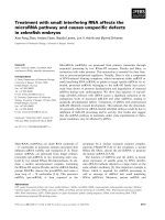
Positive cells were quantified as previously described [17]. In
brief, three sections of each specimen were examined (40×;
Leitz Orthoplan) from either the superficial zone of the carti-
lage (the superficial and upper intermediate layers) or the deep
zone (the lower intermediate and deep layers) as illustrated in
Figure 1, scored, and the resulting data integrated as a mean
for each specimen. The final results were expressed as the
percentage of chondrocytes staining positive for the antigen
(cell score) with the maximum score being 100%. Each slide
was subjected to evaluation by two observers with >95%
degree of agreement.
Determination of interleukin and MMP production
IL-1β, IL-6, MMP-1, and MMP-13 were determined by specific
ELISAs (R&D Systems) in the culture media. All determina-
tions were performed in duplicate for each cell culture.
Statistical analysis
Data are expressed as the mean ± SEM of independent spec-
imens. Statistical significance was assessed by the 2-tailed
Student's t-test, and P values ≤ 0.05 were considered signifi-
cant.
Table 1
Primer Sequence
Gene Sense Antisense
EphB4 receptor 5'-CACAGTCATCCAGCTCGTG 5'-ATCGGATGGGAATCTTTCC
ephrin B2 5'-TTCGACAACAAGTCCCTTTG 5'-CGAGTGCTTCCTGTGTCTC
IL-1β 5'-TTAGGAAGACACAAATTGC 5'-TGGGCAGACTCAAATTCCAG
IL-6 5'-CACCTCTTCAGAACGAATTG 5'-CTAGGTATACCTCAAACTCC
PAR-2 5'-GAAGCCTTATTGGTAAGGTTG 5'-CAGAGAGGAGGTCAGCCAAG
MMP-1 5'-CTGAAAGTGACTGGGAAACC 5'-AGAGTTGTCCCGATGATCTC
MMP-2 5'-CACTGTTGGTGGGAACTCAG 5'-GTGTAAATGGGTGCCATCAG
MMP-9 5'-CCTTCACTTTCCTGGGTAAG 5'-CCATTCACGTCGTCCTTATG
MMP-13 5'-CTTAGAGGTGACTGGCAAAC 5'-GCCCATCAAATGGGTAGAAG
Collagen type II 5'AGTTTCAGGTCTCTGCAGGT 5'-CCAGAAGCACCTTGGTCTC
GAPDH 5'-CAGAACATCATCCCTGCCTCT 5'-GCTTGACAAAGTGGTCGTTGAG
Arthritis Research & Therapy Vol 11 No 4 Kwan Tat et al.
Page 4 of 10
(page number not for citation purposes)
Results
Ephrin B2 and EphB4 receptor expression and
production
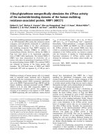
Data showed that the ephrin B2 expression level was slightly
higher in OA chondrocytes (n = 4) compared to normal (n =
5) (Figure 2a). However, the protein production of ephrin B2
was similar in normal (n = 4) and OA chondrocytes (n = 6)
(Figures 2b–f). In both cartilage types, ephrin B2 was localized
in the superficial zone (Figures 2c, 2d) and no positive cells
were detected in the deep zone (Figures 2e, 2f). The Figure 2c
inset represents a negative control done with immunoabsorp-
tion of ephrin B2 showing only background staining, and Fig-
ure 2e inset a higher magnification of some positive cells
stained for ephrin B2.
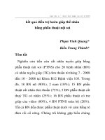
In contrast to ephrin B2, the gene expression level of the
EphB4 receptor was significantly elevated (P < 0.0001) in OA
chondrocytes (n = 8) compared to normal (n = 5) (Figure 3a).
EphB4 receptor protein production was also found at a signif-
icantly higher level (P < 0.0003) in OA (n = 4) than in normal
(n = 4) cartilage (Figure 3b). In normal cartilage, the EphB4
receptor was produced only in the superficial zone (Figures
3c, 3d), whereas in OA, EphB4 receptor positive chondro-
cytes were found throughout the cartilage (Figures 3e, 3f),
with a statistically significant increase (P < 0.01) found in both
zones (Figure 3b). As for the ephrin B2 above, the inset in Fig-
ure 3c represents a negative control done with immunoab-
sorption of EphB4 receptor showing only background
staining, and the Figure 3e inset a higher magnification of pos-
itive cells stained with the EphB4 receptor antibody.
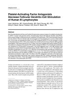
Functional consequences of ephrin B2 treatment
We then investigated the OA chondrocytes (n = 8) upon treat-
ment with ephrin B2 (50 and 100 ng/ml), the modulation of
some catabolic and anabolic factors known to be involved in
the physiological/pathophysiological chondrocyte processes.
These were IL-1β, IL-6, MMP-1, MMP-2, MMP-9, and MMP-
13, the proteinase-activated receptor-2 (PAR-2), a receptor
involved in inflammatory pathways and recently shown to play
an important role in OA [17,18], and the collagen type II. Data
revealed that ephrin B2 treatment led to a pattern of reduced
expression of several catabolic factors. Both pro-inflammatory
cytokines, IL-1β and IL-6, were significantly inhibited (P <
0.002, P < 0.04 respectively) (Figures 4a, 4b); the reduction
was dose dependent and significance reached at 100 ng/ml
of ephrin B2. MMP-1, MMP-13, and MMP-9, but not MMP-2,
were also significantly decreased with ephrin B2 at both con-
centrations (50, 100 ng/ml) tested (Figures 4c, 4d, 4e, 4f). A
similar significant inhibitory effect was obtained for PAR-2
expression upon treatment with the ephrin B2 ligand at 50 and
100 ng/ml (Figure 4g). Interestingly, ephrin B2 at 100 ng/ml
significantly increased (P < 0.03) the expression level of colla-
gen type II (Figure 4h).
In addition, experiments were done with OA chondrocytes (n
= 6 to 8) incubated in the absence or presence of ephrin B2
at 50 and 100 ng/ml with or without IL-1β at 100 pg/ml and
the protein production of IL-6, MMP-1 and MMP-13 deter-
mined. Data first showed that ephrin B2 alone had no effect on
the basal levels of IL-6, MMP-1 or MMP-13 (data not shown),
possibly due to the fact that the production of these factors by
the OA chondrocytes was at the limit of detection. The basal
level of IL-1β was also very low, yet slightly higher than the limit
of detection with a mean value of 14.6 ± 4.4 ng/mg protein
recorded. The treatment with ephrin B2 at 100 ng/ml abol-
ished such detection, indicating that ephrin B2 decreases the
protein synthesis of this cytokine.
Since in vivo OA pathophysiology is characterized by the
presence of IL-1β, protein production of these factors was fur-
ther determined in the presence of this cytokine. As MMP-2
and MMP-9 are not truly modulated by IL-1β [19,20], they
were not studied. Data as represented in Table 2 showed that
the significant stimulatory effect of IL-1β on the production of
IL-6, MMP-1, and MMP-13 was inhibited by ephrin B2, with a
statistically significant effect obtained for IL-6 (P < 0.05) and
MMP-13 (P = 0.05) at 100 ng/ml ephrin B2.
Discussion
Osteoarthritis is a debilitating disease resulting from a com-
plex degradative mechanism in the articular joint. Although
considerable advancement has been made towards a better
understanding of the pathophysiological pathways that occur
during the OA process, much remains to be accomplished in
the development of an effective DMOAD that would reduce or
stop the disease progression. In this context, identifying new
candidates able to target several joint tissues (cartilage,
subchondral bone and synovial membrane) is extremely attrac-
tive.
Figure 1
Human cartilage subdivided into two zones: superficial zone (superficial and upper intermediate layers) and deep zone (lower intermediate and deep layers)Human cartilage subdivided into two zones: superficial zone (superficial
and upper intermediate layers) and deep zone (lower intermediate and
deep layers). The subchondral bone plate is also represented.
Available online />Page 5 of 10
(page number not for citation purposes)
Our group recently showed, in human OA subchondral bone
osteoblasts [12], that ephrin B2 treatment induces a reduction
in the abnormal remodelling process as well as in several cat-
abolic factors involved in bone matrix alterations. These data
suggest that ephrin B2 could exert a protective effect on struc-
tural changes in OA articular tissues, which makes this ephrin
system an attractive and interesting therapeutic target in OA.
Since the cartilage also demonstrates a remodelling of its
extracellular matrix during the disease process and the ephrin
system is known to control extracellular matrix, we explored the
implication of this ephrin system in human OA cartilage metab-
olism and identified factors targeted in the diseased tissue.
We investigated the presence of ephrin B2 and the EphB4
receptor in human articular cartilage/chondrocytes and the
Figure 2
Ephrin B2 (a) gene expression level in human normal (n = 5) and osteoarthritic (OA) (n = 4) chondrocytes, and (b) protein production as analyzed following immunohistochemistry as described in Materials and Methods in the superficial zone in normal (n = 4) and OA (n = 6) cartilageEphrin B2 (a) gene expression level in human normal (n = 5) and osteoarthritic (OA) (n = 4) chondrocytes, and (b) protein production as analyzed
following immunohistochemistry as described in Materials and Methods in the superficial zone in normal (n = 4) and OA (n = 6) cartilage. Of note,
the arbitrary unit of the ephrin B2 ligand gene is expressed as × 10
-3
. (c) Representative immunohistological sections showing ephrin B2 in the
superficial zone of human normal and (d) OA cartilage and (e) the deep zone of human normal and (f) OA cartilage. The insets represent in (c) a
negative control done with immunoabsorption with only background staining and in (e) a higher magnification of positive cells stained for ephrin B2.
c, d, e, f and inset in c: original magnification ×100, and inset in e: original magnification ×400. Arrows indicate stained chondrocytes. Statistical sig-
nificance assessed by Student's t-test revealed no difference.
Arthritis Research & Therapy Vol 11 No 4 Kwan Tat et al.
Page 6 of 10
(page number not for citation purposes)
effects of treatment with ephrin B2 on human OA chondro-
cytes. This is the first time that this system has been studied in
chondrocytes, and our data revealed important new informa-
tion about its mechanisms of action in cartilage.
The first finding was that the EphB4 receptor is differentially
expressed and produced by normal and OA chondrocytes/
cartilage, with a significantly increased expression level in OA
compared to normal. In contrast to normal, OA cartilage
showed not only a significantly increased number of chondro-
cytes in the superficial zone producing the EphB4 receptor,
but its production was also extended to the deep zone. Ephrin
B2, however, did not appear to be modulated in human OA
cartilage. The data showing a higher level of EphB4 receptors
Figure 3
EphB4 receptor (a) gene expression level in human normal (n = 5) and osteoarthritic (OA) (n = 8) chondrocytes, and (b) total protein production as analyzed following immunohistochemistry as described in Materials and Methods in normal (n = 4) and OA (n = 4) cartilage or in the superficial or deep zones of the cartilageEphB4 receptor (a) gene expression level in human normal (n = 5) and osteoarthritic (OA) (n = 8) chondrocytes, and (b) total protein production as
analyzed following immunohistochemistry as described in Materials and Methods in normal (n = 4) and OA (n = 4) cartilage or in the superficial or
deep zones of the cartilage. Of note, the arbitrary unit of the EphB4 receptor gene is expressed as × 10
-2
. (c) Representative immunohistological
sections showing EphB4 receptor in the superficial and (d) deep zone of human normal cartilage and in the (e) superficial and (f) deep zone of OA
cartilage. The insets represent in (c) a negative control done with immunoabsorption with only background staining and in (e) a higher magnification
of positive cells stained for ephrin B2. c, d, e, f and inset in c: original magnification ×100, and inset in e: original magnification ×400. Arrows indi-
cate stained chondrocytes. Statistical significance was assessed by Student's t-test and P values are as underlined.
Available online />Page 7 of 10
(page number not for citation purposes)
Figure 4
Effect of ephrin B2 activation of the EphB4 receptor on human osteoarthritic chondrocytes (n = 8) on the gene expression level of (a) IL-1β, (b) IL-6, (c) MMP-1, (d) MMP-2, (e) MMP-9, (f) MMP-13, (g) PAR-2, and (h) collagen type IIEffect of ephrin B2 activation of the EphB4 receptor on human osteoarthritic chondrocytes (n = 8) on the gene expression level of (a) IL-1β, (b) IL-
6, (c) MMP-1, (d) MMP-2, (e) MMP-9, (f) MMP-13, (g) PAR-2, and (h) collagen type II. Cells were incubated for 18 hours. Data are expressed as
the mean ± SEM of arbitrary unit over the control which was attributed a value of 1. Statistical significance was assessed by Student's t-test and P
values are versus control.
Arthritis Research & Therapy Vol 11 No 4 Kwan Tat et al.
Page 8 of 10
(page number not for citation purposes)
in OA chondrocytes combined with those showing ephrin B2
treatment decreased inflammatory/catabolic factors and
increased collagen type II suggest that exogenous ephrin B2
treatment could be of interest in limiting the degradation
involved in abnormal cartilage breakdown.
Data first showed that ephrin B2 treatment significantly
decreased the expression levels of the proinflammatory
cytokines IL-1β and IL-6 which are highly involved in the sever-
ity and perpetuation of this disease [21-25]. Experiments also
demonstrated a similar inhibition of the collagenases MMP-1
and MMP-13, which are closely linked to the degradative prop-
erties in cartilage because of their activity not only on collagen
but also on a wide range of non-collagenous extracellular mac-
romolecules [26-34]. Data also showed that IL-1β protein pro-
duction as well as the IL-1β-induced synthesis of IL-6, MMP-1
and MMP-13 by OA chondrocytes was markedly reduced by
ephrin B2, thus strengthening the hypothesis suggesting its in
vivo beneficial and protective effect.
Although MMP-1 and MMP-13 are the most important mem-
bers of this family in relation to cartilage degradation, some
other MMPs including the gelatinases have also been sug-
gested to be involved in the OA pathological process
[19,20,35-38]. We therefore investigated the effect of the
activation of this ephrin system on MMP-2 and MMP-9. Data
revealed a significant inhibition of MMP-9, but not of MMP-2.
The lack of effect on MMP-2 is not surprising and is consistent
with the literature indicating the greater significance of MMP-
9 in joint diseases than MMP-2. Indeed, knockout mouse
experiments revealed that the absence of MMP-9, but not of
MMP-2, reduces arthritis progression [39]. Positive correlation
between the production of MMP-9, but not of MMP-2, was
also found with rapid destruction in human hip OA [40,41].
Moreover, the plasma level of MMP-9, but not of MMP-2, is
upregulated in OA compared to normal [42]. The differences
between these two MMPs could be due to the differential
pathways in cell signalling. Indeed, such differences were
seen, although on other articular cell types, in synovial and
meniscal tissues in which the production of latent and active
forms of MMP-9 was mediated partly through Jun N-terminal
kinase (JNK) and p38, whereas MMP-2 was not modulated by
such pathways. Moreover, experiments carried out on mono-
cytes and macrophages derived from rheumatoid arthritis
demonstrated that the role of CD147 in MMP production and
cell invasion enhanced MMP-9 production through extracellu-
lar signal-related kinase 1/2 (Erk1/2) and JNK, whereas MMP-
2 production was not modulated at all. Altogether, these data
strengthen our current observation about the differential mod-
ulation of MMP-2 and MMP-9 by this ephrin system [43,44].
In the joint, the inflammatory response is a major component in
sustaining the progression of OA [45]. In that respect, a factor
belonging to the PARs, PAR-2, has been shown to be involved
in arthritic inflammatory pathways, and data generated by
using a PAR-2 gene knockout mouse in the adjuvant-induced
arthritis model demonstrated its important role in chronic
arthritis [46-48]. It was also suggested that PAR-2 could be an
upstream regulator of pro-inflammatory cytokines in articular
tissue cells and responsible for their upregulation [49]. More-
over, PAR-2 was recently found to be closely linked to carti-
lage remodelling in human OA [17,18]. Interestingly, this study
showed that treatment with ephrin B2 inhibits this pro-inflam-
matory factor.
Finally, in order to complement the effect of this ephrin system
in OA chondrocytes, we also investigated whether ephrin B2
exerts an effect on a cartilage specific macromolecule, colla-
gen type II. Data indeed showed this system's ability to induce
collagen type II expression by human OA chondrocytes. Alto-
gether, these experiments demonstrated that ephrin B2 treat-
ment on human OA chondrocytes leads to decreased
catabolic/inflammatory properties at the same time as having
an anabolic effect.
As well described in the literature, the ephrin B2 ligand and
EphB4 receptors are present at the cell membrane and Hattori
et al [50] recently proposed that membranous ephrin ligands
could be cleaved by some proteases. It would therefore be
very appealing to further explore such shedding mechanism
and identify the protease(s) responsible for the cleavage in
articular tissues. Such cleavage would increase the level of
Table 2
Protein production of IL-6, MMP-1 and MMP-13 after a 72 hour incubation period on human osteoarthritic chondrocytes
IL-6
(μg/mg protein)
MMP-1
(μg/mg protein)
MMP-13
(μg/mg protein)
Control 0.3 ± 0.1 12.7 ± 3.1 0.5 ± 0.1
IL-1β (100 pg/ml) 3.6 ± 0.6
*(P < 0.002)
62.7 ± 17.7
*(P < 0.02)
3.5 ± 1.0
*(P < 0.01)
IL-1β+ephrin B2 (50 ng/ml) 2.2 ± 0.4 41.2 ± 5.7 1.9 ± 0.3
IL-1β+ephrin B2 (100 ng/ml) 2.1 ± 0.4
†(P < 0.05)
38.6 ± 5.5 2.0 ± 0.3
†(P = 0.05)
Data are expressed as mean ± SEM.
* Indicates statistically significant difference compared to control values, and † compared to IL-1β values.
Available online />Page 9 of 10
(page number not for citation purposes)
soluble ephrin B2 in OA extracellular matrix, enabling it to bet-
ter exert its effect on its specific receptor, thus contributing to
a protective effect on cartilage matrix.
Hence, in human OA cartilage treatment with ephrin B2 could
act at two different levels: (i) by limiting the extent of matrix
degradation through the inhibition of the most important inter-
leukin and MMP involved in OA cartilage breakdown, as well
as PAR-2, another inflammatory factor, and the IL-1β-induced
catabolic factors, and (ii) by promoting the production of the
cartilage specific macromolecule collagen type II. Thus, data
from this study on human chondrocytes and the previous one
on subchondral bone [12] strongly suggest this ephrin system
as a potential and very attractive therapeutic target for OA.
Conclusions
In conclusion, the data showing that treatment of OA chondro-
cytes by ephrin B2 down-regulates various catabolic factors in
cartilage at the same time as increasing a major anabolic fac-
tor, collagen type II, are of significance. These data indicate
that treatment of OA patients with ephrin B2 or that an
increase in this endogenous ligand could be an interesting
approach in the development of a specific therapeutic agent
able to act on more than one tissue of the joint.
Competing interests
The authors declare that they have no competing interests.
Authors' contributions
SKT helped to design the study, acquire data, analyse and
interpret data, prepare the manuscript and participated in the
statistical analysis. JMP and JPP helped to design the study,
and prepare the manuscript. NA helped to acquire data and
analyse and interpret data. CB helped to analyse and interpret
data and participated in the statistical analysis. ML helped to
acquire data. All authors read and approved the final manu-
script.
Acknowledgements
The authors are grateful to Saranette Cheng for preparing the immuno-
histological sections, Changshan Geng, François-Cyril Jolicoeur and
François Mineau for their expert technical assistance in real-time PCR
and cell cultures, and Virginia Wallis for the manuscript preparation. This
study was supported by internal funds of the Osteoarthritis Research
Unit of the University of Montreal Hospital Research Center, Montreal,
Quebec, Canada.
References
1. Flanagan JG, Vanderhaeghen P: The ephrins and Eph receptors
in neural development. Annu Rev Neurosci 1998, 21:309-345.
2. Zamora DO, Babra B, Pan Y, Planck SR, Rosenbaum JT: Human
leukocytes express ephrinB2 which activates microvascular
endothelial cells. Cell Immunol 2006, 242:99-109.
3. Stephen LJ, Fawkes AL, Verhoeve A, Lemke G, Brown A: A critical
role for the EphA3 receptor tyrosine kinase in heart develop-
ment. Dev Biol 2007, 302:66-79.
4. Himanen JP, Nikolov DB: Eph receptors and ephrins. Int J Bio-
chem Cell Biol 2003, 35:130-134.
5. Mundy GR, Elefteriou F: Boning up on ephrin signaling. Cell
2006, 126:441-443.
6. Zhao C, Irie N, Takada Y, Shimoda K, Miyamoto T, Nishiwaki T,
Suda T, Matsuo K: Bidirectional ephrinB2-EphB4 signaling con-
trols bone homeostasis. Cell Metab 2006, 4:111-121.
7. Himanen JP, Saha N, Nikolov DB: Cell-cell signaling via Eph
receptors and ephrins. Curr Opin Cell Biol 2007, 19:534-542.
8. Brambilla R, Schnapp A, Casagranda F, Labrador JP, Bergemann
AD, Flanagan JG, Pasquale EB, Klein R: Membrane-bound
LERK2 ligand can signal through three different Eph-related
receptor tyrosine kinases. EMBO J 1995, 14:3116-3126.
9. Sakano S, Serizawa R, Inada T, Iwama A, Itoh A, Kato C, Shimizu
Y, Shinkai F, Shimizu R, Kondo S, Ohno M, Suda T: Characteriza-
tion of a ligand for receptor protein-tyrosine kinase HTK
expressed in immature hematopoietic cells. Oncogene 1996,
13:813-822.
10. Gale NW, Yancopoulos GD: Growth factors acting via endothe-
lial cell-specific receptor tyrosine kinases: VEGFs, angiopoie-
tins, and ephrins in vascular development. Genes Dev 1999,
13:1055-1066.
11. Myshkin E, Wang B: Chemometrical classification of ephrin lig-
ands and Eph kinases using GRID/CPCA approach. J Chem
Inf Comput Sci 2003, 43:1004-1010.
12. Kwan Tat S, Pelletier JP, Amiable N, Boileau C, Duval N, Martel-
Pelletier J: Activation of the receptor EphB4 by its specific lig-
and ephrin B2 in human osteoarthritic subchondral bone oste-
oblasts: a new therapeutic approach. Arthritis Rheum 2008,
58:3820-3830.
13. Wada N, Tanaka H, Ide H, Nohno T: Ephrin-A2 regulates posi-
tion-specific cell affinity and is involved in cartilage morpho-
genesis in the chick limb bud. Dev Biol 2003, 264:550-563.
14. Altman RD, Asch E, Bloch DA, Bole G, Borenstein D, Brandt KD,
Christy W, Cooke TD, Greenwald R, Hochberg M, Howell DS,
Kaplan D, Koopman W, Longley SI, Mankin HJ, McShane DJ,
Medsger TA Jr, Meehan R, Mikkelsen W, Moskowitz RW, Murphy
W, Rothschild B, Segal L, Sokoloff L, Wolfe F: Development of
criteria for the classification and reporting of osteoarthritis.
Classification of osteoarthritis of the knee. Arthritis Rheum
1986, 29:1039-1049.
15. Boileau C, Pelletier JP, Tardif G, Fahmi H, Laufer S, Lavigne M,
Martel-Pelletier J: The regulation of human MMP-13 by
licofelone, an inhibitor of cyclooxygenases and 5-lipoxygen-
ase, in human osteoarthritic chondrocytes is mediated by the
inhibition of the p38 MAP kinase signaling pathway. Ann
Rheum Dis 2005, 64:891-898.
16. Tardif G, Hum D, Pelletier JP, Boileau C, Ranger P, Martel-Pelletier
J: Differential gene expression and regulation of the bone mor-
phogenetic protein antagonists follistatin and gremlin in nor-
mal and osteoarthritic human chondrocytes and synovial
fibroblasts. Arthritis Rheum 2004, 50:2521-2530.
17. Boileau C, Amiable N, Martel-Pelletier J, Fahmi H, Duval N, Pelletier
JP: Activation of proteinase-activated receptor 2 in human
osteoarthritic cartilage upregulates catabolic and proinflam-
matory pathways capable of inducing cartilage degradation: a
basic science study. Arthritis Res Ther 2007, 9:R121.
18. Xiang Y, Masuko-Hongo K, Sekine T, Nakamura H, Yudoh K, Nish-
ioka K, Kato T: Expression of proteinase-activated receptors
(PAR)-2 in articular chondrocytes is modulated by IL-1beta,
TNF-alpha and TGF-beta. Osteoarthritis Cartilage 2006,
14:1163-1173.
19. Duerr S, Stremme S, Soeder S, Bau B, Aigner T: MMP-2/gelati-
nase A is a gene product of human adult articular chondro-
cytes and is increased in osteoarthritic cartilage. Clin Exp
Rheumatol 2004, 22:603-608.
20. Soder S, Roach HI, Oehler S, Bau B, Haag J, Aigner T: MMP-9/
gelatinase B is a gene product of human adult articular
chondrocytes and increased in osteoarthritic cartilage. Clin
Exp Rheumatol 2006, 24:302-304.
21. Jikko A, Wakisaka T, Iwamoto M, Hiranuma H, Kato Y, Maeda T,
Fujishita M, Fuchihata H: Effects of interleukin-6 on proliferation
and proteoglycan metabolism in articular chondrocyte cul-
tures.
Cell Biol Int 1998, 22:615-621.
22. Flannery CR, Little CB, Hughes CE, Curtis CL, Caterson B, Jones
SA: IL-6 and its soluble receptor augment aggrecanase-medi-
ated proteoglycan catabolism in articular cartilage. Matrix Biol
2000, 19:549-553.
Arthritis Research & Therapy Vol 11 No 4 Kwan Tat et al.
Page 10 of 10
(page number not for citation purposes)
23. Fernandes JC, Martel-Pelletier J, Pelletier JP: The role of
cytokines in osteoarthritis pathophysiology. Biorheology 2002,
39:237-246.
24. Doss F, Menard J, Hauschild M, Kreutzer HJ, Mittlmeier T, Muller-
Steinhardt M, Muller B: Elevated IL-6 levels in the synovial fluid
of osteoarthritis patients stem from plasma cells. Scand J
Rheumatol 2007, 36:136-139.
25. Pujol JP, Chadjichristos C, Legendre F, Bauge C, Beauchef G,
Andriamanalijaona R, Galera P, Boumediene K: Interleukin-1 and
transforming growth factor-beta 1 as crucial factors in oste-
oarthritic cartilage metabolism. Connect Tissue Res 2008,
49:293-297.
26. Freije JM, Diez-Itza I, Balbin M, Sanchez LM, Blasco R, Tolivia J,
Lopez-Otin C: Molecular cloning and expression of colla-
genase-3, a novel human matrix metalloproteinase produced
by breast carcinomas. J Biol Chem 1994, 269:16766-16773.
27. Fosang AJ, Last K, Knauper V, Murphy G, Neame PJ: Degradation
of cartilage aggrecan by collagenase-3 (MMP-13). FEBS Lett
1996, 380:17-20.
28. Knauper V, Lopez-Otin C, Smith B, Knight G, Murphy G: Bio-
chemical characterization of human collagenase-3. J Biol
Chem 1996, 271:1544-1550.
29. Mitchell PG, Magna HA, Reeves LM, Lopresti-Morrow LL, Yocum
SA, Rosner PJ, Geoghegan KF, Hambor JE: Cloning, expression,
and type II collagenolytic activity of matrix metalloproteinase-
13 from human osteoarthritic cartilage. J Clin Invest 1996,
97:761-768.
30. Reboul P, Pelletier JP, Tardif G, Cloutier JM, Martel-Pelletier J: The
new collagenase, collagenase-3, is expressed and synthe-
sized by human chondrocytes but not by synoviocytes: A role
in osteoarthritis. J Clin Invest 1996, 97:2011-2019.
31. Billinghurst RC, Dahlberg L, Ionescu M, Reiner A, Bourne R,
Rorabeck C, Mitchell P, Hambor J, Diekmann O, Tschesche H,
Chen J, Van Wart H, Poole AR: Enhanced cleavage of Type II
collagen by collagenases in osteoarthritic articular cartilage. J
Clin Invest 1997, 99:1534-1545.
32. Knauper V, Cowell S, Smith B, Lopez-Otin C, O'Shea M, Morris H,
Zardi L, Murphy G: The role of the C-terminal domain of human
collagenase-3 (MMP-13) in the activation of procollagenase-3,
substrate specificity, and tissue inhibitor of metalloproteinase
interaction. J Biol Chem
1997, 272:7608-7616.
33. Hiller O, Lichte A, Oberpichler A, Kocourek A, Tschesche H:
Matrix metalloproteinases collagenase-2, macrophage
elastase, collagenase-3, and membrane type 1-matrix metal-
loproteinase impair clotting by degradation of fibrinogen and
factor XII. J Biol Chem 2000, 275:33008-33013.
34. Hashimoto G, Inoki I, Fujii Y, Aoki T, Ikeda E, Okada Y: Matrix met-
alloproteinases cleave connective tissue growth factor and
reactivate angiogenic activity of vascular endothelial growth
factor 165. J Biol Chem 2002, 277:36288-36295.
35. Mohtai M, Smith RL, Schurman DJ, Tsuji Y, Torti FM, Hutchinson
NI, Stetler-Stevenson WG, Goldberg GI: Expression of 92-kD
type IV collagenase/gelatinase (gelatinase B) in osteoarthritic
cartilage and its induction in normal human articular cartilage
by interleukin-1. J Clin Invest 1993, 92:179-185.
36. Tsuchiya K, Maloney WJ, Vu T, Hoffman AR, Huie P, Sibley R,
Schurman DJ, Smith RL: Osteoarthritis:differential expression
of matrix metalloproteinase-9 mRNA in nonfibrillated and
fibrillated cartilage. J Orthop Res 1997, 15:94-100.
37. Aigner T, Zien A, Gehrsitz A, Gebhard PM, McKenna L: Anabolic
and catabolic gene expression pattern analysis in normal ver-
sus osteoarthritic cartilage using complementary DNA-array
technology. Arthritis Rheum 2001, 44:2777-2789.
38. Volk SW, Kapatkin AS, Haskins ME, Walton RM, D'Angelo M:
Gelatinase activity in synovial fluid and synovium obtained
from healthy and osteoarthritic joints of dogs. Am J Vet Res
2003, 64:1225-33.
39. Itoh T, Matsuda H, Tanioka M, Kuwabara K, Itohara S, Suzuki R:
The role of matrix metalloproteinase-2 and matrix metallopro-
teinase-9 in antibody-induced arthritis. J Immunol 2002,
169:2643-2647.
40. Masuhara K, Bak Lee S, Nakai T, Sugano N, Ochi T, Sasaguri Y:
Matrix metalloproteinases in patients with osteoarthritis of the
hip. Int Orthop 2000, 24:92-96.
41. Masuhara K, Nakai T, Yamaguchi K, Yamasaki S, Sasaguri Y: Sig-
nificant increases in serum and plasma concentrations of
matrix metalloproteinases 3 and 9 in patients with rapidly
destructive osteoarthritis of the hip. Arthritis Rheum 2002,
46:
2625-2631.
42. Tchetverikov I, Ronday HK, Van El B, Kiers GH, Verzijl N, TeKop-
pele JM, Huizinga TW, DeGroot J, Hanemaaijer R: MMP profile in
paired serum and synovial fluid samples of patients with rheu-
matoid arthritis. Ann Rheum Dis 2004, 63:881-883.
43. Hsieh YS, Yang SF, Lue KH, Chu SC, Li TJ, Lu KH: Upregulation
of urokinase-type plasminogen activator and inhibitor and
gelatinase expression via 3 mitogen-activated protein kinases
and PI3K pathways during the early development of osteoar-
thritis. J Rheumatol 2007, 34:785-793.
44. Yang Y, Lu N, Zhou J, Chen ZN, Zhu P: Cyclophilin A up-regu-
lates MMP-9 expression and adhesion of monocytes/macro-
phages via CD147 signalling pathway in rheumatoid arthritis.
Rheumatology (Oxford) 2008, 47:1299-1310.
45. Martel-Pelletier J, Lajeunesse D, Pelletier JP: Etiopathogenesis of
osteoarthritis. In Arthritis & Allied Conditions. A Textbook of
Rheumatology 15th edition. Edited by: Koopman, Moreland. Balti-
more: Lippincott, Williams & Wilkins; 2005:2199-2226.
46. Lindner JR, Kahn ML, Coughlin SR, Sambrano GR, Schauble E,
Bernstein D, Foy D, Hafezi-Moghadam A, Ley K: Delayed onset of
inflammation in protease-activated receptor-2-deficient mice.
J Immunol 2000, 165:6504-6510.
47. Ferrell WR, Lockhart JC, Kelso EB, Dunning L, Plevin R, Meek SE,
Smith AJ, Hunter GD, McLean JS, McGarry F, Ramage R, Jiang L,
Kanke T, Kawagoe J: Essential role for proteinase-activated
receptor-2 in arthritis. J Clin Invest 2003, 111:35-41.
48. Busso N, Frasnelli M, Feifel R, Cenni B, Steinhoff M, Hamilton J, So
A: Evaluation of protease-activated receptor 2 in murine mod-
els of arthritis. Arthritis Rheum 2007, 56:101-107.
49. Kelso EB, Ferrell WR, Lockhart JC, Elias-Jones I, Hembrough T,
Dunning L, Gracie JA, McInnes IB: Expression and proinflamma-
tory role of proteinase-activated receptor 2 in rheumatoid syn-
ovium: ex vivo studies using a novel proteinase-activated
receptor 2 antagonist. Arthritis Rheum 2007, 56:765-771.
50. Hattori M, Osterfield M, Flanagan JG: Regulated cleavage of a
contact-mediated axon repellent. Science 2000,
289:1360-1365.