Báo cáo y học: " Increased expression of matrix metalloproteinase-10, nerve growth factor and substance P in the painful degenerate intervertebral disc" pot
Bạn đang xem bản rút gọn của tài liệu. Xem và tải ngay bản đầy đủ của tài liệu tại đây (863.02 KB, 8 trang )
Open Access
Available online />Page 1 of 8
(page number not for citation purposes)
Vol 11 No 4
Research article
Increased expression of matrix metalloproteinase-10, nerve
growth factor and substance P in the painful degenerate
intervertebral disc
Stephen M Richardson
1
, Paul Doyle
2
, Ben M Minogue
1
, Kanna Gnanalingham
2
and
Judith A Hoyland
1
1
Tissue Injury and Repair Group, School of Clinical and Laboratory Sciences, Stopford Building, The University of Manchester, Oxford Road,
Manchester, M13 9PT, UK
2
Department of Neurosurgery, Greater Manchester Neuroscience Centre, Salford Royal Foundation Trust, Stott Lane, Salford, M6 8HD, UK
Corresponding author: Judith A Hoyland,
Received: 9 Jun 2009 Revisions requested: 8 Jul 2009 Revisions received: 27 Jul 2009 Accepted: 20 Aug 2009 Published: 20 Aug 2009
Arthritis Research & Therapy 2009, 11:R126 (doi:10.1186/ar2793)
This article is online at: />© 2009 Richardson et al.; licensee BioMed Central Ltd.
This is an open access article distributed under the terms of the Creative Commons Attribution License ( />),
which permits unrestricted use, distribution, and reproduction in any medium, provided the original work is properly cited.
Abstract
Introduction Matrix metalloproteinases (MMPs) are known to
be involved in the degradation of the nucleus pulposus (NP)
during intervertebral disc (IVD) degeneration. This study
investigated MMP-10 (stromelysin-2) expression in the NP
during IVD degeneration and correlated its expression with pro-
inflammatory cytokines and molecules involved in innervation
and nociception during degeneration which results in low back
pain (LBP).
Methods Human NP tissue was obtained at postmortem (PM)
from patients without a history of back pain and graded as
histologically normal or degenerate. Symptomatic degenerate
NP samples were also obtained at surgery for LBP. Expression
of MMP-10 mRNA and protein was analysed using real-time
polymerase chain reaction and immunohistochemistry. Gene
expression for pro-inflammatory cytokines interleukin-1 (IL-1)
and tumour necrosis factor-alpha (TNF-α), nerve growth factor
(NGF) and the pain-associated neuropeptide substance P were
also analysed. Correlations between MMP-10 and IL-1, TNF-α
and NGF were assessed along with NGF with substance P.
Results MMP-10 mRNA was significantly increased in surgical
degenerate NP when compared to PM normal and PM
degenerate samples. MMP-10 protein was also significantly
higher in degenerate surgical NP samples compared to PM
normal. IL-1 and MMP-10 mRNA demonstrated a significant
correlation in surgical degenerate samples, while TNF-α was not
correlated with MMP-10 mRNA. NGF was significantly
correlated with both MMP-10 and substance P mRNA in
surgical degenerate NP samples.
Conclusions MMP-10 expression is increased in the
symptomatic degenerate IVD, where it may contribute to matrix
degradation and initiation of nociception. Importantly, this study
suggests differences in the pathways involved in matrix
degradation between painful and pain-free IVD degeneration.
Introduction
The human intervertebral disc (IVD) is an avascular and aneu-
ral tissue comprising a central gelatinous region (the nucleus
pulposus, or NP) surrounded by a fibrous ring of highly organ-
ised collagen fibres (the annulus fibrosus, or AF) [1]. The extra-
cellular matrix (ECM) of the NP is rich in type II collagen and
proteoglycans, predominantly aggrecan, which produces a
highly hydrated matrix capable of withstanding the loads expe-
rienced within the spine [2,3]. This ECM is constantly being
remodelled in a process driven by the constituent NP cells.
During IVD degeneration, there is an imbalance in the normal
homeostatic mechanisms, which favours matrix catabolism
and leads to a loss of disc height, coupled with ingrowth of
both nerves and blood vessels into both the AF and NP [2,4].
We have previously demonstrated that this ingrowth of nerves
ADAMTS: a disintegrin and metalloproteinase with thrombospondin motifs; AF: annulus fibrosus; ECM: extracellular matrix; GAPDH: glyceraldehyde-
3-phosphate dehydrogenase; IL-1: interleukin-1; IVD: intervertebral disc; LBP: low back pain; MMP: matrix metalloproteinase; NGF: nerve growth
factor; NP: nucleus pulposus; PCR: polymerase chain reaction; PM: postmortem; QRT-PCR: quantitative real-time polymerase chain reaction; TNF-
α: tumour necrosis factor-alpha.
Arthritis Research & Therapy Vol 11 No 4 Richardson et al.
Page 2 of 8
(page number not for citation purposes)
into the degenerate IVD is associated with low back pain
(LBP) [5]. While LBP is multi-factorial, studies have shown
that this debilitating condition affecting around 80% of adults
at some stage of life is associated with IVD degeneration in
approximately 40% of cases [6]. Indeed, in a recent study by
Cheung and colleagues [7], it was shown that there is a signif-
icant association of lumber disc degeneration imaged by mag-
netic resonance imaging with LBP.
A number of studies have demonstrated an increase in expres-
sion and activity of a range of matrix-degrading enzymes in IVD
degeneration, including the matrix metalloproteinase (MMP)
[8-11] and more recently ADAMTS (a disintegrin and metallo-
proteinase with thrombospondin motifs) families [12-16]. In
particular, we have shown that MMP-1, -3, -7, -9 and -13 are
involved in matrix catabolism during degeneration [4,12,17].
However, to date, no studies have examined the expression or
regulation of MMP-10 in IVD degeneration.
MMP-10 (also known as stromelysin-2) is a member of the
stromelysin family of enzymes, along with MMP-3 (stromelysin-
1) and MMP-11 (stromelysin-3). This family of enzymes exhibit
a wide range of substrate specificities, including both prote-
oglycans and collagens [18]. In addition to its proteolytic activ-
ity, MMP-10 has been demonstrated to be a potent activator
of a number of MMP pro-enzymes, including pro-MMP-1, -7, -
8, -9 and -13 [19,20]. MMP-10 expression has been identified
in human articular chondrocytes isolated from osteoarthritic
hip cartilage [19], where the authors also demonstrated its
ability to activate pro-MMP-1, -8 and -13, which are key
enzymes involved in both cartilage and IVD degradation
[12,19,21,22]. MMP-10, along with MMP-3, is also thought to
be capable of 'super-activating' collagenases such as MMP-1,
with studies demonstrating a significant increase in collagen
release (of up to 50%) following the addition of MMP-10 to an
IL-1-induced model of cartilage degeneration [19,23,24]. This
pivotal role of MMP-10 in multiple pro-enzyme activation is
believed to shift the balance of activity in favour of MMP activ-
ity over MMP inhibition, with resultant increases in enzymatic
activity and increased ECM degradation [24,25].
In articular chondrocytes, MMP-10 expression has been
shown to be induced by both interleukin-1 (IL-1) and tumour
necrosis factor-alpha (TNF-α), which we have previously
shown to be involved in the processes leading to IVD degen-
eration, particularly IL-1 [19,22]. Chen and colleagues [26]
also recently showed that nerve growth factor (NGF) stimula-
tion of PC-12 cells strongly induces MMP-10 gene expres-
sion. We have previously identified the expression of the
neurotrophin NGF and the pain-associated neuropeptide sub-
stance P in the human IVD and demonstrated their regulation
in NP cells by IL-1 and TNF-α [27]. NGF is also known to reg-
ulate the expression of substance P in sensory neurons
[28,29] and intrinsic airway neurons [30], which may lead to
increased nociception in painful IVD degeneration.
The aim of the current study was to examine the gene and pro-
tein expression of MMP-10 in histologically normal human IVD
and compare it with that of both non-painful and painful degen-
erate IVD. Gene expression of MMP-10 was also correlated
with that of the pro-inflammatory cytokines IL-1 and TNF-α as
well as NGF to identify potential regulatory mechanisms that
may drive MMP-10 production in this tissue. Likewise, gene
expression of substance P and its correlation with NGF were
assessed to establish any association with nociception in
those individuals with LBP.
Materials and methods
Tissue samples
Human IVD tissue was obtained at either postmortem (PM) or
following surgery, with informed consent from the patient or
relatives. Local research ethics approval (North West
Research Ethics Committee) was obtained for this work. PM
samples of normal and degenerate NP were obtained within
18 hours of donor death. Asymptomatic normal and degener-
ate discs obtained from donors at PM had no documented
clinical history of LBP. Samples of degenerate NP were
obtained from patients, diagnosed by magnetic resonance
imaging, who underwent disc replacement surgery or spinal
fusion to relieve chronic LBP. Patients suffering from classical
sciatica were excluded from the study. All samples were
obtained and processed as previously described [14].
Histological grading of nucleus pulposus tissues
To establish histological grade of degeneration, NP samples
were fixed in 4% paraformaldehyde/phosphate-buffered saline
and processed into paraffin wax. Five-micron sections from the
tissue blocks were cut and stained with haematoxylin and
eosin, and the degree of morphological degeneration was
graded according to previously published criteria [3]. The
grading system generates a score of between 0 and 12: a
grade of 0 to 3 represents a histologically normal (non-degen-
erate) disc, grades of 4 to 6 indicate mild degeneration,
grades 7 to 9 moderate degeneration and grades 10 to 12
severe degeneration.
Quantitative real-time polymerase chain reaction
Quantitative real-time polymerase chain reaction (QRT-PCR)
was conducted on 5 non-degenerate PM NP samples from 4
individuals (ages 30 to 75 years, mean 56 years), 9 degener-
ate PM NP samples from 4 individuals (ages 30 to 75, mean
59 years) and 13 surgical degenerate NP samples from 11
individuals (ages 28 to 56 years, mean 39 years). Cells were
isolated from each sample as previously reported, and RNA
was extracted using Trizol™ (Invitrogen Corporation, Carlsbad,
CA, USA) in accordance with the instructions of the manufac-
turer [14]. RNA was then treated with DNAse using the Turbo
DNA-free kit (Ambion, Inc., Austin, TX, USA) to remove any
DNA contamination. RNA (500 ng) was then reverse-tran-
scribed using Superscript II (Invitrogen Corporation) in
accordance with the instructions of the manufacturer.
Available online />Page 3 of 8
(page number not for citation purposes)
QRT-PCR was then conducted on an ABI Prism 7000
sequence detection system (Applied Biosystems, Warrington,
UK) to investigate the expression of MMP-10, IL-1β, TNF-α,
NGF and substance P in both PM and surgical samples. Glyc-
eraldehyde-3-phosphate dehydrogenase (GAPDH) pre-
designed amplification reagent (Applied Biosystems) was
used as a housekeeping gene to allow normalisation. Pre-opti-
mised primer and FAM-MGB (fluorescein-minor groove
binder) probe sets were purchased from Applied Biosystems
for MMP-10 (forward primer: CATACCCTGGGTTTTCCTC-
CAA; reverse primer: GTCCGCTGCAAAGAAGTAT-
GTTTTC; probe: CTGCATCAATTTTCC), IL-1β (forward
primer: CGGCCACATTTGGTTCTAAGA; reverse primer:
AGGGAAGCGGTTGCTCATC; probe: ACCCTCTGTCAT-
TCG), TNF-α (forward primer: CGAACATCCAACCTTC-
CCAAC; reverse primer: TGGTGGTCTTGTTGCTTAAAGTT
C; probe: CCAATCCCTTTATTACCC), NGF (ABI assay ID:
Hs00171458_m1) and substance P (ABI assay ID:
Hs00243225_m1). Twenty-microlitre reactions were pre-
pared using TaqMan Universal PCR Master Mix (Applied Bio-
systems) and 10 ng of each cDNA sample. Reactions were
performed in triplicate, and results were analysed using the 2
-
ΔCt
method and presented as relative gene expression normal-
ised to GAPDH [31].
Statistical analysis was performed using the Mann-Whitney U
test to compare the expression of each different gene between
PM normal, PM degenerate and surgical degenerate NP sam-
ples. Scatterplots were initially drawn to assess correlations
between expression of MMP-10 and IL-1, TNF-α or NGF
expression and between NGF and substance P, and then Ken-
dall's rank correlation analysis was used to identify statistically
significant correlations.
Immunohistochemistry for matrix metalloproteinase-10
expression
Immunohistochemsitry for MMP-10 expression was con-
ducted on 4 non-degenerate NP samples from 2 individuals
(ages 37 and 47 years, mean 42 years), 5 PM NP samples
from 4 individuals with mild degeneration (ages 37 to 61
years, mean 49 years), 4 PM NP samples with moderate
degeneration (ages 46 to 78 years, mean 62 years), 2 PM NP
samples with severe degeneration (ages 46 years), 10 surgi-
cal NP samples with mild degeneration (ages 20 to 74 years,
mean 42 years), 8 surgical NP samples with moderate degen-
eration (ages 33 to 69 years, mean 45 years) and 4 surgical
NP samples with severe degeneration (ages 34 to 60 years,
mean 49 years).
The immunohistochemistry protocol followed was as previ-
ously published [12,14]. No antigen retrieval was required,
and a mouse monoclonal primary antibody raised against
human MMP-10 (1:500 dilution; Abcam, Cambridge, UK) was
used. Human placental samples served as positive controls
and negative controls used mouse IgG (Dako, Ely, UK) in
place of the primary antibody at equal protein concentrations.
Following washes, sections were incubated in a 1:300 dilution
of biotinylated goat anti-mouse antiserum (Dako) for 30 min-
utes at room temperature. Binding of the secondary antibody
was disclosed with the streptavidin-biotin complex (Dako)
technique with 3,3'-diaminobenzidine tetrahydrochloride solu-
tion (Sigma-Aldrich, St. Louis, MO, USA). Sections were
counterstained with Mayer's haematoxylin (Raymond A Lamb,
Eastbourne, East Sussex, UK), dehydrated and mounted with
Pertex.
Statistical analysis
All slides were visualised by means of a Leica RMDB micro-
scope (Leica Microsystems, Wetzlar, Germany), and images
were captured by means of a digital camera and Bioquant
Nova image analysis system (Bioquant Image Analysis Corpo-
ration, Nashville, TN, USA). The proportions of immunopositive
NP cells in each grade grouping (that is, 0 to 3 non-degener-
ate, 4 to 6 mild degeneration, 7 to 9 moderate degeneration
and 10 to 12 severe degeneration) were counted and com-
pared for statistical significance using the Mann-Whitney U
test. Data were then plotted as mean ± standard error to rep-
resent the 95% confidence intervals.
Results
Gene expression of matrix metalloproteinase-10 in
human nucleus pulposus
QRT-PCR was conducted on RNA from cells extracted from
normal and degenerate NP samples obtained at PM and from
degenerated NP tissue removed during surgery for LBP.
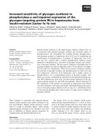
Results demonstrated similar levels of MMP-10 expression in
both PM normal and PM degenerate NP samples. However,
MMP-10 was significantly higher (P < 0.05) in the surgical
degenerate samples than in either the PM normal or PM
degenerate samples (Figure 1).
Figure 1
Expression of matrix metalloproteinase-10 in postmortem (PM) normal, PM degenerate and surgical degenerate human intervertebral discExpression of matrix metalloproteinase-10 in postmortem (PM) normal,
PM degenerate and surgical degenerate human intervertebral disc. Rel-
ative gene expression was normalised to the housekeeping gene glyc-
eraldehyde-3-phosphate dehydrogenase (GAPDH) and plotted on a
log scale. **P < 0.01.
Arthritis Research & Therapy Vol 11 No 4 Richardson et al.
Page 4 of 8
(page number not for citation purposes)
Immunohistochemical localisation of matrix
metalloproteinase-10 in human nucleus pulposus
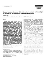
Immunopositivity was seen for MMP-10 in all samples exam-
ined and was evident in NP cells and NP cell clusters (Figure
2). Expression was predominantly localised intracellularly
within the cytoplasm of the NP cells. In PM degenerate and
surgical degenerate samples, diffuse ECM staining, which
was not present in PM normal samples, was observed. No
immunopositivity was seen in invading blood vessels or inflam-
matory cells. Positive controls conducted on placental tissue
demonstrated immunopositivity, while all IgG controls were
negative.
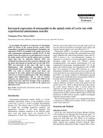
While PM normal tissues demonstrated expression in less
than 20% of constituent cells, PM degenerate tissues demon-
strated increases in the proportion of immunopositive cells
with increasing stage of degeneration (Figure 3); however, this
did not reach significance at any point. Surgical NP samples
again showed increases in the proportion of immunopositive
cells with increasing grade. At each grade, the number of
immunopositive cells was higher than that seen in PM degen-
erate tissues of the same grade, although this was not signifi-
cant. However, surgical NP samples showed significantly
higher levels of MMP-10 immunopositivity than PM normal
samples at grades 7 to 9 (moderate degeneration) and 10 to
12 (severe degeneration) (P < 0.05) (Figure 3).
Gene expression of interleukin-1 and tumour necrosis
factor-alpha and correlation with matrix
metalloproteinase-10 in human nucleus pulposus
No significant differences in the gene expression of either IL-1
or TNF-α between PM normal and PM degenerate samples
were observed (Figures 4a and 4b, respectively). However,
expressions of both IL-1 and TNF-α in surgical degenerate
samples were significantly higher than in either PM normal (P
< 0.01 and P < 0.05, respectively) or PM degenerate (P <
0.01 and P < 0.01, respectively) samples.
Kendall's rank correlation analysis revealed no significant cor-
relation between IL-1α and MMP-10 in PM degenerate sam-
ples (P = 0.076) but did reveal a significant positive
correlation in surgical degenerate samples (P = 0.02) (Figure
4c). However, TNF-α did not show a significant correlation
with MMP-10 in either PM degenerate (P = 0.49) or surgical
degenerate (P = 0.31) samples (Figure 4d). No significant cor-
relation could be identified between any of the genes and age
of the donors.
Gene expression and correlation of nerve growth factor
and substance P in human nucleus pulposus
NGF and substance P demonstrated similar levels of expres-
sion between PM normal and PM degenerate samples but sig-
nificantly higher levels of expression in surgical degenerate
samples than in either PM normal or PM degenerate samples
(P < 0.01) (Figures 5a and 5b, respectively). Kendall's rank
correlation analysis of NGF and MMP-10 expression data
demonstrated no correlation in PM degenerate tissues (P =
0.24) but did demonstrate a strong positive correlation in sur-
Figure 2
Immunohistochemical localisation of matrix metalloproteinase-10 (MMP-10) in human intervertebral discImmunohistochemical localisation of matrix metalloproteinase-10
(MMP-10) in human intervertebral disc. MMP-10 immunopositivity in (a)
postmortem (PM) normal, (b) PM degenerate and (c) surgical degener-
ate samples. An example of an IgG-negative control slide is shown (d).
Scale bar = 25 μm.
Figure 3
Histogram illustrating the percentage of matrix metalloproteinase-10 immunopositive cells in postmortem (PM) normal, PM degenerate and surgical degenerate nucleus pulposus samples classified according to histological grade of degenerationHistogram illustrating the percentage of matrix metalloproteinase-10
immunopositive cells in postmortem (PM) normal, PM degenerate and
surgical degenerate nucleus pulposus samples classified according to
histological grade of degeneration. Values are mean ± standard error of
the mean. *P < 0.05.
Available online />Page 5 of 8
(page number not for citation purposes)
gical degenerate tissues (P < 0.003) (Figure 5c). Analysis of
NGF and substance P expression data demonstrated a highly
significant positive correlation in surgical degenerate tissues
(P = 0.001) (Figure 5d) but not in either PM normal or PM
degenerate tissues.
Discussion
The NP of the normal human IVD is an avascular and aneural
environment, consisting of chondrocyte-like cells embedded
within an ECM rich in proteoglycans and collagens. This matrix
is continuously remodelled in a process controlled by the NP
cells and closely regulated by anabolic growth factors and cat-
abolic cytokines. In IVD degeneration, there is disregulation in
this finely balanced homeostatic matrix turnover mechanism,
leading to an increase in catabolic processes over anabolic
matrix formation. Over time, this results in the breakdown of
matrix until the disc loses both height and function, and in a
large proportion of cases, there is innervation and initiation of
the pain response which leads to LBP.
Studies have demonstrated the expression of a range of pro-
teolytic enzymes by NP cells, in particular MMP-1, -3, -7, -9
and -13 and ADAMTS-1, -4, -5, -9 and -15
[9,11,12,14,16,17]. These studies have demonstrated signifi-
cant increases in these enzymes during degeneration and
have suggested vital roles for each in the breakdown of the
proteoglycan and collagen-rich ECM of the NP.
To our knowledge, this is the first study to focus on the expres-
sion on MMP-10 in IVD degeneration. Importantly, we have
analysed normal NP obtained at PM and compared it with his-
tologically degenerate NP obtained at PM in patients without
a history of LBP and with degenerate NP obtained following
surgery for LBP. This enabled us to investigate any differences
in gene and protein expression between degenerate NP
obtained from individuals who were asymptomatic and those
individuals who had similar levels of histological degeneration
but who were symptomatic and underwent surgical interven-
tion for their LBP.
Interestingly, in surgical degenerate samples, there were sig-
nificantly higher levels of MMP-10 gene expression compared
with either PM normal or PM degenerate NP samples. Immu-
nohistochemical localisation also demonstrated progressive
Figure 4
Gene expression dataGene expression data. Histograms illustrating gene expression of (a) interleukin-1 (IL-1) and (b) tumour necrosis factor-alpha (TNF-α) in postmortem
(PM) normal, PM degenerate and surgical degenerate human intervertebral disc. Relative gene expression was normalised to the housekeeping
gene glyceraldehyde-3-phosphate dehydrogenase (GAPDH) and plotted on a log scale. *P < 0.05; **P < 0.01. Scatterplots illustrating correlations
in (c) IL-1 versus matrix metalloproteinase-10 (MMP-10) expression and (d) TNF-α versus MMP-10 expression in surgical degenerate samples.
Arthritis Research & Therapy Vol 11 No 4 Richardson et al.
Page 6 of 8
(page number not for citation purposes)
increases in the number of MMP-10 immunopositive cells
within both PM and surgical degenerate samples as disease
severity progressed. In the case of surgical NP samples, this
increase in immunopositivity over PM normal NP samples was
significant in both moderate and severe degeneration. This
increase in MMP-10 reflects reported similar changes in a
range of MMPs and ADAMTSs during IVD degeneration
[12,14], most notably MMP-3, which has a similar structure
and substrate specificity [19,20] and demonstrates similar
upregulation in degeneration as severity increases [11,12].
Previous studies have demonstrated the catalytic activities of
MMP-10. It has wide substrate specificity, including proteogly-
cans, laminin, fibronectin, gelatin and collagens III, IV, V and IX
[18]. In addition to this proteolytic activity, MMP-10 has been
shown to play a role in the activation of a number of other
members of the MMP family in a range of cell types, including
articular chondrocytes [19,20,32]. The activation of MMP by
other members of the MMP family is an important factor in
MMP regulation and can be a potent influence on ECM break-
down. Barksby and colleagues [19] describe the activation of
pro-MMPs by MMP-10 as 'superactivation' as the targets of
activation (pro-MMP-1, pro-MMP-8 and pro-MMP-13) have an
at least 10-fold higher specific activity than when activated by
APMA (4-aminophenylmercuric acetate), trypsin or plasmin
[19,25,33]. Such potent activation of these proteins can
therefore shift the balance of activity in favour of MMP activity
over their inhibitors with resultant ECM breakdown. In addition
to MMP-1, MMP-8 and MMP-13, MMP-10 activates MMP-7
and MMP-9. These targets of activation are significant as
numerous studies have highlighted the involvement of MMP-1,
-7, -9 and -13 in ECM degradation [11,12,15,17]. In particular,
two of these MMPs (MMP-7 and MMP-13) target type II colla-
gen and aggrecan and are highly expressed within the NP of
the degenerate IVD [12,17], which correlates with our obser-
vations regarding MMP-10 localisation to the NP. The wide
substrate specificity of MMP-10, coupled with the activity of
other MMP-10-activated MMPs, highlights a dual influence of
MMP-10 in IVD degeneration.
The results of this study also demonstrate increased expres-
sion of both IL-1 and TNF-α in surgical degenerate NP sam-
Figure 5
Gene expression dataGene expression data. Histograms illustrating gene expression of (a) nerve growth factor (NGF) and (b) substance P in postmortem (PM) normal,
PM degenerate and surgical degenerate human intervertebral disc. Relative gene expression was normalised to glyceraldehyde-3-phosphate dehy-
drogenase (GAPDH) and plotted on a log scale. **P < 0.01. Scatterplots illustrating correlations in (c) NGF versus matrix metalloproteinase-10
(MMP-10) expression and (d) NGF versus substance P expression in surgical degenerate samples.
Available online />Page 7 of 8
(page number not for citation purposes)
ples over PM normal and PM degenerate samples but no
significant differences between the latter PM groups. Interest-
ingly, this study also demonstrates a correlation between IL-1
and MMP-10 expression in the surgical degenerate samples
but not in PM normal or PM degenerate samples. Previous
studies have shown that IL-1 regulates the expression of
MMP-10 in articular chondrocytes [19,32] and this regulation
is similar to that shown for MMP-3 in NP cells [22,34]. How-
ever, while TNF-α has been demonstrated to regulate MMP-3
in NP cells [35], there is little evidence for its regulation of
MMP-10, particularly in chondrocytic cells. Our results also
demonstrated no correlation between TNF-α and MMP-10
expression in either PM or surgical degenerate NP samples.
We have previously demonstrated that IL-1 plays an important
role in the processes associated with IVD degeneration, in par-
ticular in its regulation of MMP expression [22]. IL-1 also reg-
ulates expression of NGF in NP cells [27], and the present
study has shown significant increases in NGF in surgical
degenerate NP samples, which correlates with increases in
the expression of MMP-10. The findings also demonstrate a
strong correlation between increases in NGF and increases in
the pain-associated neuropeptide substance P in surgical
degenerate samples but not in either PM normal or PM degen-
erate samples. Abe and colleagues[36] demonstrated that fol-
lowing stimulation with IL-1 and TNF-α, monolayer NP cells
increased expression of NGF, and we have previously shown
that when NP cells are cultured in alginate beads, stimulation
with IL-1 causes increases in the neurotrophins NGF and
BDNF whereas TNF-α causes increases in substance P [27].
The current findings, combined with this previous data, sug-
gest a clear association between pro-inflammatory cytokines
IL-1 and TNF-α, the increase of MMP-10, and the expression
of NGF and nociception (driven through substance P) in symp-
tomatic IVD degeneration.
These data also support the assumption that IL-1 functions
both to enhance the catabolic processes involved in IVD
degeneration and to enhance the processes associated with
innervation and the pain response that leads to LBP and symp-
tomatic IVD degeneration. Additionally, it is possible that while
TNF-α alone does not appear to significantly affect neuro-
trophin expression, it may be involved in the pain response as
it has previously been shown to regulate substance P expres-
sion in NP cells [27]. Previous studies have also demonstrated
that there is a synergistic effect between IL-1 and TNF-α in the
stimulation of NGF by fibroblasts [37]. NGF has previously
been shown to stimulate MMP-10 expression [26] and this
suggests a possible signalling cascade leading from increases
in IL-1 to increases in both NGF and MMP-10 and therefore
matrix degradation, innervation and nociception.
Furthermore, our results suggest that there may be differences
in the pathways involved in asymptomatic IVD degeneration
and symptomatic IVD degeneration that requires surgical
intervention for LBP. While in asymptomatic degenerate discs
there are clearly increases in MMP and ADAMTS family mem-
bers, there does not appear to be involvement of MMP-10 or
NGF, whereas in symptomatic IVD degeneration, the pathway
appears to involve the induced or enhanced expression of
both the neurotrophin NGF and MMP-10.
Increases in IL-1 may both directly stimulate the expression of
MMP-10 and cause indirect increases in MMP-10 expression
through stimulation of NGF expression. The increased expres-
sion of MMP-10 may therefore result in increased matrix deg-
radation directly and through 'super-activation' of other MMPs
already shown to be increased in IVD degeneration. The
increased expression of TNF-α in symptomatic degenerate
IVD may also act synergistically to stimulate both MMP-10 and
NGF expression whilst also stimulating the expression of sub-
stance P and initiating the pain response.
Conclusions
This study has demonstrated, for the first time, increased
MMP-10 expression in the symptomatic degenerate IVD when
compared with non-degenerate or asymptomatic degenerate
IVD. The correlation of MMP-10 with IL-1 and NGF, combined
with the correlation between NGF and substance P in symp-
tomatic degenerate IVDs, suggests differences in the cata-
bolic pathways between painful and pain-free IVD
degeneration. While this study focused on gene and protein
expression profiling, it emphasises the importance of MMP-10
in symptomatic IVD degeneration and highlights that a more
detailed investigation into these pathways, including analysis
of enzyme activities, is required to better understand the
underlying pathogenesis.
Competing interests
The authors declare that they have no competing interests.
Authors' contributions
SMR participated in the design of the study, molecular biology
work and analysis of results and drafted the manuscript. PD
performed the immunohistochemical studies, participated in
the molecular studies and performed the statistical analysis.
BMM participated in the molecular studies and analysis of
results. KG participated in the design of the study and co-
wrote the manuscript. JAH conceived of the study, partici-
pated in its design and coordination and co-wrote the manu-
script. All authors read and approved the final manuscript.
Acknowledgements
This research was funded by the Arthritis Research Campaign and
Research Councils UK. The Intervertebral Disc Research Group within
Tissue Injury and Repair is supported by the Manchester Academic
Health Sciences Centre and the National Institute for Health Research
Manchester Biomedical Research Centre.
Arthritis Research & Therapy Vol 11 No 4 Richardson et al.
Page 8 of 8
(page number not for citation purposes)
References
1. Richardson SM, Mobasheri A, Freemont AJ, Hoyland JA: Interver-
tebral disc biology, degeneration and novel tissue engineering
and regenerative medicine therapies. Histol Histopathol 2007,
22:1033-1041.
2. Freemont AJ, Watkins A, Le MC, Jeziorska M, Hoyland JA: Current
understanding of cellular and molecular events in interverte-
bral disc degeneration: implications for therapy. J Pathol 2002,
196:374-379.
3. Sive JI, Baird P, Jeziorsk M, Watkins A, Hoyland JA, Freemont AJ:
Expression of chondrocyte markers by cells of normal and
degenerate intervertebral discs. Mol Pathol 2002, 55:91-97.
4. Le Maitre CL, Pockert A, Buttle DJ, Freemont AJ, Hoyland JA:
Matrix synthesis and degradation in human intervertebral disc
degeneration. Biochem Soc Trans 2007, 35:652-655.
5. Freemont AJ, Watkins A, Le MC, Baird P, Jeziorska M, Knight MT,
Ross ER, O'Brien JP, Hoyland JA: Nerve growth factor expres-
sion and innervation of the painful intervertebral disc. J Pathol
2002, 197:286-292.
6. Macfarlane GJ, Thomas E, Croft PR, Papageorgiou AC, Jayson MI,
Silman AJ: Predictors of early improvement in low back pain
amongst consulters to general practice: the influence of pre-
morbid and episode-related factors. Pain 1999, 80:113-119.
7. Cheung KM, Karppinen J, Chan D, Ho DW, Song YQ, Sham P,
Cheah KS, Leong JC, Luk KD: Prevalence and pattern of lumbar
magnetic resonance imaging changes in a population study of
one thousand forty-three individuals. Spine (Phila Pa 1976)
2009, 34:934-940.
8. Kanemoto M, Hukuda S, Komiya Y, Katsuura A, Nishioka J: Immu-
nohistochemical study of matrix metalloproteinase-3 and tis-
sue inhibitor of metalloproteinase-1 human intervertebral
discs. Spine 1996, 21:1-8.
9. Rutges JP, Kummer JA, Oner FC, Verbout AJ, Castelein RJ,
Roestenburg HJ, Dhert WJ, Creemers LB: Increased MMP-2
activity during intervertebral disc degeneration is correlated to
MMP-14 levels. J Pathol 2008, 214:523-530.
10. Shen B, Melrose J, Ghosh P, Taylor F: Induction of matrix metal-
loproteinase-2 and -3 activity in ovine nucleus pulposus cells
grown in three-dimensional agarose gel culture by interleukin-
1beta: a potential pathway of disc degeneration. Eur Spine J
2003, 12:66-75.
11. Weiler C, Nerlich AG, Zipperer J, Bachmeier BE, Boos N: 2002
SSE Award Competition in Basic Science: expression of major
matrix metalloproteinases is associated with intervertebral
disc degradation and resorption. Eur Spine J 2002,
11:308-320.
12. Le Maitre CL, Freemont AJ, Hoyland JA: Localization of degrada-
tive enzymes and their inhibitors in the degenerate human
intervertebral disc. J Pathol 2004, 204:47-54.
13. Patel KP, Sandy JD, Akeda K, Miyamoto K, Chujo T, An HS, Mas-
uda K: Aggrecanases and aggrecanase-generated fragments
in the human intervertebral disc at early and advanced stages
of disc degeneration. Spine 2007, 32:2596-2603.
14. Pockert AJ, Richardson SM, Le Maitre CL, Lyon M, Deakin JA, But-
tle DJ, Freemont AJ, Hoyland JA: Modified expression of the
ADAMTS enzymes and tissue inhibitor of metalloproteinases
3 during human intervertebral disc degeneration. Arthritis
Rheum 2009, 60:482-491.
15. Roberts S, Caterson B, Menage J, Evans EH, Jaffray DC, Eisen-
stein SM: Matrix metalloproteinases and aggrecanase: their
role in disorders of the human intervertebral disc. Spine 2000,
25:3005-3013.
16. Sztrolovics R, Alini M, Roughley PJ, Mort JS: Aggrecan degrada-
tion in human intervertebral disc and articular cartilage. Bio-
chem J 1997, 326(Pt 1):235-241.
17. Le Maitre CL, Freemont AJ, Hoyland JA: Human disc degenera-
tion is associated with increased MMP 7 expression. Biotech
Histochem 2006, 81:125-131.
18. Goupille P, Jayson MI, Valat JP, Freemont AJ: Matrix metallopro-
teinases: the clue to intervertebral disc degeneration? Spine
1998, 23:1612-1626.
19. Barksby HE, Milner JM, Patterson AM, Peake NJ, Hui W, Robson
T, Lakey R, Middleton J, Cawston TE, Richards CD, Rowan AD:
Matrix metalloproteinase 10 promotion of collagenolysis via
procollagenase activation: implications for cartilage degrada-
tion in arthritis. Arthritis Rheum 2006, 54:3244-3253.
20. Nakamura H, Fujii Y, Ohuchi E, Yamamoto E, Okada Y: Activation
of the precursor of human stromelysin 2 and its interactions
with other matrix metalloproteinases. Eur J Biochem 1998,
253:67-75.
21. Haro H, Shinomiya K, Murakami S, Spengler DM: Up-regulated
expression of matrilysin and neutrophil collagenase in human
herniated discs. J Spinal Disord 1999, 12:245-249.
22. Le Maitre CL, Freemont AJ, Hoyland JA: The role of interleukin-1
in the pathogenesis of human intervertebral disc
degeneration. Arthritis Res Ther 2005, 7:R732-R745.
23. Cawston TE, Curry VA, Summers CA, Clark IM, Riley GP, Life PF,
Spaull JR, Goldring MB, Koshy PJ, Rowan AD, Shingleton WD:
The role of oncostatin M in animal and human connective tis-
sue collagen turnover and its localization within the rheuma-
toid joint. Arthritis Rheum 1998, 41:1760-1771.
24. Milner JM, Elliott SF, Cawston TE: Activation of procollagenases
is a key control point in cartilage collagen degradation: inter-
action of serine and metalloproteinase pathways. Arthritis
Rheum 2001, 44:2084-2096.
25. Murphy G, Cockett MI, Stephens PE, Smith BJ, Docherty AJ:
Stromelysin is an activator of procollagenase. A study with
natural and recombinant enzymes. Biochem J 1987,
248:265-268.
26. Chen L, Maures TJ, Jin H, Huo JS, Rabbani SA, Schwartz J, Carter-
Su C: SH2B1beta (SH2-Bbeta) enhances expression of a sub-
set of nerve growth factor-regulated genes important for neu-
ronal differentiation including genes encoding urokinase
plasminogen activator receptor and matrix metalloproteinase
3/10. Mol Endocrinol 2008, 22:454-476.
27. Purmessur D, Freemont AJ, Hoyland JA: Expression and regula-
tion of neurotrophins in the nondegenerate and degenerate
human intervertebral disc. Arthritis Res Ther 2008, 10:R99.
28. Lindsay RM, Harmar AJ: Nerve growth factor regulates expres-
sion of neuropeptide genes in adult sensory neurons. Nature
1989, 337:362-364.
29. Skoff AM, Adler JE: Nerve growth factor regulates substance P
in adult sensory neurons through both TrkA and p75
receptors. Exp Neurol 2006, 197:430-436.
30. Wu ZX, Dey RD: Nerve growth factor-enhanced airway respon-
siveness involves substance P in ferret intrinsic airway
neurons. Am J Physiol Lung Cell Mol Physiol 2006,
291:L111-L118.
31. Richardson SM, Knowles R, Tyler J, Mobasheri A, Hoyland JA:
Expression of glucose transporters GLUT-1, GLUT-3, GLUT-9
and HIF-1alpha in normal and degenerate human interverte-
bral disc. Histochem Cell Biol 2008, 129:503-511.
32. Barksby HE, Hui W, Wappler I, Peters HH, Milner JM, Richards
CD, Cawston TE, Rowan AD: Interleukin-1 in combination with
oncostatin M up-regulates multiple genes in chondrocytes:
implications for cartilage destruction and repair. Arthritis
Rheum 2006, 54:540-550.
33. Windsor LJ, Grenett H, Birkedal-Hansen B, Bodden MK, Engler JA,
Birkedal-Hansen H: Cell type-specific regulation of SL-1 and
SL-2 genes. Induction of the SL-2 gene but not the SL-1 gene
by human keratinocytes in response to cytokines and
phorbolesters. J Biol Chem 1993, 268:17341-17347.
34. Jimbo K, Park JS, Yokosuka K, Sato K, Nagata K: Positive feed-
back loop of interleukin-1beta upregulating production of
inflammatory mediators in human intervertebral disc cells in
vitro. J Neurosurg Spine 2005, 2:589-595.
35. Seguin CA, Pilliar RM, Roughley PJ, Kandel RA: Tumor necrosis
factor-alpha modulates matrix production and catabolism in
nucleus pulposus tissue. Spine 2005, 30:1940-1948.
36. Abe Y, Akeda K, An HS, Aoki Y, Pichika R, Muehleman C, Kimura
T, Masuda K: Proinflammatory cytokines stimulate the
expression of nerve growth factor by human intervertebral
disc cells. Spine 2007, 32:635-642.
37. Hattori A, Iwasaki S, Murase K, Tsujimoto M, Sato M, Hayashi K,
Kohno M: Tumor necrosis factor is markedly synergistic with
interleukin 1 and interferon-gamma in stimulating the produc-
tion of nerve growth factor in fibroblasts. FEBS Lett 1994,
340:177-180.