Báo cáo y học: "Assessment of radiographic progression in the spines of patients with ankylosing spondylitis treated with adalimumab for up to 2 years" ppt
Bạn đang xem bản rút gọn của tài liệu. Xem và tải ngay bản đầy đủ của tài liệu tại đây (356.52 KB, 8 trang )
Open Access
Available online />Page 1 of 8
(page number not for citation purposes)
Vol 11 No 4
Research article
Assessment of radiographic progression in the spines of patients
with ankylosing spondylitis treated with adalimumab for up to 2
years
Désirée van der Heijde
1
, David Salonen
2
, Barbara N Weissman
3
, Robert Landewé
4
,
Walter P Maksymowych
5
, Hartmut Kupper
6
, Shaila Ballal
7
, Eric Gibson
7
, Robert Wong
7
for the
Canadian (M03-606) study group and the ATLAS study group
1
Department of Rheumatology, C1R, Leiden University Medical Center, PO Box 9600, 2300 RC Leiden, The Netherlands
2
Department of Medical Imaging, University of Toronto, 600 University Avenue, Toronto, ON M5G 1X5, Canada
3
Brigham and Women's Hospital, 75 Francis Street, Boston, MA 02115, USA
4
Department of Internal Medicine/Rheumatology, Maastricht University Medical Center, PO Box 616, 6200 MD Maastricht, The Netherlands
5
Medicine/Rheumatic Disease Unit, University of Alberta, 562 Heritage Medical Research Building, Edmonton, AB T6G 2S2, Canada
6
Abbott GmbH & Co. KG, Knollstrasse 50, Ludwigshafen 67061, Germany
7
Formerly Abbott Laboratories, 300 Interpace Parkway B, Parsippany, NJ 07054, USA
Corresponding author: Désirée van der Heijde,
Received: 22 May 2009 Revisions requested: 8 Jul 2009 Revisions received: 7 Aug 2009 Accepted: 24 Aug 2009 Published: 24 Aug 2009
Arthritis Research & Therapy 2009, 11:R127 (doi:10.1186/ar2794)
This article is online at: />© 2009 van der Heijde et al.; licensee BioMed Central Ltd.
This is an open access article distributed under the terms of the Creative Commons Attribution License ( />),
which permits unrestricted use, distribution, and reproduction in any medium, provided the original work is properly cited.
Abstract
Introduction Ankylosing spondylitis (AS) is a chronic rheumatic
disease associated with spinal inflammation that subsequently
leads to progression of structural damage and loss of function.
The fully human anti-tumor necrosis factor (anti-TNF) antibody
adalimumab reduces the signs and symptoms and improves
overall quality of life in patients with active AS; these benefits
have been maintained through 2 years of treatment. Our
objective was to compare the progression of structural damage
in the spine in patients with AS treated with adalimumab for up
to 2 years versus patients who had not received TNF antagonist
therapy.
Methods Radiographs from patients with AS who received
adalimumab 40 mg every other week subcutaneously were
pooled from the Adalimumab Trial Evaluating Long-Term
Efficacy and Safety for Ankylosing Spondylitis (ATLAS) study
and a Canadian AS study (M03-606). Radiographic progression
from baseline to 2 years in the spine of adalimumab-treated
patients from these two studies (adalimumab cohort, n = 307)
was compared with an historic anti-TNF-naïve cohort (Outcome
in AS International Study [OASIS], n = 169) using the modified
Stoke AS Spine Score (mSASSS) method.
Results mSASSS results were not significantly different
between the adalimumab cohort and the OASIS cohort, based
on baseline and 2-year radiographs. Mean changes in mSASSS
from baseline to 2 years were 0.9 for the OASIS cohort and 0.8
for the adalimumab cohort (P = 0.771), indicating similar
radiographic progression in both groups. When results for
patients in the OASIS cohort who met the baseline disease
activity criteria for the ATLAS and Canadian studies (OASIS-
Eligible cohort) were analyzed, there was no significant
difference in mean change in mSASSS from baseline to 2 years
between OASIS-Eligible patients and adalimumab-treated
patients; the mean changes in mSASSS were 0.9 for the
OASIS-Eligible cohort and 0.8 for the adalimumab cohort (P =
0.744).
Conclusions Two years of treatment with adalimumab did not
slow radiographic progression in patients with AS, as assessed
by the mSASSS scoring system, when compared with
radiographic data from patients naïve to TNF antagonist therapy.
Trial registration Canadian study (M03-606) ClinicalTrials.gov
identifier: NCT00195819; ATLAS study (M03-607)
ClinicalTrials.gov identifier: NCT00085644.
AS: ankylosing spondylitis; ATLAS: Adalimumab Trial Evaluating Long-Term Efficacy and Safety for Ankylosing Spondylitis; BASDAI: Bath Ankylosing
Spondylitis Disease Activity Index; BASFI: Bath Ankylosing Spondylitis Functional Index; BASMI: Bath Ankylosing Spondylitis Metrology Index;
DMARD: disease-modifying antirheumatic drug; eow: every other week; ICC: intraclass correlation coefficient; MMP-3: matrix metalloproteinease-3;
mSASSS: modified Stoke Ankylosing Spondylitis Spine Score; NSAID: nonsteroidal anti-inflammatory drug; OASIS: Outcome in Ankylosing Spond-
ylitis International Study; PCS: physical component summary; PsA: psoriatic arthritis; RA: rheumatoid arthritis; SC: subcutaneously; SF-36: short-form
36 health survey; TNF: tumor necrosis factor; TSA: total spinal ankylosis.
Arthritis Research & Therapy Vol 11 No 4 van der Heijde et al.
Page 2 of 8
(page number not for citation purposes)
Introduction
Ankylosing spondylitis (AS) is a chronic rheumatic inflamma-
tory disease of the axial skeleton, large peripheral joints, and
entheses. AS is a member of the spondyloarthritides, a group
of disorders that share common clinical, serologic, radio-
graphic, and genetic features, including enthesitis, presence
of the human leukocyte antigen-B27 antigen, and radiographic
progression that may restrict spinal mobility and potentially
evolve into complete spinal ankylosis [1]. Tumor necrosis fac-
tor (TNF), a proinflammatory cytokine, is present in biopsies of
sacroiliac joints of patients with active disease, suggesting
TNF involvement in the inflammatory process of AS [2].
The TNF antagonists etaneracept [3] and infliximab [4] have
been shown to reduce the signs and symptoms of active AS
and improve disease-related quality of life. In the Adalimumab
Trial Evaluating Long-Term Efficacy and Safety for Ankylosing
Spondylitis (ATLAS) and Canadian AS (M03-606) studies,
adalimumab also demonstrated a reduction in signs and symp-
toms and improvement in disease-related quality of life in
patients with active AS [5,6]; these benefits were maintained
over 2 years of treatment [7].
Treatment with adalimumab [8], etanercept [9,10], and inflixi-
mab [11] has also been shown to reduce inflammatory activity,
inhibiting the progression of radiographic damage in rheuma-
toid arthritis (RA) and psoriatic arthritis (PsA). In vitro and in
vivo models indicate that the bone destruction is mediated by
TNF activation of osteoclasts [12-14].
Although TNF antagonists are effective in treating the signs
and symptoms of AS, a clear relationship between TNF and
spinal bone formation in patients with AS has not been estab-
lished. Recently published studies have reported that neither
etanercept [3] nor infliximab [4] inhibits structural progression
in the spine of patients with AS after 2 years of treatment, sug-
gesting that osteoproliferation in AS is independent of TNF. To
further assess the relationship between TNF and spinal bone
formation with adalimumab, we compared the radiographic
progression in patients with AS treated with adalimumab for 2
years with that of TNF antagonist-naïve patients in a separate
historical control group previously treated with conventional
nonbiologic therapy.
Materials and methods
Patients and study design
Adalimumab cohort
Data from the ATLAS and the Canadian AS trials were com-
bined to provide a database of adalimumab-treated patients
for the analysis of 2-year radiographic data. ATLAS was a
phase III, placebo-controlled, double-blind, randomized, multi-
center study conducted in the US and Europe that was
designed to demonstrate the safety and efficacy of adalimu-
mab in the treatment of patients with active AS who had an
inadequate response or intolerance to one or more nonsteroi-
dal anti-inflammatory drugs (NSAIDs) and who may have addi-
tionally failed one or more disease-modifying antirheumatic
drugs (DMARDs) [5]. Overall, 315 patients were enrolled in
ATLAS. Patients were randomly assigned in a 2:1 ratio to
receive either 40-mg adalimumab every other week (eow) sub-
cutaneously (SC) or placebo during a 24-week, placebo-con-
trolled, double-blind period. The 24-week, placebo-controlled
period of the study was followed by an open-label extension
period during which patients received 40-mg adalimumab eow
SC for up to 236 weeks. This study had coprimary endpoints
to evaluate the effect of adalimumab on the reduction of signs
and symptoms and to assess the inhibition of progression of
structural damage in the spine as measured by the mean
change in the modified Stoke AS Spine Score (mSASSS)
(range 0 to 72) from baseline to 2 years [15].
A smaller AS study conducted in Canada (M03-606) was sim-
ilar in design and shared the same endpoints as the ATLAS
study; a total of 82 patients were enrolled [6]. Data from the
315 patients from ATLAS were pooled with the 82 patients
from the Canadian study as a potential source of radiograph
data for the primary analysis (n = 397). The ATLAS and Cana-
dian studies were performed with approval from the local eth-
ics committees of the involved centers, and signed informed
consent was obtained from all study participants.
Historical control cohort
For ethical reasons, a 2-year, placebo-controlled study could
not be performed, and therefore radiographic progression in
adalimumab-treated patients was compared with radiographic
progression in a historical control cohort of TNF antagonist-
naïve patients. Established as a prevalence cohort in 1996,
the Outcome in AS International Study (OASIS) cohort con-
sists of 217 consecutive Dutch, French, and Belgian patients
with AS [16]. The OASIS cohort is representative of patients
with AS in rheumatology practice. These patients were treated
primarily with NSAIDs, and approximately 10% received treat-
ment with DMARDs. All patients were TNF antagonist-naïve.
Because some patients were lost to follow-up, pairs of base-
line and 2-year radiographs were available for 186 patients.
Primary analysis set
The primary analysis set contained all patients in the OASIS
and adalimumab-treated cohorts who had baseline and 2-year
radiographs. The primary analysis excluded patients with total
spinal ankylosis (TSA), defined as a baseline mSASSS value
of 72 (the maximum score). Patients with TSA were excluded
from the primary analysis because they could not experience
any further radiographic progression. A minimum cutoff of 1.5
years was chosen to maximize the number of adalimumab-
treated patients who could be included for evaluation;
because the first 24 weeks of both adalimumab studies were
randomized and placebo-controlled, patients enrolled for 2
years might have experienced only 18 months of adalimumab
exposure. A total of 169 patients from the OASIS study
Available online />Page 3 of 8
(page number not for citation purposes)
(OASIS cohort) and 307 patients from the adalimumab stud-
ies (adalimumab cohort) qualified for the primary analysis set.
Secondary (OASIS-Eligible) analysis set
The OASIS-Eligible set included patients in the OASIS cohort
who met the eligibility criteria for baseline disease activity as
defined in the ATLAS and Canadian studies. A total of 77
patients from the OASIS cohort qualified for the OASIS-Eligi-
ble set; this set was compared with the adalimumab cohort in
a separate analysis.
Assessment of radiographic progression
Baseline and 2-year radiographs of the lateral cervical and
lumbar spine in patients in the OASIS and the adalimumab
cohorts were scored using the mSASSS scoring method
[17,18]. Radiographs from the OASIS cohort and the adalimu-
mab cohort were combined, randomized, and read by two
independent assessors who were blinded to the origin of
cohort, treatment allocation, and sequence. Two readers and
one adjudicator were selected based on their experience with
musculoskeletal imaging and experience in reading spinal
imaging studies of patients with AS. However, none of the
assessors was familiar with the OASIS films. The assessors
read the radiographs remotely using work stations and a pro-
prietary Computer-Assisted Masked Reading system
(CAMR™) (Bio-Imaging Technologies, Inc., now part of BioCli-
nica, Newtown, PA, USA). Radiographic progression was
based on the average change in mSASSS of the two asses-
sors over 2 years. If the 2-year mSASSS progression scores
of readers 1 and 2 differed by at least 5 mSASSS units for a
patient's radiographs, the films were reread by the same read-
ers. The adjudicator evaluated patient radiographs if the dis-
crepancy of at least 5 mSASSS units between readers 1 and
2 persisted following the reread procedure.
Statistical analysis
The sample size needed was based on the assumption that
80% of patients would have evaluable radiographic x-ray films
at year 2. Thus, we expected approximately 150 patients ran-
domly assigned to adalimumab to have been available for radi-
ographic evaluation. In addition, we anticipated having
approximately 170 patients from the historical control data-
base. A two-group ranked analysis of variance with a 0.05 type
I error was employed with at least 85% power to detect the dif-
ference between an adalimumab mean of 0.2 and a historical
control mean of 1.2, a difference in means of 1.0, with the
assumption of a common standard deviation of 2.8. This cal-
culation took into account possible missing radiographs.
Demographics and baseline characteristics among the rand-
omized treatment groups were summarized and compared.
Continuous demographic variables were described by statisti-
cal characteristics (for example, number of observations,
mean, two-sided 95% confidence intervals, standard devia-
tion, minimum, first quartile, median, third quartile, and maxi-
mum) and analyzed using analysis of variance. Discrete
demographic variables described by statistical characteristics
(for example, frequency tabulations, counts, and percentages)
were analyzed using the Fisher exact test.
The primary efficacy analysis compared radiographic progres-
sion between the adalimumab cohort and the OASIS cohort
using an analysis of covariance model. The primary endpoint,
the mean change in mSASSS values from baseline to year 2,
was the dependent variable, with cohort as a factor and base-
line mSASSS values as a covariate. The primary analysis was
performed on the non-TSA patient population. Cumulative
probability plots were generated for the change in mSASSS
values from baseline to year 2 of adalimumab treatment. The
probability of any radiographic progression was modeled as a
function of the change from baseline to year 2 in the mSASSS
value using an ordinal logistic regression model.
Secondary analyses included comparison of the OASIS-Eligi-
ble set with the adalimumab cohort, assessment of correla-
tions between radiographic progression and clinical measures
of disease activity, and sensitivity analyses. Several sensitivity
analyses were performed to assess the impact of different
missing data imputations on the results of the analysis.
Intra- and inter-reader reliability was evaluated using the intra-
class correlation coefficient (ICC) for baseline and year-2 radi-
ographs. The ICC is derived from the variance components of
the linear model corresponding to the structure of the
repeated scoring of the radiographs. Decreased variability is
indicated by greater ICC values (range 0 to 1).
Results
Baseline demographic and disease characteristics
Significantly different baseline demographic and disease char-
acteristics were observed between the OASIS and adalimu-
mab cohorts (Table 1). Baseline disease activity was
significantly lower in the OASIS cohort compared with the
adalimumab cohort, as assessed by the Bath AS Disease
Activity Index (BASDAI), Bath AS Functional Index (BASFI),
total back pain, inflammation, C-reactive protein, and the
Patient's Global Assessment of disease activity. Adalimumab-
treated patients also had significantly greater mSASSS values
at baseline compared with OASIS patients. However, baseline
clinical characteristics in the OASIS cohort have been shown
not to be predictive of radiographic progression [19]. When
used for covariate adjustment in statistical models, baseline
variables had no effect on the end result of radiographic pro-
gression (data not shown).
A total of 169 patients from the OASIS study (OASIS cohort)
and 307 patients from the adalimumab studies (adalimumab
cohort) qualified for the primary analysis set. At the time of this
analysis, the 307 adalimumab-treated patients had been
treated for at least 78 weeks (approximately 1.5 years). Their
Arthritis Research & Therapy Vol 11 No 4 van der Heijde et al.
Page 4 of 8
(page number not for citation purposes)
mean adalimumab dosage was 45.6 mg eow. Ninety patients
of the original 397 in the adalimumab cohort were excluded
from the analysis because they had TSA or fewer than 1.5
years of total exposure to adalimumab.
Primary mSASSS analysis
No significant difference in radiographic progression, as
assessed by the mean change in mSASSS from baseline to
year 2, was observed between the OASIS cohort and the adal-
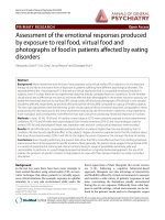
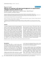
imumab cohort (Table 2 and Figure 1). More than 40% of the
patients in both cohorts experienced a change in mSASSS
from baseline to year 2 (Figure 1).
OASIS-Eligible set
There was no significant difference in the mean change in
mSASSS from baseline to year 2 between the adalimumab
cohort and OASIS-Eligible patients (that is, patients in the
OASIS cohort who met all baseline disease activity criteria for
the ATLAS and Canadian studies) (Table 2). The mean
change in mSASSS for the OASIS-Eligible cohort did not dif-
fer from the mean change in mSASSS for the full OASIS
cohort (0.9 ± 3.3 versus 0.9 ± 4.1, respectively).
Sensitivity analyses
A number of sensitivity analyses were conducted to assess
factors that could potentially affect the results of the primary
analysis. None of the sensitivity analyses revealed a significant
difference in radiographic progression between the adalimu-
mab cohort and the OASIS cohort (data not shown). For exam-
ple, a sensitivity analysis excluding the bottom C7 and T1 top
vertebral sites (which are often difficult to read owing to
obscuring of the C7 and T1 views by the shoulders on lateral
cervical films) did not change the results of the primary analy-
sis, nor did sensitivity analyses exploring alternate imputations
for missing vertebral sites.
Reader reliability
Intra- and inter-reader reliability was evaluated using the ICC.
Intrareader reliability testing was based on 56 patients
(approximately 10%) from OASIS, ATLAS, and the Canadian
Table 1
Baseline demographic and disease characteristics
Demographic characteristic OASIS Adalimumab P value
a
Number Baseline assessment Number Baseline assessment
Age, years 168 43.6 ± 12.7 307 41.8 ± 11.5 0.101
Male, percentage 169 69.2 307 76.5 0.102
Weight, kg 157 72.7 ± 12.8 307 80.0 ± 16.3 < 0.001
Height, cm 161 171.0 ± 9.3 306 172.9 ± 9.5 0.037
Concomitant medications, percentage
NSAIDs 169 77.5 307 88.3
DMARDs 169 9.5 307 21.8
Systemic glucocorticoids 169 1.8 307 9.8
Disease characteristic
Disease duration, years 163 11.3 ± 8.7 307 11.2 ± 9.3 0.946
BASDAI, 0-10 166 3.4 ± 2.1 307 6.2 ± 1.7 < 0.001
BASFI, 0-10 158 3.1 ± 2.4 307 5.3 ± 2.1 < 0.001
Total back pain, 0-10 166 3.5 ± 2.4 307 6.7 ± 1.9 < 0.001
Inflammation, 0-10
b
167 3.4 ± 2.6 307 6.7 ± 2.0 < 0.001
Patient's Global Assessment of disease activity, 0-10 165 3.7 ± 2.7 306 6.4 ± 2.0 < 0.001
C-reactive protein, mg/dL
c
160 1.5 ± 1.9 302 1.9 ± 2.5 0.036
mSASSS, 0-72 169 15.8 ± 17.6 307 19.8 ± 19.3 0.028
Values are mean ± standard deviation unless otherwise noted.
a
P values calculated using one-way analysis of variance or the Fisher exact test. No
statistical comparison of concomitant medications was completed.
b
Mean of questions 5 and 6 of the Bath Ankylosing Spondylitis Disease
Activity Index (BASDAI).
c
Using the ultrasensitive assay (normal range, 0.007 to 0.494 mg/dL). BASFI, Bath Ankylosing Spondylitis Functional
Index; DMARD, disease-modifying antirheumatic drug; mSASSS, modified Stoke Ankylosing Spondylitis Spinal Score; NSAID, nonsteroidal anti-
inflammatory drug; OASIS, Outcome in Ankylosing Spondylitis International Study.
Available online />Page 5 of 8
(page number not for citation purposes)
AS study combined. The ICC values for reader 1 were 0.982
for baseline radiographs and 0.987 for year-2 radiographs. For
reader 2, the ICC values were 0.913 for baseline radiographs
and 0.931 for year-2 radiographs. Intrareader reliability for the
change in mSASSS from baseline to year 2 was lower for
reader 1 (ICC = 0.319) than for reader 2 (ICC = 0.810)
because the intrareader analysis was conducted using only
10% of the radiographs and this figure was heavily influenced
by one outlier for reader 1. The ICC for inter-reader variability
for the change in mSASSS was 0.673. In total, of the 550 radi-
ograph cases read, 19 (3%) were adjudicated.
Radiographic progression and clinical measures of
disease activity
Another assessment evaluated whether changes in mSASSS
values from baseline to year 2 in the adalimumab cohort were
correlated with clinical measures of disease activity at baseline
or changes from baseline in clinical measures of disease activ-
ity. Changes in mSASSS were correlated with baseline scores
on several clinical outcome measures, including the Bath AS
Metrology Index (BASMI), BASFI, and short-form 36 health
survey (SF-36) physical component summary (PCS). How-
ever, there was no significant correlation between change in
radiographic progression and change from baseline for any of
the following clinical measures: BASDAI, BASMI, BASFI, C-
reactive protein, and SF-36 PCS for all patients and matrix
metalloproteinease-3 (MMP-3) (n = 37) and urinary type II col-
lagen C-telopeptide (n = 38) for patients in the Canadian
study (data not shown).
Discussion
In the present study, radiographic progression in patients with
AS treated with adalimumab 40 mg eow was compared with
radiographic progression in patients in the OASIS historical
control group. There was no difference between the adalimu-
mab and OASIS cohorts in the mean change in mSASSS from
baseline to year 2 based on the primary efficacy analysis of
patients' radiographs. Similarly, no difference between the
adalimumab cohort and the OASIS cohort was observed
when the analysis included only the subset of OASIS patients
(OASIS-Eligible set) who satisfied the minimum baseline dis-
ease activity requirements of the adalimumab studies. Addi-
tional sensitivity analyses were performed to investigate other
factors that could have potentially affected the results (for
example, vertebral imputation), but these analyses did not
reveal significant differences in radiographic progression
between adalimumab-treated patients and the control cohort.
Intra- and inter-reader reliability was evaluated using ICC val-
ues. In AS studies, ICC values generally range from 0.6 to 0.7
[20]; ICC values in the present study were within expected
ranges and did not contribute to the negative results.
The OASIS and adalimumab cohorts were heterogeneous
with respect to baseline demographic and disease character-
istics. Adalimumab-treated patients had significantly greater
Figure 1
Probability plot of 2-year progression in the modified Stoke Ankylosing Spondylitis Spine Score (mSASSS)Probability plot of 2-year progression in the modified Stoke Ankylosing
Spondylitis Spine Score (mSASSS). The cumulative probability plot
illustrates the change in mSASSS values from baseline to 2 years in the
adalimumab cohort (n = 307) and OASIS (n = 169) cohort (patients
without total spinal ankylosis). In both cohorts, over 40% of the patients
showed some change and about 10% of the patients showed a
change of at least 5 in mSASSS from baseline to year 2. No significant
differences between the adalimumab and OASIS cohorts were
observed. OASIS, Outcome in Ankylosing Spondylitis International
Study.
Table 2
mSASSS results for primary analysis set and for OASIS-Eligible patients
Cohort Number Mean change ± SD from baseline to year 2 P value
a
Primary mSASSS analysis set
OASIS 169 0.9 ± 3.3 0.771
Adalimumab 307 0.8 ± 2.6
OASIS-Eligible patients
OASIS 77 0.9 ± 4.1 0.744
Adalimumab 307 0.8 ± 2.6
a
P value calculated using analysis of covariance model with therapy as a factor and baseline modified Stoke Ankylosing Spondylitis Spinal Score
(mSASSS) as a covariate. OASIS, Outcome in Ankylosing Spondylitis International Study; SD, standard deviation.
Arthritis Research & Therapy Vol 11 No 4 van der Heijde et al.
Page 6 of 8
(page number not for citation purposes)
disease activity and mSASSS values compared with the
OASIS cohort. In addition, a greater percentage of adalimu-
mab-treated patients were taking NSAIDs at baseline. NSAID
therapy has been reported to inhibit syndesmophyte formation
and structural progression of AS; however, this finding needs
to be confirmed [21]. Differences in baseline characteristics
between the adalimumab and control cohorts had no apparent
effect on radiographic progression. A more direct and strin-
gent comparison between adalimumab and control cohorts
would ideally be performed in a randomized, double-blind, pla-
cebo-controlled trial. However, owing to the rapid
effectiveness of TNF antagonists in the treatment of AS, it
would be unethical to conduct a 2-year, placebo-controlled
trial to assess radiographic progression. Thus, the historical
control OASIS cohort is the best available comparator for
adalimumab-treated patients.
Radiographic progression in patients with AS has been
reported with TNF antagonists etanercept and infliximab [3,4].
As in the present study of adalimumab, these studies evalu-
ated changes in mSASSS from baseline to year 2 of treatment
and used the OASIS historical control group for comparison.
Baseline characteristics and radiographic progression results
in these studies and those of the present study of adalimumab
were similar (Table 3). The similar results of the three inde-
pendent cohorts of patients treated with TNF antagonists, as
well as the similar results of the OASIS cohort scored three
times independently, are striking, especially if one takes into
account the fact that each study employed a different pair of
readers. Thus, the obtained results are based on scores of six
different readers.
It is unclear why TNF antagonist therapy does not appear to
inhibit radiographic progression in patients with AS. Given the
insidious nature of spinal ankylosis, the 2-year timeframe of the
studies may have been insufficient to fully assess radiographic
damage and progression. There is one small study suggesting
that infliximab slowed the progression of structural damage
from 2 to 4 years of therapy [22]. However, that study had
notable limitations, including differences in baseline disease
activity (that is, BASDAI scores) between patients receiving
infliximab versus traditional therapies (OASIS), and differ-
ences in scoring methods [22]. Therefore, this effect requires
further investigation. It is possible that inhibition of radio-
graphic progression may take even longer periods of continu-
ous TNF antagonist therapy [23].
Initiation of anti-TNF therapy in patients with very early AS or
preradiographic spondyloarthritis may prevent radiographic
progression in the spine, but there are no data as of yet to con-
firm this hypothesis. The studies of radiographic progression
in patients treated with etanercept, infliximab, or adalimumab
included patients with long-standing AS and evidence of at
least grade 2 sacroiliitis (satisfying the modified New York cri-
teria [24]). Therefore, studies in patients with spondyloarthritis
with preradiographic sacroiliitis or early evidence of sacroiliitis
(and who do not yet satisfy the modified New York criteria)
may be more likely to demonstrate inhibition of structural dam-
age following TNF antagonist therapy. Adalimumab has been
shown to significantly suppress serum concentrations of
MMP-3, a biomarker that is a significant independent predictor
of structural damage progression in AS [25,26]. However, we
found no significant correlation between change in radio-
graphic progression and change from baseline in concentra-
tions of MMP-3 for adalimumab patients in the Canadian
study, which may reflect the small sample size. Longer-term
studies will be needed to further assess the full impact of TNF
antagonist therapy on radiographic progression.
One possible limitation of this study may be the use of the
mSASSS scoring system for quantification of disease pro-
gression. This system is limited in that it takes into account the
structural changes in the vertebral bodies and related soft tis-
sues of the cervical and lumbar spine without evaluating pos-
sible further damage at the posterior elements of the cervical
and lumbar spine, the thoracic spine, or the facet joints [20].
Table 3
Comparison of 2-year radiographic progression among tumor necrosis factor antagonists
Baseline characteristics Radiographic progression results
Cohort
Number
a
Disease duration, years
b
mSASSS value
b
Mean mSASSS change from baseline
to year 2
b
Between-cohort P value
Etanercept [2] 257 10 ± 8.5 16 ± 18.3 0.9 ± 2.5 0.996
OASIS 175 11 ± 8.5 14 ± 17.6 1.0 ± 3.2
Infliximab [3] 156 10.2 ± 8.7 17.7 ± 17.9 0.9 ± 2.6 0.541
OASIS 165 11.3 ± 8.6 15.8 ± 18.1 1.0 ± 3.2
Adalimumab 307 11.2 ± 9.3 19.8 ± 19.3 0.8 ± 2.6 0.771
OASIS 169 11.3 ± 8.6 15.8 ± 17.6 0.9 ± 3.3
a
Number of patients with a baseline and a year-2 radiograph.
b
Values are mean ± standard deviation. mSASSS, modified Stoke Ankylosing
Spondylitis Spinal Score; OASIS, Outcome in Ankylosing Spondylitis International Study.
Available online />Page 7 of 8
(page number not for citation purposes)
However, the mSASSS has been validated for AS and is cur-
rently the standard method for scoring radiographic progres-
sion [15,20]. Moreover, because this method was used in the
analysis of both the adalimumab and the OASIS cohorts, it is
doubtful that the results of this study were influenced by the
mSASSS scoring method. Moreover, the method was able to
detect changes for more than 40% of the patients.
TNF is associated with inflammation and bone destruction in
RA and PsA. TNF antagonists have been shown to reduce dis-
ease activity and inhibit the degenerative bone processes in
RA and PsA [8-11]. In contrast, TNF antagonists have not
been shown to inhibit the bone formation associated with AS
despite amelioration of the signs and symptoms of the dis-
ease. Consistent with this observation, uncoupling of inflam-
mation and bone formation has been reported in animal
models of spondyloarthritis [27-29]. Recent evidence sug-
gests that new bone formation may be more likely to occur at
the sites of spinal inflammation in patients with AS; two stud-
ies reported that more syndesmophytes developed at inflamed
vertebral edges than at noninflamed vertebral edges, although
the majority of syndesmophytes developed at vertebral edges
without inflammation at baseline [30,31]. However, one study
demonstrated the development of new syndesmophytes even
when inflammation had resolved after anti-TNF therapy [31]. It
has been proposed that each AS patient is likely to have sev-
eral spinal lesions at different stages of evolution. In addition,
it may be possible that very early lesions resolve with anti-TNF
therapy prior to the induction of reparative changes, whereas
for more mature inflammatory lesions, reparation is allowed to
proceed following resolution of inflammation with anti-TNF
therapy. The overall outcome for the individual patient is then
little change at the level of the entire spine [32]. The observa-
tion that new syndesmophytes also develop where there
appeared to have been no prior inflammation at vertebral cor-
ners also points to the possibility of non-inflammation-driven
pathways of reparation [33,34]. Further research is needed in
this area.
Conclusions
In patients with long-standing AS, 2 years of treatment with
adalimumab was effective in improving axial symptoms and
reducing spinal inflammation but did not significantly inhibit
radiographic progression. These findings are consistent with
those reported with etanercept and infliximab. Additional
studies that examine longer-term data with TNF antagonists
and earlier use of TNF antagonists to inhibit inflammation and
syndesmophyte formation are needed to better understand
the relationship between chronic inflammation and spinal
ankylosis in AS.
Competing interests
HK is an employee of an affiliate of Abbott Laboratories
(Abbott Park, IL, USA) and own shares of Abbott stock. SB,
EG, and RW were employees of Abbott Laboratories at the
time the analyses were completed and own shares of Abbott
stock. The Maastricht University Medical Center was finan-
cially supported for use of the OASIS database and the read-
ers for reading of blinded radiographs by Abbott Laboratories.
DvdH has received consulting fees, research grants, and/or
speaking fees from Abbott Laboratories, Amgen (Thousand
Oaks, CA, USA), sanofi-aventis (Paris, France), Bristol-Myers
Squibb Company (Princeton, NJ, USA), Centocor, Inc. (Hor-
sham, PA, USA), Pfizer Inc (New York, NY, USA), Roche
(Basel, Switzerland), Schering-Plough Corporation (Kenil-
worth, NJ, USA), UCB (Brussels, Belgium), and Wyeth (Madi-
son, NJ, USA). DS and WPM have received consulting fees,
speaking fees, and/or research grants from Abbott Laborato-
ries, Amgen, sanofi-aventis, Pfizer Inc, Schering-Plough Cor-
poration, and Wyeth. RL has received consulting fees,
research grants, and/or speaking fees from Abbott Laborato-
ries, Amgen, Bristol-Meyers Squibb Company, Centocor, Inc.,
Pfizer Inc, Schering-Plough Corporation, UCB, and Wyeth.
BNW declares that she has no competing interests.
Authors' contributions
SB and EG designed and performed the statistical analyses.
DS and BNW performed the blinded reading of the radio-
graphs. RL was an investigator for the OASIS study and was
the adjudicator for the radiographic reads. DvdH was the prin-
cipal investigator of the OASIS study and is the principal
investigator who assisted in designing the ATLAS study.
WPM is the principal investigator and assisted in designing
the Canadian M03-606 study. RW and HK assisted in design-
ing the ATLAS and Canadian studies and coordinated the
radiographic reads with both Bio-Imaging Technology, Inc.
(now part of BioClinica, Newtown, PA, USA) and independent
readers. All authors read and approved the final manuscript.
Acknowledgements
The authors thank the following Abbott Laboratories contributors: Shy-
anne Douma and Kerstin Krauss for coordinating the ATLAS study activ-
ities, Rebecca Hill and Annie Daudumez for coordinating the Canadian
study activities, Shafi Huda and Anna Protsenko for programming and
database management, and John Medich for helpful comments on the
radiographic analyses. Maryann Gehring (Bio-Imaging Technologies,
Inc., now part of BioClinica, Newtown, PA, USA) coordinated the logis-
tics and organization of the radiographic readings. Robin Stromberg, of
Arbor Communications, Inc. (Ann Arbor, MI, USA), and Michael A Nis-
sen, of Abbott Laboratories, provided medical writing and editing sup-
port on behalf of the authors. This work was funded by Abbott
Laboratories.
References
1. Braun J, Bollow M, Remlinger G, Eggens U, Rudwaleit M, Distler
A, Sieper J: Prevalence of spondyloarthropathies in HLA-B27
positive and negative blood donors. Arthritis Rheum 1998,
41:58-67.
2. Braun J, Bollow M, Neure L, Seipelt E, Seyrekbasan F, Herbst H,
Eggens U, Distler A, Sieper J: Use of immunohistologic and in
situ hybridization techniques in the examination of sacroiliac
joint biopsy specimens from patients with ankylosing
spondylitis. Arthritis Rheum 1995, 38:499-505.
3. Heijde D van der, Landewé , Einstein S, Ory P, Vosse D, Ni L, Lin
S-L, Tsuji W, Davis JC Jr: Radiographic progression of ankylos-
Arthritis Research & Therapy Vol 11 No 4 van der Heijde et al.
Page 8 of 8
(page number not for citation purposes)
ing spondylitis after up to two years of treatment with
etanercept. Arthritis Rheum 2008, 58:1324-1331.
4. Heijde D van der, Landewé R, Baraliakos X, Houben H, van Tuber-
gen A, Williamson P, Xu W, Baker D, Goldstein N, Braun J, Anky-
losing Spondylitis Study for the Evaluation of Recombinant
Infliximab Therapy Study Group: Radiographic findings following
two years of infliximab therapy in patients with ankylosing
spondylitis. Arthritis Rheum 2008, 58:3063-3070.
5. Heijde D van der, Kivitz A, Schiff MH, Sieper J, Dijkmans BAC,
Braun J, Dougados M, Reveille JD, Wong RL, Kupper H, Davis JC
Jr, ATLAS Study Group: Efficacy and safety of adalimumab in
patients with ankylosing spondylitis: results of a multicenter,
randomized, double-blind, placebo-controlled trial. Arthritis
Rheum 2006, 54:2136-2146.
6. Maksymowych WP, Rahman P, Keystone E, Wong R, Inman R, for
the M03-606 Study Group: Efficacy of adalimumab in active
ankylosing spondylitis (AS)-Results of the Canadian AS study
[abstract]. Arthritis Rheum 2005, 52:S217.
7. Heijde D van der, Schiff MH, Sieper J, Kivitz A, Wong RL, Kupper
H, Dijkmans BAC, Mease PJ, Davis JC Jr, ATLAS Study Group:
Adalimumab effectiveness for the treatment of ankylosing
spondylitis is maintained for up to 2 years: long-term results
from the ATLAS trial. Ann Rheum Dis 2009, 68:922-929.
8. Keystone EC, Kavanaugh AF, Sharp JT, Tannenbaum H, Hua Y,
Teoh LS, Fischkoff SA, Chartash EK: Radiographic, clinical, and
functional outcomes of treatment with adalimumab (a human
anti-tumor necrosis factor monoclonal antibody) in patients
with active rheumatoid arthritis receiving concomitant meth-
otrexate therapy: a randomized, placebo-controlled, 52-week
trial. Arthritis Rheum 2004, 50:1400-1411.
9. Mease PJ, Kivitz AJ, Burch FX, Siegel EL, Cohen SB, Ory P, Salo-
nen D, Rubenstein J, Sharp JT, Tsuji W: Etanercept treatment of
psoriatic arthritis: safety, efficacy, and effect on disease
progression. Arthritis Rheum 2004, 50:2264-2272.
10. Heijde D van der, Klareskog L, Rodriguez-Valverde V, Condreanu
C, Bolosiu H, Melo-Gomes J, Tornero-Molina J, Wajdula J, Peder-
sen R, Fatenejad S, TEMPO Study Investigators: Comparison of
etanercept and methotrexate, alone and combined, in the
treatment of rheumatoid arthritis: two-year clinical and radio-
graphic results from the TEMPO study, a double-blind, rand-
omized trial. Arthritis Rheum 2006, 54:1063-1074.
11. Kavanaugh A, Antoni CE, Gladman D, Wassenberg S, Zhou B,
Beutler A, Keenan G, Burmester G, Furst DE, Weisman MH, Kal-
den JR, Smolen J, Heijde D van der: The Infliximab Multinational
Psoriatic Arthritis Controlled Trial (IMPACT): results of radio-
graphic analyses after 1 year. Ann Rheum Dis 2006,
65:1038-1043.
12. Ritchlin CT, Haas-Smith SA, Li P, Hicks DG, Schwarz EM: Mech-
anisms of TNF-alpha- and RANKL-mediated osteoclastogene-
sis and bone resorption in psoriatic arthritis. J Clin Invest
2003, 111:821-831.
13. Redlich K, Hayer S, Ricci R, David JP, Tohidast-Akrad M, Kollias G,
Steiner G, Smolen JS, Wagner EF, Schett G: Osteoclasts are
essential for TNF-alpha-mediated joint destruction. J Clin
Invest 2002, 110:1419-1427.
14. Lam J, Takeshita S, Barker JE, Kanagawa O, Ross FP, Teitelbaum
SL: TNF-alpha induces osteoclastogenesis by direct stimula-
tion of macrophages exposed to permissive levels of RANK
ligand. J Clin Invest 2000, 106:1481-1488.
15. Heijde D van der, Landewé R: Selection of a method for scoring
radiographs for ankylosing spondylitis clinical trials, by the
Assessment in Ankylosing Spondylitis Working Group and
OMERACT. J Rheumatol 2005, 32:2048-2049.
16. Heijde D van der, Landewé R, Linden S van der: How should
treatment effect on spinal radiographic progression in
patients with ankylosing spondylitis be measured? Arthritis
Rheum 2005, 52:1979-1985.
17. Wanders AJ, Landewé RB, Spoorenberg A, Dougados M, Linden
S van der, Mielants H, Tempel H van der, Heijde DM van der: What
is the most appropriate radiologic scoring method for ankylos-
ing spondylitis? A comparison of the available methods based
on the Outcome Measures in Rheumatology Clinical Trials
filter. Arthritis Rheum 2004, 50:2622-2632.
18. Creemers MC, Franssen MJ, van't Hof MA, Gribnau FW, Putte LB
van de, van Riel PL: Assessment of outcome in ankylosing
spondylitis: an extended radiographic scoring system. Ann
Rheum Dis 2005, 64:127-129.
19. Wanders A, Landewé R, Dougados M, Mielants H, Linden S van
der, Heijde D van der: Association between radiographic dam-
age of the spine and spinal mobility for individual patients with
ankylosing spondylitis: can assessment of spinal mobility be a
proxy for radiologic evaluation? Ann Rheum Dis 2005,
64:988-994.
20. Powell A, Keeling SO, Lambert RG, Russell AS, Maksymowych
WP: Scoring of radiographic progression over 2 years with the
mSASSS in ankylosing spondylitis: does training improve
reliability? Arthritis Rheum
2007, 56:S256.
21. Wanders A, Heijde D van der, Landewé R, Béhier JM, Calin A,
Olivieri I, Zeidler H, Dougados M: Nonsteroidal antiinflammatory
drugs reduce radiographic progression in patients with anky-
losing spondylitis: a randomized clinical trial. Arthritis Rheum
2005, 52:1756-1765.
22. Baraliakos X, Listing J, Brandt J, Haibel H, Rudwaleit M, Sieper J,
Braun J: Radiographic progression in patients with ankylosing
spondylitis after 4 yrs of treatment with the anti-TNF-alpha
antibody infliximab. Rheumatology (Oxford) 2007,
46:1450-1453.
23. Sieper J, Appel H, Braun J, Rudwaleit M: Critical appraisal of
assessment of structural damage in ankylosing spondylitis.
Arthritis Rheum 2008, 58:649-656.
24. Linden S van der, Valkenburg HA, Cats A: Evaluation of diagnos-
tic criteria for ankylosing spondylitis. A proposal for modifica-
tion of the New York criteria. Arthritis Rheum 1984,
27:361-368.
25. Maksymowych WP, Rahman P, Shojania K, Olszynski WP, Thom-
son GTD, Ballal S, Wong RL, Inman RD, M03-606 Study Group:
Beneficial effects of adalimumab on biomarkers reflecting
structural damage in patients with ankylosing spondylitis. J
Rheumatol 2008, 35:2030-2037.
26. Maksymowych WP, Landewé R, Conner-Spady B, Dougados M,
Mielants H, Tempel H van der, Poole AR, Wang N, Heijde D van
der: Serum matrix metalloproteinase 3 is an independent pre-
dictor of structural damage progression in patients with anky-
losing spondylitis. Arthritis Rheum 2007, 56:1846-1853.
27. Bárdos T, Szabó Z, Czipri M, Vermes C, Tunyogi-Csapó M, Urban
RM, Mikecz K, Glant TT: A longitudinal study on an autoimmune
murine model of ankylosing spondylitis. Ann Rheum Dis 2005,
64:981-987.
28. Lories RJ, Daans M, Derese I, Matthys P, Kasran A, Tylzanowski P,
Ceuppens JL, Luyten FP: Noggin haploinsufficiency differen-
tially affects tissue responses in destructive and remodeling
arthritis. Arthritis Rheum 2006, 54:1736-1746.
29. Lories RJ, Derese I, de Bari C, Luyten FP: Evidence for uncou-
pling of inflammation and joint remodeling in a mouse model
of spondylarthritis. Arthritis Rheum 2007, 56:489-497.
30. Baraliakos X, Listing J, Rudwaleit M, Seiper J, Braun J: The rela-
tionship between inflammation and new bone formation in
patients with ankylosing spondylitis. Arthritis Res Ther 2008,
10:
R104.
31. Maksymowych WP, Chiowchanwisawakit P, Clare T, Pedersen SJ,
Østergaard M, Lambert RGW: Inflammatory lesions of the
spine on MRI predict the development of new syndesmo-
phytes in ankylosing spondylitis: evidence for coupling
between inflammation and ankylosis. Arthritis Rheum 2009,
60:93-102.
32. Maksymowych WP: What do biomarkers tell us about the
pathogenesis of ankylosing spondylitis? Arthritis Res Ther
2009, 11:101-102.
33. Baraliakos X, Listing J, Rudwaleit M, Sieper J, Braun J: Evidence
for a link between inflammation and new bone formation in
ankylosing spondylitis a detailed analysis. Ann Rheum Dis
2008, 67(Suppl II):130.
34. Heidje D van der, Landewé R, Baraliakos X, Hermann K, Houben
H, Hsu B, Baker D, Braun J: MRI-inflammation of the vertebral
unit (VU) only marginally contributes to new syndesmophyte
formation in that unit: a multi-level analysis. Ann Rheum Dis
2008, 67(Suppl II):130.