Báo cáo y học: "Mechanical effects of surgical procedures on osteochondral grafts elucidated by osmotic loading and real-time ultrasound" ppsx
Bạn đang xem bản rút gọn của tài liệu. Xem và tải ngay bản đầy đủ của tài liệu tại đây (1.46 MB, 9 trang )
Open Access
Available online />Page 1 of 9
(page number not for citation purposes)
Vol 11 No 5
Research article
Mechanical effects of surgical procedures on osteochondral grafts
elucidated by osmotic loading and real-time ultrasound
Koji Hattori
1,2
, Kota Uematsu
2
, Tomohiro Matsumoto
1
and Hajime Ohgushi
1
1
Research Institute for Cell Engineering, National Institute of Advanced Industrial Science and Technology, 3-11-46, Nakoji, Amagasaki, Hyogo 661-
0974, Japan
2
Department of Orthopaedic Surgery, Nara Medical University, 840, Shijyo-cho, Kashihara, Nara 634-8522, Japan
Corresponding author: Koji Hattori,
Received: 19 May 2009 Revisions requested: 7 Jul 2009 Revisions received: 3 Aug 2009 Accepted: 2 Sep 2009 Published: 2 Sep 2009
Arthritis Research & Therapy 2009, 11:R134 (doi:10.1186/ar2801)
This article is online at: />© 2009 Hattori et al.; licensee BioMed Central Ltd.
This is an open access article distributed under the terms of the Creative Commons Attribution License ( />),
which permits unrestricted use, distribution, and reproduction in any medium, provided the original work is properly cited.
Abstract
Introduction Osteochondral grafts have become popular for
treating small, isolated and full-thickness cartilage lesions. It is
recommended that a slightly oversized, rather than an exact-
sized, osteochondral plug is transplanted to achieve a tight fit.
Consequently, impacting forces are required to insert the
osteochondral plug into the recipient site. However, it remains
controversial whether these impacting forces affect the
biomechanical condition of the grafted articular cartilage. The
present study aimed to investigate the mechanical effects of
osteochondral plug implantation using osmotic loading and real-
time ultrasound.
Methods A full-thickness cylindrical osteochondral defect
(diameter, 3.5 mm; depth, 5 mm) was created in the lateral lower
quarter of the patella. Using graft-harvesting instruments, an
osteochondral plug (diameter, 3.5 mm as exact-size or 4.5 mm
as oversize; depth, 5 mm) was harvested from the lateral upper
quarter of the patella and transplanted into the defect. Intact
patella was used as a control. The samples were monitored by
real-time ultrasound during sequential changes of the bathing
solution from 0.15 M to 2 M saline (shrinkage phase) and back
to 0.15 M saline (swelling phase). For cartilage sample
assessment, three indices were selected, namely the change in
amplitude from the cartilage surface (amplitude recovery rate:
ARR) and the maximum echo shifts from the cartilage surface
and the cartilage-bone interface.
Results The ARR is closely related to the cartilage surface
integrity, while the echo shifts from the cartilage surface and the
cartilage-bone interface are closely related to tissue deformation
and NaCl diffusion, respectively. The ARR values of the
oversized plugs were significantly lower than those of the
control and exact-sized plugs. Regarding the maximum echo
shifts from the cartilage surface and the cartilage-bone interface,
no significant differences were observed among the three
groups.
Conclusions These findings demonstrated that osmotic loading
and real-time ultrasound were able to assess the mechanical
condition of cartilage plugs after osteochondral grafting. In
particular, the ARR was able to detect damage to the superficial
collagen network in a non-destructive manner. Therefore,
osmotic loading and real-time ultrasound are promising as
minimally invasive methods for evaluating cartilage damage in
the superficial zone after trauma or impact loading for
osteochondral grafting.
Introduction
Osteochondral grafts have become popular for the treatment
of small, isolated and full-thickness cartilage lesions [1]. Oste-
ochondral grafts have several advantages, including a high
survival rate of the grafted articular cartilage, reliable bone
union and no threat of disease transmission [1-3]. Several
osteochondral transplantation systems are commercially avail-
able in clinical practice. For most of these systems, it is recom-
mended that a slightly oversized, rather than an exact-sized,
osteochondral plug is transplanted to achieve a tight fit [4],
because plug stability is an important factor for optimal in-
growth of a transplanted plug [5]. Therefore, impacting forces
are required to insert the osteochondral plug into the recipient
site during the osteochondral grafting procedure.
ARR: amplitude recovery rate; CT: computed tomography; MRI: magnetic resonance imaging; NaCl: sodium chloride; ORT: optical coherence tom-
ography; SEM: scanning electron microscopy.
Arthritis Research & Therapy Vol 11 No 5 Hattori et al.
Page 2 of 9
(page number not for citation purposes)
It remains controversial whether the impacting forces required
to insert an osteochondral plug affect the biomechanical con-
dition of the grafted articular cartilage. We previously devel-
oped an ultrasonic evaluation system for articular cartilage.
We demonstrated that this system can be used to quantita-
tively clinically evaluate cartilage degeneration [6,7]. Using the
same ultrasonic evaluation system, Kuroki and colleagues [8]
examined the mechanical effects of the osteochondral grafting
procedure on porcine articular cartilage immediately after sur-
gery. The study indicated that osteochondral graft surgery
does not affect the stiffness, surface irregularity or thickness
of either oversized and exact-sized plugs. In contrast, Nishitani
and colleagues [9] assessed osteochondral grafting of the
human elbow using this system and showed that the cartilage
plug may become damaged during the osteochondral grafting
procedure. Nakaji and colleagues [10] evaluated the mechan-
ical properties of cartilage plugs using a tactile sensor system
and showed that the stiffness of oversized cartilage plugs did
not differ significantly from that of the normal cartilage immedi-
ately after surgery. However, it is well known that the impacting
forces required to implant an osteochondral graft can lead to
chondrocyte death and fissure formation in the surface of the
cartilage plug [11,12]. Therefore, it is speculated that the
above described evaluation methods are not suitable for the
assessment of articular cartilage damage from the impacting
forces used to implant an osteochondral graft. Therefore, a
more adjustable measurement method is required.
Ultrasound was first used to measure the osmotic swelling of
articular cartilage by Tepic and colleagues [13]. Further stud-
ies have recently been carried out by Zheng and colleagues
[14] and Wang and colleagues [15,16], who developed a new
ultrasound system for monitoring transient depth-dependent
osmotic swelling and solute diffusion in articular cartilage.
Using this system, they successfully monitored articular carti-
lage digestion by trypsin in real time. Ultrasound assessment
by osmotic loading can provide transient and depth-depend-
ent swelling information for articular cartilage in situ. There-
fore, osmotic loading and real-time ultrasound have the
potential for assessing the cartilage damage caused by the
impacting forces required to insert a plug during the osteo-
chondral graft procedure. However, it remains unknown
whether osmotic loading and real-time ultrasound can assess
the mechanical condition of a cartilage plug after osteochon-
dral grafting.
The purpose of the present study was to evaluate the mechan-
ical effects of osteochondral plug implantation using osmotic
loading and real-time ultrasound and to demonstrate the accu-
racy of ultrasound in identifying the cartilage damage after
osteochondral graft procedures. To this end, we evaluated
oversized and exact-sized cartilage plugs after osteochondral
grafting. In the present study, we also assessed the cartilage
plugs using a conventional mechanical test and observed the
cartilage surface morphology by scanning electron micros-
copy (SEM).
Materials and methods
Cartilage sample processing
Porcine knee joints (n = 30) with intact capsules and liga-
ments were purchased from a slaughterhouse. After removal
of the soft tissues, the knee joints were opened. The patellas
with visually intact surfaces were harvested, wrapped in wet
gauze soaked with physiological saline solution and stored at
-20°C until use. For sample preparation, each patella was
thawed at room temperature for one hour and immersed in
physiological saline solution (0.15 M sodium chloride (NaCl)),
before the lateral lower and upper quarters of the patella were
cut using a band saw (K-100; Hozan Tool Industrial Co. Ltd.,
Osaka, Japan). During the processing steps described below,
the cartilage surface was kept moist with physiological saline
solution without immersing the sample.
A full-thickness cylindrical osteochondral defect (diameter, 3.5
mm; depth, 5 mm) was created in the lateral lower quarter of
the patella. Using graft-harvesting instruments (MOSAIC-
PLASTY System; Smith & Nephew Inc., Andover, MA, USA),
an osteochondral plug (diameter, 3.5 or 4.5 mm; depth, 5 mm)
was harvested from the lateral upper quarter of the patella. The
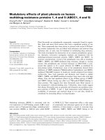
samples were divided into two groups based on the surgical
procedure (Figures 1a, b). In group I (n = 10), an exact-sized
plug (diameter, 3.5 mm; depth, 5 mm) was harvested and
implanted into the osteochondral defect in the lower quarter of
the patella. The osteochondral plug exactly matched the size
of the defect and was easily inserted with an adjustable
plunger so it was as flush as possible with the surrounding car-
tilage. In group II (n = 10), an oversized plug (diameter, 4.5
mm; depth, 5 mm) was harvested and implanted into the oste-
Figure 1
Sample preparationSample preparation. A full-thickness osteochondral defect (closed cir-
cle; diameter, 3.5 mm; depth, 5 mm) is created in the lateral lower quar-
ter of each patella. (a) Group I. An exact-sized plug (open circle) is
harvested from the lateral upper quarter of the patella and transplanted
into the defect. (b) Group II. An oversized plug (open circle) is har-
vested from the lateral upper quarter of the patella and transplanted
into the defect.
Available online />Page 3 of 9
(page number not for citation purposes)
ochondral defect in the lower quarter of the patella. The over-
sized plug was inserted into the defect in a press-fit manner.
The plug was advanced using a delivery tamp and seated as
flush as possible with the surrounding cartilage. All of the sur-
gical procedures were performed by a specialist in knee sur-
gery (KU). In the control group (n = 10), intact cartilage in the
lower quarter of the patella was used.
Ultrasound monitoring system
The ultrasound monitoring system used in this study was orig-
inally developed by Zheng and colleagues [14-16] and modi-
fied to a 10 MHz ultrasound system. The system was
developed to monitor articular cartilage in terms of the tran-
sient depth-dependent swelling behaviour and the transport of
solutes induced by changing the concentration of the bathing
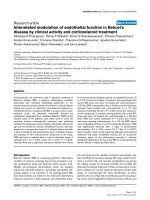
saline solution. A schematic outline of the ultrasound swelling
measurement system is shown in Figure 2. The system
included a 10 MHz transducer (diameter, 3 mm; thickness, 3
mm; flat ultrasonic wave), an ultrasonic pulser/receiver (Model
5800PR; Olympus NDT, Waltham, MA, USA), a digital oscillo-
scope (TDS 2022B; Tektronix Japan, Ltd., Tokyo, Japan) and
custom-made software (LabVIEW 8.5; National Instruments,
Austin, TX, USA) for data collection and signal processing.
Ultrasound analysis
Each articular cartilage sample was placed on the bottom of
the container and submerged in 0.15 M saline solution for
three hours. The transducer was moved to a position perpen-
dicularly above the cartilage surface of the osteochondral
graft. After the three-hour immersion, the 0.15 M saline solu-
tion was rapidly removed from the container using a syringe
and replaced with 2 M saline solution within 30 seconds, and
the sample was monitored by ultrasound for 90 minutes
(shrinkage phase). Subsequently, the 2 M saline solution was
changed back to 0.15 M saline solution within 30 seconds,
and the sample was monitored by ultrasound for 90 minutes
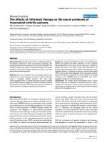
(swelling phase). The echo signals that were reflected from the
cartilage surface and the cartilage-bone interface and became
scattered inside the articular cartilage layer were continuously
recorded with a sampling period of 30 seconds (Figures 3a,
b). The ultrasound signals were also displayed in M-mode
images, with grey levels indicating the amplitudes of the ultra-
sound signals (Figures 3c to 3e). Horizontal traces of the car-
tilage surface in the M-mode images indicated the transient
displacement (shrinkage/swelling) of the samples, while simi-
lar traces of the cartilage-bone interface indicated the diffusiv-
ity of the saline solution in the cartilage. All of the experiments
were carried out at room temperature.
For cartilage sample assessment, we focused on three ultra-
sound indices, namely the change in amplitude from the carti-
lage surface and the echo shifts from the cartilage surface and
the cartilage-bone interface. The change in amplitude from the
cartilage surface refers to the change of the cartilage/saline
solution acoustic impedance. In the shrinkage phase, cartilage
is sufficiently dehydrated to relax the collagen network in the
collagen-rich superficial zone. In the swelling phase, the
impedance and amplitude increase as the proteoglycans
swell, thereby stretching the collagen and increasing the stiff-
ness [13]. Therefore, as one quantitative index of the cartilage
assessment in this study, the amplitude recovery rate (ARR)
was determined. The ARR value was expressed using the fol-
lowing equation:
Figure 2
Schematic illustration of the osmotic loading and ultrasound monitoring systemSchematic illustration of the osmotic loading and ultrasound monitoring system. The sample is fixed on the bottom of the container. NaCl = sodium
chloride.
Arthritis Research & Therapy Vol 11 No 5 Hattori et al.
Page 4 of 9
(page number not for citation purposes)
- where MAMP swelling is the mean amplitude from the carti-
lage surface in the swelling phase, and MAMP shrinkage is the
mean amplitude from the cartilage surface in the shrinkage
phase.
We also evaluated the echo shifts from the cartilage surface
and the cartilage-bone interface in both the shrinkage and
swelling phases. The echo shift from the cartilage surface indi-
cates the sample displacement, while the echo shift from the
cartilage-bone interface indicates the diffusivity of the saline
solution in the sample [14]. Therefore, as the other quantitative
indices of the cartilage assessment in this study, the maximum
echo shifts were chosen.
Morphological analysis
Two samples in each group were subjected to morphological
analysis using an SEM (Model SM-350; Topcon Technohouse
Corporation, Tokyo, Japan). The samples were fixed in 2% glu-
taraldehyde buffered with 0.1 M cacodylate, dehydrated in a
graded ethanol series, dried using the critical point technique
and coated by sputtering with a gold layer [17].
Biomechanical analysis
Eight cartilage samples were immersed in physiological saline
and tested within three hours. To determine the mechanical
properties of the grafted cartilage, an electromechanical mate-
rial testing machine (EZ-L; Shimadzu Corporation, Kyoto,
Japan) was used. Forces were applied to the grafted cartilage
at a displacement rate of 2.0 mm/min using a 3.0 mm diameter
solid aluminum indenter. A load-deformation curve was
obtained during the compression. As biomechanical parame-
ters, we defined the maximum load (breaking load: F max)
applied at fracture of the grafted cartilage.
Statistical analysis
For multiple comparisons of ultrasound findings, the groups
were analyzed using the nonparametric Kruskal-Wallis test.
When significant variance was detected, the differences
among individual groups were determined using the Mann-
Whitney U test with the Bonferroni correction. For compari-
sons between two groups in the biomechanics analyses, the
differences were analyzed by the nonparametric Mann-Whit-
ney U test. The significance level was set at P < 0.05.
Results
Ultrasonic findings
The ARR values (mean ± standard deviation) were 8.64 ±
2.70% in the control group, 7.14 ± 4.74% in group I and 3.41
± 1.58% in group II (Figure 4). A significant difference in the
ARR
100 (=
−
⎛
⎝
⎜
⎞
⎠
⎟
×
MAMP swelling MAMP shrinkage
MAMP shrinkage
%%)
Figure 3
Imaging data from the osmotic loading and real-time ultrasound systemImaging data from the osmotic loading and real-time ultrasound system. (a) Histology of a typical articular cartilage sample. (b) A-mode echogram
from an articular cartilage sample. The black arrow indicates the amplitude from the cartilage surface and the white arrow indicates the amplitude
from the cartilage-bone interface. The amplitude recovery rate was calculated from the change in the cartilage surface amplitude from the shrinkage
phase to the swelling phase. (c) M-mode image before osmotic loading. The gray levels indicate the amplitudes of the ultrasound signals. (d) Typical
M-mode image in the shrinkage phase. (e) Typical M-mode image in the swelling phase.
Available online />Page 5 of 9
(page number not for citation purposes)
ARR was observed between the control group and group II (P
= 0.008) and between group I and II (P = 0.024).
Figure 5 shows the typical time courses of the echo shifts of
the control cartilage in the shrinkage phase (Figure 5a) and
swelling phase (Figure 5b). The patterns of the echo shifts
were similar in all three groups. There was a rapid decrease in
the echo shift from the cartilage surface after 30 minutes of
immersion in 2 M NaCl (shrinkage phase), followed by a grad-
ual decrease from 30 to 90 minutes. There was a rapid
decrease in the echo shift from the cartilage-bone interface
after 30 minutes of immersion in 0.15 M NaCl (swelling
phase), followed by a gradual decrease from 30 to 90 minutes.
The maximum echo shifts are shown in Table 1. There were no
significant differences in the maximum echo shifts among the
three groups.
Morphological findings
Representative SEM images from samples in groups I and II
are shown in Figure 6. In group I, there were tiny irregularities
in the surface of the cartilage plug. However, the superficial
collagen network was not ruptured (Figure 6a). In contrast,
most of the cartilage surface in group II was damaged by the
surgical processing. The superficial collagen network was bro-
ken and the cartilage superficial layer had partially peeled
away (Figure 6b).
Biomechanical findings
A load-deformation curve is shown in Figure 7a. The F max val-
ues were 198.1 ± 42.2 N in group I and 233.2 ± 46.2 N in
group II (Figure 7b). The mean F max value was higher in group
II than in group I, but the difference was not significant (P =
0.14).
Discussion
The present study investigated the osmotic shrinkage-swelling
behaviours of oversized and exact-sized cartilage plugs in
osteochondral grafting using osmotic loading and real-time
ultrasound. The main findings of the study are that osmotic
loading and real-time ultrasound are capable of assessing the
mechanical condition of a cartilage plug after osteochondral
grafting. In particular, the ARR was able to detect damage to
the superficial collagen network in a non-destructive manner.
Therefore, osmotic loading and real-time ultrasound are prom-
ising as minimally invasive methods for evaluating cartilage
damage in the superficial zone after trauma or impact loading
for osteochondral grafting.
Figure 4
Mean amplitude recovery rate values of the three groupsMean amplitude recovery rate values of the three groups. The error bars
represent the standard deviation of each group. *P < 0.05 by the non-
parametric Kruskal-Wallis test.
Figure 5
Time courses of echo shiftsTime courses of echo shifts. (a, b) Time courses of the echo shifts from the cartilage surface (dotted line) and the cartilage-bone interface (thick line)
in the (a) shrinkage phase and (b) swelling phase.
Arthritis Research & Therapy Vol 11 No 5 Hattori et al.
Page 6 of 9
(page number not for citation purposes)
An osteochondral plug that is exactly the same size and shape
as a cartilage defect seems to be ideal for osteochondral graft-
ing. However, Makino and colleagues [18] reported that histo-
logical changes occur in the implanted cartilage, after
examining osteochondral grafts taken from the femoral con-
dyle and returned to their original sites. In their rabbit model,
the graft was not strictly the same size as the defect because
of the blade thickness of the chisel used to take the graft.
Moreover, they revealed that an oversized osteochondral graft
appeared to be almost the same as the normal adjacent carti-
lage at 4, 12 and 24 weeks after surgery [4]. Therefore, an
oversized plug can be recommended for use in the osteochon-
dral graft procedure. However, the impact load required to
insert a plug into the recipient site is higher for an oversized
plug than for an exact-sized plug.
Impact loading of articular cartilage has commonly been asso-
ciated with structural damage [19-22], loss of viability and
changes in the metabolism of chondrocytes [19,22-24], with
subsequent degeneration of the articular cartilage [25]. In
general, evaluations of damage to cartilage have been per-
formed by histological analysis of the structural integrity
[19,22], SEM imaging of the surface morphology [17], assess-
ment of tissue swelling by the water content related to disrup-
tion of collagen fibrils [19,23], assessment of chondrocyte
death [19,24] and release of cartilage macromolecular constit-
uents during subsequent tissue culture [19,22,24]. However,
these analyses require the collection of cartilage tissue sam-
ples, which will result in damage to the cartilage plug surface.
Therefore, all the above described evaluation methods should
be avoided in clinical practice.
There are several imaging modalities to assess articular carti-
lage such as radiograph, computed tomography (CT), mag-
netic resonance imaging (MRI) and optical coherence
tomography (OCT). Radiograph and CT do not image soft tis-
sue, which prevent identification of structural changes of artic-
ular cartilage. Conventional MRI has been used in clinical
practice to measure morphological change in articular carti-
lage. In comparison with MRI, the present ultrasonic approach
may allow real-time monitoring of depth-dependent osmotic
behaviours by the echo shift and the changes in amplitude.
Table 1
Echo shifts from cartilage surface and cartilage-bone interface in the shrinkage and swelling phases
Control
(n = 10)
Group I
(n = 10)
Group II
(n = 10)
P value
Shrinkage phase
Cartilage surface -82.6 ± 26.1 ns -77.4 ± 22.7 ns -70.7 ± 27.8 ns NS
Cartilage-bone interface 5.2 ± 24.8 ns 14.2 ± 24.8 ns 22.4 ± 16.6 ns NS
Swelling phase
Cartilage surface -9.2 ± 21.5 ns -2.6 ± 15.0 ns 4.4 ± 9.6 ns NS
Cartilage-bone interface -86.0 ± 18.1 ns -74.8 ± 12.9 ns -69.1 ± 19.2 ns NS
Data are presented as mean ± standard deviation. P value based on Kruskal-Wallis test. The significance level was set at P < 0.05. NS = not
significant.
Figure 6
Representative cartilage surface images obtained by scanning electron microscopyRepresentative cartilage surface images obtained by scanning electron microscopy. (a) Articular surface of a cartilage plug in group I. (b) Articular
surface of a cartilage plug in group II.
Available online />Page 7 of 9
(page number not for citation purposes)
Moreover, the present system is much less expensive in com-
parison with MRI. OCT is a novel form of optical imaging that
enables cross-sectional visualization of tissue micro architec-
ture. However, OCT is still in its early stages of development
for the assessment of articular cartilage [26,27]. Therefore,
further studies to assess articular cartilage from the view point
of biomechanics are required.
Tepic and colleagues [13] developed an ultrasonic system for
assessing osmotic swelling of articular cartilage after dehydra-
tion in humid air. However, their ultrasonic system was only
able to evaluate the whole cartilage layer and no measure-
ments were obtained for depth-dependent swelling behav-
iours. Zheng and colleagues developed a new ultrasound
system for monitoring transient depth-dependent osmotic
swelling and solute diffusion in articular cartilage [14-16].
Consequently, osmotic loading and real-time ultrasound can
provide comprehensive information about the biomechanical
behaviour of articular cartilage. The present study has demon-
strated the feasibility of this system for evaluating cartilage
damage caused by impact loading while inserting a plug dur-
ing the osteochondral graft procedure.
In this study, cartilage plugs were assessed not only by their
osmotic shrinking and swelling behaviours but also by the
changes in amplitude of the cartilage surface from the shrink-
age phase to the swelling phase. A previous study revealed
that the amplitude from the cartilage surface is related to the
tissue reflection coefficient, acoustic impedance, elastic mod-
ulus and surface condition in physics, and related to proteogly-
can depletion and collagen disruption in biology [28-30]. In
the present study, the cartilage plugs were damaged by the
impact loading required for their insertion into the defects.
Moreover, damage to the surface collagen network was con-
firmed by SEM. By using osmotic swelling, differences in the
cartilage surface integrity between oversized cartilage plugs
and intact cartilage were enhanced. As a result, the ARR of
oversized cartilage plugs was significantly lower than that of
intact cartilage. Therefore, the ARR mainly reveals the micro-
structural changes to the articular cartilage in the superficial
collagen-rich zone.
On the other hand, the echo shift from the cartilage surface is
known to reflect the sample displacement and the echo shift
from the cartilage-bone interface is known to reflect the diffu-
sivity of saline solution in the sample [14]. In the present study,
the echo shifts of oversized and exact-sized cartilage plugs
were similar to those of intact cartilage. These results suggest
that the interiors of the cartilage plugs were not damaged by
the impact loading required to insert the plugs into the defects.
Within the limitations of the measurement accuracy, the
mechanical indentation test could not detect damage to the
cartilage surface. Therefore, osmotic loading and real-time
ultrasound represent new approaches for studying the biome-
chanical and biophysical aspects associated with articular car-
tilage.
Three limitations of our study should be considered. First, we
did not examine the effects of osmotic loading on the viability
and metabolism of chondrocytes. A high concentration of
NaCl may be harmful to cartilage tissues. If this proves to be
the case, the methodology for the osmotic loading should be
changed from 2 M and 0.15 M NaCl to humid air and 0.15 M
NaCl [13]. Second, the impact loading required to insert the
osteochondral plugs could not be controlled. However, the
present study simulated an assessment of human osteochon-
dral grafts, and a surgeon who was experienced in the osteo-
chondral grafting procedure performed the harvesting and
implantation procedures. Therefore, damage to the collagen
Figure 7
Biomechanical analysisBiomechanical analysis. (a) Load-deformation curve of the sample. The maximum load applied at fracture of the sample (breaking load) is shown as
F max.(b) Breaking loads (F max) of groups I and II. The error bars represent the standard deviation of each group. P < 0.05 by the nonparametric
Mann-Whitney U test.
Arthritis Research & Therapy Vol 11 No 5 Hattori et al.
Page 8 of 9
(page number not for citation purposes)
network in the superficial layer of cartilage plugs would occur
during the osteochondral grafting procedure.
Finally, the present study was carried out to investigate the
feasibility of using osmotic loading and real-time ultrasound to
assess the shrinking and swelling behaviors of cartilage plugs
after osteochondral grafting. If the present study design were
applied to clinical practice, the length of measurement time
would come into question. However, maximum deformation of
ARR and echo shift in plug cartilage by changing the saline
concentration occurred during the first several minutes [14].
Thus, with proper miniaturization of the design, it would be
clinically practical to detect cartilage damage after the osteo-
chondal graft procedure. Therefore, for application to clinical
situations, further studies are required to determine whether
this system will prove beneficial for the assessment of human
osteochondral grafts.
Conclusions
The present study has obtained the first data for the assess-
ment of articular cartilage damage caused by the impact load-
ing required to insert an osteochondral plug using osmotic
loading and real-time ultrasound. Under osmotic loading, the
changes in the amplitude and echo shifts can support the eval-
uation of cartilage damage in osteochondral grafts. Moreover,
osmotic loading and real-time ultrasound may contribute to tis-
sue engineering in the musculoskeletal field, and the ARR and
echo shifts can be expected to become quantitative indices for
the biomechanical and biophysical properties of articular car-
tilage.
Competing interests
The authors declare that they have no competing interests.
Authors' contributions
KH conceived the study, participated in its design and per-
formed all the experiments. KU performed the harvesting and
implantation procedures of the cartilage samples. TM per-
formed the SEM assessments. HO participated in the study
design and the biomechanical analyses. All authors have read
and approved the final manuscript.
Acknowledgements
This work was supported in part by Grants-in-Aid from the Ministry of
Education, Culture, Sports, Science and Technology of Japan. The
study sponsors had no role in the study design, data collection, data
analysis or data interpretation, or in the writing of the report.
References
1. Matsusue Y, Yamamuro T, Hama H: Arthroscopic multiple oste-
ochondral transplantation to the chondral defect in the knee
associated with anterior cruciate ligament disruption. Arthros-
copy 1993, 9:318-321.
2. Hangody L, Füles P: Autologous osteochondral mosaicplasty
for the treatment of full-thickness defects of weight-bearing
joints. Ten years of experimental and clinical experience. J
Bone Joint Surg Am 2003, 85:25-32.
3. Hangody L, Ráthonyi GK, Duska Z, Vásárhelyi G, Füles P, Módis L:
Autologous osteochondral mosaicplasty. Surgical technique.
J Bone Joint Surg Am 2004, 86:65-72.
4. Makino T, Fujioka H, Terukina M, Yoshiya S, Matsui N, Kurosaka M:
The effect of graft sizing on osteochondral transplantation.
Arthroscopy 2004, 20:837-840.
5. Makino T, Fujioka H, Yoshiya S, Terukina M, Matsui N, Kurosaka M:
The effect of the small and unstable autologous osteochon-
dral graft on repairing the full-thickness large articular carti-
lage defect in a rabbit model. Kobe J Med Sci 2002, 48:97-104.
6. Hattori K, Mori K, Habata T, Takakura Y, Ikeuch K: Measurement
of the mechanical condition of articular cartilage with an ultra-
sonic probe: quantitative evaluation using wavelet transforma-
tion. Clin Biomech (Bristol, Avon) 2003, 18:553-557.
7. Hattori K, Takakura Y, Ishimura M, Habata T, Uematsu K, Ikeuch K:
Quantitative arthroscopic ultrasound evaluation of living
human cartilage. Clin Biomech (Bristol, Avon) 2004,
19:213-216.
8. Kuroki H, Nakagawa Y, Mori K, Ikeuchi K, Nakamura T: Mechanical
effects of autogenous osteochondral surgical grafting proce-
dures and instrumentation on grafts of articular cartilage. Am
J Sports Med 2004, 32:612-620.
9. Nishitani K, Nakagawa Y, Gotoh T, Kobayashi M, Nakamura T:
Intraoperative acoustic evaluation of living human cartilage of
the elbow and knee during mosaicplasty for osteochondritis
dissecans of the elbow: an in vivo study. Am J Sports Med
2008, 36:2345-2353.
10. Nakaji N, Fujioka H, Nagura I, Kokubu T, Makino T, Sakai H, Kuroda
R, Doita M, Kurosaka M: The structural properties of an osteo-
chondral cylinder graft-recipient construct on autologous
osteochondral transplantation. Arthroscopy 2006, 22:422-427.
11. Borazjani BH, Chen AC, Bae WC, Patil S, Sah RL, Firestein GS,
Bugbee WD: Effect of impact on chondrocyte viability during
insertion of human osteochondral grafts. J Bone Joint Surg Am
2006,
88:1934-1943.
12. Whiteside RA, Jakob RP, Wyss UP, Mainil-Varlet P: Impact load-
ing of articular cartilage during transplantation of osteochon-
dral autograft. J Bone Joint Surg Br 2005, 87:1285-1291.
13. Tepic S, Macirowski T, Mann RW: Mechanical properties of
articular cartilage elucidated by osmotic loading and ultra-
sound. Proc Natl Acad Sci USA 1983, 80:3331-3333.
14. Zheng YP, Shi J, Qin L, Patil SG, Mow VC, Zhou KY: Dynamic
depth-dependent osmotic swelling and solute diffusion in
articular cartilage monitored using real-time ultrasound. Ultra-
sound Med Biol 2004, 30:841-849.
15. Wang Q, Zheng YP, Niu HJ, Mak AF: Extraction of mechanical
properties of articular cartilage from osmotic swelling behav-
ior monitored using high frequency ultrasound. J Biomech Eng
2007, 129:413-422.
16. Wang Q, Zheng YP, Qin L, Huang QH, Lam WL, Leung G, Guo X,
Lu HB: Real-time ultrasonic assessment of progressive prote-
oglycan depletion in articular cartilage. Ultrasound Med Biol
2008, 34:1085-1092.
17. Jurvelin J, Kuusela T, Heikkilä R, Pelttari A, Kiviranta I, Tammi M,
Helminen HJ: Investigation of articular cartilage surface mor-
phology with a semiquantitative scanning electron micro-
scopic method. Acta Anat (Basel) 1983, 116:302-311.
18. Makino T, Fujioka H, Kurosaka M, Matsui N, Yoshihara H, Tsunoda
M, Mizuno K: Histologic analysis of the implanted cartilage in
an exact-fit osteochondral transplantation model. Arthroscopy
2001, 17:747-751.
19. Jeffrey JE, Gregory DW, Aspden RM: Matrix damage and
chondrocyte viability following a single impact load on articu-
lar cartilage. Arch Biochem Biophys 1995, 322:87-96.
20. Repo RU, Finlay JB: Survival of articular cartilage after control-
led impact. J Bone Joint Surg Am 1977, 59:1068-1076.
21. Farquhar T, Xia Y, Mann K, Bertram J, Burton-Wurster N, Jelinski L,
Lust G: Swelling and fibronectin accumulation in articular car-
tilage explants after cyclical impact. J Orthop Res 1996,
14:417-423.
22. Ewers BJ, Dvoracek-Driksna D, Orth MW, Haut RC: The extent of
matrix damage and chondrocyte death in mechanically trau-
matized articular cartilage explants depends on rate of load-
ing. J Orthop Res 2001, 19:779-784.
23. Chen CT, Burton-Wurster N, Lust G, Bank RA, Tekoppele JM:
Compositional and metabolic changes in damaged cartilage
Available online />Page 9 of 9
(page number not for citation purposes)
are peak-stress, stress-rate, and loading-duration dependent.
J Orthop Res 1999, 17:870-879.
24. D'Lima DD, Hashimoto S, Chen PC, Colwell CW Jr, Lotz MK:
Human chondrocyte apoptosis in response to mechanical
injury. Osteoarthritis Cartilage 2001, 9:712-719.
25. Donohue JM, Buss D, Oegema TR Jr, Thompson RC Jr: The
effects of indirect blunt trauma on adult canine articular carti-
lage. J Bone Joint Surg Am 1983, 65:948-957.
26. Han CW, Chu CR, Adachi N, Usas A, Fu FH, Huard J, Pan Y: Anal-
ysis of rabbit articular cartilage repair after chondrocyte
implantation using optical coherence tomography. Osteoar-
thritis Cartilage 2003, 11:111-121.
27. Adams SB Jr, Herz PR, Stamper DL, Roberts MJ, Bourquin S, Patel
NA, Schneider K, Martin SD, Shortkroff S, Fujimoto JG, Brezinski
ME: High-resolution imaging of progressive articular cartilage
degeneration. J Orthop Res 2006, 24:708-715.
28. Zheng YP, Huang YP: More intrinsic parameters should be
used in assessing degeneration of articular cartilage with
quantitative ultrasound. Arthritis Res Ther 2008, 10:125.
29. Hattori K, Ikeuchi K, Morita Y, Takakura Y: Quantitative ultrasonic
assessment for detecting microscopic cartilage damage in
osteoarthritis. Arthritis Res Ther 2005, 7:R38-46.
30. Töyräs J, Rieppo J, Nieminen MT, Helminen HJ, Jurvelin JS: Char-
acterization of enzymatically induced degeneration of articular
cartilage using high frequency ultrasound. Phys Med Biol
1999, 44:2723-2733.