Báo cáo y học: "Expression of cartilage-derived morphogenetic protein in human intervertebral discs and its effect on matrix synthesis in degenerate human nucleus pulposus cells" potx
Bạn đang xem bản rút gọn của tài liệu. Xem và tải ngay bản đầy đủ của tài liệu tại đây (1.7 MB, 10 trang )
Open Access
Available online />Page 1 of 10
(page number not for citation purposes)
Vol 11 No 5
Research article
Expression of cartilage-derived morphogenetic protein in human
intervertebral discs and its effect on matrix synthesis in
degenerate human nucleus pulposus cells
Christine L Le Maitre
1,2
, Anthony J Freemont
2
and Judith A Hoyland
2
1
Biomedical Research Centre, Biosciences, Faculty of Health and Wellbeing, Sheffield Hallam University, City Campus, Owen Building, Howard
Street, Sheffield, S1 1WB, UK
2
Tissue Injury and Repair Group, School of Clinical and Laboratory Sciences, Faculty of Medical and Human Sciences, Stopford Building, The
University of Manchester, Oxford Road, Manchester, M13 9PT, UK
Corresponding author: Judith A Hoyland,
Received: 26 Mar 2009 Revisions requested: 15 May 2009 Revisions received: 30 Jul 2009 Accepted: 15 Sep 2009 Published: 15 Sep 2009
Arthritis Research & Therapy 2009, 11:R137 (doi:10.1186/ar2808)
This article is online at: />© 2009 Le Maitre et al.; licensee BioMed Central Ltd.
This is an open access article distributed under the terms of the Creative Commons Attribution License ( />),
which permits unrestricted use, distribution, and reproduction in any medium, provided the original work is properly cited.
Abstract
Introduction Loss of intervertebral disc (IVD) matrix and
ultimately disc height as a result of 'degeneration' has been
implicated as a major cause of low back pain (LBP). The use of
anabolic growth factors as therapies to regenerate IVD matrix,
hence restoring disc height and thus reversing degenerative
disc disease, has been suggested. Cartilage-derived
morphogenetic protein (CDMP) is a growth factor which
stimulates proteoglycan production in chondrocyte-like cells
and thus could be a useful growth factor for LBP therapies.
However, little is known about the expression of CDMP or its
receptor in human IVD, nor its effects on human disc cells.
Methods Using immunohistochemistry we investigated the
localisation of CDMP in non-degenerate and degenerate human
IVDs. Additionally, we investigated the effect of CDMP on
aggrecan and type II collagen gene expression and
proteoglycan synthesis in nucleus pulposus (NP) cells derived
from degenerate IVDs.
Results We demonstrated that CDMP 1 and 2 were expressed
in the non-degenerate and degenerate IVD, particularly in cells
of the NP. A small decrease in the number of CDMP 1 and 2
immunopositive cells was seen with degeneration. Treatment of
human NP cells, (derived from degenerate IVD), with CDMP
showed an increase in aggrecan and type II collagen gene
expression and increased production of proteoglycan (GAGs).
Conclusions The data suggests that CDMP may be a useful
growth factor to stimulate proteoglycan production in the human
degenerate IVD and hence the repair of the extracellular matrix.
Introduction
Low back pain (LBP) is a major problem in the western world,
affecting approximately 11 million people in the UK for at least
one week each month [1]. It leads to a considerable loss of
working days and has a significant impact on the national
health service [2]. Imaging studies indicate a link between
degeneration of the intervertebral disc (IVD) and LBP [3,4].
However, current conservative and invasive interventions for
IVD degeneration, aimed at improving LBP, are only directed
towards symptomatic relief. Currently, there are few treat-
ments aimed at repairing the degenerate IVD, which if devel-
oped could not only relieve symptoms but prevent their
reoccurrence through restoration of normal IVD structure and
function. Modern advances in therapeutics, particularly cell
and tissue engineering, offer potential methods for inhibiting or
reversing IVD degeneration that have not previously been pos-
sible. However, to ensure success they require a greater level
of understanding of the pathobiology of IVD degeneration than
is currently available [5].
AF: annulus fibrosus; BMP: bone morphogenetic protein; BMP RII: BMP receptor 2; BSA: bovine serum albumin; CDMP: cartilage derived morpho-
genetic protein; CM: cell-associated matrix; DMEM: Dulbecco's modified eagle medium; DMMB: dimethylmethylene blue; FGF: fibroblast growth fac-
tor; FGF R3: FGF receptor 3; FRM: further removed matrix; GAGs: glycosaminoglycans; GDF: growth differentiation factor; H&E: haematoxylin and
eosin; IAF: inner annulus fibrosus; Ig: immunoglobulin; IGF: insulin-like growth factor; IGF RI: IGF receptor 1; IHC: immunohistochemistry; IVD:
intervertebral disc; LBP: low back pain; MMP: matrix metalloproteinase; NP: nucleus pulposus; OA: osteoarthritis; OAF: outer annulus fibrosus; PCR:
polymerase chain reaction; TGF: transforming growth factor; TGF RII: TGF receptor 2.
Arthritis Research & Therapy Vol 11 No 5 Le Maitre et al.
Page 2 of 10
(page number not for citation purposes)
The IVD is composed of a proteoglycan rich nucleus pulposus
(NP), which is constrained by the surrounding annulus fibro-
sus (AF) and cartilaginous endplates. During IVD degenera-
tion there is a change in cell phenotype resulting in decreased
matrix production, particularly proteoglycan synthesis, and an
increase in degradation of IVD matrix by locally produced
matrix metalloproteinases (MMPs) and ADAMTS (a disintegrin
and metalloprotease with thrombospondin motifs) [6,7]. The
overall loss of normal disc matrix results in decreased weight
bearing capacity, leading to the generation of fissures, annular
tears and the generation of pain.
Several studies have suggested the use of anabolic growth
factors to regenerate the matrix of the IVD and hence restore
disc height, thereby reversing degenerative disc disease.
Numerous growth factors have been implicated and those that
have attracted the most attention include transforming growth
factor (TGF), insulin-like growth factor (IGF), bone morphoge-
netic proteins (BMPs), cartilage derived morphogenetic pro-
teins (CDMPs) and fibroblast growth factor (FGF). All these
factors have been investigated in in vitro studies together with
some in vivo animal studies, and due to their ability to stimulate
the synthesis of matrix components of the IVD, (particularly
proteoglycans), have been postulated to be therapeutic
agents for the restoration of IVD matrix [8-15]. Our previous
study investigating the localisation of these growth factor
receptors, demonstrated expression of TGF RII, FGF R3 and
IGF RI in the endothelial cells of blood vessels, as well as the
native IVD cells. This suggests that the addition of such
growth factors may induce blood vessel ingrowth, which could
be detrimental in any treatment, because it has been reported
that this is also accompanied by nerve ingrowth [16]. In con-
trast BMP RII expression was not observed in blood vessels
suggesting that growth factors which utilise these receptors
(i.e. BMPs and CDMPs) may be preferable agents for the
regeneration of disc matrix in disc degeneration [17].
Two growth factors thought to stimulate proteoglycan synthe-
sis in chondrocyte-like cells are CDMP 1 and CDMP 2 also
known as BMP 14 and BMP 13 or growth and differentiation
factor (GDF) 5 and 6, respectively. The distribution and effects
of these growth factors have been studied in human articular
cartilage in vitro [18,19]. In addition, the effect of these growth
factors in animal models of IVD degeneration has also been
studied but their expression in or effect on human IVD cells is
still not fully understood [9,20-22].
Here we investigated the expression and localisation of CDMP
1 and 2 in non-degenerate and degenerate human IVDs to
ascertain how their expression alters with IVD degeneration.
We have previously investigated the expression of the CDMP
receptor and here we relate the expression and distribution of
CDMP to that seen previously for the receptor BMP RII [17].
Furthermore, the effect of CDMP 1 on cell proliferation, aggre-
can and collagen type II gene expression and proteoglycan
production in human NP cells derived from degenerate discs
was also investigated.
Materials and methods
Tissue samples
Human IVD tissue was obtained either during surgery or post
mortem examination with informed consent of the patient or
relatives. Local research ethics committee approval was given
for this work by the following Local Research Ethics Commit-
tees: Salford and Trafford, Bury and Rochdale, Central Man-
chester and Her Majesty's coroner.
Post mortem tissue
Discs recovered from patients within 18 hours of death con-
sisted of full thickness wedges of IVD of 120° arc removed
anteriorly. This allowed well-orientated blocks of tissue incor-
porating AF and NP to be cut for histological study. Patients
with a history of sciatica sufficient to warrant seeking medical
opinion, were excluded from the study.
Surgical tissue
Patients were selected on the basis of IVD degeneration diag-
nosed by magnetic resonance imaging and progression to
anterior resection either for spinal fusion or disc replacement
surgery for chronic LBP. Patients experiencing classical sciat-
ica were excluded from the study. Some patients underwent
fusion at more than one disc level because of spinal instability.
Occasionally the specimens retrieved from multilevel fusion
included discs with low (0 to 3 [see below for details of the
scoring system]) histological scores (i.e. morphologically nor-
mal) at one level (Table 1). Wedges of disc tissue were
removed in a manner similar to that described for cadavers.
General procedure for tissue specimens
A block of tissue, incorporating AF and NP in continuity was
fixed in 10% neutral buffered formalin, decalcified in EDTA and
processed into paraffin wax. Sections were taken for H&E
staining to score the degree of morphological degeneration
according to previously published criteria [23]. A score of 0 to
3 represents a histologically normal (non-degenerate) disc, 4
to 8 indicates evidence of intermediate degeneration and 9 to
12 indicated severe degeneration. From this histological scor-
ing, 30 discs were selected to represent a range of scores
from non-degenerate (grades 1 to 3) up to the most severe
level of histological degeneration (grade 12).
Localisation of CDMP 1 and 2
Immunohistochemistry (IHC) was used to localise the growth
factors CDMP 1 and 2 within the 30 disc samples (Table 1).
The IHC protocol followed was as previously published [6].
Briefly, 4 μm paraffin sections were dewaxed, rehydrated and
endogenous peroxidase blocked using hydrogen peroxide.
After washing in distilled water sections were treated with chy-
motrypsin enzyme antigen retrieval system (0.01% w/v chymo-
trypsin (Sigma, Gillingham, Dorset, UK) for 20 minutes at
Available online />Page 3 of 10
(page number not for citation purposes)
37°C). Following washing, non-specific binding sites were
blocked at room temperature for 45 minutes in 25% w/v don-
key serum in 1% w/v BSA (Sigma, Gillingham, Dorset, UK).
Sections were incubated overnight at 4°C with goat polyclonal
primary antibodies against human CDMP 1 (1:200 dilution,
SantaCruz biotechnology, SantaCruz, California, USA) and
CDMP 2 (1:500 dilution, SantaCruz biotechnology, San-
taCruz, California, USA). Negative controls in which goat
immunoglobulin (Ig) Gs (Dako, Ely, Cambridgeshire, UK)
replaced the primary antibody (at an equal protein concentra-
tion) were used.
After washing, sections were incubated in a 1:300 dilution of
biotinylated donkey anti-goat antiserum (SantaCruz biotech-
nology, SantaCruz, California, USA) for 30 minutes at room
temperature. Disclosure of secondary antibody binding was by
the streptavidin-biotin complex (Dako, Ely, Cambridgeshire,
UK) technique with 3,3'-diaminobenzidine tetrahydrochloride
Table 1
Patient details and grades of tissues used for immunohistochemistry analysis
Source Age (years) Clinical diagnosis Disc level Histological grade
Surgical 15 Normal L4/5 0
Surgical 41 Normal L5/S1 0
Surgical 44 Normal L4/5 0
Surgical 41 Normal L4/5 0
Surgical 41 Normal L5/S1 0
Surgical 33 Disc degeneration L4/5 1
Surgical 20 Disc degeneration L5/S1 2
Surgical 44 Disc degeneration L4/5 2
Surgical 47 Disc degeneration L4/5 2
Surgical 40 Disc degeneration L4/5 2
Surgical 39 Disc degeneration L5/S1 5
Surgical 25 Disc degeneration L4/5 5
Surgical 40 Disc degeneration L4/5 6
Surgical 25 Disc degeneration L5/S1 6
Post mortem 47 No data L4/5 6
Surgical 43 Disc degeneration L4/5 7
Surgical 37 Disc degeneration L4/5 7
Surgical 55 Disc degeneration L5/S1 7
Post mortem Not Known No Data L4/5 7
Surgical 44 Disc degeneration L5/S1 8
Surgical 33 Disc degeneration L4/5 9
Surgical 46 Disc degeneration L4/5 9
Surgical 37 Disc degeneration L4/5 9
Surgical 56 Disc degeneration L4/5 9
Surgical 33 Disc degeneration Unknown 9
Surgical 68 Disc degeneration L5/S1 10
Surgical 32 Disc degeneration L4/5 10
Surgical 45 Disc degeneration L5/S1 10
Surgical 52 Disc degeneration Unknown 10
Surgical 45 Disc degeneration L4/5 11
Arthritis Research & Therapy Vol 11 No 5 Le Maitre et al.
Page 4 of 10
(page number not for citation purposes)
solution (Sigma, Gillingham, Dorset, UK). Sections were coun-
terstained with Mayers Haematoxylin (Raymond A Lamb, East-
bourne, East Sussex, UK), dehydrated and mounted in XAM
(BDH, Poole, UK).
Image analysis
All slides were visualised using Leica RMDB research micro-
scope and images captured using a digital camera and Bio-
quant Nova image analysis system (BIOQUANT Image
Analysis Corporation, Nashville TN, USA). Each section was
divided into three areas for analysis: the NP, inner annulus
fibrosus (IAF) and outer annulus fibrosus (OAF) and analysed
separately. Within each area 200 cells were counted and the
number of immunopositive cells expressed as a proportion of
this. Averages and standard deviations were calculated for
disc sections grouped with the scores 0 to 3, 4 to 8 and 9 to
12. Data was then presented as means ± standard errors.
Statistical analysis
Data was non-parametric and thus Kruskal Wallis with all pair-
wise comparisons post hoc test Conover-Inman was used to
compare the numbers of immunopositive cells in degenerate
groups (4 to 8, and 9 to 12) to non-degenerate discs (scores
0 to 3). These tests were performed for each area of the disc
analysed (i.e. NP, IAF and OAF). In addition Wilcoxon paired
samples tests were used to compare proportions of immuno-
positive cells in the different areas of the discs (i.e. NP v/s IAF,
NP v/s OAF and IAF v/s OAF). This analysis was performed
using all disc sections regardless of level of degeneration.
Effect of CDMP on human NP samples in alginate culture
Isolation of disc cells
Samples of degenerate IVD tissue were obtained from three
patients undergoing surgery for disc replacement for the treat-
ment of chronic LBP (75-year-old male (Grade 7); 37-year-old
female (Grade 9); and 35-year-old female (Grade 10)). NP tis-
sue was separated and finely minced and digested with 2 U/
ml protease (Sigma, Gillingham, Dorset, UK) in DMEM + F12
media for 30 minutes at 37°C and washed twice in DMEM +
F12. NP cells were isolated in 0.4 mg/ml collagenase type 1
(Gibco, Paisley, UK) for four hours at 37°C.
Alginate culture
It is well recognised that cells derived from IVDs change their
morphology and phenotype in monolayer culture becoming
similar to fibroblasts. However, culturing the cells in systems
such as alginate can restore the IVD cell phenotype [24]. We
therefore used cells in alginate beads to investigate the effects
of CDMP on cell proliferation, gene expression for aggrecan
and type II collagen and proteoglycan production. Following
isolation, cells were expanded in monolayer culture for two
weeks prior to trypsinisation and resuspension in 1.2% w/v
medium-viscosity sodium alginate (Sigma, Gillingham, Dorset,
UK) in 0.15 M NaCl at a density of 4 × 10
6
cells/ml and algi-
nate beads polymerised via extrusion through a 19-gauge nee-
dle into 200 mM CaCl
2
. Following washes in 0.15 M NaCl
beads were transferred to culture plates and 2 ml of complete
culture medium was then added to each well and cultures
maintained at 37°C in a humidified atmosphere containing 5%
CO
2
.
Treatment of cells with CDMP
Following one week in this culture system, cells were treated
for two weeks with either 0 ng/ml or 10 ng/ml CDMP 1
(Autogen Bioclear, Wiltshire, UK); all treatments were per-
formed six times. Media was changed and CDMP replaced
every 48 hours. Conditioned media at each media change was
frozen at -20°C for further analysis.
Papain digest and DMMB assay
Following treatments, triplicate samples (six beads per sam-
ple) were used for quantification of DNA and glycosaminogly-
cans (GAG) content using the pico green assay (Invitrogen,
Paisley, UK) and the dimethylmethylene blue (DMMB) assay.
The beads were solubilised by incubation for 20 minutes at
4°C in dissolving buffer containing 55 mM sodium citrate, 30
mM Na
2
EDTA and 0.15 M NaCl, pH 6.8. The resulting suspen-
sion was subjected to mild centrifugation (100 g for 10 min-
utes) to separate the cells and their associated matrix in the
pellet (cell-associated matrix (CM) compartment) from mole-
cules derived from the matrix further removed from the cell sur-
face in the supernatant (further removed matrix (FRM)
compartment) as described previously [25]. The fractions
were separated into fresh tubes and digested overnight at
60°C in 500 μl 20 mM sodium phosphate buffer (pH 6.8) con-
taining 1 mM EDTA, 2 mM dithiothereitol and 100 units of
papain (Sigma, Gillingham, Dorset, UK). DMMB assay was
then performed using 25 μl of shark chondrotin sulphate
(Sigma, Gillingham, Dorset, UK) standards (62.5 μg/ml, 31.25
μg/ml, 15.625 μg/ml, 7.81 μg/ml, 3.9 μg/ml and 0 μg/ml), 5 μl
papain digested CM samples or 5 μl papain digested FRM
samples or 50 μl conditioned media collected at each media
change. Each sample was applied in duplicate in separate
wells of a 96-well plate and 200 μl of DMMB colour regent (as
described previously [26]) was added to each well. Following
mixing, absorbance at A
525 nm
was read immediately using a
Titertex Multiscan
®
MC (Thermo Fisher, Paisley, UK). The con-
centration of GAGs present within each sample and total
GAGs accumulated in the media over the two weeks was cal-
culated. DNA from papain digests of cell-associated fractions
were assayed along with calf thymus DNA standards using the
Pico Green DNA quantification kit as per manufactures'
instructions. GAG concentration was then normalised to DNA
content per bead and means and standard errors calculated.
In addition DNA content per bead was calculated as an indi-
cation of cell proliferation.
Available online />Page 5 of 10
(page number not for citation purposes)
RNA extraction, and reverse transcription
Following treatments, triplicate alginate bead samples (six
beads per sample) were used for analysis of aggrecan and
type II collagen gene expression. RNA was extracted using
TRIzol
®
l reagent (Gibco, Paisley, UK). Prior to TRIzol
®
extrac-
tion, alginate constructs were washed in 0.15 M NaCl and dis-
solved in dissolving buffer (55 mM sodium citrate, 30 mM
EDTA, 0.15 M NaCl; pH 6) at 37°C for 15 minutes and then
digested in 0.06% w/v collagenase type I (Gibco, Paisley, UK)
for 30 minutes to allow digestion of matrix. Following RNA
extraction, reverse transcription was performed using avian
myeloblastosis virus reverse transcriptase (Roche, East Sus-
sex, UK).
Real-time PCR
Real-time PCR was used to investigate the effects of CDMP
on aggrecan (FP: 3'CCG TGT GTC CAA GGA GAA GG 5';
probe: 3'FAM- CTG ATA GGC ACT GTT GAC - MGB 5'; RP:
3' GGG TAG TTG GGC AGT GAG AC 5') (Accession num-
bers: [GenBank:NM_001135.2
] (variant 1) and [Gen-
Bank:NM_013227.2
] (variant 2) primers recognise both
variants; Applied Biosystems, Warrington, UK) and type II
alpha 1 collagen (FP: 3' ATG GAG ACT GGC GAG ACT TG
5'; probe: 3' FAM - CCC AAT CCA GCA AAC G - MGB 5';
RP: GCT GCT CCA CCA GTT CTT 5') (Accession numbers:
[GenBank:NM_001844.4
] (variant 1) and [Gen-
Bank:NM_033150.2
] (variant 2) primers recognise both vari-
ants; Applied Biosystems, Warrington, UK) gene expression
using 18 s as the housekeeping gene (PDAR: Applied Biosys-
tems, Warrington, UK) and genomic DNA standard curves to
generate copy number per 100 ng cDNA as described previ-
ously [27].
Statistical analysis
Mann Whitney U tests were used to compare untreated and
CDMP-treated samples to investigate significant differences
in DNA content, GAG content and release into media and
aggrecan and type II collagen gene expression.
Results
Immunohistochemical localisation of CDMP 1 and CDMP
2
Immunopositive staining for both CDMP 1 and CDMP 2 was
restricted to the cytoplasm of native disc cells in both non-
degenerate and degenerate discs and there was no statistical
significance between non-degenerate and degenerate discs
(P > 0.05; Table 2). Staining was particularly prominent in the
cytoplasm of the chondrocyte-like cells of the NP and IAF, with
both single cells and those in clusters showing immunopositiv-
ity (Figures 1 and 2). CDMP 1 immunopositivity was observed
in a higher proportion of cells in both non-degenerate and
degenerate discs than CDMP 2 (P < 0.05). A greater propor-
tion of cells were immunopositive for CDMP 1 and CDMP 2 in
the NP than the IAF (P < 0.05), and the proportion of immuno-
positive cells in the OAF was always lower than that seen in
the NP and IAF (all targets P < 0.05). No immunopositivity was
observed in the matrix of the IVD or in the endothelial cells of
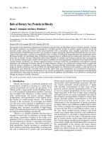
Figure 1
Examples of immunohistochemical staining for CDMPs in human intervertebral discExamples of immunohistochemical staining for CDMPs in human intervertebral disc. (row A) Cartilage derived morphogenetic protein (CDMP 1) and
(row B) CDMP 2. Images are of nucleus pulposus of grade 1 non-degenerate discs (column 1), the nucleus pulposus of grade 10 degenerate discs
(column 2) and IgG controls for each antibody. Bars = 570 μm.
Arthritis Research & Therapy Vol 11 No 5 Le Maitre et al.
Page 6 of 10
(page number not for citation purposes)
the blood vessels for either CDMP 1 or 2. IgG controls were
negative (Figure 1).
Immunohistochemical staining for BMP RII
We have previously shown BMP RII immunopositive staining
in the human IVD with a greater number of immunopositive
cells within the NP than the IAF and OAF (P < 0.05). Further-
more, in IVDs graded as intermediate degeneration there was
an increase in the proportion of immunopositive cells, which
was significant in the NP (P < 0.05) [17]
Effect of CDMP 1 on proliferation of human NP cells
derived from degenerate discs
To determine the effect of CDMP 1 on cellular proliferation
DNA content per alginate bead was calculated following two
weeks of treatment with CDMP. An increase in DNA content
(28% increase in CDMP-treated cells v/s untreated cells) was
observed in the alginate bead cultures treated with CDMP but
this did not reach significance (P = 0.35; Figure 3).
Effect of CDMP 1 on GAG production of human NP cells
derived from degenerate discs
A significant increase in overall GAG production (i.e. within the
CM, FRM and media together) was observed in NP cells
derived from degenerate discs treated with 10 ng/ml CDMP 1
for two weeks compared with untreated NP cells (P < 0.05).
An increase in GAG content of CM in CDMP-treated cultures
was observed but this did not reach significance (P = 0.43).
However, the GAG content within the FRM was significantly
increased following CDMP 1 treatment for two weeks (P <
0.05). No difference was observed in the GAG released into
the media during the two weeks treatment with CDMP from
untreated alginate bead cultures of NP cells derived from
degenerate discs (P = 0.24; Figure 4).
Figure 2
Assessment of immunopositive staining for CDMP 1 and 2 in human intervertebral discsAssessment of immunopositive staining for CDMP 1 and 2 in human intervertebral discs. Percentage of cells with immunopositivity for (a) cartilage
derived morphogenetic protein (CDMP) 1, (b) CDMP 2, according to location in the disc and grade of intervertebral disc degeneration (n = 30).
Data are presented as means ± standard error.* P < 0.05 compared with non-degenerate discs.
Table 2
Analysis of immunohistochemical data: P values for analysis of CDMP1 and 2 expression in different areas of disc in non-
degenerate v/s degenerate discs
IVD area analysed for CDMP expression Intermediate degeneration (P) Severe degeneration (P)
CDMP 1 expression in NP Non-degenerate v/s degenerate 0.302 0.106
CDMP 1 expression in IAF Non-degenerate v/s degenerate 0.336 0.112
CDMP 1 expression in OAF Non-degenerate v/s degenerate 0.461 0.362
CDMP 2 expression in NP Non-degenerate v/s degenerate 0.241 0.124
CDMP 2 expression in IAF Non-degenerate v/s degenerate 0.479 0.521
CDMP 2 expression in OAF Non-degenerate v/s degenerate 0.679 0.465
CDMP = cartilage derived morphogenetic protein; IAF = inner annulus fibrosus; IVD = intervertebral disc; NP = nucleus pulposus; OAF = outer
annulus fibrosus.
Available online />Page 7 of 10
(page number not for citation purposes)
Effect of CDMP 1 on gene expression for aggrecan and
collagen type II in human NP cells derived from
degenerate discs
A significant increase in both aggrecan (3831-fold increase)
and collagen type II (1660-fold increase) gene expression was
observed in NP cells derived from degenerate discs cultured
in alginate beads and treated with 10 ng/ml CDMP 1 for two
weeks (P < 0.05; Figure 5).
Discussion
A major cause of LBP is degeneration of the IVD, of which pro-
teoglycan loss is a key feature and has been linked to loss in
disc height, de-stabilisation of the motion segment and the
ingrowth of blood vessels and nerves resulting in generation of
pain [28,29]. Thus a potential therapeutic approach to repair
the degenerate disc would be the stimulation of normal disc
matrix production particularly increased synthesis of prote-
oglycans. A number of growth factors have been suggested as
possible therapeutic agents. However, our previous study sug-
gested that the addition of growth factors which bound to TGF
RII, FGF R3 and IGF RI may also induce unwanted blood ves-
sel ingrowth [17]. However, we demonstrated that growth fac-
tors, such as CDMP 1 and 2, which elicit their response via
BMP RII, should not induce blood vessel ingrowth.
Here we demonstrate the synthesis and localisation of CDMP
1 and CDMP 2 within human IVDs. Although a small decrease
in the proportion of cells within the NP staining for CDMP 1
Figure 3
Effect of CDMP treatment on DNA content of alginate beads containing NP cells derived from degenerate discs treated with CDMP for two weeksEffect of CDMP treatment on DNA content of alginate beads containing NP cells derived from degenerate discs treated with CDMP for two weeks.
Data are presented as means ± standard error. CDMP = cartilage derived morphogenetic protein; NP = nucleus pulposus.
Figure 4
Effect of CDMP treatment on GAG content of NP cells derived from degenerate discsEffect of CDMP treatment on GAG content of NP cells derived from degenerate discs. Data are presented as GAG content of the cell associated
matrix, further removed matrix and GAG released into the media per ug DNA (means ± standard error. * P < 0.05 compared with untreated con-
trols). CDMP = cartilage derived morphogenetic protein; GAG = glycosaminoglycan; NP = nucleus pulposus.
Arthritis Research & Therapy Vol 11 No 5 Le Maitre et al.
Page 8 of 10
(page number not for citation purposes)
and CDMP 2 was observed during degeneration this was not
significant. Similarly Bobacz and colleagues demonstrated
that both CDMP 1 and CDMP 2 were expressed in normal and
osteoarthritic (OA) articular cartilage with no change seen dur-
ing OA [18]. This suggests that the pathogenesis of disc
degeneration or OA is not associated with a reduced expres-
sion of these growth factors.
CDMP has been shown to result in increased proteoglycan
production in human mesenchymal stem cells [30], a chondro-
cyte cell line [31], and in human articular chondrocytes
[18,19]. Recently, a small number of studies have also demon-
strated proteoglycan stimulation in bovine, rabbit and mouse
disc cells [21,22]. However, to date, no studies have demon-
strated an increase in proteoglycan production in degenerate
human IVD cells following CDMP treatment. Here we investi-
gated the effect of CDMP 1 on human NP cells cultured in an
alginate bead system. Importantly an alginate bead culture
system was used as this maintains the in vivo phenotype of
IVD cells, which is lost in monolayer culture [25,32]. Our
results demonstrate that cells derived from degenerate human
discs can also respond to CDMP with an increase in GAG
production, although our study only used three patient sam-
ples. These results confirm those derived from animal disc
cells where CDMP resulted in significant increases in GAG
production [21,22]. The accumulation of GAG within alginate
beads was investigated within the compartments: CM and
FRM, together with GAG released into media. The majority of
the GAG produced by the degenerate NP cells was found in
the FRM, and this was the area which showed a significant
increase in GAG accumulation following treatment with
CDMP 1. The CM is thought to represent the highly structured
compartment encircling each cell and corresponds to the
combined pericellular and territorial matrix pools which sur-
round each cell in vivo [25,33,34]. In contrast the more loosely
organised compartment known as the FRM, accounting for
approximately 95% of the total volume of matrix, is thought to
represent the interterritorial matrix compartment in vivo
[25,34]. As this area is thought to account for the majority of
the matrix in vivo the fact that more GAGs were found in this
area of matrix following stimulation with CDMP is promising for
future therapeutic approaches.
The current study also showed that CDMP1 induced dramatic
increases in the gene expression for the matrix molecules
aggrecan and collagen type II within degenerate human NP
cells, as has been reported in mouse IVD cells [22]. During
disc degeneration the production of both aggrecan and colla-
gen type II is decreased [23,35] leading to reduced hydration
and ability to withstand load. Thus, if a growth factor could be
applied which can successfully stimulate the synthesis of
these important matrix molecules this would be of benefit for
regenerating the degenerate disc.
Previous studies investigating the effect of CDMP1 on rabbit
disc cells in monolayer [9] and mouse [22] and bovine disc
cells in alginate [21] have shown significant increases in cell
proliferation. Here we showed a small increase in proliferation
of human disc cells in alginate culture following treatment with
CDMP1 for two weeks, although, possibly due to the small
sample size, this did not reach significance. Increases in pro-
liferation could be of benefit in a therapeutic approach as a
mechanism to replace some of the cells lost through apoptosis
and senescence which are common features during disc
degeneration [27,36].
Figure 5
Effect of CDMP treatment on aggrecan and type II collagen gene expression in NP cells derived from degenerate discs treated with CDMP for two weeksEffect of CDMP treatment on aggrecan and type II collagen gene expression in NP cells derived from degenerate discs treated with CDMP for two
weeks. Absolute quantification of copy number per 250 ng cDNA normalized to the housekeeping gene 18 s. Data are presented as means ± stand-
ard error. * P < 0.05 compared with untreated controls. CDMP = cartilage derived morphogenetic protein; NP = nucleus pulposus.
Available online />Page 9 of 10
(page number not for citation purposes)
Importantly, this study, together with previous animal studies,
suggests CDMP could be a useful therapeutic agent in the
regeneration of the degenerate IVD and provides supporting
evidence for the clinical use of CDMP in human IVD degener-
ation. Indeed a phase I/II clinical trail has just started investi-
gating the efficacy and safety of recombinant GDF 5 (CDMP
1) injection into the IVD for degenerative disc disease [37].
However, it must be noted that any proposed therapy may
have to target a number of other problems that are associated
with disc degeneration. Combinations of factors may be
needed in order to promote matrix synthesis and inhibit the
increased catabolism seen within the degenerate disc
[38,39]. Furthermore, it has been shown that the nutrient sup-
ply diminishes with degeneration, which may also limit disc cell
self-renewal and function [40]. Thus, potential therapeutic
growth factors may have to be combined with therapies aimed
at restoring disc nutrition or targeted at those patients in which
the cartilaginous endplates (through which nutrients are
received) are unaffected, that is not calcified, or sclerotic [40].
Conclusions
Our data demonstrates that CDMP 1 and 2 protein is
expressed by both non-degenerate and degenerate discs
together with its receptor (BMP RII), suggesting CDMP is
involved in the normal matrix homeostasis with the human IVD.
Importantly we have demonstrated, for the first time, that
human disc cells derived from degenerate discs retain their
ability to respond to CDMP and that such treatment leads to
an increase in aggrecan and collagen type II gene expression
and increased accumulation of GAGs. Together this data sug-
gests that CDMP is an important anabolic growth factor in the
IVD and could be a suitable therapy to aid in IVD repair/regen-
eration, via stimulation of matrix synthesis.
Competing interests
The authors declare that they have no competing interests.
Authors' contributions
CLM helped conceive the study, participated in its design, per-
formed the majority of the laboratory work and all the analysis
and co-wrote the manuscript. AJF participated in interpretation
of data and contributed to the preparation of the final manu-
script. JAH conceived the study, secured funding, contributed
to its design and co-ordination, participated in interpretation of
data and contributed to the preparation of the final manuscript.
All authors read and approved the final manuscript.
Acknowledgements
The authors wish to acknowledge the support of the joint Research
Councils (MRC, BBSRC, EPSRC) UK Centre for Tissue Engineering
(34/TIE 13617). The work was undertaken in the Human Tissue Profiling
Laboratories of the School of Clinical and Laboratory Sciences that
receive core support from the ARC (ICAC grant F0551).
References
1. Borenstein DG: Epidemiology, etiology, diagnostic evaluation,
and treatment of low back pain. Curr Opin Rheumatol 2001,
13:128-134.
2. Maniadakis N, Gray A: The economic burden of back pain in the
UK. Pain 2000, 84:95-103.
3. Peterson CK, Bolton JE, Wood AR: A cross-sectional study cor-
relating lumbar spine degeneration with disability and pain.
Spine 2000, 25:218-223.
4. Luoma K, Riihimaki H, Luukkonen R, Raininko R, Viikari-Juntura E,
Lamminen A: Low back pain in relation to lumbar disc
degeneration. Spine 2000, 25:487-492.
5. Freemont AJ, Watkins A, Le Maitre C, Jeziorska M, Hoyland JA:
Current understanding of cellular and molecular events in
intervertebral disc degeneration: implications for therapy. J
Pathol 2002, 196:374-379.
6. Le Maitre CL, Freemont AJ, Hoyland JA: Localization of degrada-
tive enzymes and their inhibitors in the degenerate human
intervertebral disc. J Pathol 2004, 204:47-54.
7. Le Maitre CL, Freemont A, Hoyland J: Human disc degeneration
is associated with increased MMP 7 expression. Biotech
Histochem 2006, 81:125-131.
8. Osada R, Ohshima H, Ishihara H, Yudoh K, Sakai K, Matsui H, Tsuji
H: Autocrine/paracrine mechanism of insulin-like growth fac-
tor-1 secretion, and the effect of insulin-like growth factor-1 on
proteoglycan synthesis in bovine intervertebral discs. J Orthop
Res 1996, 14:690-699.
9. Wang H, Kroeber M, Hanke M, Ries R, Schmid C, Poller W, Rich-
ter W: Release of active and depot GDF-5 after adenovirus-
mediated overexpression stimulates rabbit and human
intervertebral disc cells. J Mol Med 2004, 82:126-134.
10. Li X, Leo BM, Beck G, Balian G, Anderson GD: Collagen and pro-
teoglycan abnormalities in the GDF-5-deficient mice and
molecular changes when treating disk cells with recombinant
growth factor. Spine 2004, 29:2229-2234.
11. Thompson JP, Oegema TR Jr, Bradford DS: Stimulation of
mature canine intervertebral disc by growth factors. Spine
1991,
16:253-260.
12. Li J, Yoon ST, Hutton WC: Effect of bone morphogenetic pro-
tein-2 (BMP-2) on matrix production, other BMPs, and BMP
receptors in rat intervertebral disc cells. J Spinal Disord Tech
2004, 17:423-428.
13. Masuda K, An HS: Growth factors and the intervertebral disc.
Spine J 2004, 4:330S-340S.
14. Masuda K, Oegema TR Jr, An HS: Growth factors and treatment
of intervertebral disc degeneration. Spine 2004,
29:2757-2769.
15. Yoon ST, Park JS, Kim KS, Li J, Attallah-Wasif ES, Hutton WC,
Boden SD: ISSLS prize winner: LMP-1 upregulates interverte-
bral disc cell production of proteoglycans and BMPs in vitro
and in vivo. Spine 2004, 29:2603-2611.
16. Freemont AJ, Hoyland JA, Baird P, Byers RJ: Expression of NGFβ
in the vasculature of the diseased intervertebral disc. J Pathol
1999, 189:A68.
17. Le Maitre CL, Richardson SM, Baird P, Freemont AJ, Hoyland JA:
Expression of receptors for putative anabolic growth factors in
human intervertebral disc: implications for repair and regener-
ation of the disc. J Pathol 2005, 207:445-452.
18. Bobacz K, Gruber R, Soleiman A, Graninger WB, Luyten FP,
Erlacher L: Cartilage-derived morphogenetic protein-1 and -2
are endogenously expressed in healthy and osteoarthritic
human articular chondrocytes and stimulate matrix synthesis.
Osteoarthritis Cartilage 2002, 10:394-401.
19. Erlacher L, Ng CK, Ullrich R, Krieger S, Luyten FP: Presence of
cartilage-derived morphogenetic proteins in articular cartilage
and enhancement of matrix replacement in vitro. Arthritis
Rheum 1998, 41:263-273.
20. Walsh AJ, Bradford DS, Lotz JC: In vivo growth factor treatment
of degenerated intervertebral discs. Spine 2004, 29:156-163.
21. Chujo T, An HS, Akeda K, Miyamoto K, Muehleman C, Attawia M,
Andersson G, Masuda K: Effects of growth differentiation fac-
tor-5 on the intervertebral disc in vitro bovine study and in
vivo rabbit disc degeneration model study. Spine 2006,
31:2909-2917.
22. Cui M, Wan Y, Anderson DG, Shen FH, Leo BM, Laurencin CT,
Balian G, Li X:
Mouse growth and differentiation factor-5 pro-
Arthritis Research & Therapy Vol 11 No 5 Le Maitre et al.
Page 10 of 10
(page number not for citation purposes)
tein and DNA therapy potentiates intervertebral disc cell
aggregation and chondrogenic gene expression. Spine J
2008, 8:287-295.
23. Sive JI, Baird P, Jeziorsk M, Watkins A, Hoyland JA, Freemont AJ:
Expression of chondrocyte markers by cells of normal and
degenerate intervertebral discs. Mol Pathol 2002, 55:91-97.
24. Wang JY, Baer AE, Kraus VB, Setton LA: Intervertebral disc cells
exhibit differences in gene expression in alginate and monol-
ayer culture. Spine 2001, 26:1747-1752.
25. Chiba K, Andersson GB, Masuda K, Thonar EJ: Metabolism of the
extracellular matrix formed by intervertebral disc cells cul-
tured in alginate. Spine 1997, 22:2885-2893.
26. Farndale RW, Buttle DJ, Barrett AJ: Improved quantitation and
discrimination of sulphated glycosaminoglycans by use of
dimethylmethylene blue. Biochim Biophys Acta 1986,
883:173-177.
27. Le Maitre CL, Freemont AJ, Hoyland JA: Accelerated cellular
senescence in degenerate intervertebral discs: A possible role
in the pathogenesis of intervertebral disc degeneration. Arthri-
tis Res Ther 2007, 9:R45.
28. Freemont AJ, Peacock TE, Goupille P, Hoyland JA, O'Brien J, Jay-
son MI: Nerve ingrowth into diseased intervertebral disc in
chronic back pain. Lancet 1997, 350:178-181.
29. Freemont AJ, Watkins A, Le Maitre C, Baird P, Jeziorska M, Knight
MT, Ross ER, O'Brien JP, Hoyland JA: Nerve growth factor
expression and innervation of the painful intervertebral disc. J
Pathol 2002, 197:286-292.
30. Bai X, Xiao Z, Pan Y, Hu J, Pohl J, Wen J, Li L: Cartilage-derived
morphogenetic protein-1 promotes the differentiation of mes-
enchymal stem cells into chondrocytes. Biochem Biophys Res
Commun 2004, 325:453-460.
31. Li J, Kim KS, Park JS, Elmer WA, Hutton WC, Yoon ST: BMP-2
and CDMP-2: stimulation of chondrocyte production of
proteoglycan. J Orthop Sci 2003, 8:829-835.
32. Le Maitre CL, Freemont AJ, Hoyland JA: The role of interleukin-1
in the pathogenesis of human intervertebral disc
degeneration. Arthritis Res Ther 2005, 7:R732-R745.
33. Hauselmann HJ, Masuda K, Hunziker EB, Neidhart M, Mok SS,
Michel BA, Thonar EJ: Adult human chondrocytes cultured in
alginate form a matrix similar to native human articular
cartilage. Am J Physiol 1996, 271:C742-C752.
34. Mok SS, Masuda K, Hauselmann HJ, Aydelotte MB, Thonar EJ:
Aggrecan synthesized by mature bovine chondrocytes sus-
pended in alginate. Identification of two distinct metabolic
matrix pools. J Biol Chem 1994, 269:33021-33027.
35. Le Maitre CL, Pockert A, Buttle DJ, Freemont AJ, Hoyland JA:
Matrix synthesis and degradation in human intervertebral disc
degeneration. Biochem Soc Trans 2007, 35:652-655.
36. Zhao CQ, Jiang LS, Dai LY: Programmed cell death in interver-
tebral disc degeneration. Apoptosis 2006, 11:2079-2088.
37. Intradiscal rhGDF-5 Phase I/II Clinical Trial [http://clinicaltri
als.gov/ct2/show/NCT00813813?term=GDF&rank=1]
38. Le Maitre CL, Freemont AJ, Hoyland JA: A preliminary in vitro
study into the use of IL-1Ra gene therapy for the inhibition of
intervertebral disc degeneration. Int J Exp Pathol 2006,
87:17-28.
39. Le Maitre CL, Hoyland JA, Freemont AJ: Interleukin-1 receptor
antagonist delivered directly and by gene therapy inhibits
matrix degradation in the intact degenerate human interverte-
bral disc: an in situ zymographic and gene therapy study.
Arthritis Res Ther 2007, 9:R83.
40. Kandel R, Roberts S, Urban JP: Tissue engineering and the
intervertebral disc: the challenges. Eur Spine J 2008, 17(Suppl
4):480-491.