Báo cáo y học: "Progress in osteoporosis and fracture prevention: focus on postmenopausal women" ppsx
Bạn đang xem bản rút gọn của tài liệu. Xem và tải ngay bản đầy đủ của tài liệu tại đây (1.09 MB, 18 trang )
Available online />Page 1 of 18
(page number not for citation purposes)
Abstract
In the past decade, we have witnessed a revolution in osteo-
porosis diagnosis and therapeutics. This includes enhanced
understanding of basic bone biology, recognizing the severe
consequences of fractures in terms of morbidity and short-term re-
fracture and mortality risk and case finding based on clinical risks,
bone mineral density, new imaging approaches, and contributors
to secondary osteoporosis. Medical interventions that reduce
fracture risk include sufficient calcium and vitamin D together with
a wide spectrum of drug therapies (with antiresorptive, anabolic,
or mixed effects). Emerging therapeutic options that target
molecules of bone metabolism indicate that the next decade
should offer even greater promise for further improving our
diagnostic and treatment approaches.
Introduction
In the past decade, we have witnessed a revolution in under-
standing bone biology. Major progress has also been achieved
in fracture risk estimation and prevention of fractures. How
does this progress translate into daily clinical practice? First,
case finding of subjects at highest risk for fractures is now
possible at the individual patient level, using clinical bone- and
fall-related risk factors, with and without bone mineral density
(BMD). Second, prevention of vertebral and nonvertebral
fractures, including hip fractures, is now possible by optimizing
calcium homeostasis and by appropriate medication in well-
selected patients with a high risk of fracture. Recent studies
indicate new possibilities for case finding, such as in vivo
structural analysis of bone microarchitecture, and new
molecular targets to rebalance bone remodeling. Here, we
review recent progress in case-finding strategies and in the
evidence that the risk of first and subsequent fractures can be
prevented in daily clinical practice.
The Fracture Risk Assessment Tool for
calculating the individual 10-year fracture risk
The clinical expression of osteoporosis is a fragility fracture,
but bone loss in and of itself is asymptomatic, which has led
to the description of osteoporosis as a ‘silent thief’. The
asymptomatic nature of bone loss suggests that osteoporosis
cannot be detected before a fragility fracture occurs, unless
BMD is measured. Indeed, BMD is related to bone strength
and low BMD is a major risk factor for fractures. However,
most patients presenting with a fracture do not have BMD-
based osteoporosis, defined according to the World Health
Organization (WHO) definition as a T score of –2.5 or below
[1]. Many qualities of bone, other than low BMD, are involved
in fracture risk such as structural and material components of
bone and the cellular activities and molecular signals that
regulate lifelong bone remodeling under control of
mechanical load, hormones, growth factors, and cytokines.
Some of these characteristics of bone are measurable in
clinical practice (for example, BMD, bone size, vertebral
deformities and fractures, and markers of bone turnover), but
many are not (for example, material properties) or are just
evolving (for example, microarchitecture by microcomputer
tomography or magnetic resonance imaging). In addition, and
independent of bone-related risks, extraskeletal risk factors
such as fall risk contribute to fracture risk and are present in
the majority of patients older than 50 years presenting with a
clinical fracture [1].
Review
Progress in osteoporosis and fracture prevention:
focus on postmenopausal women
Kenneth G Saag
1
and Piet Geusens
2
1
Division of Clinical Immunology and Rheumatology, Center for Education and Research on Therapeutics, University of Alabama at Birmingham,
820 Faculty Office Tower, 510 20th Street South, Birmingham, AL 35294-3708, USA
2
Department of Internal Medicine, Subdivision of Rheumatology, Maastricht University Medical Center, P. Debyelaan 25, Postbus 5800,
6202 AZ Maastricht, The Netherlands & Biomedical Research Institute, University Hasselt, Agoralaan, gebouw D, B-3590 Diepenbeek, Belgium
Corresponding author: Kenneth G Saag,
Published: 14 October 2009 Arthritis Research & Therapy 2009, 11:251 (doi:10.1186/ar2815)
This article is online at />© 2009 BioMed Central Ltd
AR = absolute risk; BMD = bone mineral density; CI = confidence interval; DXA = dual-energy x-ray absorptiometry; ERT = estrogen replacement
therapy; FIT = Fracture Intervention Trial; FRAX = Fracture Risk Assessment Tool; GI = gastrointestinal; ISCD = International Society of Clinical
Densitometry; MORE = Multiple Outcomes of Raloxifene Evaluation; NOF = National Osteoporosis Foundation; NOGG = National Osteoporosis
Guideline Group; NOS = National Osteoporosis Society; OPG = osteoprotegerin; PTH = parathyroid hormone; RANK = receptor activator of
nuclear factor-kappa B; RANKL = receptor activator of nuclear factor-kappa B ligand; RR = relative risk; RRR = relative risk reduction; SERM =
selective estrogen receptor modulator; VFA = vertebral fracture assessment; WHI = Women’s Health Initiative; WHO = World Health Organization.
Arthritis Research & Therapy Vol 11 No 5 Saag and Geusens
Page 2 of 18
(page number not for citation purposes)
Large-scale prospective population studies have enabled the
specification of clinical risk factors for fractures that are
independent of low BMD and have allowed quantification of
their relative risks (RRs) for predicting fractures. Thus, many
aspects of osteoporosis and fracture risk are clinically recog-
nizable (such as age, gender, and body weight), even before
a first fracture has occurred. However, RRs are difficult to
apply in daily clinical practice since their clinical significance
depends on the prevalence of fractures in the general
population. From this observation and for the purpose of
clinical application, the concept of the absolute risk (AR) of
fractures has emerged and refers to the individual’s risk for
fractures over a certain time period (for example, over the next
10 years) [2].
During the last decade, the development of the Fracture Risk
Assessment Tool (FRAX) algorithm as a clinical tool for
calculation of fracture risk in the individual patient is a major
achievement in the field of case finding [2,3]. The FRAX is
based on large-scale prospective population-based studies
and includes age, gender, body weight and body mass index,
a history of fracture, hip fracture in parents, current smoking,
excessive alcohol intake, rheumatoid arthritis, glucocorticoid
use, and other forms of secondary osteoporosis (Table 1).
The WHO developed FRAX especially for primary care
physicians for calculating the individual 10-year risk of hip
and major fractures (defined as clinical spine, forearm, hip, or
humerus fracture) in daily practice in women and men, based
on the above-mentioned clinical risk factors, with and without
results of BMD measurement in the femoral neck.
Strengths of the Fracture Risk Assessment Tool
FRAX is based on a large sample of primary data of
prospective population studies and takes into account
variability in fracture probability between geographic regions.
FRAX should not be considered a gold standard but rather a
platform technology and provides an aid to enhance patient
assessment. FRAX can be integrated in clinical practice in
many countries worldwide, both in women and men. FRAX is
therefore likely to become, in many countries, the most
popular instrument for identifying women and men at highest
risk for fractures.
FRAX has been included in guidelines as a tool for case
finding for identifying postmenopausal women at high risk for
fractures, for selecting subjects to measure BMD, and for
treatment decisions. The National Osteoporosis Foundation
(NOF) in the US and the National Osteoporosis Society
(NOS) in the UK have recently updated their guidelines on
postmenopausal osteoporosis in this context (Figure 1) [4,5].
These groups have integrated FRAX and BMD for case
finding of individuals at high risk for fracture and for treatment
decisions. Both sets of guidelines make a clear distinction
between postmenopausal women with and without a fracture
history. This is a major step forward in the clinical applicability
for postfracture treatment in patients presenting with a
fracture. Based on the fracture risk profile, the NOS, together
with the National Osteoporosis Guideline Group (NOGG)
and the Royal College of Physicians, determined treatment
thresholds at which fracture prevention became cost-effective
(Figure 2) [2,5].
Postmenopausal women with a history of fractures
The NOS advocates drug treatment in all postmenopausal
women with a history of any fragility fracture (defined as distal
radius, proximal humerus, spine [including morphometric
vertebral fracture], pelvis [pubic rami], tibia, and ankle) [5].
The NOF advocates drug treatment in postmenopausal
women with a vertebral or hip fracture (without need of BMD
or FRAX for decisions about pharmacotherapy), but after a
nonvertebral nonhip fracture, the NOF advocates performing
a dual-energy x-ray absorptiometry (DXA) measurement and
starting drug treatment in patients having osteoporosis and in
patients with osteopenia when FRAX indicates a 10-year
fracture probability of at least 3% for hip or at least 20% for
major fractures. Thus, in postmenopausal women with a
history of vertebral or hip fracture, neither set of guidelines
uses FRAX for decisions about drug treatment (and neither
does the NOS for after any fragility fracture), and both sets
consider such fracture history by itself as a starting point for
case finding and treatment decisions.
Table 1
Clinical risk factors and bone densitometry results that are
included in the Fracture Risk Assessment Tool algorithm
Age
Gender
Body mass index
History of fracture after the age of 45 to 50 years
Parent with hip fracture
Current smoking
Alcohol intake of greater than 2 units per day
Glucocorticoid use
Rheumatoid arthritis
Other causes of secondary osteoporosis:
- Untreated hypogonadism in men and women, anorexia
nervosa, chemotherapy for breast and prostate cancer,
and hypopituitarism
- Inflammatory bowel disease and prolonged immobility (for
example, spinal cord injury, Parkinson disease, stroke,
muscular dystrophy, and ankylosing spondylitis)
- Organ transplantation
- Type I diabetes and thyroid disorders (for example,
untreated hyperthyroidism and overtreated
hypothyroidism)
Results of bone densitometry using dual-energy x-ray absorptiometry of
the femoral neck.
Postmenopausal women without a fracture history
The NOS advocates applying FRAX (without BMD) in all
postmenopausal women. Women at high risk according to
FRAX without BMD are then considered candidates for drug
treatment. Women with an intermediate risk according to FRAX
without BMD are recommended to have a DXA measurement,
and when FRAX with BMD is above the intervention threshold
according to the NOGG, drug treatment should be considered.
The NOF advocates using DXA in all women older than
65 years and in postmenopausal women younger than
65 years in whom there is concern about their fracture risk
Available online />Page 3 of 18
(page number not for citation purposes)
Figure 1
Algorithms for case finding and drug treatment decisions in postmenopausal women with and without a history of fractures according to the
National Osteoporosis Foundation (NOF) in the US and the National Osteoporosis Society (NOS) in the UK. DXA, dual-energy x-ray
absorptiometry; FRAX, Fracture Risk Assessment Tool. *Previous fragility fracture, particularly of the hip, wrist and spine including morphometric
vertebral fracture. **Based on UK guidelines by NOGG.
based on the presence of clinical risk factors. This approach
suggests that all postmenopausal women under 65 years of
age should be clinically classified as having at least one of
the risk factors of FRAX. Treatment is then recommended in
patients with osteoporosis, in patients with osteopenia when
the FRAX indicates a 10-year risk of greater than 3% for hip
fractures or greater than 20% for major osteoporotic
fractures, and in other patients considered at high risk (on
glucocorticoids, total immobilization). These upgraded guide-
lines indicate that FRAX is an emerging tool in clinical
decision making about case finding, selecting patients for
DXA, and treatment decisions in postmenopausal women
without a fracture history. Patients with a fracture are con-
sidered at high enough risk to make treatment decisions
without additional need for using FRAX. It is expected that
FRAX will also be helpful in designing fracture prevention
studies and in reimbursement issues. In a study from
Switzerland, profiles of patients at increased probability of
fracture beyond currently accepted reimbursement thresholds
for bone BMD measurement by DXA and osteoporosis
treatment were identified and constitute an additional group
of patients in whom treatment should be considered [6].
Limitations of the Fracture Risk Assessment Tool
In spite of its solid scientific basis and clinical attractiveness,
FRAX has several limitations, as acknowledged by the
authors (Table 2) [2]. Meanwhile, FRAX has been integrated
in guidelines/guidance in the US, UK, Europe, Canada,
Germany, and Japan [2], but with different approaches for
diagnostic and treatment thresholds, as shown above for the
NOS and the NOF [4,5]. Fracture reduction has been
demonstrated in randomized controlled clinical trials in
patients selected on the basis of the presence of a
morphometric vertebral fracture, hip fracture, or a low BMD,
but not on the basis of FRAX. Therefore, of great interest is
the finding that fracture reduction was greater at higher
fracture probabilities based on FRAX, with or without BMD.
Antifracture efficacy was evident when baseline fracture
probabilities for major fractures were greater than 20% in
the clodronate trial (in preventing major fractures) [7] and
greater than 16% in the bazedoxifene trial (in preventing
clinical fractures), irrespective of whether BMD was used in
the fracture calculation [2]. Further studies will be needed on
the ability of treatment to reduce fracture risk in subjects at
high risk for fractures based on FRAX in the absence of a
morphometric vertebral fracture, hip fracture, or a low BMD,
which is the case in most patients presenting with a
nonvertebral fracture. Decisions on treatment thresholds will
furthermore depend on factors related to health care
providers and patients and the willingness of society to
reimburse treatment as health economic aspects are
becoming increasingly important to determine the cost-
effectiveness of treatment. Meanwhile, the NOGG of the UK
has indicated FRAX-based thresholds for measuring BMD
and for treatment decisions, with and without BMD (Figure 2).
The management algorithms proposed by the NOGG are
underpinned by a health economic analysis applied to the
epidemiology of fracture in the UK.
Arthritis Research & Therapy Vol 11 No 5 Saag and Geusens
Page 4 of 18
(page number not for citation purposes)
Figure 2
Assessment and intervention thresholds based on the 10-year risk of major fracture, as proposed in the UK [2]. BMD, bone mineral density. With
kind permission from Springer Science+Business Media [5].
Fall-related risks were explicitly excluded from the FRAX
calculations but were recognized as risks for fractures
independently of bone-related risks, especially for non-
vertebral fractures such as hip fractures. More than 80% of
women and men presenting with a clinical fracture to the
emergency unit have, beside bone-related risks, one or more
fall-related risks and have, independently from BMD, a
fourfold increased risk of a fall history during the previous
year [1]. In an integrated bone- and fall-related risk evaluation
tool for the estimation of the 5- and 10-year ARs for fractures
in patients using glucocorticoids, a history of falls had a
greater impact on fracture risk than any other evaluated risk,
and its contribution to fracture risk was similar to, and inde-
pendent of, using a high dose of glucocorticoids (prednisone
greater than 15 mg/day) [8]. Thus, with FRAX, fracture risk
calculation could be underestimated in patients with fall risks.
Subsequent fractures and postfracture
mortality cluster in time: the need for
immediate clinical attention in patients
presenting with a fracture
A history of nonvertebral fracture is associated with a
doubling of the risk of a subsequent fracture, and the
subsequent fracture risk is even quadrupled after a vertebral
fracture. However, this re-fracture risk is not constant over
time and is driven by the high, threefold to fivefold increase in
the years immediately after a first fracture, followed by a
gradual waning later on (Figure 3) [9]. This has been shown
for repeat morphometric vertebral fractures, subsequent
clinical spine, forearm, and hip fractures after hospitalization
because of a vertebral fracture, repeat low trauma fractures in
subjects older than 60 years, repeat clinical vertebral and
nonvertebral fractures from menopause onwards, and repeat
hip fractures [9-12]. As a result, it has been shown in long-
term follow-up studies that 40% to 50% of all subsequent
fractures occur within 3 to 5 years after a first fracture. The
clinical implication is that patients older than 50 years
presenting with a fracture need immediate attention to reduce
the risk of a subsequent fracture. This is a situation in which it
is important to take immediate action in fracture patients,
such as a fracture liaison service and other initiatives in the
field of postfracture care [13,14]. It also indicates that, in
such patients, treatment that has been shown to reduce
fracture risk within the short term should be started [15].
An increased risk of mortality has been found after hip,
vertebral, and several nonhip, nonvertebral fractures [16]. As
for subsequent fracture risk, this increase in mortality is
higher immediately after fracture than later on. In women and
men older than 60 years, nearly 90% of excess deaths
related to fracture over the 18 years of observation occurred
in the first 5 years. Of the 5-year excess mortality, hip,
vertebral, and nonhip, nonvertebral fractures were each
associated with approximately one third of deaths. The major
causes of death were related to cardiovascular and
respiratory comorbidity [16].
Assessment of vertebral fractures: an
opportunity to identify high-risk patients
Vertebral fractures are a special group of fractures.
Morphometric vertebral fractures are the most frequent
fractures in women and men older than 50 years [17] and
their presence is a strong predictor of future vertebral, non-
vertebral, and hip fracture risk [18]. Clinical vertebral
Available online />Page 5 of 18
(page number not for citation purposes)
Table 2
Limitations of the Fracture Risk Assessment Tool for case finding
- Factors not included in FRAX:
• The ‘dose effect’ of some risk factors
• Glucocorticoid use (dose and duration)
• Characteristics of previous fractures (location, number, and severity)
• Fall risks
• Vitamin D deficiency
• Fluctuation over time of subsequent fracture
• Markers of bone formation and bone resorption
• How to identify patients with a vertebral fracture
• Which laboratory tests are indicated (and in whom) to exclude secondary osteoporosis
- FRAX is applicable only in untreated patients.
- Inclusion of BMD results is limited to results of BMD in the femoral neck. However, total hip BMD can be used interchangeably with femoral
neck BMD in women, but not in men.
- FRAX does not indicate which intervention is indicated at which level of 10-year fracture risk of hip or major fractures (for either
nonpharmacological or drug treatment).
BMD, bone mineral density; FRAX, Fracture Risk Assessment Tool.
fractures represent one out of three to four morphometric
vertebral fractures and represent less than 10% of all
fractures in patients presenting with a fracture to the
emergency department [1]. Most morphometric vertebral
fractures are not diagnosed until clinically suspected (for
example, significant height loss, hyperkyphosis, protruding
abdomen, rib-iliac crest distance of less than 2 cm, and acute
or chronic back pain) and imaging by x-ray is performed. But
even when lateral x-rays of the spine are available, vertebral
fractures are often missed [18,19].
Vertebral fracture assessment (VFA) is a new method to
evaluate the presence of morphometric vertebral fractures
and deformities using x-ray absorptiometry (Figure 4) [19].
With appropriate DXA devices, VFA can be performed at the
occasion of a bone densitometry. Advantages are its low
irradiation, the availability of semiautomatic image analysis
tools to assist in measuring vertebral shapes of the individual
vertebrae, its plan-parallel projection, and its high negative
predictive value. Disadvantages include difficulties in measur-
ing upper thoracic vertebrae due to overlying soft tissue and
ribs.
The prevalence of previously unknown morphometric verte-
bral fractures has been studied in various at-risk populations.
In a recent study of women and men presenting with a
nonvertebral fracture, one out of four had a prevalent morpho-
metric vertebral fracture on VFA that was not recognized
previously [14]. In one other study, the prevalence of
morphometric vertebral fractures was 21% in postmeno-
pausal women with osteopenia [20]. The authors concluded
that the use of VFA contributed to better define the fracture
risk in patients presenting with a nonvertebral fracture and in
women with osteopenia and contributed to treatment
decisions by identifying patients at high risk of fractures in the
absence of BMD osteoporosis. VFA also helps to select
patients in whom x-rays of the spine are indicated to
differentiate changes in shape from normal variations and
diseases such as Scheuermann disease, pathologic
fractures, bone remodeling in the context of osteoarthritis,
and developmental short vertebral height [19]. According to
the International Society of Clinical Densitometry (ISCD),
additional x-ray imaging is needed in cases of two or more
mild (grade 1) deformities without any moderate or severe
(grade 2 or 3) deformities, when lesions in vertebrae cannot
be ascribed to benign causes, or when vertebral deformities
are found in a patient with a known history of a relevant
malignancy [19]. In patients with BMD-diagnosed osteo-
porosis, a baseline VFA is not necessary for treatment
decisions but can be helpful to identify during follow-up
whether a vertebral fracture is new or old [15]. Indications for
VFA according to the ISCD are shown in Table 3 [19].
Differential diagnosis in patients with
osteoporosis or a fragility fracture or both
Randomized controlled trials on fracture prevention in post-
menopausal women exclude patients with secondary osteo-
porosis, except in studies in glucocorticoid users. However,
patients with BMD-diagnosed osteoporosis or presenting
with a clinical fracture or both often have contributors to
secondary osteoporosis. FRAX includes a long list of causes
of secondary osteoporosis that contribute to fracture risk
independently of other clinical risks and BMD (Table 1) [2,3].
Differential diagnosis in the context of case finding therefore
includes a thorough medical history and clinical examination.
Based on FRAX, laboratory investigations can contribute to
case finding, but FRAX does not give instructions on how to
exclude other contributors to secondary osteoporosis that are
frequently found in patients with osteoporosis or fractures or
both [21,22]. In patients with BMD-based osteoporosis or
presenting with a clinical fracture or both, diagnostic evalua-
tion is necessary and should include serum 25-(OH)D3,
calcium, creatinine, thyroid-stimulating hormone, parathyroid
Arthritis Research & Therapy Vol 11 No 5 Saag and Geusens
Page 6 of 18
(page number not for citation purposes)
Figure 3
Risk of first and subsequent fracture over time. (a) Percentage of all
first fractures from menopause onwards (grey line) and fractures
subsequent to initial fractures (black line). (b) Relative risk of all
subsequent fractures calculated as a mean from the time of first
fracture (grey line) and per separate year of follow-up after a first
fracture (black line).
hormone (PTH), testosterone (in men) and, of 24-hour urine,
calcium and creatinine [21-23]. According to the clinical
picture and suspicion, other serum measurements such as
plasma cortisol, hemoglobin, white blood cell count,
serum/urine protein electrophoresis, and selected other
evaluations looking for secondary causes are indicated.
Only limited studies about the prevalence of secondary
osteoporosis in daily practice have been published during the
last decade. In patients referred for DXA in the clinical
context of an osteoporosis clinic, contributors to secondary
osteoporosis were already documented in one out of three
postmenopausal women with osteoporosis [21]. In the group
of otherwise presumably healthy women, previously undiag-
nosed contributors were found in an additional 30% of
women [21]. In women and men presenting with a clinical
fracture at the emergency unit and having BMD osteoporosis,
42% had contributors to secondary osteoporosis, mainly
vitamin D deficiency [22].
Vitamin D deficiency is endemic worldwide [24] but is not
included in the FRAX algorithm. Vitamin D deficiency was
found to be the main contributor to secondary osteoporosis
in postmenopausal women with BMD osteoporosis [21], in
women and men presenting with a clinical fracture and having
BMD osteoporosis [22], and in patients presenting with a hip
fracture [25]. Recent data indicate that vitamin D is an
independent risk for fractures [26], and meta-analyses
indicate that correction of vitamin D deficiency results in a
decreased fall and fracture risk [27,28], but the effects
depend on the dose of vitamin D and the target population
[29]. Frail older people confined to institutions may sustain
fewer hip fractures if given vitamin D with calcium. Vitamin D
alone is unlikely to prevent fracture [30].
It is still a matter of debate which dose of vitamin D
3
(or
potentially D
2
) supplementation is necessary/optimal, taking
into account baseline vitamin D status and the desired serum
levels to be achieved by supplementation [31-33]. Clearly, an
intake of 400 IU/day is not sufficient [31-34]. A daily intake of
800 to 1,600 IU in healthy adults will increase serum levels
above 75 nmol/L in half of the population [33]. Others
suggest that 1,000 to 1,200 IU/day is necessary in addition
to typical food and cutaneous inputs to achieve a target
serum level of 80 nmol/L (32 ng/mL) [31].
Lifelong milk intake is not related to fracture risk [35], but in
several reviews, the necessity of addition of calcium to
vitamin D for fracture prevention was stressed and a dose of
1,000 to 1,200 mg/day was advocated [34,36]. However, in
Available online />Page 7 of 18
(page number not for citation purposes)
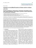
Figure 4
Example of using dual-energy x-ray absorptiometry technology for vertebral fracture assessment.
studies published in 2008, supplements of 1,000 mg
calcium/day in healthy postmenopausal women [37] and
healthy men [38] with a mean baseline calcium intake of
800 mg/day were associated with an increased risk of
vascular events, including myocardial infarction. These
studies raised considerable controversy and suggested the
need for further research [39]. In this context, it is reassuring
that, when intake of vitamin D
3
is sufficient, the need for
calcium intake is considered to be lower [32,40-42]. Indeed,
if dietary calcium is a threshold nutrient, as suggested by
Heaney [41], then the threshold for optimal calcium absorp-
tion may be at a lower calcium intake when vitamin D nutrition
is higher. Until well-designed studies address the current
uncertainties, the possible detrimental effect (for example,
hypercalcemia and its complications) of higher-than-recom-
mended calcium intake should be balanced against the likely
benefits of calcium on bone, particularly in older women [43].
It should be noted that all clinical trials with drug therapy for
osteoporosis (bisphosphonates and so on) have been con-
ducted with the concomitant use of calcium and vitamin D
supplementation.
It is generally considered that secondary causes of osteo-
porosis are more common in men than women, with the
exception of hormone deficiency, which is characteristic after
menopause, whereas andropause, depending on its
definition, is found in only a subgroup of older men or men
with osteoporosis [44]. Hypogonadism resulting from the
treatment of breast and prostate cancer is recognized as an
emerging clinical problem [45]. Cancer treatment-induced
bone loss with adjuvant endocrine therapy with an aromatase
inhibitor or androgen deprivation can be considered a risk
factor for the development of osteopenia, osteoporosis, and
bone fracture, which can be mitigated by appropriate
bisphosphonate therapy [45]. Other, less common, risk
factors for osteoporosis and fractures but commonly present
Arthritis Research & Therapy Vol 11 No 5 Saag and Geusens
Page 8 of 18
(page number not for citation purposes)
Table 3
Indications for vertebral fracture assessment using x-ray absorptiometry [19]
1. Postmenopausal women with low bone mass (osteopenia) by BMD criteria plus one of the following:
- Age of greater than or equal to 70 years.
- Historical height loss of greater than 4 cm.
- Prospective height loss of greater than 2 cm.
- Self-reported prior vertebral fracture (not previously documented).
- Two or more of the following:
Age of 60 to 69 years.
Self-reported prior nonvertebral fracture.
Historical height loss of 2 to 4 cm.
Chronic systemic diseases associated with increased risk of vertebral fractures (for example, moderate to severe COPD,
seropositive rheumatoid arthritis, and Crohn disease).
2. Men with low bone mass (osteopenia) by BMD criteria plus one of the following:
- Age of 80 years or older.
- Historical height loss of greater than 6 cm.
- Prospective height loss of greater than 3 cm.
- Self-reported vertebral fracture (not previously documented).
- Two or more of the following:
Age of 70 to 79 years.
Self-reported prior nonvertebral fracture.
Historical height loss of 3 to 6 cm.
On pharmacological androgen deprivation therapy or following orchiectomy.
Chronic systemic diseases associated with increased risk of vertebral fractures (for example, moderate to severe COPD,
seropositive rheumatoid arthritis, and Crohn disease).
3. Women or men on chronic glucocorticoid therapy (equivalent to 5 mg or more of prednisone daily for 3 months or longer).
4. Postmenopausal women or men with osteoporosis by bone density criteria (total hip, femoral neck, or lumbar spine T score of not more than
–2.5) if documentation of one or more vertebral fractures will alter clinical management.
BMD, bone mineral density; COPD, chronic obstructive pulmonary disease.
in patients with low BMD or presenting with a fracture and
that are not part of FRAX include the use of medications (for
example, anticonvulsants, primary hyperparathyroidism, renal
insufficiency, gastrectomy, Cushing syndrome, dementia, and
chronic pulmonary and/or liver diseases).
Fall prevention measures
Vitamin D supplements decrease the risk of falls, as
discussed above. Extraskeletal measures that are advocated
in guidelines include avoidance of immobility, stimulation of
weight-bearing exercise, and physiotherapy. Recent system-
atic reviews indicate that these measures still need more
research to specify their role in the prevention of fractures.
Fall prevention interventions that are likely to be effective in
older people are now available [46]. Less is known about
their effectiveness in preventing fall-related injuries, and no
data that fall prevention decreases the risk of fracture are
available. Exercise interventions reduce the risk and rate of
falls in older people living in the community [47]. The role of
hip protectors remains controversial in light of low
acceptance and low acceptability and adherence due to
discomfort and practicality [48,49].
Advances in osteoporosis pharmacotherapy:
more than a decade of progress
Beyond the need for sufficient calcium, vitamin D, and
exercise, the past decade has seen an emergence of new
data supporting a growing armamentarium of therapeutics for
osteoporosis. Pharmacological therapies useful in the preven-
tion and treatment of osteoporosis affect bone remodeling by
either inhibiting bone resorption or enhancing bone formation.
The majority of the agents currently licensed in both the US
and other countries inhibit bone resorption. Recombinant
PTH (teriparatide), on the other hand, is a bone anabolic
agent. Strontium ranelate has a dual effect on bone
remodeling: it stimulates bone formation and inhibits bone
resorption, as shown in animal models, but is not available in
the US. Despite an increasing number of well-designed
studies providing evidence for pharmacotherapies in redu-
cing primary or secondary fracture risk, many high-risk
patients are not treated [50], and for patients who initiate
therapy, adherence to therapy is commonly below 50% at 1
to 2 years [51].
Estrogen
Estrogen has a direct effect on bone mass through receptors
on osteoclasts and other bone cells and it results in lowered
bone turnover and resorption. Observational studies have
suggested a 25% to 70% risk reduction for fractures
associated with the use of estrogen replacement therapy
(ERT) [52-55]. Results from the Women’s Health Initiative
(WHI), a study of over 16,000 postmenopausal women,
convincingly confirmed a significant risk reduction of hip
fractures attributed to combined conjugated equine estrogen
and medroxyprogesterone (RR = 0.66, 95% confidence
interval [CI] 0.45 to 0.98) [56] as well as estrogen alone in
those women who had undergone hysterectomy [57]. In
addition to its beneficial effects on bone, ERT raises high-
density lipoproteins and lowers low-density lipids in post-
menopausal women [58,59]. Although a number of obser-
vational studies, including the Nurses Health Study [60], have
reported a 35% to 80% reduction in cardiovascular events
and prolonged survival among women with coronary heart
disease compared with nonusers [61-65], results from the
WHI and other studies of both primary and secondary cardio-
vascular prevention refute this conclusion [56,62,66,67].
Data from the WHI found a nearly 30% increased risk of
coronary heart disease and an over 40% increased risk of
stroke.
Beyond heart disease, three significant concerns with
estrogen are an increased risk of thromboembolic events [68],
hyperplastic effects on the endometrium (potentially leading
to endometrial cancer), and a heightened risk for breast
cancer. The WHI [56] and other studies [69] have shown a
26% to 35% increased risk of breast cancer. Some [70], but
not all [71], studies suggest that invasive breast tumors that
develop among estrogen users have a more favorable
histologic prognosis and that lobular cancer is more common
than ductal cancer [72].
The decision to initiate ERT should be individualized and
based on a balanced assessment of risk and benefits by the
physician and patient [73,74]. Lower-dose estrogen can
increase bone mass, may have a lower adverse effect profile,
and raises interest in further study of this possible approach
[75,76]. The proven increased risks for breast cancer and
hypercoagulability and the higher risks of both primary and
secondary cardiovascular disease (at least among older
women) offset bone benefits and have substantially
diminished enthusiasm for long-term higher-dose estrogen
historically used by many patients. Although questions about
the relative benefit and risks of different estrogen types,
routes of administration (oral versus transdermal),
administration protocols (opposed by progestins versus
unopposed), and variable risk profiles based on a woman’s
age and comorbidities persist, current recommendations
support restricting the use of estrogen in most women to the
perimenopausal period [77,78] and not with the primary aim
to prevent fractures in the context of treatment of
osteoporosis. Furthermore, the growing array of alternative
bone-directed medications now available further restrict the
estrogen niche.
Selective estrogen receptor modulators
Selective estrogen receptor modulators (SERMs) are non-
steroidal synthetic compounds that have estrogen-like
properties on the bone and cardiovascular systems yet are
estrogen antagonists to the breast and, in some cases, the
endometrium. The first SERM developed both for breast
cancer prevention and for osteoporosis, raloxifene, is now
licensed in many countries for osteoporosis [79]. After 3
Available online />Page 9 of 18
(page number not for citation purposes)
years of follow-up in the Multiple Outcomes of Raloxifene
Evaluation (MORE), a multicenter study of over 7,700 post-
menopausal women with at least one vertebral fracture or
osteoporosis on the basis of a T score of –2.5 or below,
60 mg/day of raloxifene reduced vertebral fracture risk by
30% [80]. This decline in fracture risk at the spine was of a
magnitude similar to that seen with more potent antiresorptive
agents such as the aminobisphosphonates and emphasized
the importance of attenuation of bone turnover, in addition to
effects on BMD, for fracture risk reduction [81,82]. Similar to
tamoxifen, the risk of invasive breast cancer was decreased
by 72% during the MORE study [83,84], particularly among
women with higher estradiol levels [85,86]. Hot flashes and
other menopausal symptoms may recur on raloxifene. Also
similar to estrogen, with raloxifene, there is an increase in
lower-extremity edema as well as a roughly threefold
increased risk of deep venous thrombosis [80]. Additional
SERMs, such as bazedoxifene and lasofoxifene, are under
development. Bazodoxifene decreases vertebral fracture risk
to a degree similar to that of raloxifene (approximately 40%
over a 3-year period [87]) and, in a post hoc analysis, reduced
the risk of nonspine fractures in a subgroup of patients with
high risk for fractures based on the FRAX algorithm [2].
Preliminary results from the PEARL (Postmenopausal
Evaluation And Risk reduction with Lasofoxifene) trial showed
significant reductions compared with placebo in vertebral and
nonvertebral (but not hip) fracture risk as well as in estrogen
receptor breast cancer with the 0.5 mg dose [88]. This is the
only SERM, to date, that has primary data on nonvertebral
fracture risk reduction. Of potential concern, a small rise in
overall mortality was reported in the 0.25 mg dose but not in
the 0.5 mg dose.
Calcitonin
Randomized controlled trials of both injectable [89-91] and
intranasal [92-95] calcitonin for treatment of established
postmenopausal osteoporosis have consistently shown either
stabilization of BMD or small, but significant, increases in
vertebral BMD of approximately 1% to 3% on 200 IU daily for
over 3 to 5 years. Beneficial BMD effects at the hip have not
yet been reported. Modest increases in vertebral BMD with
intranasal calcitonin are accompanied by significant declines
in biochemical measures of bone resorption [96]. A 5-year
multicenter study of 1,255 postmenopausal women showed
a 36% reduction in vertebral fractures in the 200 IU, but not
in the 100 or 400 IU, dosage group. Interpretation of study
results was further limited by an approximately 50% dropout
rate [97,98]. Nasal calcitonin is generally well tolerated, with
occasional rhinitis. Headache, flushing, nausea, and diarrhea
have been reported more commonly with subcutaneous
rather than with intranasal calcitonin. On the basis of data
that are somewhat weaker than those of osteoporosis drugs
(including the absence of data on hip or nonvertebral fracture
risk reduction) along with emerging new therapeutic agents,
calcitonin has been relegated to a second- or third-line agent
for osteoporosis prevention and treatment.
Bisphosphonates
Bisphosphonates are potent inhibitors of bone resorption and
fractures when administered orally or by intravenous infusion
[99]. Variations in the structure of the amino side chains of
these drugs affect their pharmacological activity. All oral
bisphosphonates are poorly absorbed, with bioavailability of
less than 1%. These agents bind tightly to hydroxyapatite
crystals of bone, where they have a variable but generally
long skeletal retention (approximately 10 years for alendro-
nate). Over prolonged administration, a regional paracrine
effect of continuously deposited and recycled bisphos-
phonates may partially account for a lack of rapid loss of
BMD gains at some, but not all, skeletal sites when these
agents are discontinued [100-102]. The nitrogen-containing
bisphosphonates (that is, alendronate, risedronate, and
zolendronate) have variable affinity for bone and function as
antiresorptive agents by variable enzyme inhibition, impairing
cholesterol metabolism of the osteoclast and leading to
cytoskeletal alterations and premature osteoclast cell death
via apoptosis [103,104].
As a class, oral bisphosphonates may lead to gastrointestinal
(GI) intolerance, particularly at low pH [105]. Most reported
GI symptoms have been nonulcer dyspepsia, and in most
clinical trials, there have not been significant differences
between those exposed to bisphosphonates and those
receiving placebo [106,107]. There have been rare reports of
severe esophagitis [108] and case reports of esophageal
cancer in patients taking oral bisphosphonates [109]. Some
small studies suggest that GI side effects may be fewer with
risedronate than alendronate [110].
The most common bisphosphonates licensed and used
internationally are alendronate, risedronate, ibandronate, and
zoledronic acid. These drugs are used in osteoporosis, Paget
disease, myositis ossificans progressiva, heterotopic ossifica-
tion, multiple myeloma, other malignancies with bone
metastasis, and hypercalcemia. Alendronate, risedronate, and
zoledronic acid have all been shown to improve BMD among
patients receiving glucocorticoids [111-114].
Alendronate was the first aminobisphosphonate approved by
the US Food and Drug Administration for the treatment and
prevention of osteoporosis. Postmenopausal women
receiving 10 mg/day of alendronate showed a lumbar spine
BMD increase of 7% to nearly 9% over a 2-year period
[115,116]. Smaller, but still significant, changes were seen at
the femoral neck and trochanter. In early postmenopausal
women, 5 mg/day of alendronate prevented the loss of BMD
at the spine, hip, and total body [117]. In a separate study,
the 5 mg/day dose prevented bone loss to nearly the same
extent as an estrogen-progestin combination (estrogen effect
was 1% to 2% greater than 5 mg) [118]. Increases in spinal
BMD with alendronate continue for up to 7 years of daily
therapy [119]. Daily alendronate has a similar benefit and
adequate tolerability even among older female residents of
Arthritis Research & Therapy Vol 11 No 5 Saag and Geusens
Page 10 of 18
(page number not for citation purposes)
long-term care facilities [120]. A once-weekly preparation of
alendronate has greatly exceeded daily administration based
on BMD efficacy, improved ease of use, and tolerability that is
equivalent to or better than daily therapy [121,122]. Among
2,027 older women with at least one prior vertebral fracture
and low femoral neck BMD in the Fracture Intervention Trial
(FIT), alendronate had significant 47% and 51% reductions in
morphometric vertebral and hip fractures, respectively [123].
In FIT subjects without prevalent vertebral fractures, alendro-
nate 10 mg decreased radiographic vertebral fractures by
44% [124]. A multinational study of alendronate similarly
identified a 47% risk reduction for nonvertebral fractures
[125]. A long-term extension to the FIT study found that, with
the exception of clinical vertebral fractures, fracture risk
reduction at other skeletal sites was statistically indistin-
guishable in those receiving 5 years on followed by 5 years
off of alendronate versus a full 10 years of therapy [100].
Further preliminary evaluation of these data has revealed that
women with a femoral neck BMD T score of –2.5 or below at
the 5-year mark had a higher risk of subsequent fractures
[126]. Thus, the decision about whether to stop therapy with
alendronate after a finite period of time is a topic of current
controversy and in need of additional scientific data. In
addition to prior duration of therapy, past adherence to
therapy informs the risk of subsequent fractures [127].
Combination approaches of bisphosphonates with either
estrogen or SERMs have shown equivalent or better BMD
than with either therapy alone [128,129], although concerns
of oversuppression of bone remodeling and potential risk of
inadequate repair of bone microdamage persist [130].
Alendronate can attenuate the loss of BMD seen after
stopping hormone replacement therapy [131].
Alendronate clinical trials have shown no significant increases
in serious adverse effects or significant GI adverse effects
between treatment groups and placebo [106,132]. In a study
of glucocorticoid-induced osteoporosis, there was a small
increase in nonserious upper GI adverse effects in those
taking 10 mg but not 5 mg or placebo [111]. Results of bone
histomorphometry indicate that alendronate decreases bone
turnover in a dose-dependent manner but does not impair
mineralization [121,133,134].
Risedronate is a pyridinyl bisphosphonate that increases
bone mass and prevents fractures [135]. In separate US
[136] and multinational [137] VERT (Vertebral Efficacy with
Risedronate Therapy) studies, 1,226 and 2,458 postmeno-
pausal women with at least one prior vertebral fracture were
treated with 5 mg of risedronate. Women receiving risedro-
nate experienced significantly fewer new vertebral (41% US
and 49% multinational) and nonvertebral (39% and 33%
reduction, respectively) fractures over a 3-year period [136].
In the Hip Intervention Program study, risedronate 5 mg
significantly reduced hip fractures among women with
confirmed low bone mass but not among those selected
primarily on the basis of fall risks without documented
osteoporosis [138]. Similar to alendronate, combined treat-
ment with risedronate and estrogen resulted in additive
improvement in BMD and further reduction in bone turnover
[139]. Although the increases in BMD seen with risedronate
were more modest compared with those of alendronate in
one head-to-head comparator study [140], a fairly similar
fracture effectiveness is believed to be due in part to the
decrease in bone resorption, as evidenced by significant
suppression of biochemical markers [141,142]. Risedronate
is taken daily, weekly, or (more recently) monthly and is
generally well tolerated, with no significant differences in
upper GI adverse events between those receiving placebo
and risedronate [136,143].
Ibrandronate either orally (daily or monthly schedules) or
intravenously successfully reduced markers of bone turnover,
increased BMD [144,145], and reduced fractures of the
vertebra (relative risk reduction [RRR] = 52%) [146].
Secondary analyses of persons in ibandronate studies with
initial BMD at or below –3.0 showed that ibandronate had a
protective effect on hip fracture risk reduction as well.
Zolendronic acid (zolendronate) is administered as a yearly
intravenous infusion and significantly reduced both vertebral
(RRR = 70%) and hip (RRR = 41%) fractures in a large
multinational study [147]. A subsequent study examined
women and men who had experienced a prior hip fracture
and showed a significant reduction in subsequent clinical
fractures along with a reduction in mortality [148]. Side
effects may include an acute-phase response with myalgias
and flu-like symptoms in 10% to 15% of patients receiving
their first dose. These symptoms most commonly resolve
within several days and are attenuated with acetaminophen,
prior oral bisphosphonates, and repeated doses of the
intravenous therapy. Patients receiving intravenous zoledronic
acid must have adequate renal function (creatinine clearance
of greater than 30 mL/minute) prior to getting this agent.
On the basis of largely uncontrolled reports of osteonecrosis
of the jaw and newer questions about atypical femoral
fractures, there is increasing scrutiny of particularly longer-
term therapy with bisphosphonates as a class [149]. Osteo-
necrosis of the jaw has been reported in an estimated 2% of
cancer patients receiving higher doses of predominately
intravenous bisphosphonates for patients with malignancies
in particular [150]. Cases also have been described in
patients receiving bisphosphonates for osteoporosis. Although
mechanisms are not confirmed for these two adverse
outcomes, if a relationship is supported by further studies,
this will have further impact on the idea of a ‘drug holiday’
[151].
In summary, there have been a large number of studies
documenting the efficacy of several bisphosphonates in
terms of BMD gains and, of more importance, with regard to
Available online />Page 11 of 18
(page number not for citation purposes)
reduction of both vertebral and nonvertebral fractures. As a
class, bisphosphonates are the most efficacious anti-
resorptive agents currently available for bone. Despite a
significant duration of worldwide use of bisphosphonates, a
number of questions such as the necessary duration of
therapy, long-term safety, and use among women of child-
bearing potential as well as among children remain [152].
Parathyroid hormone
When bone is exposed to elevated PTH levels continuously
(e.g., hyperparathyroidism) it acts in a catabolic fashion. In
contrast, exogenously administered intermittent PTH is
anabolic stimulating skeletal remodeling and raising BMD
both in rodent models and in human studies [153,154]. PTH
(residues 1 to 34) (teriparatide) significantly decreased the
risk of vertebral (65% risk reduction in those on 20 μg/day)
and nonvertebral fractures and increased BMD at all sites
investigated, except for the radial shaft [155]. As a potential
explanation for initial decreases in cortical bone density at
sites such as the wrist, PTH initially increases intracortical
porosity. It also leads to periosteal new bone formation and
increases cross-sectional area, potentially increasing cortical
bone strength [156,157]. Teriparatide increases BMD in the
spine of men by nearly 6% [158]. There is an enhanced effect
on bone mass when PTH is sequentially followed by
alendronate [159] or estrogen [160]. When PTH is
compared directly with alendronate, there is a greater BMD
increase seen with PTH in postmenopausal [161] as well as
glucocorticoid-associated [162] osteoporosis. Although
BMD increases with PTH occur even in the presence of
potent antiresorptive agents such as alendronate [163],
antecedent oral bisphosphonates started concurrently with
PTH may attenuate bone mass improvement seen with PTH
[164,165]. PTH administered subcutaneously once a day has
been associated with asymptomatic hypercalcemia,
occasional nausea, and headache. Clinical trials of
teriparatide were terminated early by the finding of
osteosarcoma in Fisher rats [155]. Selective parathyroid
receptor agonists and antagonists are under investigation
and may play a future role in osteoporosis [166]. Due in part
to the development of osteosarcoma in Fisher rats in the
initial fracture trials (leading to their premature
discontinuation), teriparatide at 20 μg/day is recommended
for only a 24-month administration. It is used most commonly
in adults with severe osteoporosis, many of whom have had
fractures while on other antiosteoporotic agents or have had
intolerance to bisphosphonates.
Strontium ranelate
Daily intake of strontium ranelate has been shown to reduce
the risk of vertebral and nonvertebral fractures in post-
menopausal women with osteoporosis or a prevalent
vertebral fracture or both. In a post hoc analysis in women
over 74 years old with low BMD at the femoral neck, it
reduces the risk of hip fractures [167,168]. Fracture
reduction was still found after 5 years of treatment [169]. In a
post hoc analysis in women older than 80 years, strontium
ranelate reduced the risk of vertebral and nonvertebral
fractures [170]. Strontium ranelate prevented quality-of-life
impairment in postmenopausal women with established
vertebral osteoporosis [171].
Examples of new osteoporosis targets and
new mechanisms of action
Denosumab
The discovery of the receptor activator of the nuclear factor-
kappa B ligand RANKL/RANK/osteoprotegerin (OPG)
pathway has opened new ways to target osteoclastic bone
resorption. Clinical trials indicate that denosumab, a RANKL-
specific recombinant humanized monoclonal antibody, is
effective in suppressing bone resorption, resulting in an
increase in BMD in postmenopausal women with low BMD
[172-175]. The effect of denosumab on BMD and markers of
bone remodeling was more pronounced than with weekly
alendronate [176]. The effects on fracture reduction in
postmenopausal osteoporosis are awaited from the recently
finished FREEDOM (Fracture REduction Evaluation of
Denosumab in Osteoporosis Every 6 Months) study of nearly
8,000 women [177]. As compared with placebo, denosumab
reduced the risk of new radiographic vertebral fracture by
68%, with a cumulative incidence of 2.3% in the denosumab
group versus 7.2% in the placebo group (risk ratio 0.32, 95%
CI 0.26 to 0.41; P <0.001). Denosumab significantly
reduced the risk of hip fracture by 40% and also reduced
significantly the risk of nonvertebral fracture by 20%. There
was no increase in the risk of cancer, infection, cardio-
vascular disease, delayed fracture healing, or hypocalcemia,
and there were no cases of osteonecrosis of the jaw.
In clinical trials with denosumab, overall adverse events were
similar to placebo or comparators, indicating a favorable
safety profile in these diseases, which are, up until now,
available up to 4 years. Since the RANKL/RANK/OPG path-
way is involved in the development of the immune system,
data on long-term safety, particularly with respect to bacterial
infection and neoplasms, will be needed [172-176,178].
Catepsin K inhibition
Cathepsin K is the most abundant cysteine protease
expressed in the osteoclast and is believed to be instrumental
in the bone matrix degradation necessary for bone resorption.
Cathepsin K inhibitors represent a novel target for developing
agents to treat osteoporosis and other disorders
characterized by increased bone resorption [179].
Antisclerostin antibodies
The discovery that the Wnt signaling is a major pathway in
osteoblast activity has resulted in a revolution in our
understanding of the molecular mechanisms that are involved
in bone formation. Preclinical studies have shown that
sclerostin has a pivotal role as a negative regulator of bone
formation in the aging skeleton and, furthermore, suggest that
Arthritis Research & Therapy Vol 11 No 5 Saag and Geusens
Page 12 of 18
(page number not for citation purposes)
antibody-mediated inhibition of sclerostin represents a
promising new therapeutic approach for the anabolic
treatment of bone-related disorders, such as postmenopausal
osteoporosis [180].
Summary
In the past decade, we have witnessed a veritable revolution
in osteoporosis diagnosis and therapeutics. Much of the
success achieved has been motivated by an enhanced
understanding of basic bone biology, a topic reviewed in a
recent publication in the Arthritis Research & Therapy
anniversary series. While bone density maintains great
respect as one of the most valid and reliable measures of
fracture risk, a renewed appreciation for the importance of
other risk factors has led to new interest in AR models such
as FRAX. New imaging approaches, including lateral VFA,
have been added to the diagnostic armamentarium of bone
health evaluation in an effort to identify fractures earlier. There
is an increased appreciation of the severe consequences of
prevalent fractures, not only of the hip but also of the much
more common spine fractures. In particular, data substan-
tiating the heightened risk of short-term re-fracture have
increased the interest in secondary osteoporosis prevention.
As international focus on osteoporosis has grown,
accentuated diagnosis of alternate metabolic bone disorders
has followed, and astute clinicians must be aware that there
are many causes for low bone mass beyond osteoporosis.
The use of sufficient calcium and attention to adequate
vitamin D provide a necessary but often insufficient starting
place for osteoporosis prevention and treatment. Amino-
bisphosphonates, taken orally or intravenously, have become
the international mainstay of osteoporosis therapy. Questions
exist about the very-long-term safety and the potential need
for a drug holiday with some, if not all, of these compounds,
despite their common use. Alternate therapeutic approaches
that target suppression of bone resorption include historical
use of sex steroids, SERMs, and now, less commonly, nasal
calcitonin analogs. The mechanism of fracture reduction with
daily strontium ranelate needs further study. Teriparatide is
the first anabolic agent licensed for osteoporosis treatment
but it must be given as a daily subcutaneous injection and
used for a defined period of time. Therapeutic approaches on
the horizon include biologic agents targeting RANKL,
antibodies to sclerositin (a natural inhibitor of Wnt-mediated
bone formation), and approaches to inhibit proteolytic
enzymes such as catepsin K. While the past decade and a
half has been a very exciting time in clinical osteoporosis
care, the next decade should offer even greater promise for
further improving our diagnostic and treatment approaches.
Competing interests
KGS is a consultant, speaker or research grant recipient for
Amgen, Lilly, Merck, Novartis, Proctor and Gamble, and
Sanofi-Aventis. PG declares that they have no competing
interests.
References
1. van Helden S, van Geel AC, Geusens PP, Kessels A, Nieuwenhui-
jzen Kruseman AC, Brink PR: Bone and fall-related fracture
risks in women and men with a recent clinical fracture. J Bone
Joint Surg Am 2008, 90:241-248.
2. Kanis JA, Oden A, Johansson H, Borgstrom F, Strom O,
McCloskey E: FRAX((R)) and its applications to clinical prac-
tice. Bone 2009, 44:734-743.
3. FRAX - WHO Fracture Risk Assessment Tool [f.
ac.uk/FRAX].
4. National Osteoporosis Foundation - Clinician’s Guide to Pre-
vention and Treatment of Osteoporosis [ />professionals/Clinicians_Guide.htm].
5. Kanis JA, McCloskey EV, Johansson H, Strom O, Borgstrom F,
Oden A: Case finding for the management of osteoporosis
with FRAX–assessment and intervention thresholds for the
UK. Osteoporos Int 2008, 19:1395-1408.
6. Lippuner K, Johansson H, Kanis JA, Rizzoli R: FRAX(R) assess-
ment of osteoporotic fracture probability in Switzerland.
Osteoporos Int 2009 Jun 11. [Epub ahead of print].
7. McCloskey EV, Johansson H, Oden A, Vasireddy S, Kayan K,
Pande K, Jalava T, Kanis JA: Ten-year fracture probability identi-
fies women who will benefit from clodronate therapy—addi-
tional results from a double-blind, placebo-controlled
randomised study. Osteoporos Int 2009, 20:811-817.
8. van Staa TP, Geusens P, Pols HA, de Laet C, Leufkens HG,
Cooper C: A simple score for estimating the long-term risk of
fracture in patients using oral glucocorticoids. QJM 2005, 98:
191-198.
9. van Geel TA, van Helden S, Geusens PP, Winkens B, Dinant GJ:
Clinical subsequent fractures cluster in time after first frac-
tures. Ann Rheum Dis 2009, 68:99-102.
10. Center JR, Bliuc D, Nguyen TV, Eisman JA: Risk of subsequent
fracture after low-trauma fracture in men and women. JAMA
2007, 297:387-394.
11. van Geel AC, Geusens PP, Nagtzaam IF, Schreurs CM, van der
Voort DJ, Rinkens PE, Kester AD, Dinant GJ: Timing and risk
factors for clinical fractures among postmenopausal women:
a 5-year prospective study. BMC Med 2006, 4:24.
12. van Helden S, Cals J, Kessels F, Brink P, Dinant GJ, Geusens P:
Risk of new clinical fractures within 2 years following a frac-
ture. Osteoporos Int 2006, 17:348-354.
13. Chevalley T, Hoffmeyer P, Bonjour JP, Rizzoli R: An osteoporosis
clinical pathway for the medical management of patients with
low-trauma fracture. Osteoporos Int 2002, 13:450-455.
14. Gallacher SJ, Gallagher AP, McQuillian C, Mitchell PJ, Dixon T:
The prevalence of vertebral fracture amongst patients pre-
senting with non-vertebral fractures. Osteoporos Int 2007, 18:
185-192.
15. Geusens PP, Roux CH, Reid DM, Lems WF, Adami S, Adachi JD,
Sambrook PN, Saag KG, Lane NE, Hochberg MC: Drug Insight:
choosing a drug treatment strategy for women with osteo-
porosis-an evidence—based clinical perspective. Nat Clin
Pract Rheumatol 2008, 4:240-248.
16. Bliuc D, Nguyen ND, Milch VE, Nguyen TV, Eisman JA, Center JR:
Available online />Page 13 of 18
(page number not for citation purposes)
This article is part of a special collection of reviews, The
Scientific Basis of Rheumatology: A Decade of
Progress, published to mark Arthritis Research &
Therapy’s 10th anniversary.
Other articles in this series can be found at:
/>The Scientific Basis
of Rheumatology:
A Decade of Progress
Mortality risk associated with low-trauma osteoporotic frac-
ture and subsequent fracture in men and women. JAMA 2009,
301:513-521.
17. Sambrook P, Cooper C: Osteoporosis. Lancet 2006, 367:2010-
2018.
18. Lems WF: Clinical relevance of vertebral fractures. Ann Rheum
Dis 2007, 66:2-4.
19. Schousboe JT, Vokes T, Broy SB, Ferrar L, McKiernan F, Roux C,
Binkley N: Vertebral Fracture Assessment: the 2007 ISCD Offi-
cial Positions. J Clin Densitom 2008, 11:92-108.
20. Netelenbos JC, Lems WF, Geusens PP, Verhaar HJ, Boermans
AJ, Boomsma MM, Mulder PG, Papapoulos SE: Spine radi-
ographs to improve the identification of women at high risk
for fractures. Osteoporos Int 2009, 20:1347-1352.
21. Tannenbaum C, Clark J, Schwartzman K, Wallenstein S, Lapinski
R, Meier D, Luckey M: Yield of laboratory testing to identify
secondary contributors to osteoporosis in otherwise healthy
women. J Clin Endocrinol Metab 2002, 87:4431-4437.
22. Dumitrescu B, van Helden S, ten Broeke R, Nieuwenhuijzen-
Kruseman A, Wyers C, Udrea G, van der Linden S, Geusens P:
Evaluation of patients with a recent clinical fracture and
osteoporosis, a multidisciplinary approach. BMC Muscu-
loskelet Disord 2008, 9:109.
23. Bone Health and Osteoporosis: A Report of the Surgeon
General [ />24. Kuchuk NO, van Schoor NM, Pluijm SM, Chines A, Lips P:
Vitamin D status, parathyroid function, bone turnover, and
BMD in postmenopausal women with osteoporosis: global
perspective. J Bone Miner Res 2009, 24:693-701.
25. Pieper CF, Colon-Emeric C, Caminis J, Betchyk K, Zhang J,
Janning C, Shostak J, LeBoff MS, Heaney RR, Lyles KW: Distrib-
ution and correlates of serum 25-hydroxyvitamin D levels in a
sample of patients with hip fracture. Am J Geriatr Pharma-
cother 2007, 5:335-340.
26. Cauley JA, Lacroix AZ, Wu L, Horwitz M, Danielson ME, Bauer
DC, Lee JS, Jackson RD, Robbins JA, Wu C, Stanczyk FZ, LeBoff
MS, Wactawski-Wende J, Sarto G, Ockene J, Cummings SR:
Serum 25-hydroxyvitamin D concentrations and risk for hip
fractures. Ann Intern Med 2008, 149:242-250.
27. Bischoff-Ferrari HA, Willett WC, Wong JB, Giovannucci E, Diet-
rich T, Dawson-Hughes B: Fracture prevention with vitamin D
supplementation: a meta-analysis of randomized controlled
trials. JAMA 2005, 293:2257-2264.
28. Bischoff-Ferrari HA, Dawson-Hughes B, Willett WC, Staehelin
HB, Bazemore MG, Zee RY, Wong JB: Effect of Vitamin D on
falls: a meta-analysis. JAMA 2004, 291:1999-2006.
29. Izaks GJ: Fracture prevention with vitamin D supplementation:
considering the inconsistent results. BMC Musculoskelet
Disord 2007, 8:26.
30. Avenell A, Gillespie WJ, Gillespie LD, O’Connell D: Vitamin D
and vitamin D analogues for preventing fractures associated
with involutional and post-menopausal osteoporosis.
Cochrane Database Syst Rev 2009, (2):CD000227.
31. Heaney RP: Vitamin D: criteria for safety and efficacy. Nutr Rev
2008, 66 (10 Suppl 2):S178-181.
32. Bischoff-Ferrari HA: How to select the doses of vitamin D in
the management of osteoporosis. Osteoporos Int 2007, 18:
401-407.
33. Dawson-Hughes B: The role of vitamin D in fracture preven-
tion. IBMS BoneKEy 2005, 2:6-10.
34. Boonen S, Vanderschueren D, Haentjens P, Lips P: Calcium and
vitamin D in the prevention and treatment of osteoporosis - a
clinical update. J Intern Med 2006, 259:539-552.
35. Kanis JA, Johansson H, Oden A, De Laet C, Johnell O, Eisman JA,
Mc Closkey E, Mellstrom D, Pols H, Reeve J, Silman A, Tenen-
house A: A meta-analysis of milk intake and fracture risk: low
utility for case finding. Osteoporos Int 2005, 16:799-804.
36. Boonen S, Lips P, Bouillon R, Bischoff-Ferrari HA, Vander-
schueren D, Haentjens P: Need for additional calcium to
reduce the risk of hip fracture with vitamin d supplementa-
tion: evidence from a comparative metaanalysis of random-
ized controlled trials. J Clin Endocrinol Metab 2007, 92:
1415-1423.
37. Bolland MJ, Barber PA, Doughty RN, Mason B, Horne A, Ames R,
Gamble GD, Grey A, Reid IR: Vascular events in healthy older
women receiving calcium supplementation: randomised con-
trolled trial. BMJ 2008, 336:262-266.
38. Reid IR, Ames R, Mason B, Reid HE, Bacon CJ, Bolland MJ,
Gamble GD, Grey A, Horne A: Randomized controlled trial of
calcium supplementation in healthy, nonosteoporotic, older
men. Arch Intern Med 2008, 168:2276-2282.
39. Andrews N: Calcium supplementation and vascular disease: a
legitimate new worry? IBMS BoneKEy 2008, 5:124-129.
40. Roux C, Bischoff-Ferrari HA, Papapoulos SE, de Papp AE, West
JA, Bouillon R: New insights into the role of vitamin D and
calcium in osteoporosis management: an expert roundtable
discussion. Curr Med Res Opin 2008, 24:1363-1370.
41. Heaney RP: The Vitamin D requirement in health and disease.
J Steroid Biochem Mol Biol 2005, 97:13-19.
42. Cranney A, Weiler HA, O’Donnell S, Puil L: Summary of evi-
dence-based review on vitamin D efficacy and safety in rela-
tion to bone health. Am J Clin Nutr 2008, 88:513S-519S.
43. Sabbagh Z, Vatanparast H: Is calcium supplementation a risk
factor for cardiovascular diseases in older women? Nutr Rev
2009, 67:105-108.
44. Kaufman JM, Vermeulen A: The decline of androgen levels in
elderly men and its clinical and therapeutic implications.
Endocr Rev 2005, 26:833-876.
45. Saad F, Adachi JD, Brown JP, Canning LA, Gelmon KA, Josse
RG, Pritchard KI: Cancer treatment-induced bone loss in
breast and prostate cancer. J Clin Oncol 2008, 26:5465-5476.
46. Gillespie LD, Gillespie WJ, Robertson MC, Lamb SE, Cumming
RG, Rowe BH: Interventions for preventing falls in elderly
people. Cochrane Database Syst Rev 2009, (2):CD000340.
47. Gillespie LD, Robertson MC, Gillespie WJ, Lamb SE, Gates S,
Cumming RG, Rowe BH: Interventions for preventing falls in
older people living in the community. Cochrane Database Syst
Rev 2009, (2):CD007146.
48. Parker MJ, Gillespie LD, Gillespie WJ: Hip protectors for pre-
venting hip fractures in the elderly. Cochrane Database Syst
Rev 2005, (4):CD001255.
49. Warnke A, Meyer G, Bender R, Muhlhauser I: Predictors of
adherence to the use of hip protectors in nursing home resi-
dents. J Am Geriatr Soc 2004, 52:340-345.
50. Siris ES, Bilezikian JP, Rubin MR, Black DM, Bockman RS, Bone
HG, Hochberg MC, McClung MR, Schnitzer TJ: Pins and plaster
aren’t enough: a call for the evaluation and treatment of
patients with osteoporotic fractures. J Clin Endocrinol Metab
2003, 88:3482-3486.
51. Tosteson AN, Grove MR, Hammond CS, Moncur MM, Ray GT,
Hebert GM, Pressman AR, Ettinger B: Early discontinuation of
treatment for osteoporosis. Am J Med 2003, 115:209-216.
52. Cauley JA, Seeley DG, Ensrud K, Ettinger B, Black D, Cummings
SR: Estrogen replacement therapy and fractures in older
women. Study of Osteoporotic Fractures Research Group.
Ann Intern Med 1995, 122:9-16.
53. Kanis JA, Johnell O, Gullberg B, Allander E, Dilsen G, Gennari C,
Lopes Vaz AA, Lyritis GP, Mazzuoli G, Miravet L, Passeri M, Cano
RP, Rapado A, Ribot C: Evidence for efficacy of drugs affecting
bone metabolism in preventing hip fracture. BMJ 1992, 305:
1124-1128.
54. Paganini-Hill A, Ross RK, Gerkins VR, Henderson BE, Arthur M,
Mack TM: Menopausal estrogen therapy and hip fractures.
Ann Intern Med 1981, 95:28-31.
55. Weiss NS, Ure CL, Ballard JH, Williams AR, Daling JR:
Decreased risks of fractures of the hip and lower forearm
with postmenopausal use of estrogen. N Engl J Med 1980,
303:1195-1198.
56. Rossouw JE, Anderson GL, Prentice RL, LaCroix AZ, Kooperberg
C, Stefanick ML, Jackson RD, Beresford SA, Howard BV, Johnson
KC, Kotchen JM, Ockene J; Writing Group for the Women’s
Health Initiative Investigators: Risks and benefits of estrogen
plus progestin in healthy postmenopausal women: principal
results from the Women’s Health Initiative Randomized Con-
trolled Trial. JAMA 2002, 288:321-333.
57. The Women’s Health Initiative Steering C: Effects of conjugated
equine estrogen in postmenopausal women with hysterec-
tomy: the Women’s Health Initiative Randomized Controlled
Trial. JAMA 2004, 291:1701-1712.
58. Binder EF, Williams DB, Schechtman KB, Jeffe DB, Kohrt WM:
Effects of hormone replacement therapy on serum lipids in
elderly women: a randomized, placebo-controlled trial. Ann
Intern Med 2001, 134:754-760.
59. Walsh BW, Schiff I, Rosner B, Greenberg L, Ravnikar V, Sacks
Arthritis Research & Therapy Vol 11 No 5 Saag and Geusens
Page 14 of 18
(page number not for citation purposes)
FM: Effects of postmenopausal estrogen replacement on the
concentrations and metabolism of plasma lipoproteins. N
Engl J Med 1991, 325:1196-1204.
60. Grodstein F, Chen J, Pollen DA, Albert MS, Wilson RS, Folstein
MF, Evans DA, Stampfer MJ: Postmenopausal hormone
therapy and cognitive function in healthy older women. J Am
Geriatr Soc 2000, 48:746-752.
61. Grodstein F, Stampfer MJ, Colditz GA, Willett WC, Manson JE,
Joffe M, Rosner B, Fuchs C, Hankinson SE, Hunter DJ, Hen-
nekens CH, Speizer FE: Postmenopausal hormone therapy
and mortality. N Engl J Med 1997, 336:1769-1775.
62. Hulley S, Grady D, Bush T, Furberg C, Herrington D, Riggs B, Vit-
tinghoff E: Randomized trial of estrogen plus progestin for
secondary prevention of coronary heart disease in post-
menopausal women. Heart and Estrogen/progestin Replace-
ment Study (HERS) Research Group. JAMA 1998, 280:
605-613.
63. Newton KM, LaCroix AZ, McKnight B, Knopp RH, Siscovick DS,
Heckbert SR, Weiss NS: Estrogen replacement therapy and
prognosis after first myocardial infarction. Am J Epidemiol
1997, 145:269-277.
64. O’Keefe JH Jr., Kim SC, Hall RR, Cochran VC, Lawhorn SL,
McCallister BD: Estrogen replacement therapy after coronary
angioplasty in women. J Am Coll Cardiol 1997, 29:1-5.
65. Sullivan JM, El-Zeky F, Vander Zwaag R, Ramanathan KB: Effect
on survival of estrogen replacement therapy after coronary
artery bypass grafting. Am J Cardiol 1997, 79:847-850.
66. Barrett-Connor E: Postmenopausal estrogen and prevention
bias. Ann Intern Med 1991, 115:455-456.
67. Grodstein F, Stampfer MJ, Manson JE, Colditz GA, Willett WC,
Rosner B, Speizer FE, Hennekens CH: Postmenopausal estro-
gen and progestin use and the risk of cardiovascular disease.
N Engl J Med 1996, 335:453-461.
68. Grady D, Wenger NK, Herrington D, Khan S, Furberg C, Hunning-
hake D, Vittinghoff E, Hulley S: Postmenopausal hormone
therapy increases risk for venous thromboembolic disease:
the Heart and Estrogen/progestin Replacement Study. Ann
Intern Med 2000, 132:689-696.
69. Diamond TH, Champion B, Clark WA: Management of acute
osteoporotic vertebral fractures: a nonrandomized trial com-
paring percutaneous vertebroplasty with conservative
therapy. Am J Med 2003, 114:257-265.
70. Gapstur SM, Morrow M, Sellers TA: Hormone replacement
therapy and risk of breast cancer with a favorable histology:
results of the Iowa Women’s Health Study. JAMA 1999, 281:
2091-2097.
71. Stallard S, Litherland JC, Cordiner CM, Dobson HM, George WD,
Mallon EA, Hole D: Effect of hormone replacement therapy on
the pathological stage of breast cancer: population based,
cross sectional study. BMJ 2000, 320:348-349.
72. Chen C-L, Weiss NS, Newcomb P, Barlow W, White E:
Hormone replacement therapy in relation to breast cancer.
JAMA 2002, 287:734-741.
73. Col NF, Eckman MH, Karas RH, Pauker SG, Goldberg RJ, Ross
EM, Orr RK, Wong JB: Patient-specific decisions about
hormone replacement therapy in postmenopausal women.
JAMA 1997, 277:1140-1147.
74. Keating NL, Cleary PD, Rossi AS, Zaslavsky AM, Ayanian JZ: Use
of hormone replacement therapy by postmenopausal women
in the United States. Ann Intern Med 1999, 130:545-553.
75. Lindsay R, Gallagher JC, Kleerekoper M, Pickar JH: Effect of
lower doses of conjugated equine estrogens with and without
medroxyprogesterone acetate on bone in early post-
menopausal women. JAMA 2002, 287:2668-2676.
76. Prestwood KM, Kenny AM, Kleppinger A, Kulldorff M: Ultralow-
dose micronized 17b-estradiol and bone density and bone
metabolism in older women: a randomized controlled trial.
JAMA 2003, 290:1042-1048.
77. Manson JE, Martin KA: Postmenopausal hormone-replacement
therapy. N Engl J Med 2001, 345:34-40.
78. Nelson H, Helfand M, Woolf S, Allan J: Screening for post-
menopausal osteoporosis: A review of the evidence for the
U.S. preventive services task force. Ann Intern Med 2002, 137:
529-541.
79. Khovidhunkit W, Shoback DM: Clinical effects of raloxifene
hydrochloride in women. Ann Intern Med 1999, 130:431-439.
80. Ettinger B, Black DM, Mitlak BH, Knickerbocker RK, Nickelsen T,
Genant HK, Christiansen C, Delmas PD, Zanchetta JR,
Stakkestad J, Glüer CC, Krueger K, Cohen FJ, Eckert S, Ensrud
KE, Avioli LV, Lips P, Cummings SR: Reduction of vertebral frac-
ture risk in postmenopausal women with osteoporosis
treated with raloxifene: results from a 3-year randomized clin-
ical trial. Multiple Outcomes of Raloxifene Evaluation (MORE)
Investigators. JAMA 1999, 282:637-645.
81. Riggs BL, Melton III LJ: Bone turnover matters: the raloxifene
treatment paradox of dramatic decreases in vertebral frac-
tures without commensurate increases in bone density. J
Bone Min Res 2002, 17:11-14.
82. Sarkar S, Mitlak BH, Wong M, Black DM, Harper KD: Relation-
ships between bone mineral density and incident vertebral
fracture risk with raloxifene therapy. J Bone Min Res 2002, 17:
1-10.
83. Cauley JA, Norton L, Lippman ME, Eckert S, Krueger KA, Purdie
DW, Farrerons J, Karasik A, Mellstrom D, Ng KW, Stepan JJ,
Powles TJ, Morrow M, Costa A, Silfen SL, Walls EL, Schmitt H,
Muchmore DB, Jordan VC, Ste-Marie LG: Continued breast
cancer risk reduction in postmenopausal women treated with
raloxifene: 4-year results from the MORE trial. Breast Cancer
Res Treat 2001, 65:125-134.
84. Cummings SR, Eckert S, Krueger KA, Grady D, Powles TJ, Cauley
JA, Norton L, Nickelsen T, Bjarnason NH, Morrow M, Lippman ME,
Black D, Glusman JE, Costa A, Jordan VC: The effect of ralox-
ifene on risk of breast cancer in postmenopausal women:
results from the MORE randomized trial. Multiple Outcomes
of Raloxifene Evaluation. JAMA 1999, 281:2189-2197.
85. Cummings SR, Duong T, Kenyon E, Cauley JA, Whitehead M,
Krueger KA; Multiple Outcomes of Raloxifene Evaluation (MORE)
Trial: Serum estradiol level and risk of breast cancer during
treatment with raloxifene. JAMA 2002, 287:216-220.
86. Grady D, Cauley JA, Geiger MJ, Kornitzer M, Mosca L, Collins P,
Wenger NK, Song J, Mershon J, Barrett-Connor E; Raloxifene Use
for The Heart Trial Investigators: Reduced incidence of invasive
breast cancer with raloxifene among women at increased
coronary risk. J Natl Cancer Inst 2008, 100:854-861.
87. Silverman SL, Christiansen C, Genant HK, Vukicevic S, Zanchetta
JR, de Villiers TJ, Constantine GD, Chines AA: Efficacy of baze-
doxifene in reducing new vertebral fracture risk in post-
menopausal women with osteoporosis: results from a 3-year,
randomized, placebo-, and active-controlled clinical trial. J
Bone Miner Res 2008, 23:1923-1934.
88. Cummings SR, Eastell R, Ensrud K, Reid DM, Vukicevic S,
LaCroix A, Sriram U, Thompson D, Thompson JR, Delmas PD:
The effects of lasofoxifene on fractures and breast cancer: 3-
year results from the PEARL trial [abstract]. J Bone Miner Res
2008, 1288:S81.
89. Ljunghall S, Gärdsell P, Johnell O, Larsson K, Lindh E, Obrant K,
Sernbo I: Synthetic human calcitonin in postmenopausal
osteoporosis: a placebo-controlled, double-blind study. Calcif
Tissue Int 1991, 49:17-19.
90. MacIntyre I, Stevenson JC, Whitehead MI, Wimalawansa SJ,
Banks LM, Healy MJ: Calcitonin for prevention of post-
menopausal bone loss. Lancet 1988, 1:900-902.
91. Mazzuoli GF, Passeri M, Gennari C, Minisola S, Antonelli R, Val-
torta C, Palummeri E, Cervellin GF, Gonnelli S, Francini G:
Effects of salmon calcitonin in postmenopausal osteoporosis:
a controlled double-blind clinical study. Calcif Tissue Int 1986,
38:3-8.
92. Ellerington MC, Hillard TC, Whitcroft SI, Marsh MS, Lees B,
Banks LM, Whitehead MI, Stevenson JC: Intranasal salmon cal-
citonin for the prevention and treatment of postmenopausal
osteoporosis. Calcif Tissue Int 1996, 59:6-11.
93. Overgaard K, Riis BJ, Christiansen C, Hansen MA: Effect of sal-
catonin given intranasally on early postmenopausal bone
loss. BMJ 1989, 299:477-479.
94. Reginster JY, Deroisy R, Lecart MP, Sarlet N, Zegels B, Jupsin I,
de Longueville M, Franchimont P: A double-blind, placebo-con-
trolled, dose-finding trial of intermittent nasal salmon calci-
tonin for prevention of postmenopausal lumbar spine bone
loss. Am J Med 1995, 98:452-458.
95. Thamsborg G, Storm TL, Sykulski R, Brinch E, Nielsen HK,
Sorensen OH: Effect of different doses of nasal salmon calci-
tonin on bone mass. Calcif Tissue Int 1991, 48:302-307.
96. Nielsen NM, von der Recke P, Hansen MA, Overgaard K, Chris-
tiansen C: Estimation of the effect of salmon calcitonin in
Available online />Page 15 of 18
(page number not for citation purposes)
established osteoporosis by biochemical bone markers.
Calcif Tissue Int 1994, 55:8-11.
97. Chesnut CH 3rd, Silverman S, Andriano K, Genant H, Gimona A,
Harris S, Kiel D, LeBoff M, Maricic M, Miller P, Moniz C, Peacock
M, Richardson P, Watts N, Baylink D: A randomized trial of
nasal spray salmon calcitonin in postmenopausal women with
established osteoporosis: the Prevent Recurrence of Osteo-
porotic Fractures Study. Am J Med 2000, 109:267-276.
98. Silverman SL, Chesnut C, Andriano K, Genant H, Gimona A,
Maricic M, Stock J, Baylink D: Salmon calcitonin nasal spray (NS-
CT) reduces risk of vertebral fracture(s) (VF) in established
osteoporosis and has continuous efficacy with prolonged treat-
ment: accrued 5 year worldwide data of The PROOF Study
[abstract]. J Bone Miner Res 1998, 23 (Suppl 5):S174.
99. Cummings SR, Nevitt MC: Non-skeletal determinants of frac-
tures: the potential importance of the mechanics of falls.
Study of Osteoporotic Fractures Research Group. Osteoporos
Int 1994, 4 (Suppl 1):67-70.
100. Black DM, Schwartz AV, Ensrud KE, Cauley JA, Levis S, Quandt
SA, Satterfield S, Wallace RB, Bauer DC, Palermo L, Wehren LE,
Lombardi A, Santora AC, Cummings SR; FLEX Research Group:
Effects of continuing or stopping alendronate after 5 years of
treatment: the Fracture Intervention Trial Long-term Extension
(FLEX): a randomized trial. JAMA 2006, 296:2927-2938.
101. Stock JL, Bell NH, Chesnut CH 3rd, Ensrud KE, Genant HK,
Harris ST, McClung MR, Singer FR, Yood RA, Pryor-Tillotson S,
Wei L, Santora AC 2nd: Increments in bone mineral density of
the lumbar spine and hip and suppression of bone turnover
are maintained after discontinuation of alendronate in post-
menopausal women. Am J Med 1997, 103:291-297.
102. Rossini M, Gatti D, Zamberlan N, Braga V, Dorizzi R, Adami S:
Long-term effects of a treatment course with oral alendronate
of postmenopausal osteoporosis. J Bone Miner Res 1994, 9:
1833-1837.
103. Russell RG, Rogers MJ, Frith JC, Luckman SP, Coxon FP, Benford
HL, Croucher PI, Shipman C, Fleisch HA: The pharmacology of
bisphosphonates and new insights into their mechanisms of
action. J Bone Miner Res 1999, 14 (Suppl 2):53-65.
104. Rodan GA: Mechanisms of action of bisphosphonates. Annu
Rev Pharmacol Toxicol 1998, 38:375-388.
105. Peter CP, Handt LK, Smith SM: Esophageal irritation due to
alendronate sodium tablets: possible mechanisms. Dig Dis
Sci 1998, 43:1998-2002.
106. Bauer DC, Black D, Ensrud K, Thompson D, Hochberg M, Nevitt
M, Musliner T, Freedholm D: Upper gastrointestinal tract safety
profile of alendronate: The Fracture Intervention Trial. Arch
Intern Med 2000, 160:517-525.
107. Donahue JG, Chan KA, Andrade SE, Beck A, Boles M, Buist DS,
Carey VJ, Chandler JM, Chase GA, Ettinger B, Fishman P,
Goodman M, Guess HA, Gurwitz JH, LaCroix AZ, Levin TR, Platt
R: Gastric and duodenal safety of daily alendronate. Arch
Intern Med 2002, 162:936-942.
108. de Groen PC, Lubbe DF, Hirsch LJ, Daifotis A, Stephenson W,
Freedholm D, Pryor-Tillotson S, Seleznick MJ, Pinkas H, Wang
KK: Esophagitis associated with the use of alendronate. N
Engl J Med 1996, 335:1016-1021.
109. Wysowski DK: Reports of esophageal cancer with oral bispho-
sphonate use. N Engl J Med 2009, 360:89-90.
110. Lanza FL, Hunt RH, Thomson ABR, Provenza JM, Blank MA, for
the Risedronate Endoscopy Study Group: Endoscopic compari-
son of esophageal and gastroduodenal effects of risedronate
and alendronate in postmenopausal women. Gastroenterology
2000, 119:631-638.
111. Saag KG, Emkey R, Schnitzer TJ, Brown JP, Hawkins F, Goe-
maere S, Thamsborg G, Liberman UA, Delmas PD, Malice MP,
Czachur M, Daifotis AG: Alendronate for the prevention and
treatment of glucocorticoid-induced osteoporosis. Glucocorti-
coid-Induced Osteoporosis Intervention Study Group. N Engl
J Med 1998, 339:292-299.
112. Cohen S, Levy RM, Keller M, Boling E, Emkey RD, Greenwald M,
Zizic TM, Wallach S, Sewell KL, Lukert BP, Axelrod DW, Chines
AA: Risedronate therapy prevents corticosteroid-induced
bone loss: a twelve-month, multicenter, randomized, double-
blind, placebo-controlled, parallel-group study. Arthritis Rheum
1999, 42:2309-2318.
113. Reid DM, Hughes RA, Laan RF, Sacco-Gibson NA, Wenderoth
DH, Adami S, Eusebio RA, Devogelaer JP: Efficacy and safety of
daily risedronate in the treatment of corticosteroid-induced
osteoporosis in men and women: a randomized trial. Euro-
pean Corticosteroid-Induced Osteoporosis Treatment Study.
J Bone Miner Res 2000, 15:1006-1013.
114. Reid DM, Devogelaer JP, Saag K, Roux C, Lau CS, Reginster JY,
Papanastasiou P, Ferreira A, Hartl F, Fashola T, Mesenbrink P,
Sambrook PN; HORIZON investigators: Zoledronic acid and
risedronate in the prevention and treatment of glucocorticoid-
induced osteoporosis (HORIZON): a multicentre, double-
blind, double-dummy, randomised controlled trial. Lancet
2009, 373:1253-1263.
115. Chesnut CH 3rd, McClung MR, Ensrud KE, Bell NH, Genant HK,
Harris ST, Singer FR, Stock JL, Yood RA, Delmas PD: Alen-
dronate treatment of the postmenopausal osteoporotic
woman: effect of multiple dosages on bone mass and bone
remodeling. Am J Med 1995, 99:144-152.
116. Liberman UA, Weiss SR, Bröll J, Minne HW, Quan H, Bell NH,
Rodriguez-Portales J, Downs RW Jr., Dequeker J, Favus M: Effect
of oral alendronate on bone mineral density and the incidence
of fractures in postmenopausal osteoporosis. The Alen-
dronate Phase III Osteoporosis Treatment Study Group. N
Engl J Med 1995, 333:1437-1443.
117. McClung M, Clemmesen B, Daifotis A, Gilchrist NL, Eisman J,
Weinstein RS, Fuleihan G el-H, Reda C, Yates AJ, Ravn P: Alen-
dronate prevents postmenopausal bone loss in women
without osteoporosis. A double-blind, randomized, controlled
trial. Alendronate Osteoporosis Prevention Study Group. Ann
Intern Med 1998, 128:253-261.
118. Hosking D, Chilvers CE, Christiansen C, Ravn P, Wasnich R,
Ross P, McClung M, Balske A, Thompson D, Daley M, Yates AJ:
Prevention of bone loss with alendronate in postmenopausal
women under 60 years of age. Early Postmenopausal Inter-
vention Cohort Study Group. N Engl J Med 1998, 338:485-
492.
119. Tonino RP, Meunier PJ, Emkey R, Rodriguez-Portales JA, Menkes
CJ, Wasnich RD, Bone HG, Santora AC, Wu M, Desai R, Ross
PD: Skeletal benefits of alendronate: 7-year treatment of
postmenopausal osteoporotic women. J Clin Endocrinol Metab
2000, 85:3109-3115.
120. Greenspan SL, Schneider DL, McClung MR, Miller PD, Schnitzer
TJ, Bonin R, Smith ME, DeLucca P, Gormley GJ, Melton ME:
Alendronate improves bone mineral density in elderly women
with osteoporosis residing in long-term care facilities: a ran-
domized, double-blind, placebo-controlled trial. Ann Intern
Med 2002, 136:742-746.
121. Rizzoli R, Greenspan SL, Bone G 3rd, Schnitzer TJ, Watts NB,
Adami S, Foldes AJ, Roux C, Levine MA, Uebelhart B, Santora AC
2nd, Kaur A, Peverly CA, Orloff JJ; Alendronate Once-Weekly
Study Group: Two-year results of once-weekly administration
of alendronate 70 mg for the treatment of postmenopausal
osteoporosis. J Bone Min Res 2002, 17:1988-1996.
122. Schnitzer T, Bone HG, Crepaldi G, Adami S, McClung M, Kiel D,
Felsenberg D, Recker RR, Tonino RP, Roux C, Pinchera A, Foldes
AJ, Greenspan SL, Levine MA, Emkey R, Santora AC 2nd, Kaur A,
Thompson DE, Yates J, Orloff JJ: Therapeutic equivalence of
alendronate 70 mg once-weekly and alendronate 10 mg daily
in the treatment of osteoporosis. Aging (Milano) 2000, 12:1-
12.
123. Black DM, Cummings SR, Karpf DB, Cauley JA, Thompson DE,
Nevitt MC, Bauer DC, Genant HK, Haskell WL, Marcus R, Ott
SM, Torner JC, Quandt SA, Reiss TF, Ensrud KE: Randomised
trial of effect of alendronate on risk of fracture in women with
existing vertebral fractures. Fracture Intervention Trial
Research Group. Lancet 1996, 348:1535-1541.
124. Cummings SR, Black DM, Thompson DE, Applegate WB, Barrett-
Connor E, Musliner TA, Palermo L, Prineas R, Rubin SM, Scott
JC, Vogt T, Wallace R, Yates AJ, LaCroix AZ: Effect of alen-
dronate on risk of fracture in women with low bone density
but without vertebral fractures: results from the Fracture
Intervention Trial. JAMA 1998, 280:2077-2082.
125. Pols HA, Felsenberg D, Hanley DA, Stepán J, Muñoz-Torres M,
Wilkin TJ, Qin-sheng G, Galich AM, Vandormael K, Yates AJ,
Stych B: Multinational, placebo-controlled, randomized trial of
the effects of alendronate on bone density and fracture risk in
postmenopausal women with low bone mass: results of the
FOSIT study. Fosamax International Trial Study Group. Osteo-
poros Int 1999, 9:461-468.
Arthritis Research & Therapy Vol 11 No 5 Saag and Geusens
Page 16 of 18
(page number not for citation purposes)
126. Schwartz AV, Bauer DC, Cauley JA, Ensrud K, Palermo L, Wallace
RB, Hochberg MC, Feldstein C, Lombardi A, Cummings SR,
Black DM: Efficacy of continued alendronate for fractures in
women without prevalent vertebral fracture: the FLEX Trial
[abstract]. J Bone Min Res 2007, 22 (Suppl 1):S1057.
127. Curtis JR, Westfall AO, Cheng H, Delzell E, Saag KG: Risk of hip
fracture after bisphosphonate discontinuation: implications
for a drug holiday. Osteoporos Int 2008, 19:1613-1620.
128. Lindsay R, Cosman F, Lobo RA, Walsh BW, Harris ST, Reagan
JE, Liss CL, Melton ME, Byrnes CA: Addition of alendronate to
ongoing hormone replacement therapy in the treatment of
osteoporosis: a randomized, controlled clinical trial. J Clin
Endocrinol Metab 1999, 84:3076-3081.
129. Greenspan SL, Resnick NM, Parker RA: Combination therapy
with hormone replacement and alendronate for prevention of
bone loss in elderly women. A randomized controlled trial.
JAMA 2003, 289:2525-2533.
130. Ettinger B, Bilezikian JP: For osteoporosis, are two antiresorp-
tive drugs better than one? J Clin Endocrinol Metab 2002, 87:
983-984.
131. Ascott-Evans BH, Guanabens N, Kivinen S, Stuckey BG, Magaril
CH, Vandormael K, Stych B, Melton ME: Alendronate prevents
loss of bone density associated with discontinuation of
hormone replacement therapy: a randomized controlled trial.
Arch Intern Med 2003, 163:789-794.
132. Lowe CE, Depew WT, Vanner SJ, Paterson WG, Meddings JB:
Upper gastrointestinal toxicity of alendronate. Am J Gastroen-
terol 2000, 95:634-640.
133. Balena R, Toolan BC, Shea M, Markatos A, Myers ER, Lee SC,
Opas EE, Seedor JG, Klein H, Frankenfield D, Quartuccio H, Fio-
ravanti C, Clair J, Brown E, Hayes WC, Rodan GA The effects of
2-year treatment with the aminobisphosphonate alendronate
on bone metabolism, bone histomorphometry, and bone
strength in ovariectomized nonhuman primates. J Clin Invest
1993, 92:2577-2586.
134. Chavassieux PM, Arlot ME, Reda C, Wei L, Yates AJ, Meunier PJ:
Histomorphometric assessment of the long-term effects of
alendronate on bone quality and remodeling in patients with
osteoporosis. J Clin Invest 1997, 100:1475-1480.
135. Mortensen L, Charles P, Bekker PJ, Digennaro J, Johnston CC Jr.:
Risedronate increases bone mass in an early post-
menopausal population: two years of treatment plus one year
of follow-up. J Clin Endocrinol Metab 1998, 83:396-402.
136. Harris ST, Watts NB, Genant HK, McKeever CD, Hangartner T,
Keller M, Chesnut CH 3rd, Brown J, Eriksen EF, Hoseyni MS,
Axelrod DW, Miller PD: Effects of risedronate treatment on ver-
tebral and nonvertebral fractures in women with post-
menopausal osteoporosis: a randomized controlled trial.
Vertebral Efficacy With Risedronate Therapy (VERT) Study
Group. JAMA 1999, 282:1344-1352.
137. Reginster J, Minne HW, Sorensen OH, Hooper M, Roux C, Brandi
ML, Lund B, Ethgen D, Pack S, Roumagnac I, Eastell R: Random-
ized trial of the effects of risedronate on vertebral fractures in
women with established postmenopausal osteoporosis. Ver-
tebral Efficacy with Risedronate Therapy (VERT) Study Group.
Osteoporos Int 2000, 11:83-91.
138. McClung MR, Geusens P, Miller PD, Zippel H, Bensen WG, Roux
C, Adami S, Fogelman I, Diamond T, Eastell R, Meunier PJ, Regin-
ster JY; Hip Intervention Program Study Group: Effect of rise-
dronate on the risk of hip fracture in elderly women. N Engl J
Med 2001, 344:333-340.
139. Harris ST, Eriksen EF, Davidson M, Ettinger MP, Moffett Jr AH Jr.,
Baylink DJ, Crusan CE, Chines AA: Effect of combined rise-
dronate and hormone replacement therapies on bone mineral
density in postmenopausal women. J Clin Endocrinol Metab
2001, 86:1890-1897.
140. Rosen CJ, Hochberg MC, Bonnick SL, McClung M, Miller P, Broy
S, Kagan R, Chen E, Petruschke RA, Thompson DE, de Papp AE;
Fosamax Actonel Comparison Trial Investigators: Treatment with
once-weekly alendronate 70 mg compared with once-weekly
risedronate 35 mg in women with postmenopausal osteo-
porosis: a randomized double-blind study. J Bone Miner Res
2005, 20:141-151.
141. Curtis JR, Westfall AO, Cheng H, Saag KG, Delzell E: Rise-
dronatE and ALendronate Intervention over Three Years
(REALITY): minimal differences in fracture risk reduction.
Osteoporos Int 2009, 20:973-978.
142. Eastell R, Barton I, Hannon RA, Chines A, Garnero P, Delmas PD:
Relationship of early changes in bone resorption to the reduc-
tion in fracture risk with risedronate. J Bone Min Res 2003, 18:
1051-1056.
143. Taggart H, Bolognese MA, Lindsay R, Ettinger MP, Mulder H,
Josse RG, Roberts A, Zippel H, Adami S, Ernst TF, Stevens KP:
Upper gastrointestinal tract safety of risedronate: a pooled
analysis of 9 clinical trials. Mayo Clin Proc 2002, 77:262-270.
144. Riis BJ, Ise J, von Stein T, Bagger Y, Christiansen C: Iban-
dronate: a comparison of oral daily dosing versus intermittent
dosing in postmenopausal osteoporosis. J Bone Min Res
2001, 16:1871-1878.
145. Schimmer RC, Bauss F: Effect of daily and intermittent use of
ibandronate on bone mass and bone turnover in post-
menopausal osteoporosis: a review of three phase II studies.
Clin Ther 2003, 25:19-34.
146. Chesnut III CH, Skag A, Christiansen C, Recker R, Stakkestad JA,
Hoiseth A, Felsenberg D, Huss H, Gilbride J, Schimmer RC,
Delmas PD; Oral Ibandronate Osteoporosis Vertebral Fracture
Trial in North America and Europe (BONE): Effects of oral iban-
dronate administered daily or intermittently on fracture risk in
postmenopausal osteoporosis. J Bone Miner Res 2004,
19:1241-1249.
147. Black DM, Delmas PD, Eastell R, Reid IR, Boonen S, Cauley JA,
Cosman F, Lakatos P, Leung PC, Man Z, Mautalen C, Mesenbrink
P, Hu H, Caminis J, Tong K, Rosario-Jansen T, Krasnow J, Hue TF,
Sellmeyer D, Eriksen EF, Cummings SR; HORIZON Pivotal Frac-
ture Trial: Once-yearly zoledronic acid for treatment of post-
menopausal osteoporosis. N Engl J Med 2007, 356:
1809-1822.
148. Lyles KW, Colón-Emeric CS, Magaziner JS, Adachi JD, Pieper CF,
Mautalen C, Hyldstrup L, Recknor C, Nordsletten L, Moore KA,
Lavecchia C, Zhang J, Mesenbrink P, Hodgson PK, Abrams K,
Orloff JJ, Horowitz Z, Eriksen EF, Boonen S; for the HORIZON
Recurrent Fracture Trial: Zoledronic acid in reducing clinical
fracture and mortality after hip fracture. N Engl J Med 2007,
357:nihpa40967.
149. Lenart BA, Neviaser AS, Lyman S, Chang CC, Edobor-Osula F,
Steele B, van der Meulen MC, Lorich DG, Lane JM: Association
of low-energy femoral fractures with prolonged bisphospho-
nate use: a case control study. Osteoporos Int 2009, 20:1353-
1362.
150. Hoff AO, Toth BB, Altundag K, Johnson MM, Warneke CL, Hu M,
Nooka A, Sayegh G, Guarneri V, Desrouleaux K, Cui J, Adamus A,
Gagel RF, Hortobagyi GN: Frequency and risk factors associ-
ated with osteonecrosis of the jaw in cancer patients treated
with intravenous bisphosphonates. J Bone Miner Res 2008,
23:826-836.
151. Geusens P: Bisphosphonates for postmenopausal osteoporo-
sis: determining duration of treatment. Curr Osteoporos Rep
2009, 7:12-17.
152. Whyte MP, Wenkert D, Clements KL, McAlister WH, Mumm S:
Bisphosphonate-induced osteopetrosis. N Engl J Med 2003,
349:457-463.
153. Misof BM, Roschger P, Cosman F, Kurland ES, Tesch W,
Messmer P, Dempster DW, Nieves J, Shane E, Fratzl P,
Klaushofer K, Bilezikian J, Lindsay R: Effects of intermittent
parathyroid hormone administration on bone mineralization
density in iliac crest biopsies from patients with osteoporosis:
a paired study before and after treatment. J Clin Endocrinol
Metab 2003, 88:1150-1156.
154. Reginster JY, Taquet AN, Fraikin G, Gosset C, Zegels B: Parathy-
roid hormone in the treatment of involutional osteoporosis:
back to the future. Osteoporos Int 1997, 7 (Suppl 3):S163-168.
155. Neer RM, Arnaud CD, Zanchetta JR, Prince R, Gaich GA, Regin-
ster JY, Hodsman AB, Eriksen EF, Ish-Shalom S, Genant HK,
Wang O, Mitlak BH: Effect of parathyroid hormone (1-34) on
fractures and bone mineral density in postmenopausal
women with osteoporosis. N Engl J Med 2001, 344:1434-
1441.
156. Parfitt AM: Parathyroid hormone and periosteal bone expan-
sion. J Bone Min Res 2002, 17:1741-1743.
157. Zanchetta JR, Bogado CE, Ferretti JL, Wang O, Wilson MG, Sato
M, Gaich GA, Dalsky GP, Myers SL: Effects of teriparatide
[recombinant human parathyroid hormone (1-34)] on cortical
bone in postmenopausal women with osteoporosis. J Bone
Min Res 2003, 18:539-543.
Available online />Page 17 of 18
(page number not for citation purposes)
158. Orwoll ES, Scheele WH, Paul S, Adami S, Syversen U, Diez-
Perez A, Kaufman JM, Clancy AD, Gaich GA: The effect of teri-
paratide [human parathyroid hormone (1-34)] therapy on
bone density in men with osteoporosis. J Bone Min Res 2003,
18:9-17.
159. Rittmaster RS, Bolognese M, Ettinger MP, Hanley DA, Hodsman
AB, Kendler DL, Rosen CJ: Enhancement of bone mass in
osteoporotic women with parathyroid hormone followed by
alendronate. J Clin Endocrinol Metab 2000, 85:2129-2134.
160. Cosman F, Nieves J, Woelfert L, Formica C, Gordon S, Shen V,
Lindsay R: Parathyroid hormone added to established
hormone therapy: effects of vertebral fracture and mainte-
nance of bone mass after parathyroid hormone withdrawal. J
Bone Min Res 2001, 16:925-931.
161. Body J-J, Gaich GA, Scheele WH, Kulkarni PM, Miller PD, Peretz
A, Dore RK, Correa-Rotter R, Papaioannou A, Cumming DC,
Hodsman AB: A randomized double-blind trial to compare the
efficacy of teriparatide [recombinant human parathyroid
hormone (1-34)] with alendronate in postmenopausal women
with osteoporosis. J Clin Endocrinol Metab 2002, 87:4528-
4535.
162. Saag KG, Shane E, Boonen S, Marín F, Donley DW, Taylor KA,
Dalsky GP, Marcus R: Teriparatide or alendronate in glucocor-
ticoid-induced osteoporosis. N Engl J Med 2007, 357:2028-
2039.
163. Cosman F, Nieves J, Woelfert L, Shen V, Lindsay R: Alendronate
does not block the anabolic effect of PTH in postmenopausal
osteoporotic women. J Bone Miner Res 1998, 13:1051-1055.
164. Black DM, Greenspan SL, Ensrud KE, Palermo L, McGowan JA,
Lang TF, Garnero P, Bouxsein ML, Bilezikian JP, Rosen CJ; PaTH
Study Investigators: The effects of parathyroid hormone and
alendronate alone or in combination in postmenopausal
osteoporosis. N Engl J Med 2003, 349:1207-1215.
165. Finkelstein JS, Hayes A, Hunzelman JL, Wyland JJ, Lee H, Neer
RM: The effects of parathyroid hormone, alendronate, or both
in men with osteoporosis. N Engl J Med 2003, 349:1216-1226.
166. Stewart AF: PTHrP(1-36) as a skeletal anabolic agent for the
treatment of osteoporosis. Bone 1996, 19:303-306.
167. Meunier PJ, Roux C, Seeman E, Ortolani S, Badurski JE, Spector
TD, Cannata J, Balogh A, Lemmel EM, Pors-Nielsen S, Rizzoli R,
Genant HK, Reginster JY: The effects of strontium ranelate on
the risk of vertebral fracture in women with postmenopausal
osteoporosis. N Engl J Med 2004, 350:459-468.
168. Reginster JY, Seeman E, De Vernejoul MC, Adami S, Compston J,
Phenekos C, Devogelaer JP, Curiel MD, Sawicki A, Goemaere S,
Sorensen OH, Felsenberg D, Meunier PJ: Strontium ranelate
reduces the risk of nonvertebral fractures in postmenopausal
women with osteoporosis: Treatment of Peripheral Osteo-
porosis (TROPOS) study. J Clin Endocrinol Metab 2005, 90:
2816-2822.
169. Reginster JY, Felsenberg D, Boonen S, Diez-Perez A, Rizzoli R,
Brandi ML, Spector TD, Brixen K, Goemaere S, Cormier C,
Balogh A, Delmas PD, Meunier PJ: Effects of long-term stron-
tium ranelate treatment on the risk of nonvertebral and verte-
bral fractures in postmenopausal osteoporosis: results of a
five-year, randomized, placebo-controlled trial. Arthritis Rheum
2008, 58:1687-1695.
170. Seeman E, Vellas B, Benhamou C, Aquino JP, Semler J, Kaufman
JM, Hoszowski K, Varela AR, Fiore C, Brixen K, Reginster JY,
Boonen S: Strontium ranelate reduces the risk of vertebral
and nonvertebral fractures in women eighty years of age and
older. J Bone Miner Res 2006, 21:1113-1120.
171. Marquis P, Roux C, de la Loge C, Diaz-Curiel M, Cormier C, Isaia
G, Badurski J, Wark J, Meunier PJ: Strontium ranelate prevents
quality of life impairment in post-menopausal women with
established vertebral osteoporosis. Osteoporos Int 2008, 19:
503-510.
172. Bekker PJ, Holloway DL, Rasmussen AS, Murphy R, Martin SW,
Leese PT, Holmes GB, Dunstan CR, DePaoli AM: A single-dose
placebo-controlled study of AMG 162, a fully human mono-
clonal antibody to RANKL, in postmenopausal women. J Bone
Miner Res 2004, 19:1059-1066.
173. Lewiecki EM, Miller PD, McClung MR, Cohen SB, Bolognese MA,
Liu Y, Wang A, Siddhanti S, Fitzpatrick LA; AMG 162 Bone Loss
Study Group: Two-year treatment with denosumab (AMG 162)
in a randomized phase 2 study of postmenopausal women
with low BMD. J Bone Miner Res 2007, 22:1832-1841.
174. McClung MR, Lewiecki EM, Cohen SB, Bolognese MA, Woodson
GC, Moffett AH, Peacock M, Miller PD, Lederman SN, Chesnut
CH, Lain D, Kivitz AJ, Holloway DL, Zhang C, Peterson MC,
Bekker PJ; AMG 162 Bone Loss Study Group: Denosumab in
postmenopausal women with low bone mineral density. N
Engl J Med 2006, 354:821-831.
175. Miller PD, Bolognese MA, Lewiecki EM, McClung MR, Ding B,
Austin M, Liu Y, San Martin J, Amg Bone Loss Study Group:
Effect of denosumab on bone density and turnover in post-
menopausal women with low bone mass after long-term con-
tinued, discontinued, and restarting of therapy: a randomized
blinded phase 2 clinical trial. Bone 2008, 43:222-229.
176. Brown JP, Prince RL, Deal C, Recker RR, Kiel DP, de Gregorio
LH, Hadji P, Hofbauer LC, Alvaro-Gracia JM, Wang H, Austin M,
Wagman RB, Newmark R, Libanati C, San Martin J, Bone HG:
Comparison of the effect of denosumab and alendronate on
BMD and biochemical markers of bone turnover in post-
menopausal women with low bone mass: a randomized,
blinded, phase 3 trial. J Bone Miner Res 2009, 24:153-161.
177. Cummings S, San Martin J, McClung M, Siris E, Eastell R, Reid IR,
Delmas P, Zoog HB, Austin M, Wang A, Kutilek S, Adami S,
Zanchetta J, Libanati C, Siddhanti S, Christiansen C; FREEDOM
Trial: Denosumab for prevention of fractures in post-
menopausal women with osteoporosis. N Engl J Med 2009
361:756-765.
178. Geusens P: Emerging treatments for postmenopausal osteo-
porosis - focus on denosumab. Clin Interv Aging 2009, 4:241-
250.
179. Stoch SA, Wagner JA: Cathepsin K inhibitors: a novel target
for osteoporosis therapy. Clin Pharmacol Ther 2008, 83:172-
176.
180. Li X, Ominsky MS, Warmington KS, Morony S, Gong J, Cao J,
Gao Y, Shalhoub V, Tipton B, Haldankar R, Chen Q, Winters A,
Boone T, Geng Z, Niu QT, Ke HZ, Kostenuik PJ, Simonet WS,
Lacey DL, Paszty C: Sclerostin antibody treatment increases
bone formation, bone mass, and bone strength in a rat model
of postmenopausal osteoporosis. J Bone Miner Res 2009, 24:
578-588.
Arthritis Research & Therapy Vol 11 No 5 Saag and Geusens
Page 18 of 18
(page number not for citation purposes)