Báo cáo y học: "Plasma cytokine profiles in systemic sclerosis: associations with autoantibody subsets and clinical manifestations" pptx
Bạn đang xem bản rút gọn của tài liệu. Xem và tải ngay bản đầy đủ của tài liệu tại đây (573.65 KB, 11 trang )
Open Access
Available online />Page 1 of 11
(page number not for citation purposes)
Vol 11 No 5
Research article
Plasma cytokine profiles in systemic sclerosis: associations with
autoantibody subsets and clinical manifestations
Pravitt Gourh, Frank C Arnett, Shervin Assassi, Filemon K Tan, Mei Huang, Laura Diekman,
Maureen D Mayes, John D Reveille and Sandeep K Agarwal
Division of Rheumatology and Clinical Immunogenetics, Department of Internal Medicine, University of Texas Health Science Center at Houston, 6431
Fannin M.S.B. 5.278, Houston, TX 77030, USA
Corresponding author: Sandeep K Agarwal,
Received: 8 Jun 2009 Revisions requested: 7 Jul 2009 Revisions received: 19 Aug 2009 Accepted: 2 Oct 2009 Published: 2 Oct 2009
Arthritis Research & Therapy 2009, 11:R147 (doi:10.1186/ar2821)
This article is online at: />© 2009 Gourh et al.; licensee BioMed Central Ltd.
This is an open access article distributed under the terms of the Creative Commons Attribution License ( />),
which permits unrestricted use, distribution, and reproduction in any medium, provided the original work is properly cited.
Abstract
Introduction Systemic sclerosis (SSc) (scleroderma) is a
complex autoimmune disease that clinically manifests as
progressive fibrosis of the skin and internal organs. Anti-
centromere antibodies (ACAs), anti-topoisomerase antibodies
(ATAs), and anti-RNA polymerase III antibodies (ARAs) are three
mutually exclusive SSc-associated autoantibodies that correlate
with distinct clinical subsets characterized by extent of
cutaneous involvement and pattern of organ involvement. The
current report sought to determine whether plasma cytokine
profiles differ in SSc patients grouped according to these SSc-
associated autoantibody subsets.
Methods Plasma from 444 SSc patients and 216 healthy
controls was obtained from the Scleroderma Family Registry
and University of Texas Rheumatology Division. Patients were
classified according to the presence of ACAs, ATAs, ARAs, or
none of the above (antibody-negative). Levels of 13 cytokines
were determined using multiplex assays.
Results Compared with females, healthy control males had
higher plasma levels of IL-2 (P = 0.008), IL-5 (P = 0.01) and IL-
8 (P = 0.01). In addition, in controls, IL-6 (P = 0.02) and IL-17
(P = 0.01) levels increased with advancing age. After adjusting
for age and gender, SSc patients had higher circulating levels of
TNFα (P < 0.0001), IL-6 (P < 0.0001), and IFNγ (P = 0.05) and
lower IL-17 (P = 0.0005) and IL-23 (P = 0.014). Additional
analyses demonstrated that disease duration also influenced
these cytokine profiles. IL-6 was elevated in ATA-positive and
ARA-positive patients, but not in ACA-positive patients. IL-8 was
uniquely increased in the ATA-positive subset while both ATA-
positive and ACA-positive subsets had elevated IFNγ and IL-10.
IL-5 was only significantly increased in the ACA-positive subset.
Lastly, patients with interstitial lung disease had elevated IL-6
and patients with pulmonary hypertension had elevated IL-6 and
IL-13.
Conclusions Plasma cytokine profiles differ in SSc patients
based on the presence of SSc-associated autoantibodies.
Plasma cytokine profiles in SSc patients may also be affected by
disease duration and the pattern of internal organ involvement.
Introduction
Systemic sclerosis (SSc) (scleroderma) is a chronic, multisys-
tem autoimmune disease clinically characterized by progres-
sive fibrosis of the skin and internal organs. Pathologically,
SSc exhibits three cardinal features: inflammation and autoim-
munity, vasculopathy, and excessive extracellular matrix pro-
duction and deposition. How the disease process is triggered
remains to be established, but current paradigms point
towards immune dysregulation as a central process in the
pathogenesis of SSc.
Multiple lines of evidence support the importance of immune
dysregulation in the pathogenesis of SSc. Skin biopsies of
early scleroderma skin demonstrate perivascular infiltrates of
mononuclear inflammatory cells, including CD4
+
T cells, which
produce cytokines and chemokines that induce tissue
Ab-Neg: antibody-negative; ACA: anti-centromere antibody; ARA: anti-RNA polymerase antibody; ATA: anti-topoisomerase antibody; ELISA: enzyme-
linked immunosorbent assay; IFN: interferon; IL: interleukin; SSc: systemic sclerosis; Th1: T-helper type 1; Th2: T-helper type 2; TNF: tumor necrosis
factor.
Arthritis Research & Therapy Vol 11 No 5 Gourh et al.
Page 2 of 11
(page number not for citation purposes)
damage, recruit additional inflammatory cells, and promote
extracellular matrix production and fibrosis [1]. Whole genome
gene expression profiling of peripheral blood has demon-
strated the presence of a type-I interferon signature in SSc [2].
There have been conflicting reports in the literature regarding
the role of T cells and the T-helper type 1 (Th1)/T-helper type
2 (Th2) cytokine balance in SSc. Some studies support Th1
activation in the peripheral blood with production of IFNγ,
while others predict a preferential involvement of Th2 cells in
SSc with increased levels of IL-4 and IL-13 [3-5]. Lastly, sev-
eral reports have demonstrated increased circulating levels of
cytokines in plasma of patients with SSc compared with con-
trols with conflicting results [4,6-11]. These conflicting results
may be due to the samples being collected in different stages
of the disease process. Alternatively, these conflicting results
could reflect the heterogeneity amongst SSc patients.
The presence of multiple SSc-associated autoantibodies has
been well described [12-15]. Interestingly, the SSc-associ-
ated autoantibodies correlate with distinct clinical subsets
characterized by the extent of cutaneous involvement and the
pattern of organ involvement [15]. For example, pulmonary
arterial hypertension is more common in patients with anti-cen-
tromere antibodies (ACAs), pulmonary fibrosis is more com-
mon in patients with anti-topoisomerase antibodies (ATAs),
and scleroderma renal crisis is more common in patients with
anti-RNA polymerase III antibodies (ARAs) [15]. Whether the
clinical differences observed in these autoantibody subsets
also reflect differences in immune dysregulation is not known.
In the current report, a comprehensive panel of cytokines was
assessed in a large cohort of SSc patients and controls to
determine whether SSc patient have differences in plasma
cytokines and whether these profiles correlate with autoanti-
body subsets of SSc.
Materials and methods
Systemic sclerosis patients and controls
Patients and unrelated controls were selected from the Scle-
roderma Family Registry and DNA Repository and University of
Texas Rheumatology Division, dating from 1986 to present
[16]. All SSc patients fulfilled American College of Rheumatol-
ogy preliminary criteria for disease classification [17] or had at
least three of the five features of CREST (calcinosis, Ray-
naud's phenomenon, esophageal dysfunction, sclerodactyly,
and telangiectasias). All SSc patients were classified based
on the presence of scleroderma-associated autoantibodies
including ACAs, ATAs, and ARAs or the absence of these
three antibodies (Ab-Neg). SSc patients negative for antinu-
clear antibodies were excluded from this study. From these
groups, a total of 444 SSc patients were randomly chosen
from a cohort of 665 SSc patients. Two hundred and sixteen
healthy controls were also randomly selected. Samples used
in the study were obtained at the earliest time point available.
The patients were classified as having limited or diffuse cuta-
neous SSc according to published criteria [18]. SSc-associ-
ated pulmonary fibrosis was defined as the presence of typical
findings on chest high-resolution computerized tomography,
regular chest computerized tomography or radiograph, or a
restrictive pattern on pulmonary function testing. Pulmonary
hypertension was defined as estimated peak right ventricular
systolic pressure ≥ 40 mmHg on echocardiography or pulmo-
nary arterial systolic pressure ≥ 40 mmHg by right heart cath-
eterization. Scleroderma renal crisis was characterized by the
presence of new-onset accelerated systemic hypertension
with evidence of renal impairment. Myositis was defined as
inflammatory myositis referenced in the patient's chart or as
objective muscle weakness and elevated creatine kinase
levels.
All study subjects provided written informed consent and the
study was approved by the institutional review board of the
University of Texas Health Science Center at Houston.
Autoantibody analysis
Sera were tested for antinuclear antibodies using indirect
immunofluorescence with HEp-2 cells as the antigen sub-
strate (Antibodies Inc., Davis, CA, USA). ACAs were deter-
mined by their distinctive indirect immunofluorescence pattern
on HEp-2 cells. Autoantibodies to topoisomerase I were deter-
mined by passive immunodiffusion against calf thymus extract
(Inova Diagnostics, San Diego, CA, USA). ARAs were deter-
mined by enzyme-linked immunoassay (MBL Co. Ltd, Nagoya,
Japan) using a cutoff value defined as 2.5 standard deviations
above the mean of 40 controls.
Enzyme-linked immunosorbent assay
Plasma was collected in ethylenediamine tetraacetic acid
blood collection tubes and stored at -80°C for bulk analysis.
Cytokine ELISAs were performed using electrochemilumines-
cent multiplex assays (Meso Scale Discovery, Gaithersburg,
MD, USA) to determine the plasma levels of 13 cytokines
(IFNγ, TNFα, IL-1β, IL-2, IL-4, IL-5, IL-6, IL-8, IL-10, IL-12p70,
IL-13, IL-17 and IL-23) [19]. Calibration curves were prepared
in the supplied assay diluents for human serum, with a range
of 2,500 to 1.2 pg/ml. Cytokine concentrations were deter-
mined with MSD Workbench 3.0 software (Meso Scale Dis-
covery, Gaithersburg, MD, USA), using curve fit models (log-
log or four-parameter log-logistic).
Statistical analysis
Statistical analyses were performed using SAS 9.1.3 software
(SAS Institute Inc., Cary, NC, USA). Cytokine data were log-
transformed due to the non-normal distribution of plasma
cytokines in both the healthy controls and the SSc population
[20]. The cytokine values were compared between two groups
using an unpaired Student's t test. Logistic regression analysis
was used to compare plasma cytokine levels when controlling
for age and gender. Association of plasma cytokines with
Available online />Page 3 of 11
(page number not for citation purposes)
clinical manifestations of SSc was performed using logistic
regression with adjustment for age and gender.
Results
Demographics
The cohort consisted of 216 healthy controls and 444 sclero-
derma patients of similar ages (Table 1). Of the scleroderma
patients, 241 patients had limited SSc and 200 patients had
diffuse SSc. All patients were antinuclear antibody-positive,
and SSc-associated antibodies were present in 330 patients:
109 patients with ACAs, 112 patients with ATAs, and 109
patients with ARAs. One hundred and fourteen patients were
negative for all three SSc-associated antibodies (Ab-Neg).
Furthermore, no patients had more than one of the above SSc-
associated antibodies present.
The mean disease duration for all SSc patients was 6.9 years
from the time of first non-Raynaud's phenomenon manifesta-
tion. ACA-positive patients had the longest disease duration,
with a mean duration of 9.3 years. ATA-positive patients had a
mean disease duration of 8.0 years, and ARA-positive patients
and Ab-Neg patients had disease durations of 5.4 years and
4.9 years, respectively. The differences in disease duration in
patients grouped according to SSc-associated antibodies
were statistically significant (P < 0.0001 by analysis of
variance).
Age and gender changes in plasma cytokines in healthy
controls
To determine whether plasma cytokines levels were affected
by age and gender, plasma cytokine levels were determined
on plasma from 216 healthy controls. Plasma cytokine levels
were not normally distributed across the cohort of healthy con-
trols, so the data were log
n
-transformed [20].
Compared with females (n = 99), male healthy controls (n =
117) had higher circulating levels of IL-2 (P = 0.008), IL-5 (P
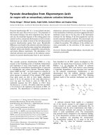
= 0.01) and IL-8 (P = 0.01) (Figure 1). In contrast, male con-
trols had lower circulating levels of IL-13 (P = 0.03) and IL-23
(P = 0.006). Male controls tended to have increased circulat-
ing levels of IL-1β, IL-4 and IL-10 (P < 0.10) but no significant
differences were observed in circulating levels of TNFα, IFNγ,
IL-6, IL-12p70, and IL-17.
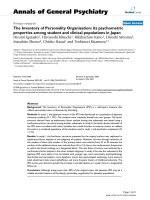
To determine whether increasing age alters the circulating
plasma cytokine levels, healthy controls were grouped accord-
ing to age in 20-year intervals. As seen in Figure 2, IL-6 (P =
0.02) and IL-17 (P = 0.01) levels increased with advancing
age. There was also a trend for increasing levels of IL-8 with
age (P = 0.07). No changes were noted in IL-1β, IL-2, IL-4, IL-
5, IL-10, IL-12p70, IL-13, IL-23, IFNγ, and TNFα.
Together these data demonstrate that circulating cytokine lev-
els are different in healthy males and females and are depend-
ent on age.
Changes in plasma cytokines in scleroderma patients
Circulating cytokine levels were determined in plasma from
SSc patients and were compared with those for control
patients (Table 2). Compared with healthy control subjects,
SSc patients had higher circulating levels of TNFα (P <
0.0001), IL-6 (P < 0.0001) and IL-13 (P = 0.05) but lower cir-
culating levels of IL-17 (P = 0.0009) and IL-23 (P = 0.04). No
differences were observed in circulating levels of IL-1β, IL-2,
IL-4, IL-5, IL8, IL-10, and IFNγ.
Table 1
Demographics and clinical data of the cohort
Controls (n = 216) Scleroderma patients (n = 444)
Age (years) 51 ± 14 years 53 ± 12.1
Gender
Male 117 (54%) 56 (13%)
Female 99 (46%) 388 (87%)
Scleroderma phenotype
Limited 241 (54%)
Diffuse 200 (45%)
Disease duration (years) 6.9 ± 0.3
Systemic sclerosis-association autoantibodies
Anti-centromere 109 (25%)
Anti-topoisomerase 112 (25%)
Anti-RNA polymerase III 109 (25%)
Data presented as mean ± standard deviation or n (%).
Arthritis Research & Therapy Vol 11 No 5 Gourh et al.
Page 4 of 11
(page number not for citation purposes)
Given the observation that age and gender can influence
plasma cytokine levels in healthy controls, comparisons
between scleroderma patients and healthy controls were per-
formed adjusting for age and gender using logistic regression
analysis (Table 2). After adjusting for age and gender, patients
with SSc had higher circulating levels of TNFα and IL-6 and
lower circulating levels of IL-17 and IL-23. In addition, SSc
patients had higher circulating levels of IFNγ; however, the pre-
vious unadjusted change in plasma IL-13 was no longer
significant.
Together these data demonstrate that SSc patients have an
increase in circulating levels of TNFα, IL-6, and IFNγ and a
decrease in IL-17 and IL-23.
Effect of disease duration on cytokine profiles in
scleroderma patients
Immune dysregulation is commonly thought to be important in
the early pathogenesis of SSc. Whether the cytokine altera-
tions that might be observed early in the disease process per-
sist as the disease progresses, however, is unclear. To
determine whether disease duration influenced the patterns of
plasma cytokine profiles, patients were grouped according to
disease duration: 0 to 5 years (n = 196 patients), 5 to 10 years
(n = 107 patients), and >10 years (n = 94 patients). The dis-
ease duration was not known for 47 SSc patients, and these
patients were excluded from this analysis.
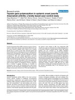
TNFα and IL-6 were significantly increased in SSc patients
with a disease duration of 0 to 5 years (P < 0.0001 and P <
0.0001, respectively) and a duration of 5 to 10 years (P <
0.0001 and P = 0.007, respectively) compared with controls
(Figure 3). TNFα and IL-6 levels were similar between controls
Figure 1
Gender effects on plasma cytokine levelsGender effects on plasma cytokine levels. Male healthy controls have higher circulating levels of IL-2 (P = 0.008), IL-5 (P = 0.01) and IL-8 (P =
0.01), and lower circulating levels of IL-13 (P = 0.03) and IL-23 (P = 0.006). Plasma cytokine levels (in pg/ml) were log
n
-transformed. Data pre-
sented as mean ± standard error of the mean.
Available online />Page 5 of 11
(page number not for citation purposes)
and SSc patients with disease duration >10 years. Alterations
in circulating levels of other cytokines became apparent after
controlling for disease duration as well as age and gender. For
example, circulating levels of IL-5, IL-10 and IFNγ were only
elevated in patients with disease duration >10 years com-
pared with healthy controls (P = 0.04, P = 0.008, and P =
0.03, respectively). In contrast, IL-13 was slightly increased in
patients with disease duration <5 years (P = 0.03). Lastly, IL-
17 levels remained decreased in patients independent of dis-
ease duration, but IL-23 levels were only decreased in SSc
patients with disease duration of 0 to 5 years or 5 to 10 years
compared with controls.
These data suggest that the circulating cytokine profiles are
different in patients based on disease duration, and suggest
that alterations in immune balance may change during different
stages of SSc.
Cytokine profiles of scleroderma patients based on the
presence of scleroderma-associated autoantibodies
The presence of scleroderma-associated autoantibodies is
associated with distinct clinical phenotypes of SSc [15]; how-
ever, it is not known whether these different subsets have dif-
ferent alterations in immune function. Comparisons of plasma
cytokines were performed in each group based on the pres-
ence of scleroderma-associated autoantibodies (ACAs, ATAs,
ARAs, Ab-Neg), controlling for age and gender using logistic
regression analysis.
As seen in Figures 4 and 5, all four groups of SSc patients had
a significant increase in TNFα and a decrease in IL-23. The
ATA-positive, ARA-positive and Ab-Neg subsets had a statis-
tically significant increase in IL-6, which was not observed in
the ACA-positive group. Furthermore, IL-17 was significantly
decreased in all groups compared with controls, except in the
Ab-Neg group. IL-8 was uniquely increased in the ATA-posi-
tive subset. Interestingly, both ATA-positive and ACA-positive
subsets had an increase in IFNγ and IL-10, but IL-5 was only
significantly increased in the ACA-positive subset. Lastly, the
Figure 2
Age effects on plasma cytokine levelsAge effects on plasma cytokine levels. IL-6 (P = 0.02) and IL-17 (P = 0.01) levels increased with advancing age. There was also a trend for increas-
ing levels of IL-8 with age (P = 0.07). Plasma cytokine levels (in pg/ml) were log
n
-transformed. Data presented as mean ± standard error of the
mean.
Arthritis Research & Therapy Vol 11 No 5 Gourh et al.
Page 6 of 11
(page number not for citation purposes)
ARA-positive and Ab-Neg group did not have alterations in cir-
culating IFNγ, IL-8 or IL-10.
Together these data suggest that the presence of SSc-asso-
ciated autoantibodies may identify patients with different pat-
terns of circulating cytokines and that different pathways of
immune dysregulation may underlie the development of SSc.
Association of cytokine profiles with clinical
manifestations of scleroderma
To determine whether circulating plasma cytokine profiles
were associated with clinical involvement in SSc, plasma
cytokines in patients with and without interstitial lung disease,
pulmonary hypertension, SSc renal crisis, Sjogren's syn-
drome/sicca symptoms, myositis or primary biliary cirrhosis
were compared with controls, using logistic regression to con-
trol for age and gender (Table 3). Patients were excluded from
this analysis if the specific status was not known.
Patients with higher circulating IL-6 were more likely to have
interstitial lung disease (odds ratio = 1.33, 95% confidence
interval = 1.08 to 1.62). Patients with pulmonary hypertension
were more likely to have higher IL-6 (odds ratio = 1.31, 95%
confidence interval = 1.04 to 1.67) and IL-13 (odds ratio =
1.42, 95% confidence interval = 1.04 to 1.94). Patients with
SSc renal crisis were more likely to have elevated TNFα levels
(odds ratio = 2.22, 95% confidence interval = 1.11 to 4.45).
Interestingly, patients with Sjogren's syndrome/sicca symp-
toms were more likely to have lower IL-8 (odds ratio = 0.89,
95% confidence interval = 0.81 to 0.99) and IL-1β (odds ratio
= 0.75, 95% confidence interval = 0.63 to 0.88).
Finally, we performed a logistic regression analysis using the
individual components of the Medsger Damage Index (gen-
eral, peripheral vascular, skin score, joint/tendon, muscle, gas-
trointestinal tract, and heart) as a continuous variable[21].
Interestingly, IL-6 was associated with total skin scores
(regression coefficient = 0.77; 95% confidence interval =
0.05 to 1.49) and IL-17 levels were associated with joint/ten-
don scores (regression coefficient = 0.27; 95% confidence
interval = 0.10 to 0.43).
Together these data demonstrate that there are distinct differ-
ences in the plasma cytokine profiles in patients representing
different clinical manifestations of SSc.
Discussion
In the current report, the circulating plasma cytokine profile
was determined in patients with SSc compared with controls
using a large cross-sectional cohort of SSc patients and
healthy controls. We observed that SSc patients have higher
levels of TNFα, IL-6 and IFNγ, but lower levels of IL-17 and IL-
23. We also observe that the disease duration and the pres-
ence of SSc autoantibodies have an influence on these
cytokine profiles. Lastly, it was discerned that specific clinical
manifestations of SSc, such as interstitial lung disease, pulmo-
Table 2
Plasma cytokine levels of healthy controls and scleroderma patients
Healthy controls (n = 216) Scleroderma patients (n = 444) P value
pg/ml log
n
cytokine pg/ml log
n
cytokine
Unpaired t test
Adjusted for age and gender
TNFα 8.9 ± 0.5 1.90 ± 0.06 14.1 ± 0.7 2.27 ± 0.05 <0.0001 <0.0001
IL1β 7.0 ± 1.2 0.36 ± 0.11 5.8 ± 0.8 0.38 ± 0.07 NS NS
IL-6 5.2 ± 0.8 0.55 ± 0.11 9.7 ± 1.2 1.24 ± 0.07 <0.0001 <0.0001
IL-8 77.7 ± 20.1 1.32 ± 0.15 84.3 ± 13.8 1.58 ± 0.11 NS NS
IL-2 2.8 ± 0.7 0.05 ± 0.08 2.4 ± 0.4 0.02 ± 0.05 NS NS
IFNγ 2.9 ± 0.5 - 0.17 ± 0.08 8.2 ± 4.6 - 0.06 ± 0.06 NS 0.05
IL-12p70 16.2 ± 4.3 0.66 ± 0.12 53.2 ± 12.8 0.71 ± 0.09 NS NS
IL-4 5.1 ± 0.8 0.38 ± 0.11 5.4 ± 1.2 0.32 ± 0.07 NS NS
IL-5 1.2 ± 0.2 - 0.27 ± 0.05 11.1 ± 5.1 - 0.14 ± 0.06 NS NS
IL-13 24.6 ± 1.7 2.97 ± 0.06 51.5 ± 12.0 3.12 ± 0.05 0.05 NS
IL-10 18.5 ± 11.6 0.62 ± 0.10 30.6 ± 10.0 0.83 ± 0.08 NS 0.07
IL-17 1.9 ± 0.2 0.04 ± 0.08 1.2 ± 0.1 - 0.22 ± 0.04 0.0009 0.0005
IL-23 4.7 ± 1.1 0.46 ± 0.12 3.7 ± 0.5 0.18 ± 0.07 0.04 0.014
Plasma cytokine levels are presented as both absolute levels (pg/ml) as well as natural log-transformed (log
n
cytokine) to normalize the data for
analysis. Data presented as the mean ± standard error of the mean. P value determined either by unpaired t test or by logistic regression
controlling for age and gender; NS, not significant with P > 0.05.
Available online />Page 7 of 11
(page number not for citation purposes)
nary hypertension or SSc renal crisis, are also associated with
alterations in distinct plasma cytokine levels.
Collectively, these data bring to light the complex immun-
opathogenesis of SSc and echo the clinical heterogeneity that
is seen within SSc. The SSc-associated autoantibodies are
clinically useful to risk-stratify patients for the systemic involve-
ment of SSc. Accordingly, we observe that there are also dif-
ferences in circulating cytokine levels when using these
autoantibodies to classify SSc patients. For example, ATA-
positive patients are the only subset with an increase in IL-8.
ATA-positive and ACA-positive patients both have an increase
in IFNγ and IL-10, while only ACA-positive patients present an
increase in IL-5. Based on the current data, the ARA-positive
subset appears to be a distinct subset. Similar to ATAs and
ACAs, ARAs show an increase in TNFα and IL-6 as well as a
decrease in IL-17 and IL-23. The ARA-positive subset, how-
ever, does not have alterations in the Th1/Th2 cytokines that
are observed in the ATA-positive subset or the ACA-positive
subset.
Alterations in plasma cytokine levels, including IL-6, TNFα, IL-
10 and IL-4, have been reported by several groups in the past,
with varying results [6-10]. The differences are probably due
to the heterogeneity within SSc, differences in disease dura-
tion, differences in gender, disease activity, and small sample
size of these studies. The current report utilizes the largest
cohort of SSc patients and healthy controls to date. This
Figure 3
Effect of disease duration on plasma cytokine profiles in scleroderma patients compared with controlsEffect of disease duration on plasma cytokine profiles in scleroderma patients compared with controls. Plasma cytokine levels (in pg/ml) were log
n
-
transformed. Data presented as mean ± standard error of the mean. *P < 0.05. **P < 0.01. ****P < 0.0001.
Arthritis Research & Therapy Vol 11 No 5 Gourh et al.
Page 8 of 11
(page number not for citation purposes)
enables controlling for variables such as gender and age,
which we have shown can influence circulating cytokine levels
even in healthy controls as well as in SSc patients. Similar to
previous reports, we demonstrate an increase in circulating
levels of IL-6 [6,7]. Interestingly, these changes appear to be
less significant in patients with longer disease duration,
although we cannot rule out a contribution of disease activity.
IL-6 is a pleiomorphic cytokine produced by T cells, B cells,
monocytes, endothelial cells, and fibroblasts, and is involved in
regulating many cellular processes including multilineage blast
cell colony formation, T-cell differentiation and fibroblast
behavior [22]. IL-6 is therefore probably a key cytokine in the
immunopathogenesis of SSc.
Scleroderma has often been considered a Th2 cytokine dis-
ease [3,23]. Th1 cytokines such as IFNγ have antifibrotic
effects, while Th2 cytokines such as IL-4 and IL-13 have profi-
brotic effects [24]. Indeed, Th2 cells have been cloned from
SSc skin with greater frequency than Th1 cells, although not
exclusively [3]. Other reports have not, however, consistently
demonstrated a Th2 cytokine profile in SSc patients
[11,25,26]. In the current report, the ATA-positive and ACA-
positive subsets have an increase in the Th1 cytokine IFNγ.
Both subsets present an increase in the Th2 cytokine IL-5, but
it is only statistically significant in the ACA-positive subset.
These data do not point to a selective increase in Th2
cytokines. Additional studies using more sensitive measures of
the Th1/Th2 cytokine balance are needed to better address
this aspect.
T-helper type 17 cells have recently been implicated as key T
cells in the pathogenesis of autoimmune diseases, such as
multiple sclerosis, rheumatoid arthritis and ankylosing spond-
ylitis [27-29]. IL-17 and IL-23 have been reported to be
increased in the plasma of patients in two small Japanese
cohorts of SSc patients [30,31]. In the current large cohort,
when controlling for age, gender, and autoantibody status, we
observed a decrease in circulating IL-17 and IL-23, especially
in patients with shorter disease duration. We cannot defini-
tively explain these differences but possible explanations are
the sample sizes of the cohorts and the differences in genetic
background of Japanese versus the current cohort of North
American Caucasians, which may influence the cytokine pro-
file. Given the potential importance of the T-helper type 17
pathway in autoimmune diseases, future efforts should focus
Figure 4
Four plasma cytokine profiles in systemic sclerosis-association autoantibody subsets of scleroderma patients compared with controlsFour plasma cytokine profiles in systemic sclerosis-association autoantibody subsets of scleroderma patients compared with controls. Plasma
cytokine levels (in pg/ml) were log
n
-transformed. Data presented as mean ± standard error of the mean. *P < 0.05. **P < 0.01. ***P < 0.001. ****P
< 0.0001.
Available online />Page 9 of 11
(page number not for citation purposes)
on delineating the role of T-helper type 17 cells in SSc
immunopathogenesis.
A final observation of potential interest is the association of cir-
culating cytokines with systemic manifestations of SSc. Simi-
lar to prior reports, we observed an association of IL-6 with
interstitial lung disease [6]. We also noted that IL-6 and IL-13
levels were increased in patients with pulmonary hypertension,
and that TNFα levels were associated with the presence of
renal crisis. While alterations in cytokine levels were associ-
ated with clinical manifestations of SSc, prospective studies
are needed to determine whether measuring these cytokine
levels in patients would be helpful in predicting the develop-
ment of these detrimental clinical manifestations.
The current report has several limitations that should be
acknowledged. While the plasma samples were obtained from
a large cohort of SSc patients and healthy controls, accurate
data for immunosuppressive medications were not available at
the time of the present publication. Another limitation is the
Figure 5
Nine plasma cytokine profiles in systemic sclerosis-association autoantibody subsets of scleroderma patients compared with controlsNine plasma cytokine profiles in systemic sclerosis-association autoantibody subsets of scleroderma patients compared with controls. Plasma
cytokine levels (in pg/ml) were log
n
-transformed. Data presented as mean ± standard error of the mean. *P < 0.05. **P < 0.01.
Arthritis Research & Therapy Vol 11 No 5 Gourh et al.
Page 10 of 11
(page number not for citation purposes)
cross-sectional design of the current study. The data reported
herein identify disease duration as a factor that influences
cytokine profiles. This might be particularly relevant with
regards to the ACA-positive group, which has longer disease
duration than the other autoantibody groups. A prospective
study with sequential plasma samples would be beneficial to
better understand the immune changes that are associated
with the development of SSc as well as the progression of the
disease and disease activity. It should also be noted that the
SSc-associated autoantibody group (Ab-Neg) remains a het-
erogeneous group of patients - as defined by the presence of
other autoantibodies such as anti-fibrillarin, anti-PM-Scl or
anti-Th/To [32] - and whether these less common subsets also
have differences in the plasma cytokine profiles remains to be
determined.
Conclusions
SSc is associated with alterations in circulating plasma
cytokines. These alterations are influenced by gender, age,
disease duration and the presence of specific SSc-associated
autoantibodies. These results highlight the complex immun-
opathogenesis of SSc and point to several potential targets
that could be considered for monitoring disease progression
and treatment of SSc.
Competing interests
The authors declare that they have no competing interests.
Authors' contributions
PG, FCA, FKT, MDM, JDR and SKA designed the project. PG,
SA, MH, LD, MDM, JDR and SKA acquired the data. PG, FCA,
SA, FKT, MH, LD and SKA analyzed the data. All authors read
and approved the final manuscript.
Acknowledgements
The present study was supported by the Scleroderma Foundation New
Investigator Award (to SKA), NIH/NIAMS-K08AR054404 (to SKA), the
NIH/NIAMS Center of Research Translation in Scleroderma
(P50AR054144) (to FCA), the NIH/NIAMS Scleroderma Family Regis-
try and DNA Repository (N01-AR-0-2251) (to MDM), and the University
of Texas Health Science Center at Houston Center for Clinical and
Translational Sciences (Houston CTSA Program) (NIH/NCRR
3UL1RR024148) (to FCA and SKA).
References
1. Fleischmajer R, Perlish JS, Reeves JR: Cellular infiltrates in scle-
roderma skin. Arthritis Rheum 1977, 20:975-984.
2. Tan FK, Zhou X, Mayes MD, Gourh P, Guo X, Marcum C, Jin L,
Arnett FC Jr: Signatures of differentially regulated interferon
gene expression and vasculotrophism in the peripheral blood
cells of systemic sclerosis patients. Rheumatology (Oxford)
2006, 45:694-702.
3. Mavalia C, Scaletti C, Romagnani P, Carossino AM, Pignone A,
Emmi L, Pupilli C, Pizzolo G, Maggi E, Romagnani S: Type 2
helper T-cell predominance and high CD30 expression in sys-
temic sclerosis. Am J Pathol 1997, 151:1751-1758.
4. Needleman BW, Wigley FM, Stair RW: Interleukin-1, interleukin-
2, interleukin-4, interleukin-6, tumor necrosis factor alpha, and
interferon-gamma levels in sera from patients with
scleroderma. Arthritis Rheum 1992, 35:67-72.
5. Molteni M, Della BS, Mascagni B, Bazzi S, Zulian C, Compasso S,
Lessi M, Scorza R: Increased interferon-gamma (IFN-gamma)
levels produced in vitro by alloactivated T lymphocytes in sys-
temic sclerosis and Raynaud's phenomenon. Clin Exp
Immunol 1999, 116:164-168.
6. Hasegawa M, Sato S, Fujimoto M, Ihn H, Kikuchi K, Takehara K:
Serum levels of interleukin 6 (IL-6), oncostatin M, soluble IL-6
receptor, and soluble gp130 in patients with systemic
sclerosis. J Rheumatol 1998, 25:308-313.
7. Hasegawa M, Sato S, Ihn H, Takehara K: Enhanced production
of interleukin-6 (IL-6), oncostatin M and soluble IL-6 receptor
by cultured peripheral blood mononuclear cells from patients
with systemic sclerosis. Rheumatology (Oxford) 1999,
38:612-617.
8. Sato S, Hasegawa M, Takehara K: Serum levels of interleukin-6
and interleukin-10 correlate with total skin thickness score in
patients with systemic sclerosis. J Dermatol Sci 2001,
27:140-146.
9. Hasegawa M, Fujimoto M, Kikuchi K, Takehara K: Elevated serum
tumor necrosis factor-alpha levels in patients with systemic
sclerosis: association with pulmonary fibrosis. J Rheumatol
1997, 24:663-665.
10. Hasegawa M, Fujimoto M, Kikuchi K, Takehara K: Elevated serum
levels of interleukin 4 (IL-4), IL-10, and IL-13 in patients with
systemic sclerosis. J Rheumatol 1997,
24:328-332.
11. Sato S, Hanakawa H, Hasegawa M, Nagaoka T, Hamaguchi Y,
Nishijima C, Komatsu K, Hirata A, Takehara K: Levels of inter-
leukin 12, a cytokine of type 1 helper T cells, are elevated in
Table 3
Plasma cytokine associations with clinical involvement of scleroderma
Present Absent Unknown Cytokine Odds ratio 95% confidence interval P value
Interstitial lung disease 120 196 128 IL-6 1.33 1.08 to 1.62 0.006
Pulmonary hypertension 58 255 131 IL-6 1.31 1.04 to 1.67 0.02
IL-13 1.42 1.04 to 1.94 0.03
SSc renal crisis 20 294 130 TNFα 2.22 1.11 to 4.45 0.02
Sjogren's syndrome 118 199 127 IL-8 0.89 0.81 to 0.99 0.03
IL-1β 0.75 0.63 to 0.88 0.0006
Myositis 17 297 130 None
Primary biliary cirrhosis 5 309 140 IL-17 3.46 1.35 to 8.9 0.01
Cytokine levels were normalized using log
n
transformation. Logistic regression was used to compare cytokine levels to control subjects for gender
and age and odd ratios reflect each cytokine as a continuous variable.
Available online />Page 11 of 11
(page number not for citation purposes)
sera from patients with systemic sclerosis. J Rheumatol 2000,
27:2838-2842.
12. Arnett FC, Reveille JD, Valdez BC: Autoantibodies to a nucleolar
RNA helicase protein in patients with connective tissue
diseases. Arthritis Rheum 1997, 40:1487-1492.
13. Arnett FC, Reveille JD, Goldstein R, Pollard KM, Leaird K, Smith
EA, LeRoy EC, Fritzler MJ: Autoantibodies to fibrillarin in sys-
temic sclerosis (scleroderma). An immunogenetic, serologic,
and clinical analysis. Arthritis Rheum 1996, 39:1151-1160.
14. Reveille JD, Fischbach M, McNearney T, Friedman AW, Aguilar
MB, Lisse J, Fritzler MJ, Ahn C, Arnett FC: Systemic sclerosis in
3 US ethnic groups: a comparison of clinical, sociodemo-
graphic, serologic, and immunogenetic determinants. Semin
Arthritis Rheum 2001, 30:332-346.
15. Meyer OC, Fertig N, Lucas M, Somogyi N, Medsger TA Jr: Dis-
ease subsets, antinuclear antibody profile, and clinical fea-
tures in 127 French and 247 US adult patients with systemic
sclerosis. J Rheumatol 2007, 34:104-109.
16. Wu SP, Leng L, Feng Z, Liu N, Zhao H, McDonald C, Lee A, Arnett
FC, Gregersen PK, Mayes MD, Bucala R: Macrophage migration
inhibitory factor promoter polymorphisms and the clinical
expression of scleroderma. Arthritis Rheum 2006,
54:3661-3669.
17. Preliminary criteria for the classification of systemic sclerosis
(scleroderma). Subcommittee for scleroderma criteria of the
American Rheumatism Association Diagnostic and Therapeu-
tic Criteria Committee. Arthritis Rheum 1980, 23:581-590.
18. LeRoy EC, Black C, Fleischmajer R, Jablonska S, Krieg T, Medsger
TA Jr, Rowell N, Wollheim F: Scleroderma (systemic sclerosis):
classification, subsets and pathogenesis. J Rheumatol 1988,
15:202-205.
19. Chowdhury F, Williams A, Johnson P: Validation and comparison
of two multiplex technologies, Luminex and Mesoscale Dis-
covery, for human cytokine profiling. J Immunol Methods 2009,
340:55-64.
20. Olivier J, Johnson WD, Marshall GD: The logarithmic transfor-
mation and the geometric mean in reporting experimental IgE
results: what are they and when and why to use them? Ann
Allergy Asthma Immunol 2008, 100:
333-337.
21. Bombardieri S, Medsger TA Jr, Silman AJ, Valentini G: The
assessment of the patient with systemic sclerosis.
Introduction. Clin Exp Rheumatol 2003, 21:S2-S4.
22. Naka T, Nishimoto N, Kishimoto T: The paradigm of IL-6: from
basic science to medicine. Arthritis Res 2002, 4(Suppl
3):S233-S242.
23. Giacomelli R, Cipriani P, Lattanzio R, Di FM, Locanto M, Parzanese
I, Passacantando A, Ciocci A, Tonietti G: Circulating levels of
soluble CD30 are increased in patients with systemic sclerosis
(SSc) and correlate with serological and clinical features of
the disease. Clin Exp Immunol 1997, 108:42-46.
24. McGaha TL, Bona CA: Role of profibrogenic cytokines secreted
by T cells in fibrotic processes in scleroderma. Autoimmun Rev
2002, 1:174-181.
25. Valentini G, Baroni A, Esposito K, Naclerio C, Buommino E, Farzati
A, Cuomo G, Farzati B: Peripheral blood T lymphocytes from
systemic sclerosis patients show both Th1 and Th2 activation.
J Clin Immunol 2001, 21:210-217.
26. Giacomelli R, Cipriani P, Fulminis A, Barattelli G, Matucci-Cerinic
M, D'Alo S, Cifone G, Tonietti G: Circulating gamma/delta T
lymphocytes from systemic sclerosis (SSc) patients display a
T helper (Th) 1 polarization. Clin Exp Immunol 2001,
125:310-315.
27. Cua DJ, Sherlock J, Chen Y, Murphy CA, Joyce B, Seymour B,
Lucian L, To W, Kwan S, Churakova T, Zurawski S, Wiekowski M,
Lira SA, Gorman D, Kastelein RA, Sedgwick JD: Interleukin-23
rather than interleukin-12 is the critical cytokine for autoim-
mune inflammation of the brain. Nature 2003, 421:744-748.
28. Bettelli E, Carrier Y, Gao W, Korn T, Strom TB, Oukka M, Weiner
HL, Kuchroo VK: Reciprocal developmental pathways for the
generation of pathogenic effector TH17 and regulatory T cells.
Nature 2006, 441:235-238.
29. Bettelli E, Korn T, Kuchroo VK: Th17: the third member of the
effector T cell trilogy. Curr Opin Immunol 2007, 19:652-657.
30. Murata M, Fujimoto M, Matsushita T, Hamaguchi Y, Hasegawa M,
Takehara K, Komura K, Sato S: Clinical association of serum
interleukin-17 levels in systemic sclerosis: is systemic sclero-
sis a Th17 disease? J Dermatol Sci 2008,
50:240-242.
31. Komura K, Fujimoto M, Hasegawa M, Ogawa F, Hara T, Muroi E,
Takehara K, Sato S: Increased serum interleukin 23 in patients
with systemic sclerosis. J Rheumatol 2008, 35:120-125.
32. Reveille JD, Fischbach M, McNearney T, Friedman AW, Aguilar
MB, Lisse J, Fritzler MJ, Ahn C, Arnett FC: Systemic sclerosis in
3 US ethnic groups: a comparison of clinical, sociodemo-
graphic, serologic, and immunogenetic determinants. Semin
Arthritis Rheum 2001, 30:332-346.