Báo cáo y học: "Chemokine receptor expression and functional effects of chemokines on B cells: implication in the pathogenesis of rheumatoid arthritis" ppt
Bạn đang xem bản rút gọn của tài liệu. Xem và tải ngay bản đầy đủ của tài liệu tại đây (1.15 MB, 11 trang )
Available online />
Research article
Vol 11 No 5
Open Access
Chemokine receptor expression and functional effects of
chemokines on B cells: implication in the pathogenesis of
rheumatoid arthritis
Toshihiro Nanki1,2, Kazuki Takada1, Yukiko Komano1,2, Tomohiro Morio3, Hirokazu Kanegane4,
Atsuo Nakajima5,6, Peter E Lipsky7 and Nobuyuki Miyasaka1,8
1Departments of Medicine and Rheumatology, Graduate School, Tokyo Medical and Dental University, 1-5-45, Yushima, Bunkyo-ku, Tokyo, 1138519, Japan
2Department of Pharmacovigilance, Graduate School, Tokyo Medical and Dental University, 1-5-45, Yushima, Bunkyo-ku, Tokyo, 113-8519, Japan
3Department of Pediatrics and Developmental Biology, Graduate School, Tokyo Medical and Dental University, 1-5-45, Yushima, Bunkyo-ku, Tokyo,
113-8519, Japan
4Department of Pediatrics, Graduate School of Medicine, University of Toyama, 2630, Sugitani, Toyama, 930-0194, Japan
5Department of Joint Disease and Rheumatism, Nippon Medical School, 1-1-5, Sendagi, Bunkyo-ku, Tokyo, 113-8603, Japan
6Department of Rheumatology, Tokyo Metropolitan Police Hospital, 4-22-1, Nakano, Nakano-ku, Tokyo, 164-8541, Japan
7National Institute of Arthritis and Musculoskeletal and Skin Diseases, National Institutes of Health, 9000 Rockville Pike, Bethesda, MD 20892, USA
8Global Center of Excellence (GCOE) Program; International Research Center for Molecular Science in Tooth and Bone Diseases, Tokyo Medical
and Dental University, 1-5-45, Yushima, Bunkyo-ku, Tokyo, 113-8519, Japan
Corresponding author: Toshihiro Nanki,
Received: 14 May 2009 Revisions requested: 30 Jun 2009 Revisions received: 10 Sep 2009 Accepted: 5 Oct 2009 Published: 5 Oct 2009
Arthritis Research & Therapy 2009, 11:R149 (doi:10.1186/ar2823)
This article is online at: />© 2009 Nanki et al.; licensee BioMed Central Ltd.
This is an open access article distributed under the terms of the Creative Commons Attribution License ( />which permits unrestricted use, distribution, and reproduction in any medium, provided the original work is properly cited.
Abstract
Methods Cell surface molecule expression was analyzed by
flow cytometry. Cellular migration was assessed using
chemotaxis chambers. Cellular proliferation was examined by
3H-thymidine incorporation. Tumor necrosis factor (TNF)
production was assayed by enzyme-linked immunosorbent
assay.
healthy donors and RA. Synovial B cells more frequently
expressed CCR5, but less often expressed CCR6, CCR7 and
CXCR5 compared to peripheral blood in RA. Further functional
analyses were performed on peripheral blood B cells from
healthy donors. Migration of peripheral blood B cells, especially
CD27+ B cells, was enhanced by CC chemokine ligand
(CCL)20, CCL19, CCL21 and CXCL12. All four chemokines
alone induced B cell proliferation; with CCL21 being the most
effective. CCL21 also enhanced the proliferation of antiimmunoglobulin (Ig)M-stimulated B cells and blockade of CCR7
inhibited this effect. CCL20, CCL21 and CXCL12 enhanced
TNF production by anti-IgM mAb-stimulated B cells. Finally,
stimulation with CXCL12, but not CCL20, CCL19 and CCL21,
enhanced inducible costimulator-ligand (ICOSL) expression by
peripheral blood B cells of healthy donors and RA, but did not
increase B cell-activating factor receptor or transmembrane
activator and CAML-interactor.
Results Significant numbers of peripheral blood B cells of
healthy donors and subjects with RA expressed CC chemokine
receptor (CCR)5 and CXCR3, and most B cells expressed
CCR6, CCR7, CXCR4 and CXCR5. CCR5 expression was
more frequent on CD27+ than CD27- peripheral blood B cells of
Conclusions The data suggest that CCR5, CCR6, CCR7,
CXCR3, CXCR4 and CXCR5 may be important for the B cell
migration into the synovium of RA patients, and also their local
proliferation, cytokine production and ICOSL expression in the
synovium.
Introduction Accumulation of B cells in the rheumatoid arthritis
(RA) synovium has been reported, and it has been thought that
these cells might contribute to the pathogenesis of RA by
antigen presentation, autoantibody production, and/or
inflammatory cytokine production. Chemokines could enhance
the accumulation of B cells in the synovium. The aims of this
study were to determine chemokine receptor expression by B
cells both in the peripheral blood of normal donors and subjects
with RA, and at the inflammatory site in RA, and the effects of
chemokines on B cell activation.
BAFF-R: B cell-activating factor receptor; BSA: bovine serum albumin; CCL: CC chemokine ligand; CCR: CC chemokine receptor; DMEM: Dulbecco's Modified Eagle Medium; ELISA: enzyme-linked immunosorbent assay; FCS: fetal calf serum; FITC: fluorescein isothiocyanate; ICOS: inducible costimulator; ICOSL: inducible costimulator-ligand; Ig: immunoglobulin; mAb: monoclonal antibody; PBMCs: peripheral blood mononuclear cells;
PBS: phosphate-buffered saline; PE: phycoerythrin; RA: rheumatoid arthritis; RPMI: Roswell Park Memorial Institute; SEM: standard error of the mean;
TACI: transmembrane activator and CAML-interactor; TNF: tumor necrosis factor.
Page 1 of 11
(page number not for citation purposes)
Arthritis Research & Therapy
Vol 11 No 5
Nanki et al.
Introduction
Rheumatoid arthritis (RA) is characterized by chronic inflammation of multiple joints. As B cell depletion by treatment with
rituximab, an anti-CD20 monoclonal antibody (mAb), is beneficial for RA patients [1,2], B cells are considered to play
important roles in the pathogenesis of RA. In this regard, the
synovial tissue of RA patients shows abundant accumulation
of inflammatory cells, including T cells, macrophages, dendritic cells and B cells [3-6]. Synovial B cells could present
antigens to T cells. Importantly, rheumatoid factor-expressing
B cells that are found within the synovium [7] can present any
antigen in the context of an immune complex and, thereby, trigger T cells specific for a variety of foreign antigens [8]. Notably, the severity of RA correlates with levels of rheumatoid
factor [9]. Furthermore, activated B cells produce inflammatory cytokines, such as TNF [10]. Therefore, synovial B cells
could contribute to the pathogenesis of RA by antigen presentation, autoantibody production, and inflammatory cytokine
production. One of the mechanisms for accumulation of B
cells in synovial tissues relates to the interaction with chemokines produced in the RA synovium and chemokine receptors
expressed by the B cells [6].
Chemokines are classified into C, CC, CXC, and CX3C subclasses based on the conserved cysteine motifs [11], and are
involved in cellular migration, activation of adhesion molecules,
cellular proliferation, cytokine production and regulation of
apoptosis [12,13]. Chemokines contribute to homeostatic
migration as well as entry into acute and chronic inflammatory
sites. Expression of chemokines and chemokine receptors in
the RA synovial tissue has been extensively analyzed, and
chemokines are thought to be potential therapeutic targets
[14,15]. However, the role of chemokines specifically on B
cells in RA has not been completely delineated.
In this study, we examined chemokine receptor expression by
peripheral blood in both normal donors and subjects with RA,
and also synovial B cells from subjects with RA, and determined the functional effects of chemokines on B cells.
Materials and methods
Samples
Peripheral blood samples were obtained from healthy donors
and subjects with RA after obtaining informed consent. RA
was diagnosed according to the criteria of the American College of Rheumatology [16]. Synovial tissues were obtained at
the time of total knee joint replacement from RA patients.
Signed consent forms were obtained prior to the operation.
The study protocol was approved in advance by the Ethics
Committee of the Tokyo Medical and Dental University.
Chemokine receptor expression
Peripheral blood mononuclear cells (PBMCs) were isolated by
ficoll-hypaque (Immuno-Biological Laboratories, Gunma,
Japan) gradient centrifugation. The synovial tissue was minced
Page 2 of 11
(page number not for citation purposes)
and incubated with 0.3 mg/ml collagenase (Sigma, St. Louis,
MO, USA) for one hour at 37°C in Dulbecco's Modified Eagle
Medium (DMEM) (Sigma, St. Louis, MO, USA). Partially
digested pieces of the tissue were pressed through a metal
screen to obtain single cell suspensions. The following mAbs
were used for FACS analysis: phycoerythrin (PE) Cy5-conjugated anti-CD19 mAb (J4.119; Beckman Coulter, San Jose,
CA, USA), fluorescein isothiocyanate (FITC)-conjugated antiCD27 (M-T271; Ancell, Bayport, MN, USA) mAb, PE-conjugated anti-CC chemokine receptor (CCR)5 (2D7; BD Bioscience, San Jose, CA, USA), -CCR6 (53103; R&D Systems,
Minneapolis, MN, USA), -CCR7 (150503; R&D Systems, Minneapolis, MN, USA), -CXCR3 (49801; R&D Systems, Minneapolis, MN, USA), -CXCR4 (12G5; R&D Systems,
Minneapolis, MN, USA) and -CXCR5 (51505.111; R&D Systems, Minneapolis, MN, USA) mAbs, and isotype-matched
control mAbs. PBMCs or synovial tissue cells were incubated
with the mAbs for 20 minutes, and then rinsed with PBS-3%
fetal calf serum (FCS; Sigma, St. Louis, MO, USA). More than
5000 stained cells were analyzed with a FACSCalibur (BD
Bioscience, San Jose, CA, USA).
Migration assay
Cell migration was assessed in 24-well chemotaxis chambers
(6.5 mm diameter, 5 μm pore polycarbonate transwell culture
insert; Costar, Cambridge, MA, USA). ECV304 cells (2 × 10⁵)
were cultured in the chemotaxis chambers for 48 to 72 hours
in medium 199 (Sigma, St. Louis, MO, USA) with 10% FCS.
The migration medium (Roswell Park Memorial Institute
(RPMI)1640 medium (Sigma, St. Louis, MO, USA):medium
199 = 1:1, 0.5% BSA) supplemented where indicated with
various concentrations of chemokines (CC chemokine ligand
(CCL)20, CCL19, CCL21, and CXCL12: PeproTech, Rocky
Hill, NJ, USA) was added to the lower wells. ECV304 coated
chemotaxis chambers were placed in each well, and 5 × 10⁵
PBMCs suspended in migration medium were added to the
upper wells. After two hours of incubation, the membrane was
removed, and migrated cells were stained with PE Cy5-conjugated anti-CD19 mAb (J4.119) and FITC-conjugated antiCD27 mAb (M-T271). The cells were counted by FACSCalibur.
Proliferation assay
Peripheral blood CD19+ B cells were purified by magneticactivated cell sorting microbead-coupled mAb and magnetic
cell separation columns (Miltenyi Biotec, Auburn, CA, USA).
Purity of CD19+ B cells was determined by flow cytometry,
and was more than 95%. To block CCR7, B cells were incubated with 5 μg/ml anti-CCR7 mAb (150503; R&D Systems,
Minneapolis, MN, USA) or control mAb for 30 minutes. Then,
the 5 × 10⁵ B cells were incubated in 96-well with the indicated chemokines with or without pre-coated anti-IgM mAb (2
μg UHB; SouthernBiotech, Birmingham, AL, USA) in
RPMI1640 with 10% FCS at 37°C for 48 hours. 3H-thymidine
(1 μCi; Amersham Biosciences, Little Chalfont, Buckingham-
Available online />
shire, UK) was added and the B cells were incubated for 24
hours. Afterward, the incorporated radioactivity was quantified. After the 72-hour incubation, viabilities of the cells, determined by trypan blue exclusion, were 87.3% and 80.3%
without and with anti-IgM stimulation, respectively.
TNF production
Purified 5 × 10⁵ peripheral blood B cells were stimulated with
the indicated chemokines with or without coating of wells with
anti-IgM mAb (2 μg UHB) in 96-well in RPMI1640 with 10%
FCS at 37°C for 24 hours. Afterward, the concentration of
TNF in the culture supernatant was assayed using an ultra sensitive ELISA kit (BioSource International, Camarillo, CA).
Cell surface molecule expression
PBMCs were cultured with the indicated chemokine in
RPMI1640+10% FCS for 24 hrs. Afterward, the cells were
stained with PE Cy5-conjugated anti-CD19 mAb (J4.119) and
FITC-conjugated anti-inducible costimulator-ligand (ICOSL)
mAb (MIH12; eBioscience, San Diego, CA, USA), PE-conjugated anti-B cell-activating factor receptor (BAFF-R; 8A7; eBioscience, San Diego, CA, USA), -transmembrane activator
and CAML-interactor (TACI) mAb (11H3; eBioscience, San
Diego, CA, USA), or isotype-matched control mAb. The
stained cells were analyzed with a FACSCaliber.
Statistical analysis
Paired t test was used to compare paired samples of CD27and CD27+ peripheral blood B cells, and peripheral blood and
synovial B cells from the same subjects for chemokine receptor expression and migration. Differences in migration, fold
increase of proliferation and TNF production were examined
for statistical significance using the unpaired t test. All data
were expressed as mean ± standard error of the mean (SEM).
A P value less than 0.05 denoted the presence of a statistically
significant difference.
Results
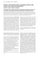
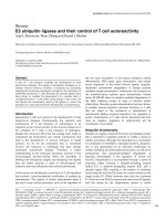
Chemokine receptor expression by B cells
Chemokine receptor expression by naive CD27- B cells and
memory CD27+ B cells from the peripheral blood of healthy
donors was analyzed by flow cytometry. As shown in Figure
1a, most peripheral blood B cells of healthy donors expressed
CCR6, CCR7, CXCR4 and CXCR5. About 60% of the B cells
expressed CXCR3, and less than 20% of the B cells
expressed CCR5. These results are similar to previous reports
[17-20]. We compared the chemokine receptor expression
between CD27- and CD27+ B cells. The frequencies of
CCR6, CCR7, CXCR3 and CXCR5 expression were not different between CD27- and CD27+ B cells of normal donors.
However, the proportion of CCR5-expressing peripheral
blood CD27+ B cells was significantly higher than that of
CD27- B cells in normal controls. The percentage of CD27+ B
cells expressing CXCR4 was less than CXCR4-expressing
CD27- B cells in normal controls.
Next, we analyzed the chemokine receptor expression by
CD27- B cells and CD27+ B cells from peripheral blood and
synovial tissue of subjects with RA. The frequency of CD27expressing peripheral blood B cells was not significantly different between subjects with RA and healthy donors (data not
shown). The proportion of the chemokine receptor expression
of RA peripheral blood B cells was similar to that of healthy
donors without any statistically significant differences. As with
healthy donors, CCR5 expression by RA peripheral blood
CD27+ B cells was more frequent than that of CD27- B cells,
and CXCR4 expression by CD27+ B cells was less frequent
than that of CD27- B cells. In addition, the proportions of
CCR6, CCR7 and CXCR5 expression were significantly less
by CD27+ compared with CD27- B cells in subjects with RA.
We also compared the chemokine receptor expression
between peripheral blood and synovial tissue B cells of RA
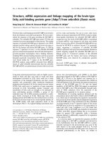
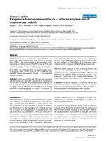
(Figure 1b). The frequency of CD27+ by synovial B cells was
significantly higher than that of peripheral blood B cells in RA
subjects (Figure 2) (peripheral blood, 30.0 ± 5.1% (mean ±
SEM); synovial B cells, 62.3 ± 4.7%; P < 0.005, n = 11), as
we have previously reported [21], suggesting that a specific
subset of B cells might be recruited to the inflammatory site in
RA. The proportion of synovial B cells that expressed CCR5
was significantly higher than that of either peripheral blood
CD27- or CD27+ B cells of subjects with RA. The proportion
of CXCR3-expressing CD27+ B cells in the synovium was
higher than peripheral blood. In addition, the frequency of synovial B cells that expressed CCR6 and CCR7 was less than
that expressed by peripheral blood CD27- B cells, but not
CD27+ B cells. The proportion of synovial B cells that
expressed CXCR5 was less than that in peripheral blood.
CXCR4 expression was no different between peripheral blood
and synovial B cells.
Migration
As frequencies of the analyzed chemokine receptor expression by peripheral blood B cells were not significantly altered
by RA, we next examined functional effects of chemokine ligands for the chemokine receptors using peripheral blood B
cells of healthy donors. Most peripheral B cells expressed
CCR6, CCR7 and CXCR4, and a significant number of RA
synovial B cells expressed also them. Therefore, we selected
four chemokines, CCL20, a ligand for CCR6, CCL19 and
CCL21, ligands for CCR7, and CXCL12, a ligand for CXCR4.
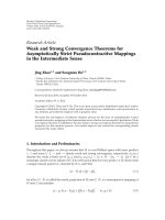
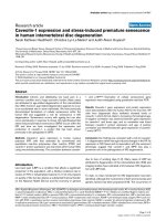
First, we analyzed the effects the chemokines on migration of
peripheral blood B cells. Each of the four chemokines induced
migration of both CD27- and CD27+ B cells (Figure 3a). However, the migration induced by CCL21 was most prominent.
Comparison of the migratory effects of the chemokines on
peripheral blood CD27- and CD27+ B cells in each individual
showed that for each of the chemokines, the chemotactic
response of CD27+ B cells was significantly greater than with
CD27- B cells (Figure 3b).
Page 3 of 11
(page number not for citation purposes)
Arthritis Research & Therapy
Vol 11 No 5
Nanki et al.
Figure 1
Chemokine receptor expression by B cells Peripheral blood mononuclear cells (PBMCs) from healthy donors (n = 4 to 7) and rheumatoid arthritis
cells.
(RA) patients (n = 17 to 18) and synovial cells from RA patients (n = 10 to 11) were stained with CD19, CD27, and CCR5, CCR6, CCR7, CXCR3,
CXCR4 or CXCR5, and the expression of the various markers was analyzed by flow cytometry. CD19+ B cells were gated, and the frequency of
expression of each chemokine receptor is shown. Data represent mean ± standard error of the mean. *P < 0.05, **P < 0.01, ***P < 0.001, ****P <
0.0001. ST = synovial tissue.
Page 4 of 11
(page number not for citation purposes)
Available online />
Figure 2
CD27 expression by peripheral blood and synovial tissue B cells of subjects with RA CD19+ B cells were gated, and representative histograms
RA.
from two patients with rheumatoid arthritis (RA) show the cells stained with anti-CD27 monoclonal antibody (mAb) (solid lines) and isotype-matched
control (dotted lines).
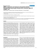
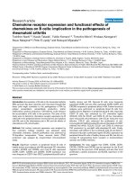
Proliferation
The effect of chemokines on B cell proliferation was next analyzed in normal donors. Although the effect was weak, CCL20,
CCL19, CCL21 and CXCL12 induced significant B cell proliferation (Figure 4a). Among the chemokines, 1000 ng/ml
CCL21 was the most effective stimulus of proliferation. Stimulation with anti-IgM mAb induced B cell proliferation (fold
increase: 5.5 ± 0.8). Stimulation with a low concentration of
CCL20 (10 ng/ml) decreased the proliferation of anti-IgMstimulated peripheral blood B cells. In contrast, a high concentration of CCL21 (1000 ng/ml) significantly enhanced the antiIgM-stimulated B cell proliferation (Figure 4b). Notably,
CCL21-induced proliferation was inhibited by anti-CCR7 mAb
by blocking the corresponding receptor (Figure 4c).
TNF production
We also analyzed the effect of chemokine stimulation on TNF
production by peripheral blood B cells of healthy donors.
Without anti-IgM stimulation, B cells secreted small amounts
of TNF (less than 1 pg/ml by this assay), and stimulation with
CCL20, CCL19, CCL21 and CXCL12 did not change the
TNF production (Figure 5). In contrast, anti-IgM mAb stimulation increased TNF production by B cells. Moreover, co-stimulation of anti-IgM activated B cells by CCL20, CCL21, and
CXCL12 enhanced TNF production, whereas CCL19
decreased TNF production.
Cell surface molecule expression
Finally, we examined the effects of the chemokines on the
expression of the cell surface molecules ICOSL, BAFF-R and
TACI by peripheral blood B cells of normal donors and sub-
Page 5 of 11
(page number not for citation purposes)
Arthritis Research & Therapy
Vol 11 No 5
Nanki et al.
Figure 3
B cell migration in response to chemokines Peripheral blood mononuclear cells (PMBCs) from healthy donors were cultured in the presence of varchemokines.
ious concentrations of CCL20, CCL19, CCL21, or CXCL12 for two hours. The cells migrated through ECV304-coated transwells were stained with
CD19 and CD27, and the numbers of cells were assessed. The percentage of migrated cells was calculated by dividing the number of migrated
CD27- or CD27+ B cells by the number of total cultured CD27- or CD27+ B cells for six to seven donors. (a) Values are mean ± standard error of the
mean. *P < 0.05, **P < 0.01, ***P < 0.005, vs no chemokine. (b) Each symbol represents an individual subject. *P < 0.05, **P < 0.01, ***P < 0.005,
****P < 0.0001.
Page 6 of 11
(page number not for citation purposes)
Available online />
Figure 4
B cell proliferation in response to chemokine stimulation. Purified B cells from peripheral blood mononuclear cells (PBMCs) of normal donors were
stimulation
stimulated with the indicated chemokines for 48 hours (a) without and (b) with anti-IgM stimulation. (c) To block CCR7, the B cells were pre-incubated with anti-CCR7 monoclonal antibody (mAb) or control mAb for 30 minutes. 3H-thymidine was added and B cells were incubated for 24 hours.
The incorporated radioactivity was quantified. Fold increase in 3H-thymidine incorporation in response to chemokine stimulation for four to eight
donors was calculated. Values are mean ± standard error of the mean. (a, b) *P < 0.05, **P < 0.005, ***P < 0.0005, vs no chemokine stimulation.
(c) *P < 0.05, **P < 0.005.
Page 7 of 11
(page number not for citation purposes)
Arthritis Research & Therapy
Vol 11 No 5
Nanki et al.
Figure 5
TNF production by chemokine stimulation. Purified peripheral blood B cells from normal donors were incubated with the indicated chemokines with
stimulation
or without anti-IgM monoclonal antibody (mAb) for 24 hours. The concentration of TNF in the culture supernatant was measured by ELISA. Data are
mean ± standard error of the mean values of three independent experiments analyzed in duplicate. *P < 0.05, **P < 0.005, ***P < 0.0005, vs no
chemokine stimulation.
jects with RA. ICOSL was expressed by unstimulated peripheral B cells of both normals and subjects with RA, and
CXCL12 enhanced the expression of ICOSL on both normal
and RA B cells. In contrast, the effect of CCL20, CCL19 and
CCL21 was not significant (Figures 6a and 6b). BAFF-R and
TACI were also expressed by unstimulated peripheral B cells
of normal donors and subjects with RA. However, stimulation
with either CCL20, CCL19, CCL21 or CXCL12 did not alter
expression.
Discussion
In this study, we showed that significant numbers of peripheral
blood and RA synovial B cells express CCR5, CCR6, CCR7,
CXCR3, CXCR4, and CXCR5. The ligand chemokines,
CCL3, CCL4 and CCL5 for CCR5, CCL20 for CCR6,
CCL19 and CCL21 for CCR7, CXCL9, CXCL10 and
CXCL11 for CXCR3, CXCL12 for CXCR4, and CXCL13 for
CXCR5 has been reported to be expressed in the RA synovium [22-29]. Therefore, interactions between the chemokines and the chemokine receptors might contribute to B cell
migration into the synovial tissue in patients with RA.
In the RA synovium, the proportion of memory CD27+ B cells
was increased compared with peripheral blood of RA patients.
The results also showed that CCR5 was expressed more frequently by peripheral blood CD27+ B cells compared with
CD27-, and the proportion of synovial B cells expressing
CCR5 was increased compared with peripheral blood. These
results suggest that interaction between CCR5 and the ligand
chemokines could contribute to the accumulation of CD27+ B
cells in the synovium. Alternatively, because the migration of
Page 8 of 11
(page number not for citation purposes)
CD27+ B cells to all the chemokines analyzed was greater
than that of CD27- B cells, the increased proportion of CD27+
B cells in the synovium might be related to their higher chemotactic activity. In contrast, the expression of CCR6, CCR7
and CXCR5 was downregulated by the synovial B cells. As
most peripheral blood B cells express these chemokine receptors, it is not likely that the chemokine receptor-negative B
cells selectively migrated into the synovium. Rather chemokine
receptor expression might be downregulated after ligation of
the corresponding ligand chemokine. Alternatively, stimulation
with cytokines or adhesion molecules may downregulate
chemokine receptor expression in the synovium.
The present study showed that stimulation with chemokine
regulates peripheral blood B cell proliferation. Previous studies showed the presence of germinal center-like structures in
the RA synovium [30], somatic hypermutation of the Ig variable
region genes, B cell clonal expansion, and a skewed Ig repertoire in the synovium [31,32]. Collectively, these results suggest that synovial B cells might be antigenically stimulated at
the inflammatory site. Based on such B cell stimulation in the
synovium, the interaction between chemokines and chemokine receptors, especially CCL21 and CCR7, might also contribute to B cell proliferation. There is an evidence that follicular
dendritic cells in the RA synovium produce CXCL13, a ligand
for CXCR5 [29]. Interaction with the expressed CXCL13 and
CXCR5 on B cells might contribute to the formation of the germinal center-like structures in the synovium.
Stimulation with CCL20, CCL21 and CXCL12 enhanced TNF
production by anti-IgM mAb-stimulated peripheral blood B
Available online />
Figure 6
Cell surface expression of ICOSL and BAFF receptors. Peripheral blood mononuclear cells (PBMCs) from (a) healthy donors and (b) subjects with
receptors
rheumatoid arthritis (RA) were stimulated with the indicated chemokines for 24 hours. Afterward, the cells were stained with monoclonal antibody
(mAbs) to CD19 and inducible costimulator-ligand (ICOSL), B cell-activating factor receptor (BAFF-R) or transmembrane activator and CAML-interactor (TACI), and the expression was analyzed by flow cytometry. Representative expression patterns by CD19+ cells are shown from three similar
independent experiments.
Page 9 of 11
(page number not for citation purposes)
Arthritis Research & Therapy
Vol 11 No 5
Nanki et al.
cells suggesting that chemokine stimulation in the RA synovium might also increase TNF production by synovial B cells.
It is widely known that TNF plays important roles in the pathogenesis of RA and blockade of this cytokine is an effective
therapy for RA [33]. Moreover, CXCL12 upregulated ICOSL
expression on peripheral blood B cells. ICOSL could interact
with inducible costimulator (ICOS), which is expressed by
activated T cells [34]. We showed previously that ICOS
expression was upregulated on RA synovial T cells [35]. Thus,
upregulated ICOSL on CXCL12-stimulated B cells could augment T cell stimulation in the synovium. Taken together, interaction between chemokine and chemokine receptor might
play roles not only on B cell migration into the synovium, but
also B cell activation in the synovium. In this regard, we
reported previously that CXCL12 enhanced cellular proliferation and expression of cytokines and activation markers by
peripheral blood T cells [36,37], and that CCL2, CCL5 and
CXCL12 upregulated the expression of cytokines and chemokines by fibroblast-like synoviocytes from RA [38]. Thus,
chemokine stimulation in the RA synovial tissue could play an
important role on the chronic immune activation found in this
tissue.
Conclusions
3.
4.
5.
6.
7.
8.
9.
10.
11.
12.
13.
14.
CCR5, CCR6, CCR7, CXCR3, CXCR4, and CXCR5 might
be important for B cell migration into the synovium of RA.
Chemokines are suggested to contribute to B cell migration as
well as their proliferation, cytokine production and ICOSL
expression in the RA synovium.
16.
Competing interests
17.
The authors declare that they have no competing interests.
Authors' contributions
TN designed the study, and carried out data analysis, interpretation, and manuscript preparation. KT and YK participated in
the data analysis and interpretation, and assisted in manuscript preparation. TM, HK, AN, PEL, and NM assisted in data
interpretation and manuscript preparation. All authors read
and approved the final manuscript.
Acknowledgements
We thank Fumiko Inoue and Aya Sato for the excellent technical support. This work was supported in part by grants-in-aid from the Ministry
of Health, Labor and Welfare, and the Ministry of Education, Science,
Sports and Culture, Japan, and the Japanese Ministry of Education, Global Center of Excellence (GCOE) Program, International Research
Center for Molecular Science in Tooth and Bone Diseases.
References
1.
2.
Edwards JC, Cambridge G: Sustained improvement in rheumatoid arthritis following a protocol designed to deplete B lymphocytes. Rheumatology (Oxford) 2001, 40:205-211.
Cohen SB, Emery P, Greenwald MW, Dougados M, Furie RA,
Genovese MC, Keystone EC, Loveless JE, Burmester GR, Cravets
MW, Hessey EW, Shaw T, Totoritis MC: Rituximab for rheumatoid arthritis refractory to anti-tumor necrosis factor therapy:
Results of a multicenter, randomized, double-blind, placebo-
Page 10 of 11
(page number not for citation purposes)
15.
18.
19.
20.
21.
22.
23.
24.
25.
controlled, phase III trial evaluating primary efficacy and safety
at twenty-four weeks. Arthritis Rheum 2006, 54:2793-2806.
Lundy SK, Sarkar S, Tesmer LA, Fox DA: Cells of the synovium
in rheumatoid arthritis. T lymphocytes. Arthritis Res Ther 2007,
9:202.
Kinne RW, Stuhlmuller B, Burmester GR: Cells of the synovium
in rheumatoid arthritis. Macrophages. Arthritis Res Ther 2007,
9:224.
Lutzky V, Hannawi S, Thomas R: Cells of the synovium in rheumatoid arthritis. Dendritic cells. Arthritis Res Ther 2007, 9:219.
Mauri C, Ehrenstein MR: Cells of the synovium in rheumatoid
arthritis. B cells. Arthritis Res Ther 2007, 9:205.
Randen I, Brown D, Thompson KM, Hughes-Jones N, Pascual V,
Victor K, Capra JD, Forre O, Natvig JB: Clonally related IgM rheumatoid factors undergo affinity maturation in the rheumatoid
synovial tissue. J Immunol 1992, 148:3296-3301.
Roosnek E, Lanzavecchia A: Efficient and selective presentation
of antigen-antibody complexes by rheumatoid factor B cells. J
Exp Med 1991, 173:487-489.
van Zeben D, Hazes JM, Zwinderman AH, Cats A, Voort EA van
der, Breedveld FC: Clinical significance of rheumatoid factors
in early rheumatoid arthritis: results of a follow up study. Ann
Rheum Dis 1992, 51:1029-1035.
Duddy ME, Alter A, Bar-Or A: Distinct profiles of human B cell
effector cytokines: a role in immune regulation? J Immunol
2004, 172:3422-3427.
Zlotnik A, Yoshie O: Chemokines: a new classification system
and their role in immunity. Immunity 2000, 12:121-127.
Yoshie O, Imai T, Nomiyama H: Chemokines in immunity. Adv
Immunol 2001, 78:57-110.
Jin T, Xu X, Hereld D: Chemotaxis, chemokine receptors and
human disease. Cytokine 2008, 44:1-8.
Koch AE: Chemokines and their receptors in rheumatoid
arthritis: future targets? Arthritis Rheum 2005, 52:710-721.
Tak PP: Chemokine inhibition in inflammatory arthritis. Best
Pract Res Clin Rheumatol 2006, 20:929-939.
Arnett FC, Edworthy SM, Bloch DA, McShane DJ, Fries JF, Cooper
NS, Healey LA, Kaplan SR, Liang MH, Luthra HS, Medsger TA Jr,
Mitchell DM, Neustadt DH, Pinals RS, Schaller JG, Sharp JT,
Wilder RL, Hunder GG: The American Rheumatism Association
1987 revised criteria for the classification of rheumatoid arthritis. Arthritis Rheum 1988, 31:315-324.
Brandes M, Legler DF, Spoerri B, Schaerli P, Moser B: Activationdependent modulation of B lymphocyte migration to chemokines. Int Immunol 2000, 12:1285-1292.
Armengol MP, Cardoso-Schmidt CB, Fernandez M, Ferrer X, PujolBorrell R, Juan M: Chemokines determine local lymphoneogenesis and a reduction of circulating CXCR4+ T and CCR7 B and
T lymphocytes in thyroid autoimmune diseases. J Immunol
2003, 170:6320-6328.
Jones D, Benjamin RJ, Shahsafaei A, Dorfman DM: The chemokine receptor CXCR3 is expressed in a subset of B-cell lymphomas and is a marker of B-cell chronic lymphocytic leukemia.
Blood 2000, 95:627-632.
Durig J, Schmucker U, Duhrsen U: Differential expression of
chemokine receptors in B cell malignancies. Leukemia 2001,
15:752-756.
Souto-Carneiro MM, Mahadevan V, Takada K, Fritsch-Stork R,
Nanki T, Brown M, Fleisher TA, Wilson M, Goldbach-Mansky R,
Lipsky PE: Alterations in peripheral blood memory B cells in
patients with active rheumatoid arthritis are dependent on the
action of tumour necrosis factor. Arthritis Res Ther 2009,
11:R84.
Hosaka S, Akahoshi T, Wada C, Kondo H: Expression of the
chemokine superfamily in rheumatoid arthritis. Clin Exp Immunol 1994, 97:451-457.
Robinson E, Keystone EC, Schall TJ, Gillett N, Fish EN: Chemokine expression in rheumatoid arthritis (RA): evidence of
RANTES and macrophage inflammatory protein (MIP)-1β production by synovial T cells.
Clin Exp Immunol 1995,
101:398-407.
Ruth JH, Shahrara S, Park CC, Morel JC, Kumar P, Qin S, Koch
AE: Role of macrophage inflammatory protein-3α and its ligand CCR6 in rheumatoid arthritis.
Lab Invest 2003,
83:579-588.
Page G, Lebecque S, Miossec P: Anatomic localization of
immature and mature dendritic cells in an ectopic lymphoid
Available online />
26.
27.
28.
29.
30.
31.
32.
33.
34.
35.
36.
37.
38.
organ: correlation with selective chemokine expression in
rheumatoid synovium. J Immunol 2002, 168:5333-5341.
Ueno A, Yamamura M, Iwahashi M, Okamoto A, Aita T, Ogawa N,
Makino H: The production of CXCR3-agonistic chemokines by
synovial fibroblasts from patients with rheumatoid arthritis.
Rheumatol Int 2005, 25:361-367.
Buckley CD, Amft N, Bradfield PF, Pilling D, Ross E, ArenzanaSeisdedos F, Amara A, Curnow SJ, Lord JM, Scheel-Toellner D,
Salmon M: Persistent induction of the chemokine receptor
CXCR4 by TGFβ1 on synovial T cells contributes to their accumulation within the rheumatoid synovium. J Immunol 2000,
165:3423-3429.
Nanki T, Hayashida K, El-Gabalawy HS, Suson S, Shi K, Girschick
HJ, Yavuz S, Lipsky PE: Stromal cell-derived factor-1-CXC
chemokine receptor 4 interactions play a central role in CD4 T
cell accumulation in rheumatoid arthritis synovium. J Immunol
2000, 165:6590-6598.
Shi K, Hayashida K, Kaneko M, Hashimoto J, Tomita T, Lipsky PE,
Yoshikawa H, Ochi T: Lymphoid chemokine B cell-attracting
chemokine-1 (CXCL13) is expressed in germinal center of
ectopic lymphoid follicles within the synovium of chronic
arthritis patients. J Immunol 2001, 166:650-655.
Kim HJ, Berek C: B cells in rheumatoid arthritis. Arthritis Res
2000, 2:126-131.
Lee SK, Bridges SL Jr, Koopman WJ, Schroeder HW Jr: The
immunoglobulin kappa light chain repertoire expressed in the
synovium of a patient with rheumatoid arthritis. Arthritis
Rheum 1992, 35:905-913.
Schroder AE, Greiner A, Seyfert C, Berek C: Differentiation of B
cells in the nonlymphoid tissue of the synovial membrane of
patients with rheumatoid arthritis. Proc Natl Acad Sci USA
1996, 93:221-225.
Lipsky PE, Heijde DM van der, St Clair EW, Furst DE, Breedveld
FC, Kalden JR, Smolen JS, Weisman M, Emery P, Feldmann M,
Harriman GR, Maini RN: Infliximab and methotrexate in the
treatment of rheumatoid arthritis. Anti-Tumor Necrosis Factor
Trial in Rheumatoid Arthritis with Concomitant Therapy Study
Group. N Engl J Med 2000, 343:1594-1602.
Hutloff A, Dittrich AM, Beier KC, Eljaschewitsch B, Kraft R, Anagnostopoulos I, Kroczek RA: ICOS is an inducible T-cell co-stimulator structurally and functionally related to CD28. Nature
1999, 397:263-266.
Nanki T, Shimaoka T, Hayashida K, Taniguchi K, Yonehara S, Miyasaka N: Pathogenic role of the CXCL16-CXCR6 pathway in
rheumatoid arthritis. Arthritis Rheum 2005, 52:3004-3014.
Nanki T, Lipsky PE: Cutting edge: stromal cell-derived factor-1
is a costimulator for CD4+ T cell activation. J Immunol 2000,
164:5010-5014.
Nanki T, Lipsky PE: Stimulation of T Cell Activation By CXCL12/
Stromal Cell Derived Factor-1 Involves a G-Protein Mediated
Signaling Pathway. Cell Immunol 2001, 214:145-154.
Nanki T, Nagasaka K, Hayashida K, Saita Y, Miyasaka N: Chemokines regulate IL-6 and IL-8 production by fibroblast-like synoviocytes from patients with rheumatoid arthritis. J Immunol
2001, 167:5381-5385.
Page 11 of 11
(page number not for citation purposes)