Colonoscopy Principles and Practice - part 10 potx
Bạn đang xem bản rút gọn của tài liệu. Xem và tải ngay bản đầy đủ của tài liệu tại đây (975.59 KB, 63 trang )
Chapter 49: Infections and Other Noninflammatory-Bowel-Disease Colitides 593
large. Therapy is dependent on prevention of impaction
(Fig. 49.19).
Preparation artifacts
The preparation for colonoscopy can induce mucosal
changes in the rectum which could be mistaken for IBD.
They are not friable, and regress within days of the pro-
cedure (Fig. 49.20).
References
1 Reinus JF, Brandt LJ. Diarrhea. In: Raskin, JB, Nord, HJ, eds.
Colonoscopy: Principles and Techniques. New York: Igaku-
Shoin, 1995: 241–63.
2 Nostrant TT, Kumar NB, Appelman HD. Histopathology
differentiates acute self-limited colitis from ulcerative col-
itis. Gastroenterology 1987; 92: 318–28.
3 Surawicz CM, Haggitt RC, Husseman M, McFarland LV.
Mucosal biopsy diagnosis of colitis: acute self-limited colitis
and idiopathic inflammatory bowel disease. Gastroentero-
logy 1994; 107: 755–63.
4 Cohen MB, Giannella RA. Bacterial infections. pathophy-
siology, clinical features, and treatment. In: Phillips SF,
Pemberton JH, Shorter RG, eds. The Large Intestine: Physi-
ology, Pathophysiology, and Disease. New York: Raven, 1991:
395–428.
5 Maguire TM, Wensel RH, Malcolm N et al. Massive gast-
rointestinal hemorrhage, cecal ulcers and salmonella colitis.
J Clin Gastroenterol 1985; 7: 249–50.
6 Wolf DC, Gianella RA. Antibiotic therapy for bacterial
enterocolitis: a comprehensive review. Am J Gastroenterol
1993; 88: 1667–83.
7 Levine MM, DuPont HL, Formal SB et al. Pathogenesis of
Shigella dysenteriae I (Shiga) dysentery. J Infect Dis 1973; 127:
261–70.
8 Rout WR, Formal SB, Giannella RA, Dammin GJ.
Pathophysiology of Shigella diarrhea in the rhesus monkey:
intestinal transport, morphological and bacteriological stud-
ies. Gastroenterology 1975; 68: 270–8.
9 Barrett-Connor EB, Connor JD. Extraintestinal mani-
festations of shigellosis. Am J Gastroenterol 1970; 53: 234–
45.
10 Speelman P, Kabir I, Islam M. Distribution and spread of
colonic lesions in shigellosis: a colonoscopic study. J Infect
Dis 1984; 150: 899–903.
11 Blaser MJ, Wells JG, Feldman RA, Pollard RA, Allen JR.
Campylobacter enteritis in the United States: a multicenter
study. Ann Intern Med 1983; 98: 360–5.
12 Robinson DA. Infective doses of Campylobacter jejuni in
milk. Br J Med 1981; 282: 1584.
13 Black RE, Levine MM, Clements ML, Hughes TP, Blaser MJ.
Experimental Campylobacter jejuni infection in humans. J
Infect Dis 1988; 157: 472–9.
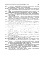
Fig. 49.18 Angioectasias of radiation proctopathy over the
prostate gland. (Courtesy of David M. Martin MD, “Atlas of
Gastrointestinal Endoscopy,” www.EndoAtlas.com.)
Fig. 49.20 Typical red-ring signs of a phospho-soda prep. as
seen in the rectum. (Courtesy of David M. Martin MD, “Atlas
of Gastrointestinal Endoscopy,” www.EndoAtlas.com.)
Fig. 49.19 Stercoral ulcer due to fecal impaction.
594 Section 12: Clinical Use of Colonoscopy
14 Karmali MA, Fleming PC. Campylobacter enteritis in chil-
dren. J Pediatr 1979; 94: 527–53.
15 McKinley MJ, Taylor M, Sangree MH. Toxic megacolon
with Campylobacter colitis. Connecticut Med 1980; 44: 496–7.
16 Mee AS, Shield M, Burke M. Campylobacter colitis: differenti-
ation from acute inflammatory bowel disease. J R Soc Med
1985; 78: 217–23.
17 Waye JD. The differential diagnosis of inflammatory and
infectious colitis. In: Sivak MV, ed. Gastroenterologic Endo-
scopy, 1st edn. Philadelphia: WB Saunders, 1987: 881–99.
18 Loss RW Jr, Mangla JC, Pereira M. Campylobacter colitis pre-
senting as inflammatory bowel disease with segmental
colonic ulcerations. Gastroenterology 1980; 79: 138–40.
19 Hannuksela M, Ahvonen P. Skin manifestations in human
yersiniosis. Ann Clin Res 1975; 7: 368–73.
20 Spira TJ, Kabins SA. Yersinia enterocolitica septicemia with
septic arthritis. Arch Intern Med 1976; 136: 1305–8.
21 Vantrappen G, Ponette E, Geboes K, Bertrand P. Yersinia
enteritis and enterocolitis: gastroenterological aspects. Gast-
roenterology 1977; 72: 220–7.
22 Simmonds SD, Noble MA, Freeman HJ. Gastrointestinal
features of culture-positive Yersinia enterocolitica infection.
Gastroenterology 1987; 92: 112–17.
23 Matsumoto T, Iida M, Matsui T et al. Endoscopic findings in
Yersinia enterocolitica enterocolitis. Gastrointest Endosc 1990;
36: 583–7.
24 Vantrappen G, Geboes K, Ponette E. Yersinia enteritis. Med
Clin North Am 1982; 66: 639–53.
25 Hoogkamp-Korstanje JA. Antibiotics in Yersinia enterocolit-
ica infections. J Antimicrob Chemother 1987; 20: 123–31.
26 DuPont HL, Formal SB, Hornick RB et al. Pathogenesis of
Escherichia coli diarrhea. N Engl J Med 1971; 285: 1–9.
27 Tullock EF Jr, Ryan KJ, Formal SB, Franklin FA. Invasive
enteropathic Escherichia coli dysentery. An outbreak in 28
adults. Ann Intern Med 1973; 79: 13–17.
28 Snyder JD, Wells JG, Yashuk J, Puhr N, Blake PA. Outbreak
of invasive Escherichia coli gastroenteritis on a cruise ship.
Am J Trop Med Hyg 1984; 33: 281–4.
29 Griffin PM, Ostroff SM, Tauxe RV et al. Illness associated
with Escherichia coli O157: H7 infections. A broad clinical
spectrum. Ann Intern Medical 1988; 109: 705–12.
30 Griffin PM, Olmstead LC, Petras RE.
Escherichia coli O157.
H7-associated colitisaa clinical and histological study of 11
cases. Gastroenterology 1990; 99: 142–9.
31 Tarr PI, Neill MA, Clausen CR, Watkins SL, Christie DL,
Hickman RO. Escherichia coli O157. H7 and the hemolytic
uremic syndrome: importance of early cultures in establish-
ing the etiology. J Infect Dis 1990; 162: 553–6.
32 Martin DL, MacDonald DL, White KE, Soler JT, Osterholm
MT. The epidemiology and clinical aspects of the hemolytic
uremic syndrome in Minnesota. N Engl J Med 1990; 323:
1161–7.
33 Ryan CA, Tauxe RV, Hosek GW et al. Escherichia coli O157:
H7 diarrhea in a nursing home: clinical, epidemiological,
and pathological findings. J Infect Dis 1986; 154: 631–8.
34 Bentley G, Webster JH. Gastro-intestinal tuberculosis: a 10-
year review. Br J Surg 1967; 54: 90–6.
35 Goldberg J. Colonoscopic diagnosis of colonic tuberculosis.
Gastrointest Endosc 1984; 30: 216.
36 Gomez-Rubio M, de Cuenca B, Opio V, Ulloa J, Garcia J.
Colonic tuberculosis. An unusual endoscopic diagnosis.
Endoscopy 1993; 25: 377.
37 Tabrisky J, Lindstrom RR, Peters R, Lachman RS. Tuber-
culosis enteritis: review of a protean disease. Am J Gastro-
enterol 1975; 63: 49–57.
38 Ferentzi CV, Sieck JO, Ali MA. Colonoscopic diagnosis and
medical treatment of ten patients with colonic tuberculosis.
Endoscopy 1988; 20: 62–5.
39 Shah S, Thomas V, Mathan M et al. Colonoscopic study
of 50 patients with colonic tuberculosis. Gut 1992; 33:
347–51.
40 Eboda MA, Akande B. Massive lower gastrointestinal hem-
orrhage from abdominal tuberculosis. Trop Geogr Med 1991;
43: 307–9.
41 Chutkan RK, Balba NH. Infectious Diseases of the Colon. In:
DiMarino AJ, Benjamin SB, eds. Gastrointestinal Disease: an
Endoscopic Approach, 2nd edn. Thorofare, NJ: Slack, 2002:
795–814.
42 Hamer DH, Gorbach SL. Infectious diarrhea and bacterial
food poisoning. In: Feldman M, Friedman LS, Sleisenger
MH, eds. Sleisenger and Fordtran’s Gastrointestinal and Liver
Disease: Pathophysiology/Diagnosis/Management, 7th edn. Phil-
adelphia: WB Saunders, 2002: 1864–913.
43 Bhargava DK, Tandon HD, Chawla TC, Shriniwas Tandon
BN, Kapur BM. Diagnosis of ileocecal and colonic tubercu-
losis by colonoscopy. Gastrointest Endosc 1985; 31: 68–70.
44 Pettengell KE, Pirie D, Simjee AE. Colonoscopic features of
early intestinal tuberculosis. Report of 11 cases. S Afr Med J
1991; 79: 279–80.
45 Breiter JR, Hajjar JJ. Segmental tuberculosis of the colon
diagnosed by colonoscopy. Am J Gastroenterol 1981; 76:
369–73.
46 Zyngier FR, Liberal MH, Dechoum A. Tuberculous colitis
manifested by skip-lesions of the colon. Gastrointest Endosc
1986; 32: 375.
47 Franklin GO, Mohapatra M, Perrillo RP. Colonic tubercu-
losis diagnosed by colonoscopic biopsy. Gastroenterology
1979; 76: 362–4.
48 Bhargava DK, Shriniwas Chawla TC, Tandon BN, Kapur
BM. Intestinal tuberculosis: bacteriological study of tissue
obtained by colonoscopy and during surgery. J Trop Med
Hyg 1985; 88: 249–52.
49 Bartlett JG. Pseudomembranous enterocolitis and anti-
biotic-associated diarrhea. In: Feldman M, Friedman LS,
Sleisenger MH, eds. Sleisenger and Fordtran’s Gastrointestinal
and Liver Disease: Pathophysiology/Diagnosis/Management
, 7th
edn. Philadelphia: WB Saunders, 2002: 1914–31.
50 Tedesco FJ, Corless JK, Brownstein RE. Rectal sparing in
antibiotic-associated pseudomembranous colitis: a prospect-
ive study. Gastroenterology 1982; 83: 1259–60.
51 Burbige EJ, Radigan JJ. Antibiotic-associated colitis with
normal-appearing rectum. Dis Colon Rectum 1981; 24: 198–
200.
52 Bartlett JG, Tedesco FJ, Shull S, Lowe B, Chang T. Symp-
tomatic relapse after oral vancomycin therapy of antibiotic-
associated pseudomembranous colitis. Gastroenterology 1980;
78: 431–4.
53 Bartlett JG. Treatment of antibiotic-associated pseudomem-
branous colitis. Rev Infect Dis 1984; 6: S235–41.
54 Wenisch C, Parschalk B, Hasenhundl M, Hirschl AM,
Graninger W. Comparison of vancomycin, teicoplanin,
metronidazole, and fusidic acid for the treatment of
Clostridium difficile-associated diarrhea. Clin Infect Dis 1996;
22: 813–18.
Chapter 49: Infections and Other Noninflammatory-Bowel-Disease Colitides 595
and comparison to collagenous colitis. Dig Dis Sci 1989; 34:
1730–8.
69 Noyer CM, Brandt LJ. Systemic, iatrogenic, and unusual
disorders of the colon. In: DiMarino AJ, Benjamin SB, eds.
Gastrointestinal Disease: An Endoscopic Approach, 2nd edn.
Thorofare, NJ: Slack, 2002: 915–40.
70 Brandt LJ, Boley SJ, Goldberg L et al. Colitis in the elderly: a
reappraisal. Am J Gastroenterol 1981; 76: 239–45.
71 Berman LG, Burdick D, Heitzman ER, Prior JT. A critical
reappraisal of sigmoid peridiverticulitis. Surg Gynecol
Obstet 1968; 127: 481–91.
72 Stollman NS, Raskin JB. Diverticular disease. In: DiMarino
AJ, Benjamin SB, eds. Gastrointestinal Disease: An Endoscopic
Approach, 2nd edn. Thorofare, NJ: Slack, 2002: 859–79.
73 Imperiali G, Meucci G, Alvisi C et al. Segmental colitis asso-
ciated with diverticula: a prospective study. Am J Gastro-
enterol 2000; 95: 1014–16.
74 McFarland LV, Elmer GW, Surawicz CM. Breaking the cycle:
treatment strategies for 163 cases of recurrent Clostridium
difficile disease. Am J Gastroenterol 2002; 97: 1769–75.
75 Baert F, Schmit A, D’Haens G et al. Budesonide in colla-
genous colitis: a double-blind placebo-controlled trial with
histologic follow-up. Gastroenterology 2002; 122: 20–5.
76 Harig JM, Soergel KH, Komorowski RA, Wood CM.
Treatment of diversion colitis with short chain fatty acid
irrigation. N Engl J Med 1989; 320: 23–8.
77 Gibson GR, Whitacre EB, Ricotti CA. Colitis induced by
nonsteroidal anti-inflammatory drugs. Arch Intern Med
1992; 152: 625–32.
78 Clarkston W, Bonacini M, Peterson I. Colitis due to
Histoplasma capsulatum in the acquired immune deficiency
syndrome. Am J Gastroenterol 1991; 86: 913.
79 Blaser, MJ, Smith, PD, Ravdin, JI. Viral infections. In: Blaser
MJ, Smith PD, Ravdin JI et al., eds. Infections of the GI Tract.
New York: Raven Press, 1995.
80 Wilcox CM, Schwartz DA, Costonis G, Thompson SE 3rd.
Chronic unexplained diarrhea in human immunodeficiency
virus infection: determination of the best diagnostic ap-
proach. Gastroenterology 1996; 110: 30–7.
81 Horsburgh CR. Epidemiology of Mycobacterium avium com-
plex disease. Am J Med 1997; 102: 11–15.
55 Blumencranz H, Kasen L, Romeu J, Waye JD, LeLeiko NS.
The role of endoscopy in suspected amebiasis. Am J Gas-
troenterol 1983; 78: 15–18.
56 Luterman L, Alsumait AR, Daly DS, Goresky CA. Colo-
noscopic features of cecal amebomas. Gastrointest Endosc
1985; 31: 204–6.
57 Nebel OT, el-Masry NA, Castell DO, Farid Z, Fornes MF,
Sparks HA. Schistosomal colonic polyposis: endoscopic and
histologic characteristics. Gastrointest Endosc 1974; 20: 99–
101.
58 Mohamed AR, al Karawi M, Yasawy MI. Schistosomal
colonic disease. Gut 1990; 31: 439–42.
59 Surawicz CM, Myerson D. Self-limited cytomegalovirus
colitis in immunocompetent individuals. Gastroenterology
1988; 94: 194–9.
60 Akdamar K, Martin RJ, Ichinose H. Syphilitic proctitis. Am J
Dig Dis
1977; 22: 701–4.
61 Martin EG, Kallet HI. Primary syphilis of the anorectal
region. JAMA 1925; 84: 1556.
62 Nazemi MM, Musher DM, Schell KF, Milo S. Syphilitic
proctitis in a homosexual. JAMA 1975; 231: 389.
63 Rompalo AM, Price CB, Roberts PL, Stamm WE. Poten-
tial value of rectal-screening cultures for Chlamydia tra-
chomatis in homosexual men. J Infect Dis 1986; 153:
888–92.
64 Fried R, Surawicz C. Proctitis and sexually transmissible
intestinal disease. In: Feldman M, Friedman LS, Sleisenger
MH, eds. Sleisenger and Fordtran’s Gastrointestinal and Liver
Disease: Pathophysiology/Diagnosis/Management, 7th edn. Phil-
adelphia: WB Saunders, 2002: 2263–75.
65 Levine JS, Smith PD, Brugge WR. Chronic proctitis in male
homosexuals due to lymphogranuloma venereum. Gastro-
enterology 1980; 79: 563–5.
66 Giardiello FM, Hansen FC III, Lazenby AJ et al. Collagenous
colitis in the setting of nonsteroidal anti-inflammatory
drugs and antibiotics. Dig Dis Sci 1990; 35: 257–60.
67 Tanaka M, Mazzoleni G, Riddell RH. Distribution of col-
lagenous colitis: utility of flexible sigmoidoscopy. Gut 1992;
33: 65–70.
68 Giardiello FM, Lazenby AJ, Bayless TM et al. Lymphocytic
(microscopic) colitis: clinicopathologic study of 18 patients
596
Introduction
Sir William Heneage Ogilvie first described acute colonic
pseudo-obstruction (ACPO) in 1948 in two patients with
far-advanced intraabdominal malignancies [1]. He was
the first to postulate an underlying imbalance between
the sympathetic and parasympathic innervation of the
colon as the cause of this disorder. Ogilvie’s patients,
however, developed subacute symptoms over the course
of 2 months and thus represent an atypical presenta-
tion of what we now recognize as ACPO. The hallmark
features of ACPO consist of acute colonic dilation in
the absence of a mechanical etiology. This condition is
increasingly recognized and is associated with substan-
tial morbidity and mortality.
Epidemiology and predisposing factors
The exact incidence of ACPO in hospitalized patients
is unknown. Vanek and Al-Salti [2] analyzed 400 cases
of ACPO and found that it occurred most commonly
in the sixth decade and was more common in men
than women. More than 90% of patients had signific-
ant comorbid disease, thought to be contributing to the
syndrome. About 50% of cases occurred in the post-
operative state. The diverse underlying medical and
surgical problems associated with ACPO are listed in
Table 50.1.
Pathophysiology
The pathophysiology of ACPO is still not entirely under-
stood but there is evidence of an imbalance between the
sympathetic and parasympathic nervous system, which
leads to a functional obstruction caused by atony of the
distal colon followed by progressive dilation of the
cecum and ascending colon [1,3].
Ogilvie favored the sympathetic deprivation theory,
leading to unopposed parasympathic stimulation and
thereby resulting in “excessive and probably incoor-
dinated contraction” of the distal colon [1] mimicking
obstruction. More recent theories postulate either an
impairment of the sacral parasympathetic outflow [3–5]
or excessive sympathetic stimulation [6,7]. The clinical
presentation of ACPO resembles Hirschsprung’s disease,
supporting the hypothesis of impaired parasympathetic
function [5], which is also supported by the commonly
observed transition point at the level of the splenic
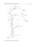
flexure. The parasympathetic innervation of the colon
distal to the splenic flexure is via the pelvic splanchnic
nerves whereas the more proximal colon is innervated
by the vagus (Fig. 50.1).
The proponents of the sympathetic stimulation theory
[6,7] argue that right-sided colonic motility is impaired
Chapter 50
Acute Colonic Pseudo-obstruction
Hubert Nietsch and Michael B. Kimmey
Table 50.1 Causes of acute colonic pseudo-obstruction
[2,11,17,18,33,45–50].
Neurologic
Parkinson’s disease
Alzheimer’s disease
Cerebrovascular accident
Multiple sclerosis
Spinal cord disease
Craniotomy
Cardiovascular
Myocardial infarction
Congestive heart failure
Post cardiac arrest
Respiratory
Pneumonia
Mechanical ventilation
Acute respiratory distress syndrome
Metabolic
Hyponatremia
Hypocalcemia
Hypomagnesemia
Liver failure
Renal failure
Hypothyroidism
Infective/inflammatory
Acute cholecystitis
Pelvic abscess
Spontaneous bacterial peritonitis
Acute pancreatitis
Sepsis
Herpes zoster
Appendicitis
Neoplasia
Retroperitoneal
Metastatic cancer
Post surgical
Cardiac surgery
Cesarian section
Gynecologic surgery
Pelvic surgery
Organ transplantation
Orthopedic surgery
Post traumatic
Pelvic trauma
Spinal cord injury
Femoral fracture
Drugs
Narcotics
Tricyclic antidepressants
Phenothiazines
Antiparkinson agents
Calcium channel blockers
Benzodiazepines
Clonidine
Vincristine
Colonoscopy Principles and Practice
Edited by Jerome D. Waye, Douglas K. Rex, Christopher B. Williams
Copyright © 2003 Blackwell Publishing Ltd
Chapter 50: Acute Colonic Pseudo-obstruction 597
by excessive sympathetic inhibition. This theory is sup-
ported by animal experiments performed in the 1920s
and 1930s [8] showing increased colonic peristalsis after
spinal anesthesia, which leads to a temporary paralysis
of sympathetic input. This was the rationale for the
induction of spinal anesthesia as a successful treatment
of adynamic ileus in Europe in the 1920s.
Clinical presentation
ACPO is usually seen in middle-aged to elderly critically
ill patients in the intensive care unit or postoperatively
and is exacerbated by immobility and narcotic pain
medications. Symptoms usually develop gradually over
3–7 days. Significant abdominal distension is seen in
all patients, associated with pain (83%), vomiting (57%),
constipation (51%), and fever (37%). The bowel sounds
are variable and can be normal or hyperactive (40%),
hypoactive (31%), high-pitched (17%), or absent (12%)
[2]. If peritoneal signs are present, transmural ischemia
or perforation should be suspected.
Diagnosis
Abdominal examination shows significant distension
in all patients, with variable degrees of tenderness. The
presence or quality of bowel sounds is also variable.
Peritoneal signs are suggestive of transmural ischemia
or perforation and mandate surgical consultation.
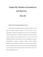
The diagnosis is confirmed by plain abdominal radio-
graphs, which typically show significant distension of
the colon with predominance of the right side in the
absence of mechanical obstruction (Fig. 50.2). A cut-off
sign at the splenic flexure is frequently observed [5].
Initial studies suggested that a cecal diameter of
greater than 12 cm increases the risk of perforation sub-
stantially [9]. The case series by Vanek and Al-Salti
[2] reported no cecal perforation with a cecal diameter
< 12 cm, 7% perforation risk with cecal diameters of
12–14 cm, and 23% perforation risk with a cecal dia-
meter > 14 cm. However, more recent reports suggest
that the duration of significant cecal dilation is more
predictive of ischemia than the cecal diameter per se
[10].
A water-soluble contrast enema should be cauti-
ously performed to confirm the functional etiology, if
a mechanical obstruction (absence of rectal air) can-
not be entirely excluded by the initial radiographs.
Barium should not be administered, because this could
Superior
mesenteric
ganglion
T10
11
12
L1
2
T10
11
12
L1
2
Vagus nerves
Celiac
ganglion
Inferior
mesenteric
ganglion
Pudendal nerves
S2
3
4
S2
3
4
Pelvic plexus
T10-12
Fig. 50.1 Schematic diagram illustrating colonic innervation.
Parasympathetic pathways (stimulatory/prokinetic):
prevertebral ganglia and sacral nerves (red) and vagus
(yellow). Sympathetic pathways (inhibitory): thoracic spinal
cord to inferior mesenteric plexus and pelvic plexus (green).
Fig. 50.2 Abdominal radiograph of patient with acute colonic
pseudo-obstruction following internal fixation of a fractured
femur.
598 Section 12: Clinical Use of Colonoscopy
complicate surgery if perforation is present and endo-
scopic decompression if this is required.
Complications
The dreaded complication of progressive colonic
dilation is transmural ischemia followed by perforation.
However, the frequency of this complication, which
requires emergency colonic resection, has significantly
decreased in recent case series. The risk of perforation
was initially reported to be as high as 13% with a mortal-
ity of 43% [11]. A summary of more recent studies shows
a perforation risk of 3% [12]. The surgical mortality may
be as high as 40–50%, if perforation occurs [13].
Management
Supportive medical care
Initial management for the first 24–48 h is conservative,
with close attention to correcting any fluid and elec-
trolyte imbalances that may be present. The medica-
tion list should be carefully scrutinized and drugs that
might delay intestinal transit, such as anticholinergics
or opiates, should be discontinued if possible [14]. The
abdominal examination needs to be followed carefully
and daily abdominal radiographs obtained to monitor
for progressive dilation and evidence of perforation. The
introduction of a nasogastric tube for decompression is
advisable for most patients, and in selected cases a rectal
tube might also be of help. Mobilization of the patient
with frequent turning might facilitate the passage of
flatus. Success rates of supportive management are vari-
able but can be as high as 96%, as reported in a cohort
of cancer patients from Sloan-Kettering Cancer Center
[15].
Pharmacotherapy
When colonic dilation persists or progresses despite con-
servative therapy, specific pharmacotherapy to stimul-
ate the parasympathic innervation of the colon should be
attempted (Table 50.2). Catchpole [16] first proposed the
combined use of a sympathetic blocker (guanethidine)
followed by a cholinesterase inhibitor (neostigmine) to
correct the sympathetic/parasympathic imbalance. Sub-
sequent small case series suggested that a majority of
patients with ACPO could be effectively treated using
neostigmine [17–19].
A double-blind, randomized, placebo-controlled clin-
ical trial reported by Ponec and colleagues [20] con-
clusively showed a dramatic improvement of clinical
status and colonic distension in the majority of patients
treated with intravenous neostigmine, making endo-
scopic intervention unnecessary in most cases. In this
study, patients were treated with 2 mg of neostigmine
administered over a few minutes by slow intravenous
push. Patients were monitored continuously by electro-
cardiography with atropine available at the bedside, as
symptomatic bradycardia is the most significant adverse
effect of this treatment. Of 11 patients who received
neostigmine, 10 (91%) had prompt colonic decompres-
sion with a median time to response of only 4 min,
whereas none of the patients receiving placebo (saline)
had a clinical response. Seven patients in the placebo
group and the one patient in the neostigmine group who
failed initial response received open-label neostigmine
3 h after the initial infusion, with prompt colonic de-
compression noted in all patients. Only two patients
developed recurrent symptoms requiring colonoscopic
decompression [20].
Several studies have since confirmed the safety
of neostigmine for the treatment of ACPO, reporting
Number of Initial decompression Recurrence
Medication Reference patients (%) (%)
Erythromycin Armstrong et al. [27] 2 100 0
Bonacini et al. [28] 1 100 0
Rovira et al. [29] 2 100 0
Cisapride MacColl et al. [7] 1 100 0
Pelkmans et al. [30] 2 0 0
Mazloum et al. [31] 1 100 0
Neostigmine Hutchinson & Griffiths [17] 11 72 0
Stephenson et al. [18] 12 83 16
Turegano-Fuentes et al. [19] 16 75 0
Ponec et al. [20] 21 81 11
Paran et al. [21] 11 82 0
Trevisani et al. [22] 28 93 0
Abeyta et al. [23] 8 87 0
Van der Spoel et al. [24] 24 79 0
Table 50.2 Reports of
pharmacotherapy of acute colonic
pseudo-obstruction.
Chapter 50: Acute Colonic Pseudo-obstruction 599
successful colonic decompression in 79–93% of cases.
Several different neostigmine infusion protocols have
been used, including 2-mg and 2.5-mg intravenous
boluses and 2.5 mg administered over 60 min [21–24], all
with similar success rates.
Recurrence of colonic distension following successful
decompression using neostigmine occurs in up to 16% of
patients. In these situations, neostigmine can be safely
readministered, leading to colonic decompression in
approximately two-thirds of cases [21,23].
The observed adverse effects of neostigmine, like
other cholinesterase inhibitors, include excessive saliva-
tion (38%), vomiting (9%), abdominal pain (62%), brady-
cardia (9%), and bronchospasm. Patients must therefore
be closely monitored during drug administration with
continuous electrocardiography and atropine available
at the bedside [20]. Symptomatic bradycardia responds
to administration of atropine, but this also leads to a re-
versal of any benefit of neostigmine in relieving colonic
dilation. The coadministration of glycopyrrolate, an
antimuscarinic anticholinergic agent, seems to decrease
the incidence of bradycardia without reducing neostig-
mine’s efficacy [25,26].
Suitable candidates for neostigmine administration
are hence patients with ACPO who have no evidence
of mechanical bowel obstruction, a resting heart rate
greater than 60 beats per minute with a systolic blood
pressure greater than 90 mmHg, and no active bron-
chospasm [14,20]. Neostigmine is contraindicated in
patients on β-blockers and those who have significant
acidosis or recent myocardial ischemia, because of the
risk of inducing cardiac arrhythmias [18].
Anecdotal case reports with other prokinetic agents
show variable success rates in the treatment of ACPO.
Intravenous erythromycin, which acts as a motilin re-
ceptor agonist, showed some success in a total of five
reported cases [27–29]. The efficacy of intravenous cisap-
ride, no longer available in the USA, was highly variable
in case reports of five patients [7,30,31].
The new 5-hydroxytryptamine 5-HT
4
receptor agon-
ists (tegaserod, prucalopride) might be theoretically
useful for stimulating colonic motility in the setting of
ACPO, but no data are yet available with the use of these
medications in ACPO [14].
Colonoscopic decompression
Pharmacologic treatment of ACPO has markedly re-
duced the need for urgent colonoscopic decompression.
While previously considered to be the treatment of
choice for progressive colonic dilation, it is now usually
reserved for patients who have failed treatment with
neostigmine (Fig. 50.3). No randomized comparative
studies of colonoscopic decompression with neostigmine
or other treatment modalities are available. A sum-
mary of 11 retrospective studies involving 264 patients
shows a high initial success rate for colonoscopic decom-
pression (64–100%), with an average recurrence rate of
23% (range 13–65%) (Table 50.3). Complications were
reported in 3% [32,33]. The largest single-center series
from the Mayo Clinic shows a similar experience in 50
patients, with an overall success rate of 88% complicated
Conservative
measures
(NG, rectal tube,
stop narcotics,
mobilize patient)
Success
Stop
Fail
Bradycardia,
active bronchospasm,
renal failure?
Yes
No
Colonoscopic
decompression
Fail
Neostigmine
2.0 mg IV
Fig. 50.3 Algorithm for management of acute colonic pseudo-
obstruction.
Number of Initial Complications
Reference patients success Recurrence (death) Surgery
Kukora & Dent [51] 6 5 (83%) 0 0 1 (17%)
Nivatvongs et al. [5] 22 19 (86%) 4 (21%) 0 4 (18%)
Strodel et al. [35] 44 32 (73%) 5 (13%) 5% (2%) 9 (20%)
Starling [52] 17 17 (100%) 3 (18%) 12% 0
Bode [55] 22 20 (91%) 4 (20%) 5% (5%) 3 (14%)
Nakhgevany [53] 10 9 (90%) 0 0 1 (10%)
Fausel & Goff [54] 12 11 (91%) 3 (25%) 0 2 (17%)
Nano et al. [47] 17 13 (76%) 6 (46%) 0 0
Gosche et al. [32] 19 17 (89%) 11 (65%) 5% 2 (11%)
Jetmore et al. [33] 45 29 (64%) 13 (29%) 0 5 (11%)
Geller et al. [34] 50 39 (78%) 9 (18%) 2% (2%) 1 (2%)
Total 264 83% 23% 3% (1%) 11%
Table 50.3 Reports of colonoscopic
decompression of acute colonic
pseudo-obstruction.
600 Section 12: Clinical Use of Colonoscopy
by one procedure-related perforation. The overall hos-
pital mortality is 30% [34].
Colonoscopic decompression is technically more chal-
lenging compared with routine colonoscopy since the
colon is unprepared and the patients are often critically
ill, necessitating performance of the procedure in an
intensive care unit. Enemas are not very helpful in pre-
paration for colonoscopy and should be done gently, if
at all, due to the risk of perforation. Liquid stool must
be suctioned and irrigated at the time of colonoscopy in
most cases. Air insufflation should be kept to a minimum
to prevent further cecal dilation, which could potentially
precipitate perforation. It is important to reach the hepatic
flexure in order to achieve significant decompression,
although cecal intubation is not required [35]. Jetmore
and colleagues [33] reported that colonic decompression
was almost twice as successful if the ascending colon
was reached (initial success 71% vs. 37%). If mucosal
changes suggestive of acute ischemia are encountered,
the procedure should be terminated and the patient
referred for emergency colectomy. The overall decrease
in cecal diameter following colonoscopic decompression
is generally quite modest, with an average reduction of
only 2 cm [36].
Up to 40% of patients develop recurrence of colonic
distension after initial successful colonoscopic decom-
pression. This led to the introduction of decompression
tubes, which are inserted at the time of the initial proced-
ure. Harig and colleagues [37] performed a randomized
trial in 20 patients comparing endoscopic decompres-
sion alone vs. additional insertion of a modified enter-
oclysis catheter and demonstrated a reduction in the
recurrence of colonic dilation from 44% to 0%. Decom-
pression tubes remained in place for an average of 3–4
days without any reported complications.
The two most commonly used decompression tubes
are a modified enteroclysis catheter, with additional
side holes at the tip or a 14F colon decompression kit
(Wilson-Cook Medical, Winston-Salem, NC). A flexible
guidewire is placed through the endoscope channel and
the tip is directed into the cecum under fluoroscopy. The
endoscope is then slowly withdrawn leaving the wire in
place. Fluoroscopy is helpful in keeping the wire straight
during complete withdrawal of the colonoscope. The
decompression tube is then advanced under fluoro-
scopic guidance, using traction on the wire to keep it
straight while the tube is advanced. The decompression
tube is than taped to the patient’s buttock and connected
to low intermittent suction. It is advisable to flush the
tube with water every 4 h to prevent clogging with stool.
Fig. 50.4 (a) Abdominal radiograph of patient with acute
colonic pseudo-obstruction following bone marrow
transplantation for leukemia. (b) Abdominal radiograph of
same patient immediately following colonoscopy at which
time a 14F decompression tube was placed.
(a) (b)
Chapter 50: Acute Colonic Pseudo-obstruction 601
The patient’s clinical status should be followed care-
fully with daily abdominal radiographs (Fig. 50.4). The
catheters are usually left in situ for 2–4 days, until colon
decompression is complete and underlying reversible
contributors to ACPO are reversed. Use of larger tubes
up to 40F in diameter (Levacuator, Mallinckrodt Medical,
St Louis, MO) has been described in case reports, with
more rapid decompression and less tube clogging [38]. A
minority of patients may not respond to these measures
and if there is suspicion of acute ischemia or perforation
the patient should be referred for immediate surgery.
Percutaneous cecostomy
In the absence of ischemia or perforation, percutaneous
cecostomy (PCC) should be considered as a minimally
invasive alternative to surgery in those critically ill
patients where induction of general anesthesia poses a
significant risk. Both transperitoneal and retroperitoneal
approaches for PCC have been described [39–41]. The
early work by VanSonnenberg and colleagues [42]
showed the technical feasibility and safety of PCC tubes.
The theoretically safer retroperitoneal approach did not
lead to a lower risk of peritonitis than the anterior trans-
peritoneal approach [42]. The technique was recently
refined by using additional T-fasteners, which allow for
better colonic apposition to the abdominal wall, thereby
potentially reducing the risk of fecal soilage of the
abdominal cavity [43]. No studies comparing the efficacy
and safety of pharmacotherapy, endoscopic intervention,
radiographically guided PCC, and surgery are available.
Surgery
Peritoneal signs or free air on abdominal radiography
are clear indications for laparotomy and colectomy [2].
The definitive surgical management depends on the
viability of the cecum and ascending colon at the time
of exploration. Partial colectomy is indicated for trans-
mural ischemia and perforation but carries a high
mortality in these critically ill patients. Surgical decom-
pression in the absence of perforation, through an open
or laparoscopic cecostomy, is an alternative to colectomy
if the local expertise is not available to perform com-
puted tomography-guided PCC [44].
Prognosis
The overall mortality of ACPO remains approximately
30%, despite the recent advances in its management
[2,34]. This reflects the severity of the underlying disease
process leading to ACPO and is not directly related to
the colonic complications. The impact of pharmacologic
therapy on the outcome of patients with ACPO has not
yet been fully assessed.
References
1 Ogilvie H. Large-intestine colic due to sympathetic depriva-
tion: a new clinical syndrome. Br Med J 1948; 2: 671–3.
2 Vanek VW, Al-Salti M. Acute pseudo-obstruction of the
colon (Ogilvie syndrome): an analysis of 400 cases. Dis Colon
Rectum 1986; 29: 203–10.
3 Spira I, Rodrigues R, Wolff W. Pseudo-obstruction of the
colon. Am J Gastroenterol 1976; 65: 397–408.
4 Bachulis BL, Smith PE. Pseudo-obstruction of the colon. Am
J Surg 1978; 136: 66–72.
5 Nivatvongs S, Vermeulen F, Fang D. Colonoscopic decom-
pression of acute pseudo-obstruction of the colon. Am J Surg
1982; 196: 598–600.
6 Lee JT, Taylor BM, Singleton BC. Epidural anesthesia for
acute pseudo-obstruction of the colon (Ogilvie’s syndrome).
Dis Colon Rectum 1988; 31: 686–91.
7 MacColl C, MacCannell KL, Baylis B, Lee SS. Treatment of
acute colonic pseudo-obstruction (Ogilvie’s syndrome)
with cisapride. Gastroenterology 1990; 98: 773–6.
8 Burstein CL. Effect of spinal anesthesia on intestinal activ-
ity. Proc Soc Exp Biol Med 1939; 42: 291–3.
9 Gierson ED, Storm FK, Shaw W, Coyne SK. Caecal rupture
due to colonic ileus. Br J Surg 1975; 62: 393–6.
10 Johnson CD, Rice RP, Kelvin FM et al. The radiologic evalu-
ation of gross cecal distension: emphasis on cecal ileus. AJR
Am J Roentgenol 1985; 145: 1211–17.
11 Soreide O, Bjerkeset T, Fossdol J. Pseudo-obstruction of the
colon (Ogilvie syndrome): a genuine clinical condition?
Review of the literature (1948–75) and a report of 5 cases.
Dis Colon Rectum 1977; 20: 487–91.
12 Rex DK. Colonoscopy and acute colonic pseudo-obstruc-
tion. Gastrointest Endosc Clin North Am 1997; 7: 499–508.
13 Wojtalik RS, Lindenauer SM, Kahn SS. Perforation of the
colon associated with adynamic ileus. Am J Surg 1973; 125:
601–6.
14 De Giorgio R, Barbara G, Stanghellin V et al. Review article:
the pharmacologic treatment of acute colonic pseudo-
obstruction. Aliment Pharmacol Ther 2001; 15: 1717–27.
15 Sloyer AF, Panella VS, Demas BE et al. Ogilvie’s syndrome:
successful management without colonoscopy. Dig Dis Sci
1988; 33: 1391–6.
16 Catchpole BN. Ileus: use of sympathetic blocking agents in
its treatment. Surgery 1969; 66: 811–20.
17 Hutchinson R, Griffiths C. Acute colonic pseudo-obstruc-
tion: a pharmacologic approach. Ann R Coll Surg Engl 1992;
74: 364–7.
18 Stephenson BM, Morgan AR, Salaman JR, Wheeler MH.
Ogilvie’s syndrome: a new approach to an old problem. Dis
Colon Rectum 1995; 38: 424–7.
19 Turegano-Fuentes F, Munoz-Jimenez F, Del Valle-Hernandez
E et al. Early resolution of Ogilvie’s syndrome with intra-
venous neostigmine. Dis Colon Rectum 1997; 40: 1353–7.
20 Ponec RJ, Saunders MD, Kimmey MB. Neostigmine for the
treatment of acute colonic pseudo-obstruction. N Engl J Med
1999; 341: 137–41.
21 Paran H, Silverberg D, Mayo A, Shwartz I, Neufeld D,
Freund U. Treatment of acute colonic pseudo-obstruction
with neostigmine. J Am Coll Surg 2000; 190: 315–18.
22 Trevisani GT, Hyman NH, Church JM. Neostigmine: safe
and effective treatment for acute colonic pseudo-obstruction.
Dis Colon Rectum 2000; 43: 599–603.
602 Section 12: Clinical Use of Colonoscopy
39 Crass JE, Simmons RL, Mathis PF, Charles WM. Per-
cutaneous decompression of the colon using CT guidance in
Ogilvie syndrome. AJR Am J Roentgenol 1985; 144: 475–6.
40 Casola G, Withers C, vanSonnenberg E, Herba MJ, Saba RM,
Brown RA. Percutaneous cecostomy for decompression of
the massively distended cecum. Radiology 1986; 158: 793–4.
41 Haaga JR, Bick JR, Zollinger RM. CT-guided percutaneous
catheter cecostomy. Gastrointest Radiol 1987; 12: 166–8.
42 VanSonnenberg E, Varney RR, Casola G et al. Percutaneous
cecostomy for Ogilvie syndrome: laboratory observations
and clinical experience. Radiology 1990; 175: 679–82.
43 Chevallier P, Marcy PY, Francois E et al. Controlled
transperitoneal percutaneous cecostomy as a therapeutic
alternative to endoscopic decompression of Ogilvie’s syn-
drome. Am J Gastroenterol 2002; 97: 471–4.
44 Groff W. Colonoscopic decompression and intubation of
the cecum for Ogilvie’s syndrome. Dis Colon Rectum 1983;
26: 503–6.
45 Bender GN, Do-Dai DD, Briggs LM. Colonic pseudo-
obstruction: decompression with tricomponent coaxial
system under fluoroscopic guidance. Radiology 1991; 188:
395–8.
46 Dorudi S, Berry AR, Kettlewell MG. Acute colonic pseudo-
obstruction. Br J Surg 1992; 79: 99–103.
47 Nano D, Rindiville T, Pavly M et al. Colonoscopic therapy of
acute pseudo-obstruction of the colon. J Gastroenterol 1987;
82: 145–8.
48 Romeo DP, Solomon GD, Hover AR. Acute colonic pseudo-
obstruction: a possible role for colocolonic reflex. J Clin
Gastroenterol 1985; 7: 256–60.
49 Wanebo H, Mathewson C, Conolly B. Pseudo-obstruction of
the colon. Surg Gynecol Obstet 1971; 133: 44–8.
50 Wegener M, Boersch G. Acute colonic pseudo-obstruction
(Ogilvie’s syndrome): presentation of 14 of our own cases
and analysis of 1027 cases reported in the literature. Surg
Endosc 1987; 1: 169–74.
51 Kukora JS, Dent TL. Colonoscopic decompression of massive
non-obstructive cecal dilation. Arch Surg 1977; 112: 512–17.
52 Starling JR. Treatment of nontoxic megacolon by colono-
scopy. Surgery 1983; 94: 243–5.
53 Nakhgevany KB. Colonoscopic decompression of the colon
in patients with Ogilvie’s syndrome. Am J Surg 1984; 148:
317–20.
54 Fausel CS, Goff JS. Non-operative management of acute
idiopathic colonic pseudo-obstruction (Ogilvie’s Syndrome).
West J Med 1985; 143: 50–4.
55 Bode WE, Beart RW Jr, Spencer RJ, Culp CE, Wolff BG,
Taylor BM. Colonoscopic decompression for acute pseu-
doobstruction of the colon (Ogilvie’s syndrome). Report of
22 cases and review of the literature. Am J Surg 1984; 147:
243–5.
23 Abeyta BJ, Albrecht RM, Schermer CR. Retrospective study
of neostigmine for the treatment of acute colonic pseudo-
obstruction. Am Surg 2001; 67: 265–8.
24 Van der Spoel JI, Oudemans-van Straaten HM, Stoutenbeck
CP, Bosman RJ, Zandstra DF. Neostigmine resolves crit-
ical illness-related colonic ileus in intensive care patients
with multiple organ failure: a prospective, double-blind,
placebo-controlled trial. Intensive Care Med 2001; 27: 822–7.
25 Ostheimer GW. A comparison of glycopyrrolate and atrop-
ine during reversal of nondepolarizing neuromuscular
block with neostigmine. Anesth Analg 1977; 56: 182–6.
26 Mirakur RK, Briggs LP, Clarke RS, Dundee JW, Johnston
HM. Comparison of atropine and glycopyrrolate in a mix-
ture with pyridostigmine for the antagonism of neuromus-
cular block. Br J Anaesth 1981; 53: 1315–20.
27 Armstrong DN, Ballantyne GH, Modlin IM. Erythromycin
for reflex ileus in Ogilvie’s syndrome (letter). Lancet
1991;
337: 378.
28 Bonacini M, Smith OJ, Pritchard T. Erythromycin as therapy
for acute colonic pseudo-obstruction (Ogilvie’s syndrome).
J Clin Gastroenterol 1991; 13: 475–6.
29 Rovira A, Lopez A, Cambray C, Gimeno C. Acute colonic
pseudo-obstruction (Ogilvie’s syndrome) treated with ery-
thromycin. Intensive Care Med 1997; 23: 798.
30 Pelkmans PA, Michielsen PP, Jorens PG, Van Maercke YM.
Cisapride in Ogilvie’s syndrome. Gastroenterology 1990; 99:
1194–5.
31 Mazloum BW, Barnes JB, Lee M. Cisapride as a successful
treatment for acute intestinal pseudo-obstruction. South
Med J 1996; 89: 828–30.
32 Gosche JR, Sharpe JN, Larson GM. Colonic decompression
for pseudo-obstruction of the colon. Am Surg 1989; 55:
111–15.
33 Jetmore AB, Timmcke AE, Gathright JB Jr, Hicks TC, Ray JE,
Baker JW. Ogilvie’s syndrome: colonoscopic decompres-
sion and analysis of predisposing factors. Dis Colon Rectum
1992; 35: 1135–42.
34 Geller A, Petersen BT, Gostout CJ. Endoscopic decompres-
sion for acute colonic pseudo-obstruction. Gastrointest
Endosc 1996; 44: 144–50.
35 Strodel WE, Nostrant TT, Eckhauser FE, Dent TL.
Therapeutic and diagnostic colonoscopy in nonobstructive
colonic dilation. Ann Surg 1983; 197: 416–21.
36 Pham TN, Cosman BC, Chu P, Savides TJ. Radiographic
changes after colonoscopic decompression for acute
pseudo-obstruction. Dis Colon Rectum 1999; 42: 1586–91.
37 Harig JM, Fumo DE, Loo FD et al. Treatment of acute non-
toxic megacolon during colonoscopy: tube placement versus
simple decompression. Gastrointest Endosc 1988; 34: 23–7.
38 Yarze JC, Winchell EC. A novel device for colonic tube
decompression. Am J Gastroenterol 2000; 95: 2136.
603
Introduction
Radiation proctopathy can be a disabling delayed out-
come of radiation therapy directed at pelvic malig-
nancies. Rectal outlet bleeding can be severe enough to
result in anemia and transfusion dependency. Bleeding
typically develops from 6 months to 1 year after com-
pletion of radiation therapy and is due to friable mucosal
angiectasias. Although many approaches to controlling
bleeding from chronic radiation proctopathy have been
attempted, ranging from topical enema formulations
to surgical diversion of the rectum, endoscopic coagula-
tion therapy is effective and the easiest to use successful
therapy. This chapter discusses the issues surrounding
the development of chronic bleeding due to radiation
proctopathy and focuses on endoscopic methods of
treatment.
Radiation proctopathy (a better term than “radiation
proctitis”) is a frustrating problem for patients and man-
aging physicians. Radiation therapy (external beam and
intracavitary) is a common modality of treatment for
pelvic malignancies, especially with supervoltage tech-
niques and computerization for modeling dosimetry.
Malignancies of the uterus, prostate, cervix, bladder, and
rectum as well as lymphomas are treated with pelvic
radiation. The rapidly dividing mucosa of the gastroin-
testinal tract is vulnerable to radiation, with the entire
colon, rectum, and pelvic small bowel susceptible to
injury. Although the rectal mucosa is more resistant to
the damaging effects of radiation compared with the rest
of the colon and small bowel, because of its proximity to
the uterine cervix and prostate, the rectum is the most
common gastrointestinal organ to be affected by pelvic
radiation (> 90%) [1]. In addition to the close anatomic
relation of the rectum to the pelvic organs, the rectum is
in a fixed position within the pelvic field of radiation.
Fixed organs are generally more likely to be damaged
by radiation compared with mobile organs such as the
small bowel, where peristalsis causes different portions
of the intestine to move in and out of the field of
radiation.
Acute radiation injury is common and typically occurs
during radiation [2]. The findings within the rectum are
consistent, with a proctitis with mucosal edema, ulcera-
tion, erythema, and spontaneous bleeding. Histologic
findings include mucosal cell loss, acute inflammation,
eosinophilic crypt abscesses, and endothelial swelling of
arterioles. Most patients recover but some progress to a
chronic stage. Radiation proctopathy is diagnosed when
there are rectal mucosal changes and clinical symptoms
that develop 3–6 months after completion of therapy
[3,4]. The frequency of this late complication varies from
5 to 20% in different series [4,5].
The clinical features of radiation proctopathy include
diarrhea, tenesmus, rectal pain, rectal bleeding (low
grade or severe), stricture, and fistulae into adjacent
organs [6]. Rectal bleeding can be daily or episodic, with
multiple passages of blood and clot. Incontinence of
blood is a common complaint.
The endoscopic findings of radiation proctopathy
include mucosal pallor, friability, spontaneous oozing,
angiectasia, and rarely ulceration (Fig. 51.1) [7]. The
angiectasias are the hallmark findings distinctive for this
disorder. These endoscopic features begin at the dentate
line and typically occupy the distal rectum (Fig. 51.2).
An occasional patient may have sigmoid involvement,
typically women whose radiation has been directed
higher in the pelvis, which has implications regard-
ing treatment strategy and outcomes (Fig. 51.3) [8]. The
histology of this late sequela includes fibrosis within
the lamina propria and endarteritis of the arterioles
[2].
Treatment approaches for radiation
proctopathy
Rectal bleeding is the most vexing problem for which
endoscopic treatment is sought. A variety of treatment
regimens have been attempted without objective data
to support efficacy. Steroids (oral and by retention
enema), sulfasalazine, 5-aminosalicylic acid preparations
(oral and enema), sucralfate enemas, sodium pentosan-
polysulfate PPS (synthetic sulfated polysaccharides),
hyperbaric oxygen, short-chain fatty acids, nutritional
therapy, and even angiographic embolization (despite
the ischemic origins postulated and even observed)
are among the various treatments attempted for radi-
ation proctopathy [3,9–13]. Sucralfate enemas have been
Chapter 51
Radiation Proctopathy
Christopher J. Gostout
Colonoscopy Principles and Practice
Edited by Jerome D. Waye, Douglas K. Rex, Christopher B. Williams
Copyright © 2003 Blackwell Publishing Ltd
604 Section 12: Clinical Use of Colonoscopy
lation or panmucosal injury (e.g. topical formalin). Both
general methods are intended to eventually induce
scarification of the mucosa to prohibit the reformation
of angiectasias. The Nd:YAG and argon laser have been
the most commonly used early reported methods fol-
lowed by bipolar electrocoagulation and argon plasma
coagulation [8,17–29]. Dilute formalin can be instilled
into the rectum via an enema or directly applied dur-
ing proctoscopy or flexible sigmoidoscopy [30–36]. On
follow-up after endoscopic therapy, the number of angi-
Fig. 51.1 Angiectasias of radiation proctopathy can vary in
presentation within the distal rectum: (a) dense vascular
lesions with coalescence; (b) scattered infrequent lesions.
Fig. 51.2 Angiectasias typically extend down to the dentate
line and can be approached from (a) retroflexed or (b,c)
straight viewing positions.
shown to offer benefit in a small randomized and short
follow-up trial compared with oral sulfasalazine and
steroid enemas [9].
Surgery is reserved for intractable cases as a last resort
and also for obstruction, perforations, and fistulae [6].
Surgical treatment is approached individually and has
consisted of diverting colostomy and resection with
potential coloanal pull-through anastomosis [14]. The
morbidity of surgery is significant and complications as
high as 79% have been reported [15].
Endoscopic therapy has become the favored inter-
vention for control of bleeding. Laser phototherapy was
first described by Leuchter and colleagues in 1982 [16]
and since then confirmed by different experiences to be
a useful method to treat the friable angiectasias. The
rationale of endoscopic therapy has been to eradicate
the many angiectatic lesions using either direct coagu-
(a)
(b)
(c)
(b)
(a)
Chapter 51: Radiation Proctopathy 605
ectatic lesions are noticeably diminished or completely
eradicated and mucosal friability may also disappear.
Criteria for selection of ideal patients for endos-
copic coagulation have been described and are shown
in Table 51.1. Assessment of the efficacy of endoscopic
therapy can be based on the criteria listed in Table 51.2.
However, “patient satisfaction” has not been directly
assessed by quality-of-life measures.
Endoscopic therapy
Endoscopic therapy can be carried out in the outpatient
setting. It is important to perform an initial complete
colonoscopy to assess the extent of involvement (rectum
and/or sigmoid) and to seek other causes of bleeding. A
formal bowel preparation is needed when electrocoagu-
lation (bipolar or argon plasma coagulation) is to be used
in order to eliminate the risk of gaseous explosion.
In the patient with bleeding from radiation procto-
pathy, the angiectasias within the distal rectum are
extremely friable, with bleeding induced by the slightest
contact of any instrument or device. This degree of
friability generated the interest in noncontact therapy
with laser photocoagulation as an alternative to the
traditional thermal contact methods of endoscopic treat-
ment. Because of its portability, safety, and excellent
results, the argon plasma coagulator has become an
alternative noncontact method to the laser.
There are three critical aspects of endoscopic therapy
that are applicable to all the treatment methods and
worthy of emphasis prior to the discussion of each treat-
ment approach. Consideration of these key points will
improve the outcome of the experience for both the
endoscopist and the patient.
1 Endoscope selection has not been formally studied. The
use of a gastroscope has intuitive advantages, chiefly
the small caliber of the insertion tube. This minimizes
unwanted contact-induced bleeding due to straight and
retroflexed tip positions, permitting greater atraumatic
maneuverability within the rectum. The narrow radius
of the retroflexed tip also enhances access to the lesions
at and immediately above the dentate line.
2 During thermal therapy, use the least amount of coagulat-
ing energy (Fig. 51.4). This will avoid creating deep,
slowly resolving, and invariably problematic thermal
ulcers (Fig. 51.5). Such ulceration can cause bleeding that
may exceed the bleeding experienced prior to endo-
scopic therapy. Bleeding is from the margins of these
ulcers and is not amenable to any endoscopic interven-
tion. The ulcers are usually associated with troublesome
rectal and perineal pain. There is no treatment for the
symptomatic thermal ulcer other than time to allow
healing. Overtreatment should be avoided when coagu-
lated areas bleed lest deep thermal injury result. Often
bleeding will stop by washing and waiting for reactive
edema to appear. Nothing further should be done if the
treated site appears to be adequately coagulated with
a uniform white coagulum. Minimization of excessive
thermal energy will eliminate the development of stric-
tures as well.
3 The goal is to treat all the angiectasias in each session.
Changing the patient’s position from the more com-
mon left lateral decubitus may allow access to lesions
obscured by pooled materials. Cleansing accumulating
blood and clot continuously will avoid obscuring the
treatment site and also prevent inadvertent coagulation
of adherent blood mistaken for vascular lesions, as only
vascular lesions should be coagulated. More widespread
Table 51.1 Criteria for selection of ideal patients for
endoscopic coagulation.
Chronic hematochezia
Transfusion-dependent anemia for 6 months or longer
Bleeding refractory to medical management
No active nonrectal bleeding source
No tumor recurrence
No postradiation fistulae, ulceration, or strictures
Fig. 51.3 Segmental involvment of the distal sigmoid above
and separate from the distal rectal lesions.
Table 51.2 Assessment of the efficacy of treatment for chronic
radiation proctopathy.
Decrease in rectal bleeding
Patient satisfaction (quality-of-life improvement)
Increase in hemoglobin level
Reduction in transfusion requirements
Reduction in hospital admissions
Improvement in endoscopic appearance
606 Section 12: Clinical Use of Colonoscopy
coagulation of surrounding mucosa will increase the risk
for stricture and ulceration. Angiectasias must be treated
down to the dentate line. Failure to do so is a common
reason for “refractory bleeding.”
Once bleeding has been controlled, patients may
direct their attention to nonbleeding symptoms, which
include frequent stooling, tenesmus and, particularly,
urgency.
Laser therapy
The Nd:YAG laser with a wavelength of 1.06 nm has a
depth of penetration of up to 5 mm compared with 2 mm
for the argon and KTP (potassium titanyl phosphate)
532 nm lasers. The monochromatic light energy from
these lasers is absorbed more efficiently by the darker
ectatic blood vessels as opposed to the surrounding non-
vascular mucosa [8]. Argon laser energy is preferentially
absorbed by red-colored or pigmented tissues as is the
light energy of the KTP device [37].
With the Nd:YAG laser, the lowest power setting
should be used with a maximum pulse duration of 0.5 s.
A starting power of 40 W per pulse can be used, with fur-
ther reductions by 5 W if there is cavitation or charring
at any treatment site. The tip is maintained at a distance
of 1 cm or less from the mucosal surface. All visible
lesions are coagulated in a proximal to distal sequence.
Dependent portions are treated first to avoid pooling
of blood and suboptimal access to the vascular lesions.
Tangential distal lesions, if difficult to approach by
the noncontact method, can be conveniently treated by
contact coagulation using a heater probe (Olympus
America, Mellville, NY) or bipolar electrocautery probe.
Angiectasias clustered at and just above the dentate
line present the greatest challenge to noncontact laser
photocoagulation. They are best approached from a
retroflexed position. Frequent decompression of the
colon to prevent gaseous distension is necessary for
patient comfort. As mentioned above, all visible lesions
should be treated in each treatment session. The argon
laser can be used at a power setting of 3–8 W with similar
short pulse durations.
After the initial endoscopic coagulation session, the
patient should be given a sufficient amount of time to
allow the coagulated areas to heal. The treatment sites
will ulcerate and can bleed. This usually occurs several
days to a week following the treatment and after an ini-
tial period of absent bleeding. It is important to inform
patients of this sequence and encourage patience. A
practical interval for follow-up that will allow healing
of treatment sites, cessation of treatment-induced bleed-
ing, and an accurate assessment of residual lesions is
3 months. If at any point the patient notices resolution of
bleeding or a marked reduction of bleeding to trivial and
episodic amounts, with cessation of transfusion needs
and anemia, then supplemental treatment can be avoided.
Fig. 51.4 (a) Before and (b) after
argon plasma coagulation. Note that
a white coagulum ablates the
angiectasia. Charring and cavitating
the mucosa should be avoided.
Fig. 51.5 Thermal ulceration complicating argon plasma
coagulation. This ulceration is typically deep, accompanied by
anal pain, and gives rise to refractory bleeding. Some may heal
in time.
(a) (b)
Chapter 51: Radiation Proctopathy 607
Results of laser treatment
The largest series of 47 patients reported a decrease
of daily rectal bleeding from 87% of patients to 11%
(P < 0.001) [8]. The median duration of rectal bleed-
ing before treatment was 11 months despite previous
medical treatment (98%) or bypass colostomy (6%).
The median hemoglobin level increased from 9.7 to
11.7 g/dL (P < 0.001). Transfusion dependence decreased
from 57% of patients to 9% after laser treatment (P <
0.01). In another series of eight patients using Nd:YAG
laser therapy, there was a decrease in the average trans-
fusion requirements and hospital admissions through-
out the entire follow-up period subsequent to the first
laser treatment [17]. In a series of 14 patients treated by
argon laser photocoagulation, no recurrence of bleed-
ing was reported in 50% of patients and only minor
infrequent bleeding in the remaining patient group dur-
ing follow-up [18].
Transmural necrosis and fibrosis with perforation
or stricture formation are more common with Nd:YAG
laser due to its inherently deeper penetration. Complica-
tion rates of 5–15% have been reported with the more
widely used Nd:YAG laser for a variety of indications in
the rectum, colon, and small bowel [18]. The Mayo laser
group [8] experienced a 6% complication rate with no
deaths; 4% of patients ultimately required surgery for
control of bleeding. Nonfatal complications involved
hypotension with subendocardial infarction, a seizure,
and a rectovaginal fistula. Fistula was the only com-
plication directly attributed to the laser treatment and
was managed with rectosigmoid resection and an end-
sigmoid colostomy. Of 47 patients, 39 (83%) were fol-
lowed for longer than 6 months and of these 36 who
responded to treatment continued to be in remission.
Long-term remission is the usual outcome, although
female gender and sigmoid involvement were associ-
ated with poor outcome in the Mayo series. Gynecologic
cancers requiring expanded radiation along with female
pelvic anatomy may cause more proximal lesions in the
sigmoid. The multiple bends of the sigmoid colon and
the usually extensive number of vascular lesions over-
whelm attempts at any coagulation modality. In patients
with known sigmoid involvement, it is feasible to first
concentrate therapy exclusively within the rectum since
continued clinically significant bleeding from the sig-
moid colon can then be managed by surgical resection.
No immediate or later complications have been reported
after argon laser therapy.
Preliminary results with photodynamic therapy
performed by the Mayo laser group on patients with
refractory bleeding limited to the rectum have been
very encouraging. In theory, presensitizing the vascular
lesions with a parenteral injection of a photosensitizing
agent, such as hematoporphyrin derivative, before
inducing selective autodestruction after exposure to a
preselected wavelength of laser light has great appeal. It
is possible that this alternative form of laser therapy,
although costly, may offer a less invasive and even better
outcome in the more difficult patients, including those
with involvement proximal to the rectum.
Argon plasma coagulation
Argon plasma coagulation (APC) has replaced laser
coagulation therapy for radiation proctopathy for many
practices. The device is portable and therefore available
for use in any procedure room, provided that measures
are taken to eliminate or dramatically reduce the electrical
interference the device can produce in the endoscopic
video imaging system. The advantages of this modality
include noncontact coagulation and shallow depth of
injury. As a result, treated areas of radiation proctopathy
heal more quickly compared with the Nd:YAG laser
and the endpoint of therapy can be reached sooner. The
recommended settings include a power range within
30–45 W and a gas flow rate of 0.9 L/min. Care should be
taken to avoid unnecessary contact between the APC
probe and the rectal mucosal surface in order to main-
tain a shallow coagulation injury from the monopolar
coagulating energy. Higher power or, more import-
antly, prolonged coagulation of a focal area will result
in deep injury and a subsequent thermal ulcer. Ulcers
in radiated mucosa are slow to heal and will frustrate
care. The end-firing probe is more desirable than the
side-firing probe, which often results in contact therapy.
Those lesions at and just above the dentate line can be
treated with the endoscope in a retroverted position,
unlike laser therapy. This is possible because of the ad-
vantageous electrical plasma arcing toward the mucosa
with the probe tip in any position relative to the intended
area of treatment. As with laser therapy, treatment is
interrupted regularly to decompress the colon.
Results of argon plasma coagulation
There are a number of experiences in the literature,
most retrospective, that have reported on the number
of treatment sessions observed until clinical improve-
ment, as measured by direct endoscopic observation
and use of bleeding scores, units of blood transfused,
hemoglobin change, and complications. One of the earli-
est and largest experiences with APC reported dramatic
improvement in bleeding scores and an increase in
hemoglobin of 1.9 g/dL in anemic patients with no seri-
ous complications [20]. Overall success in controlling
bleeding has ranged from 70 to 95%, with complete
cessation of bleeding ranging from 47 to 80% [23–29].
Power settings in these reports have ranged from 40 to
50 W. Success in control of bleeding has occurred with
608 Section 12: Clinical Use of Colonoscopy
one to four treatment sessions, with control of bleeding
reported as long as 36 months after completed therapy
[27]. Complications have included pneumoperitoneum,
refractory ulceration, and rectal stenosis. Recurrence
of lesions have been infrequently reported after long
periods of remission.
Bipolar and heater probe coagulation
Although less preferable because of contact-induced
bleeding and tissue adherence to the tip of the coagu-
lating probe, bipolar and heater probe coagulation
can be performed with successful results [21,22]. The
Gold probe (Boston Scientific Corporation, Microvasive
Endoscopy, MA) is advantageous compared with the
original multipolar probe because of the larger coagulat-
ing surface and less tissue adherence. These probes work
well in coagulating vascular lesions in the very distal
rectum, at and just above the dentate line, with the endo-
scope in a retroflexed position. Treating these extremely
distal lesions adequately often makes a major difference
to long-term outcome. The power settings are 12–16 W
with a continuous pulse mode for the bipolar probe, and
10–15 J for the heater probe. There have been no com-
plications other than anal pain during coagulation near
the dentate line [22]. Of note, patients treated by these
contact thermal modalities appeared to require more fre-
quent treatment sessions compared with the laser and
argon plasma devices.
Topical formalin
Initially used to control bleeding from the bladder in
radiation-induced hemorrhagic cystitis, formalin treat-
ment for radiation proctopathy was first reported by
Rubinstein and colleagues in 1986 [30]. A dilute (4%)
formaldehyde solution is used, which has been demon-
strated in animal models to be free of toxic adverse
effects [38]. Reported experiences have directly instilled
formalin in up to 50-mL aliquots, exposing the rectal
mucosa for a limited time, from 30 s to 15 min, followed
by rinsing [30,31,33–36]. Alternative methods have
involved painting the mucosa with a formalin-soaked
swab via an anoscope or rigid proctoscope or applying
guaze-soaked pads for up to 45 min [32]. Comparison
studies are underway (Mayo Clinic Developmental
Endoscopy Unit) to prospectively compare formalin
with argon plasma coagulation.
Unlike coagulation therapy, the endoscopic ob-
servations during and immediately after treatment are
minimal. There is usually a diminution in the amount
of friability and bleeding during the treatment and
sometimes a blanching of the vascular lesions. Formalin
can bind to proteins and, by doing so, causes cellular
necrosis. Eventually, considerable edema develops that
can reduce the rectal lumen by greater than 50%, al-
though it is asymptomatic. Animal studies have shown
no change in rectal compliance [38]. Over a span of days,
superficial mucosal ulceration develops that resembles a
proctitis. Formalin should not be used in patients who
have any preexisting ulceration, since the superimposed
chemical injury involving the ulcers induces consider-
able pain.
Results of formalin therapy
Success in the control of bleeding has ranged from 71 to
100%, with the majority of patients experiencing control
after one treatment session [30–36]. Follow-up has been
reported after 4–64 months [34]. Most surgical experi-
ences have involved treatment under general anesthesia,
although in our experience the procedure can be
performed readily with or without conscious seda-
tion. Described complications include lower abdominal
cramps during treatment, anal and perineal pain after
treatment, self-limited fissures, severe chemical colitis,
and a rectovaginal fistula [30–36]. Anal pain after treat-
ment has been reported in up to 25% of patients [30–36].
Summary (Table 51.3)
At present, there is little evidence to support the benefits
of medical therapy. The scant but encouraging experi-
ence with sucralfate enemas suggests that an initial trial
Problem Treatment
Minimal bleeding (infrequent, scant), no anemia Sucralfate enemas (topical formalin)
Refractory bleeding (daily), clots and incontinence, Endoscopic coagulation (topical
± anemia formalin)
Refractory bleeding, failed coagulation (formalin), Photodynamic therapy
sigmoid involvement, anemia
Refractory bleeding, failed photodynamic therapy, Surgery
sigmoid involvement, complications, anemia
Table 51.3 Treatment
recommendations for radiation
proctopathy.
Chapter 51: Radiation Proctopathy 609
should be considered for those patients who experi-
ence nuisance rectal outlet bleeding, unassociated with
anemia [39]. For patients who are anemic due to bleed-
ing, endoscopic coagulation therapy is the first line of
treatment. Argon plasma coagulation has performed so
well that it can be endorsed as the preferred coagulation
treatment method. Since the argon plasma coagulator
and the laser are not universally available, meticulous
contact coagulation with shallow injury devices such as
the heater probe or any of the bipolar electrocautery
probes can be used. Careful use of these devices may
require a few extra treatment sessions compared with
the noncontact therapies. Patients who remain refractory
to endoscopic therapy, especially those with segmental
involvement of the colon proximal to the rectum, are
candidates for surgical extirpation of the involved seg-
ment or bypass surgery to facilitate management of
the frequent loss of blood. Photodynamic therapy may
offer an excellent alternative to surgery for the refract-
ory patient when there is more extensive involve-
ment. Additional prospective experience with topical
formalin, including the identification of an ideal endo-
scopic method of application, may bring this modality
into the mainstream and has the potential to change this
treatment schema.
References
1 Strockbine MF, Hancock JE, Fletcher GH. Complications in
831 patients with squamous cell carcinoma of the intact
uterine cervix treated with 3000 rads or more whole pelvic
irradiation. Am J Roentgenol 1970; 108: 293–304.
2 Haboubi NY, Schofield PF, Rowland PL. The light and
electron microscopic features of early and late phase
radiation-induced proctitis. Am J Gastroenterol 1988; 83:
1140–4.
3 Babb RR. Radiation proctitis: a review. Am J Gastroenterol
1996; 91: 1309–11.
4 Kinsella TJ, Bloomer WD. Tolerance of the intestine to radi-
ation therapy. Surg Gynecol Obstet 1980; 151: 273–84.
5 Buchi K. Radiation proctitis: therapy and prognosis. JAMA
1991; 265: 1180.
6 Jao SW, Beart RW, Gunderson LL. Surgical treatment of
radiation injuries of the colon and rectum. Am J Surg 1986;
151: 272–7.
7 Reichelderfer M, Morrissey JF. Colonoscopy in radiation
colitis. Gastrointest Endosc 1980; 26: 41–3.
8 Viggiano TR, Zighelboim J, Ahlquist DA et al. Endoscopic
Nd: YAG laser coagulation of bleeding from radiation proc-
topathy. Gastrointest Endosc 1993; 39: 513–17.
9 Kochhar R, Sharma SC, Gupta BB et al. Rectal sucralfate in
radiation proctitis. Lancet 1988; ii: 400.
10 Grigsby PW, Pilepich MV, Pearson CL. Preliminary results
of a phase I/II study of sodium pentosanpolysulfate in the
treatment of chronic radiation-induced proctitis. Am J Clin
Oncol 1990; 13: 28–31.
11 Charneau J, Bouachour G, Person B et al. Severe hemor-
rhagic radiation proctitis advancing to gradual cessation
with hyperbaric oxygen. Dig Dis Sci 1991; 36: 373–5.
12 Al-Sabbagh R, Sinicrope FA, Sellin JH et al. Evaluation of
short chain fatty acid enemas: treatment of radiation proc-
titis. Am J Gastroenterol 1996; 91: 1814–16.
13 Athanasoulis CA, Walkman AC, Barnes AB, Herbst AL.
Angiographic control of pelvic bleeding from treated carci-
noma of the cervix. Gynecol Oncol 1976; 4: 144–50.
14 Gazet JC. Parks coloanal pull-through anastomosis for
severe, complicated radiation proctitis. Dis Colon Rectum
1993; 36: 135–8.
15 Gilinsky NH, Burns DG, Barbezat GO et al. The natural his-
tory of radiation-induced proctosigmoiditis: an analysis of
88 patients. Q J Med 1983; 205: 40–53.
16 Leuchter RS, Petrilli ES, Dwyer RM et al. Nd:YAG laser ther-
apy of rectosigmoid bleeding due to radiation injury. Obstet
Gynecol 1982; 59: 655–75.
17 Alexander TJ, Dwyer RM. Endoscopic Nd:YAG laser treat-
ment of severe radiation injury of the lower gastrointestinal
tract: long-term follow-up. Gastrointest Endosc 1988; 34:
407–11.
18 Taylor JG, DiSario JA, Buchi KN. Argon laser therapy for
hemorrhagic radiation proctitis: long-term results. Gastro-
intest Endosc 1993; 39: 641–4.
19 O’Connor JJ. Argon laser treatment of radiation procto-
pathy. Arch Surg 1989; 124: 749.
20 Silva RA, Correia AJ, Dias LM, Viana HL, Viana RL. Argon
plasma coagulation therapy for hemorrhagic radiation
proctosigmoiditis. Gastrointest Endosc 1999; 50: 221–4.
21 Maunowry V, Brunetaud JM, Cortot A. Bipolar electroco-
agulation treatment for hemorrhagic radiation injury of the
lower digestive tract.
Gastrointest Endosc 1991; 37: 493–4.
22 Jensen DM, Machicado GA, Cheng S, Jensen ME, Jutabha R.
A randomized prospective study of endoscopic bipolar
electrocoagulation and heater probe treatment of chronic
rectal bleeding from radiation telangiectasia. Gastrointest
Endossc 1997; 45: 20–5.
23 Smith S, Wallner K, Dominitz JA et al. Argon plasma coagu-
lation for rectal bleeding after prostate brachytherapy. Int J
Radiat Oncol Biol Phys 2001; 51: 636–42.
24 Villavicencio RT, Rex DK, Rahmani E. Efficacy and com-
plications of argon plasma coagulation for hematochezia re-
lated to radiation therapy. Gastrointest Endosc 2002; 55: 70–4.
25 Taieb S, Rolachon A, Cenni JC et al. Effective use of argon
plasma coagulation in the treatment of server radiation
proctitis. Dis Colon Rectum 2001; 44: 1766–71.
26 Tjandra JJ, Sengupta S. Argon plasma coagulation is an
effective treatment for refractory hemorrhagic radiation
proctitis. Dis Colon Rectum 2001; 44: 1759–65.
27 Kaassis M, Oberti E, Burtin P, Boyer J. Argon plasma coagu-
lation for the treatment of hemorrhagic radiation proctitis.
Endoscopy 2000; 32: 673–6.
28 Tam W, Moore J, Schoeman M. Treatment of radiation proc-
titis with argon plasma coagulation. Endoscopy 2000; 32:
667–72.
29 Fantin AC, Binek J, Suter WR, Meyenberger C. Argon
beam coagulation for treatment of symptomatic radiation-
induced proctitis. Gastrointest Endosc 1999; 49: 515–18.
30 Rubinstein E, Ibsen T, Rasmussen RB et al. Formalin treat-
ment of radiation-induced hemorrhagic proctitis. Am J
Gastroenterol 1986; 81: 44–5.
31 Saclarides TJ, King DG, Franklin JL, Doolas A. Formalin
instillation for refractory radiation-induced hemorrhagic
proctitis. Report of 16 patients. Dis Colon Rectum 1996; 39:
196–9.
610 Section 12: Clinical Use of Colonoscopy
32 Biswal BM, Lal P, Rath GK et al. Intrarectal formalin applica-
tion: an effective treatment for grade III hemorrhagic radi-
ation proctopathy. Radiother Oncol 1995; 35: 212–15.
33 Seow-Chen F, Goh H-S, Eu K-W et al. A simple and effective
treatment for hemorrhagic radiation proctopathy using for-
malin. Dis Colon Rectum 1993; 36: 135–8.
34 Counter SF, Froese DP, Hart MJ. Prospective evaluation of
formalin therapy for radiation proctopathy. Am J Surg 1999;
177: 396–8.
35 Luna-Perez P, Rodriguez-Ramirez SE. Formalin installation
for refractory radiation-induced hemorrhagic proctitis. J
Surg Oncol 2002; 80 (Suppl.): 41–4.
36 Pikarsky AJ, Belin B, Efron J et al. Complications following
formalin installation in the treatment of radiation induced
proctitis. Int J Colorectal Dis 2000; 15: 96–9.
37 Taylor JG, Disario JA, Bjorkman DJ. KTP laser therapy for
bleeding from chronic radiation proctopathy. Gastrointest
Endosc 2000; 52: 353–7.
38 Myers JA, Hollinger EF, Mall JW et al. Mechanical, histo-
logic and biochemical effects of canine rectal formalin instil-
lation. Dis Colon Rectum 1998; 41: 153–8.
39 Swaroop VS, Gostout CJ. Endoscopic treatment of chronic
proctopathy. J Clin Gastroenterol 1998; 27: 36–40.
611
Introduction
A variety of conditions may lead to the formation of
benign and malignant strictures of the colon and rectum
(Table 52.1). Colonoscopy facilitates the clinical and
histologic study of stenotic areas in the large bowel.
Therapeutic interventions through the colonoscope may
be performed as an adjunct or alternative to surgery in
selected patients with symptoms related to colorectal
strictures. This chapter reviews the use of colonoscopy in
the management of benign and malignant strictures of
the colon and rectum.
Colonoscopy in the diagnosis of
colorectal strictures
Colonoscopy allows direct visualization and inspec-
tion of colorectal strictures. The endoscopic appearance
of the stricture may, in most instances, provide the cor-
rect diagnosis. For example, endoscopic features of a
malignant stricture include an obvious mass, ulceration,
and bleeding, whereas benign strictures usually appear
smooth and symmetrical, although the visual appear-
ance is not always accurate. The combination of endo-
scopic, clinical (prior cancer or surgery), and radiologic
features on computed tomography (CT) (presence or
absence of mass or inflammatory changes) allows a fairly
accurate diagnosis of benign or malignant disease to be
made.
During endoscopic evaluation or treatment of an
obstructive colonic stricture the endoscopist must be
careful not to overinsufflate air, since the segment be-
tween the stricture and a competent ileocecal valve can
become overdistended resulting in a proximal pneu-
matic colon rupture, even though the instrument did
not pass beyond the stricture. This is the “closed loop
phenomenon,” which must be considered whenever a
narrow colon stricture is inspected [1]. Tissue sampling
during colonoscopy allows for a positive diagnosis of
malignancy to be made in a high percentage of patients.
Direct forceps biopsy is the standard method of tissue
acquisition. Sampling of the entire portion of the stric-
ture may produce a higher yield than sampling of only
the distal portion, but can be technically difficult be-
cause a severely narrowed lumen may prevent passage
of the endoscope. It may be necessary to dilate a sus-
pected malignant stricture to allow passage of the endo-
scope through the stricture so that complete endoscopic
evaluation with tissue sampling is possible (Fig. 52.1).
Even if the endoscope can be insinuated into the stric-
ture, it is unusual to be able to angulate the scope tip
within the narrowed segment to permit adequate tissue
sampling of the walls. Another alternative to stric-
ture dilation is the use of smaller-diameter endoscopes
such as a pediatric or upper endoscope. Although not
routinely used, brush cytology sampling may increase
the diagnostic yield of malignancy over biopsy alone
[2].
Colorectal strictures occurring in the setting of es-
tablished chronic ulcerative colitis should be assumed
to be malignant in nature. Predictors of a malignant
stricture in the setting of ulcerative colitis include long
duration of disease (> 10 years), proximal location, and
symptomatic large-bowel obstruction [3]. Colorectal
strictures in documented Crohn’s colitis may also be
Chapter 52
Benign and Malignant Colorectal
Strictures
Todd H. Baron
Table 52.1 Etiology of colorectal strictures.
Benign
Diverticular disease
Anastomosis (including ileocolonic)
Inflammatory bowel disease
Radiation
Ischemia
Nonsteroidal antiinflammatory drugs
Infectious
Severe acute pancreatitis
Endometriosis
Malignant
Primary colorectal cancer
Recurrent colorectal cancer
Intracolonic metastases
Lymphoma
Extrinsic compression
Ovarian cancer
Prostate cancer
Drop metastases
Nodal metastases
Colonoscopy Principles and Practice
Edited by Jerome D. Waye, Douglas K. Rex, Christopher B. Williams
Copyright © 2003 Blackwell Publishing Ltd
612 Section 12: Clinical Use of Colonoscopy
malignant; duration of disease more than 10 years, short
strictures, and involvement of more than one-third of
the large bowel appear to be associated with malig-
nancy [4].
Most symptomatic benign colorectal strictures are
fibrotic in nature. However, patients with inflammatory
bowel disease or diverticular disease may have a com-
ponent of luminal compromise as a result of chronic
fibrotic changes that become symptomatically obstruct-
ive when acute inflammatory changes are superimposed
on the underlying pathology. Once the acute inflammat-
ory component resolves, usually with medical therapy,
obstructive symptoms tend to resolve. Balloon dilation
of a stricture in the setting of acute inflammatory changes
is likely not to be as effective as when the obstruction is
fibrotic.
In the setting of immunosuppression, infections such
as cytomegalovirus may produce colonic strictures,
some of which resemble primary colorectal malignancy
[5]. Therefore, in immunosuppressed patients, biopsies
should be obtained and processed appropriately for the
detection of infectious agents.
Severe acute pancreatitis may result in acute and/or
chronic inflammatory changes of the colon, with fixation
and obstruction [6]. It is important for the endoscopist
to recognize this well-described but underappreciated
entity, since dilation is not appropriate for the treatment
of these strictures.
Endoscopic therapy of colorectal
strictures
Benign disease
Dilation
Most of the data concerning the endoscopic dilation of
symptomatic benign strictures comes from experience
with anastomotic strictures. Dilation may be achieved
using balloon dilators or rigid (bougie) over-the-wire
dilators, with or without electrosurgical devices. The
data for each disease process are discussed separately.
Anastomotic strictures
Colonic anastomotic strictures occur in up to 22% of
patients following bowel resection and anastomosis [7].
Factors promoting development of anastomotic stric-
tures include ischemia, anastomotic dehiscence, pre-
operative or postoperative radiation therapy, or cancer
recurrence (when resection is for malignant disease).
The success rate of endoscopic dilation depends on sev-
eral factors. Pucciarelli and colleagues [8] analyzed the
outcome following dilation of anastomotic strictures.
Factors associated with a successful response to dilation
were high anastomosis (> 8 cm from the anal verge), no
adjuvant radiation therapy, minimal or no dehiscence,
no neoplastic recurrence, simple stricture morphology,
and short stenosis (< 1 cm). These authors found when
radiation therapy, local neoplastic recurrence, and large
dehiscence were present, there was nearly a 100% prob-
ability of dilation failure. When these three factors were
absent, the probability of dilation failure was 5%. An
Fig. 52.1 Submucosal recurrence of colorectal cancer: (a) on
initial inspection the stricture appears benign; (b) after stricture
dilation and further passage of the endoscope, obvious
features of malignancy are seen.
(a) (b)
Chapter 52: Benign and Malignant Colorectal Strictures 613
important anatomic concept is that some anastomotic
strictures are membranous and more responsive to endo-
scopic therapy, while others are transmural and concen-
tric and less responsive to endoscopic therapy.
The first endoscopic therapy in which balloon dilation
was performed for a postoperative colonic stricture was
described in 1984. Since then, there have been numer-
ous reports using through-the-scope (TTS) hydrostatic
balloon dilators for dilation of anastomotic strictures.
Kozarek [9] published the results of a survey of 3000
endoscopists who were queried about their use of
hydrostatic dilation of benign strictures. Of 64 patients
who underwent colonic stricture dilation, 44 had anasto-
motic strictures. Immediate objective and symptomatic
relief was reported in 83% and 73% of patients respect-
ively, while objective and symptomatic relief at more
than 3 months were persistent in 73% and 86%. Addi-
tionally, the size of the balloon is an important para-
meter for success. Of all patients undergoing colonic
stricture dilation, immediate symptomatic improvement
following balloon dilation was less than 50% when bal-
loons with a diameter under 40 French (13 mm) were
used, while a success rate of greater than 90% was
achieved with balloon dilators having a diameter of at
least 51 French (17 mm). Achalasia-dilating balloons
with a diameter of 30–40 mm have been used to dilate
rectal anastomotic colonic strictures [10], with good
long-term results in 16 of 18 (94%) patients. Overall, the
success rates following balloon dilation of anastomotic
strictures range from 70 to 90%[7].
Over-the-wire plastic dilators (Savary–Gilliard) are
also used to treat anastomotic strictures in the left colon,
particularly those close to the anus. Werre and col-
leagues [11] described this technique in 15 patients. After
endoscopic placement of a guidewire across the stric-
ture, 10–19 mm dilators were passed through the narrow
segment under fluoroscopic guidance. Ten patients had
a complete response after three or less sessions, whereas
five patients underwent four or more procedures with-
out a complete response, suggesting that if patients do
not respond to dilation within a few sessions, they are
not likely to respond. There are no prospective trials
comparing balloon dilation to Savary dilators. In a retro-
spective comparative study, balloon dilation was found
to produce a better response after a single session than
bougie dilation (77% vs. 52% respectively) [12]. Both
methods of dilation may lead to complications of per-
foration and bleeding. There are no specific guidelines
on diameters of dilators and number of dilations per
session, as there are for esophageal dilation. Although
there are no supportive data, it is assumed that more
aggressive dilation may be performed in the rectum as
opposed to more proximal disease. Additionally, there
are no data to support injection of corticosteroids into
anastomotic strictures to improve the long-term out-
come following dilation.
In summary, the response rate following dilation of
anastomotic strictures is variable and dependent upon
several factors. With proper patient selection, balloon or
rigid dilation is the initial nonoperative treatment of
choice (Fig. 52.2).
Fig. 52.2 Anastomotic stricture: (a) smooth membranous-type
anastomotic rectal stricture; (b) hydrostatic through-the-scope
balloon dilation using a 20-mm balloon (the membranous
nature is more obvious during dilation).
(a)
(b)
614 Section 12: Clinical Use of Colonoscopy
Electroincision
The use of an endoscopic electroincision technique has
been described as a method to treat benign anastomotic
colorectal stenoses. Brandimarte and Tursi [13] described
39 patients with central membranous anastomotic “stric-
tures” defined by barium enema and endoscopy. A
needle-knife electrocautery device, as used for endo-
scopic retrograde cholangiopancreatography (ERCP)
precutting techniques, was used to incise the stricture
radially in six directions. No other therapy (such as bal-
loon dilation) was performed. Patients were followed
clinically and endoscopically for a mean of 25 months
without recurrence of stricture or symptoms. The use of
this electroincision technique to augment balloon dila-
tion therapy has also been proposed as a treatment for
refractory strictures. In a series of 35 patients, Truong
and colleagues [14] performed electroincision, cutting
radially in four directions followed by balloon dilation.
Two-thirds of the patients required one treatment. A
good response was obtained in all patients following
one to three sessions. Recently, the incision technique
has been described using Nd:YAG laser with excellent
results in 9 of 10 patients [15].
Because of the potential complications, electroincision
should be performed only by experienced endoscopists
in selected patients with membranous-type strictures.
Inflammatory bowel disease
Most of the data regarding endoscopic dilation of colonic
strictures occurring in the setting of inflammatory bowel
disease is derived from the treatment of recurrent
Crohn’s disease with stricturing at the site of colocolonic
or ileocolonic anastomoses. Nearly all of these reports
have used TTS balloons [16]. Couckuyt and colleagues
[17] prospectively evaluated the outcome of 55 patients
with clinically symptomatic ileocolonic strictures fol-
lowing endoscopic TTS balloon dilation with balloons
ranging in diameter from 18 to 25 mm. Long-term suc-
cess was achieved in 62% of patients, although perfora-
tion occurred in six patients (11%). In another study
where the maximum balloon diameter was 18 mm, sim-
ilar results were achieved with no perforations [18]. The
addition of corticosteroid injections may improve the
outcome following endoscopic therapy [19,20]. One case
of successful wire-guided bougienage dilation of an
ileocolonic anastomotic stricture after failed TTS balloon
dilation has been reported [21].
There are only a few reports of successful balloon
dilation of colonic Crohn’s strictures in the absence of
previous surgery [20,22,23], with the goal being an 18-mm
dilator, achieved over several sessions. In one series
of 10 Crohn’s patients under-going endoscopic therapy,
six had colonic strictures not involving the ileum, five
of which were not postoperative [20]. Endoscopic “dila-
tion” was performed using needle-knife electroincision
followed by injection of triamcinolone. Unfortunately,
the details of follow-up are unavailable and this
approach cannot be recommended.
In summary, endoscopic dilation for the treatment
of ileocolonic and colonic strictures in the setting of
inflammatory bowel disease is a reasonable nonsurgical
alternative, although up to one-third of patients will
eventually require surgery. The ideal dilation strategy
and the need for adjuvant corticosteroid injection are
unknown.
Nonsteroidal antiinflammatory drug-induced strictures
One of the adverse effects of nonsteroidal antiinflam-
matory drugs (NSAIDs) is the development of colonic
strictures. These strictures are usually symmetrical, 2–
4 mm thick, may be multiple, and may occur in the right
colon. There have been only a few reports of endoscopic
therapy, but it appears that large-diameter TTS balloon
dilation (15–20 mm) is safe and effective for treating
NSAID-induced strictures [24–26].
Miscellaneous strictures
There are few or no data on the use of endoscopic
dilation for the other benign strictures outlined in
Table 52.1. In the report by Kozarek [9], 5 of 44 patients
undergoing colonic dilation had diverticular strictures.
All five patients had objective relief at more than
3 months following balloon dilation.
Self-expandable metal stents
Self-expandable metal stents (SEMS) are approved by
the Food and Drug Administration (FDA) only for the
treatment of malignant colorectal obstruction. How-
ever, there are reports of their use in benign obstructive
colorectal diseases. The main safety concern with the
use of metal stents for benign disease is the long-term
consequences of implantation. When a stent is used
as a bridge to surgery to relieve acute colonic obstruc-
tion and allow a one-stage operation (see preoperative
decompression of malignant strictures later in this chap-
ter), long-term safety is not a concern since the device is
removed at the time of operation. However for the long-
term nonoperative management of benign strictures,
there are few data on their safety and they should be
used only as the last option for patients with poor oper-
ative risk. There is a high rate of spontaneous migration
of SEMS from benign strictures, which usually occurs in
the first month after insertion. Although not designed
for endoscopic removal, SEMS are potentially remov-
able and it is strongly recommended that in benign
Chapter 52: Benign and Malignant Colorectal Strictures 615
disease a cautious attempt should be made to remove
them within 4–8 weeks of implantation before they are
completely imbedded in the tissue. Whether SEMS spon-
taneously migrate or are removed, a lasting benefit from
stent dilation may be seen.
Anastomotic strictures
In patients with anastomotic strictures unresponsive to
endoscopic dilation, there are case reports of temporary
expandable metal stent placement to dilate the stricture.
In one case the stent migrated distally 6 days after inser-
tion [27]. Endoscopically, the colonic lumen remained
widely patent at last follow-up 12 months later. In
another case, the stent was endoscopically removed 3
months after insertion and no further treatment was
required over an 18-month follow-up [28].
Inflammatory bowel disease
There is one report where SEMS were placed in two
patients for the treatment of refractory Crohn’s strictures
as an alternative to surgical strictureplasty [29]. One
patient had a symptomatic descending colonic stricture
and one patient had small-bowel obstruction due to an
ileocolonic stricture. The SEMS spontaneously migrated
in less than 1 month and 5 months respectively. In a sub-
sequent report on the follow-up of these patients, both
remained without stricture recurrence at 3 years and
4.5 years respectively [30].
Diverticular disease
In a series of patients who underwent endoscopic SEMS
placement for treatment of colonic obstruction [31], three
patients had diverticular disease as the cause of acute
obstruction. SEMS were placed successfully in all three
patients, with resolution of obstruction and subsequent
one-stage operative resection with primary anastomosis.
Another group has described this scenario as well [32].
The use of SEMS for long-term nonoperative manage-
ment of fibrous diverticular strictures has not been
reported.
Radiation-induced strictures
There are two case reports of SEMS placement for treat-
ment of colonic obstruction from chronic radiation-
induced colonic strictures. In the initial report [33], a
stent was placed in a patient with complete rectosigmoid
obstruction. The stent spontaneously migrated distally
from the stricture 19 days after placement. There was
clinical and radiographic resolution of the stricture at
follow-up of 43 weeks. In the other case, the stent
remained in place for 4 months until the patient died
from underlying medical illness unrelated to the stent
[34].
Malignant disease
Colonic obstruction secondary to malignancy is the
number one cause for emergency large-bowel surgery,
accounting for as much as 85% of such procedures. There
are two clinical scenarios for endoscopic treatment of
malignant colorectal strictures: preoperative decompres-
sion and palliation. Additionally, there are two major
endoscopic modalities for decompressing the obstructed
colon: laser therapy and SEMS. Each of the two endo-
scopic treatment options and clinical scenarios are dis-
cussed separately.
General comments
Laser
Laser therapy of primary colorectal neoplasms has been
performed for over 15 years. Laser therapy is most useful
for treating patients who have intrinsic lesions in the
distal colon that are bulky, polypoid, and exophytic
(Fig. 52.3). One drawback is the inability to treat intrinsic
scirrhous lesions and extrinsically compressive lesions.
Laser therapy, however, has an advantage over SEMS in
the ability to control bleeding from primary colorectal
cancer. Since laser therapy has become largely sup-
planted by other modalities, its overall use is declining.
Whether newer endoscopic tumor-ablative modalities,
such as argon beam plasma coagulation (delivered at
Fig. 52.3 Polypoid primary rectal cancer is an ideal lesion for
laser therapy.
616 Section 12: Clinical Use of Colonoscopy
high settings), can produce results similar to laser ther-
apy remains to be seen.
Self-expandable metal stents
SEMS are composed of a variety of metal alloys
with varying shapes and sizes depending on the indi-
vidual manufacturer and organ of placement. The radial
expansile forces and degree of shortening differ between
stent types [35]. Tissue reactions to SEMS in vivo are
known based on animal data as well as autopsy and
surgical findings in humans [36]. Once deployed, the
tissue response to SEMS seems to be consistent through-
out the gastrointestinal tract. The stent material becomes
incorporated into both the tumor and surrounding
tissue by pressure necrosis. In the areas uninvolved by
tumor above and below the stenosis, the stent imbeds
deep into the wall of the organ. This reaction anchors the
stent and helps to prevent stent migration. With the use
of fully covered stents this integration does not always
occur and there is a higher rate of stent migration. At
the present time, SEMS specifically designed for use
within the colon are uncovered. Covered esophageal
stents have been used in the colon to combat problems
with tumor ingrowth and to close fistulae [37].
SEMS may produce imaging artifacts on both CT
and magnetic resonance imaging (MRI) localized to the
area around the stent that may prevent accurate inter-
pretation. Most SEMS materials appear safe for MRI,
although factors such as stent shape, orientation to the
magnetic field, and type of alloy composition influence
signal intensity in vitro. Therefore, information concern-
ing magnetic reactivity should be obtained before MRI is
performed in a patient who has undergone colorectal
stent placement [38,39].
Preoperative decompression
The traditional management of patients with either
subtotal or complete malignant colonic obstruction of
the left colon involves creation of a diverting colostomy.
In some series, 30% of patients with primary colorectal
carcinoma presented with large-bowel obstruction [40].
These patients cannot undergo a one-stage operative re-
section of the lesion and primary colonic reanastomosis
because stool in the uncleansed colon proximal to the ob-
struction leads to breakdown of the colonic anastomosis.
The standard two-stage operative procedure consists of
the initial surgery with diverting colostomy and resec-
tion of the primary tumor; reanastomosis of the colon
is performed as a second-stage operation. Patients pre-
senting with complete colonic obstruction tend to be
acutely ill with more advanced disease compared with
patients without obstruction. The goal of preoperative
endoscopic decompression is to allow clinical stabiliza-
tion of the patient and subsequent colonic preparation
so that a one-stage operation can be performed and a
colostomy avoided. After successful endoscopic colonic
decompression, the patient’s comorbid medical illnesses
and extent of malignancy can be addressed. Addi-
tionally, preoperative decompression allows preoperat-
ive chemoradiation therapy to be administered. If the
patient is a poor candidate for surgical resection because
of underlying illnesses, such as severe coronary artery
disease, or has unresectable or widely metastatic disease
discovered by imaging studies, laser therapy and/or
SEMS can serve as the palliative approach.
Laser therapy
Although most series have described laser therapy as
a palliative modality, it has the potential to serve as
a bridge to surgery. Arrigoni and colleagues [41] used
endoscopic modalities to recanalize the lumen of
patients with acute large-bowel obstruction due to
colorectal cancer. Using a combination of TTS balloon
(18 mm) or Savary dilation (12–18 mm), snare debulk-
ing, and Nd:YAG laser therapy, emergency colostomy
was avoided in 16 of 17 patients by successful restoration
of the colonic lumen and relief of bowel obstruction.
No complications occurred as a result of endoscopic
therapy. Although no patients in this series ultimately
underwent surgical resection, the data demonstrate the
ability to decompress the acutely obstructed colon with
this approach.
Self-expandable metal stents
The use of SEMS as a bridge to surgery is becoming more
widely accepted. Metal stents have luminal diameters
of 20–30 mm and remain in place until surgery when
they are removed en bloc with the tumor[31]. Segmental
colonic resection after successful stent placement and
decompression has until recently been performed by
open surgery but a recent series of laparoscopic stent
and tumor resection [42] has been reported.
There are several small series describing successful
preoperative placement of colonic SEMS with subsequent
one-stage resections[31]. A recent large multicenter series
of patients with primary colon carcinoma evaluated
the effectiveness of preoperative placement of 20- and
22-mm diameter SEMS [43]. Successful stent placement,
with clinical resolution of large-bowel obstruction
within 96 h, was achieved in 66 of 71 (93%) patients;
65 patients underwent elective single-stage surgery with
a primary colonic anastomosis at a mean of 8.6 days
following stent placement. One severe complication,
intestinal perforation, occurred. Although the stents
were inserted by interventional radiologists, the data
can be extrapolated to endoscopic placement.
Chapter 52: Benign and Malignant Colorectal Strictures 617
Two studies have compared the outcome of patients
undergoing endoscopic placement of SEMS for relief
of acute large-bowel obstruction followed by elective
resection to those patients undergoing surgical interven-
tion without stent placement [44,45]. A retrospective
study [44] reported13 consecutive patients with colorec-
tal carcinoma who received SEMS compared with a
similar group that had traditional surgical management
at the same institution. Stent placement and subsequent
clinical resolution of large-bowel obstruction was
achieved in 12 of 13 patients; in three the stents remained
for palliation. A single-stage operation was performed in
eight of the nine remaining patients in the stent group.
Only 2 of 13 patients treated with colonic SEMS required
colostomy compared with 10 of 13 patients in the tra-
ditional surgical group. When cost data were analyzed,
a cost saving of 28.8% was seen in the SEMS group
because of a decrease in total hospital days, days spent in
the intensive care unit, and fewer surgical procedures. A
more recent prospective study demonstrated similar
findings [45] in 72 patients with primary colorectal can-
cer and obstruction; SEMS were used when personnel
were available to place them. If not available, traditional
surgery was performed. A primary anastomosis with
avoidance of colostomy was achieved significantly more
often (85% vs. 41%) in the SEMS group. Despite these
promising results, there are no prospective randomized
studies of SEMS vs. surgery for preoperative decom-
pression. It remains to be seen whether long-term results
such as tumor recurrence rates are altered by the use of
preoperative colonic stent placement.
Preoperative radiation therapy prolongs survival
after rectal cancer [46]. Stent placement for obstructing
primary rectal cancer can allow the necessary time to
provide this treatment. In one reported case, a full course
of chemoradiation therapy was completed following
which the tumor and stent were resected. No adverse
pathologic effects were seen in the resected specimen
[47].
Palliation of malignant colonic obstruction
Laser therapy
Laser therapy is useful for palliation of both colonic
obstruction and bleeding from primary colorectal can-
cer. In patients with obstruction, it appears that laser
therapy is most effective in treating small tumors. With
tumors smaller than 3 cm in diameter there can be a high
probability of symptomatic improvement from obstruct-
ive symptoms [48]. Patients with large tumors require
several sessions to maintain an adequate lumen. The
response rate in large tumors is not 100% and patients
with extensive disease may not be improved with laser
therapy.
Two large series of laser therapy for palliation of
colorectal cancer have been published. The largest study
[49] included 272 patients undergoing palliative therapy
for rectosigmoid cancers, with a high immediate success
rate (85%) and low major complication rate (2%) for
palliation of obstructive symptoms. Another study [50]
evaluated the long-term outcome of laser palliation
of rectal cancer in 219 patients. Long-term follow-up
was obtained until death (mean 6.7 months). Results
were analyzed based upon the predominant symptom
of obstruction, bleeding or other symptoms (soiling,
tenesmus, and diarrhea). Significantly more patients in
the obstruction group (25%) eventually required palliat-
ive colostomy. Patients with obstruction required signi-
ficantly more treatment sessions compared with the
other groups. Palliation of bleeding was achieved in 83%
of patients. Major complications of perforation (4.1%),
fistula (3.2%), bleeding (4.1%), and abscess formation
(1.7%) were seen. This study demonstrates that the out-
come of laser therapy depends on whether the modality
is used to treat obstruction or bleeding.
Overall, successful palliation is achieved in 80–90% of
patients using laser. An average of approximately three
procedures is required to achieve sufficient and lasting
relief of obstructive symptoms. Serious complications
(bleeding, perforation, severe pain) occur in up to 10–
15% of patients [51–53].
Self-expandable metal stents
Patients with colorectal carcinoma and colonic obstruc-
tion who have extensive local or metastatic disease
are poor operative candidates for surgical resection, as
are patients with obstruction secondary to noncolonic
pelvic malignancies (e.g. bladder or ovarian carcinoma)
or metastatic diseases (e.g. breast carcinoma). These
patients are candidates for colonic SEMS placement for
palliation [54–56]. Several other series have demon-
strated successful palliation of obstruction with avoid-
ance of colostomy in 85–100% of patients. In some series,
the stents effectively palliated obstruction for more than
1 year [57–59].
The largest series of endoscopic stent placement for
palliation of obstructive primary rectal and rectosig-
moid obstruction was published by Spinelli and Mancini
[60]. Stents were successfully placed in 36 of 37 patients.
Three early migrations occurred. Of the remaining 33
patients, 28 had good long-term resolution of obstruc-
tion without need for further treatment.
Nearly all the published series have used uncovered
stents. One study found an unacceptably high rate
of migration using fully covered stents [61]. How-
ever, in a recent study using partially covered stents
for palliation of malignant left-sided obstruction, only
two stent migrations occurred in 16 patients [62]. At