Dermatology therapy essentials - part 2 pdf
Bạn đang xem bản rút gọn của tài liệu. Xem và tải ngay bản đầy đủ của tài liệu tại đây (2.69 MB, 64 trang )
Arteritis of the aged 65
A
Differential diagnosis
Cyanosis; diffuse melanosis from meta-
static melanoma; hyperpigmentation from
other drugs, such as minocycline, gold, or
phenothiazine derivative
Therapy
Discontinuation of exposure to silver;
avoidance of sun exposure; chelating agents
such as dimercaprol (BAL)
References
Humphreys SD, Routledge PA (1998) The toxicol-
ogy of silver nitrate. Adverse Drug Reactions &
To xicological Reviews 17(2–3):115–143
Argyrosis
᭤ Argyria
Arrid XX
᭤ Aluminium chlorohydrate
Arsenical keratosis
Synonym(s)
None
Definition
Punctate keratoses of the palms and soles,
occurring after long-term exposure to inor-
ganic trivalent form of arsenic
Pathogenesis
Inorganic arsenic retained in the body for
long periods after exposure, because of
poor detoxification mechanisms; affecting
many enzymes by combining with sulfhy-
dryl groups; acting as a cancer promoter,
through its action on chromosomes
Clinical manifestation
Punctate, non-tender, hard, yellowish, often
symmetric, corn-like papules, mainly on
the palms and soles; pressure points com-
monly involved; sometimes coalescing to
form large, verrucous plaques
Differential diagnosis
Keratosis palmaris et plantaris; clavus;
wart; nevoid basal cell carcinoma syn-
drome; porokeratosis; psoriasis of the
palms and soles; lichen planus; Darier dis-
ease; Bazex syndrome; pityriasis rubra pila-
ris
Therapy
Acitretin; destructive modalities such as
electrosurgery, liquid nitrogen cryother-
apy, and laser vaporization
References
Ye rebakan O, Ermis O, Yilmaz E, Basaran E (2002)
Treatment of arsenical keratosis and Bowen's
disease with acitretin. International Journal of
Dermatology 41(2):84–87
Arteriovenous malformation
᭤ Vascular malformation
Arteritis cranialis
᭤ Te m p o r a l arteritis
Arteritis of the aged
᭤ Te m p o r a l arteritis
PART1.MIF Page 65 Wednesday, October 29, 2003 4:13 PM
66 Arteritis temporalis
Arteritis temporalis
᭤ Te m p o r a l arteritis
Arthritis urethritica
᭤ Reiter syndrome
Ascher syndrome
᭤ Ascher’s syndrome
Ascher’s syndrome
Synonym(s)
Ascher syndrome; double lip and nontoxic
thyroid enlargement syndrome; struma-
double lips syndrome; thyroid blepharoch-
alasis syndrome; Fuchs’ syndrome III;
Laffer-Ascher syndrome
Definition
Disorder consisting of blepharochalasis,
double lip, and non-toxic goiter
Pathogenesis
Unknown
Clinical manifestation
Blepharochalasis (excessive upper lid skin);
duplication of the upper lip; euthyroid
goiter
Differential diagnosis
Grave’s disease; angioedema
Therapy
Surgical correction of excess eyelid skin and
lip
References
Sanchez MR, Lee M, Moy JA, Ostreicher R (1993)
Ascher syndrome: a mimicker of acquired an-
gioedema. Journal of the American Academy of
Dermatology 29(4):650–651
Ash-leaf macule
Definition
Sharply circumscribed, round-to-oval area
of macular hypopigmentation seen at birth
in patients with tuberous sclerosis
References
Arbuckle HA, Morelli JG (2000) Pigmentary dis-
orders: update on neurofibromatosis-1 and tu-
berous sclerosis. Current Opinion in Pediatrics
12(4):354–358
Ashy dermatosis
Synonym(s)
Ashy dermatosis of Ramirez; erythema dys-
chromicum perstans; dermatosis
cenicienta; erythema chronicum figuratum
melanodermicum; lichen pigmentosus
Definition
Eruption of gray-blue macules over the
trunk; closely linked to lichen planus
Pathogenesis
Unknown
Clinical manifestation
Asymptomatic, gray-blue patches of varia-
ble shape and size, distributed symmetri-
cally on the face, trunk, and upper extremi-
ties; elevated, erythematous border in the
early stages; oral cavity and genitals spared
Differential diagnosis
Lichen planus; lichenoid drug eruption;
tuberculoid leprosy; pinta; hemochromato-
sis
PART1.MIF Page 66 Wednesday, October 29, 2003 4:13 PM
Asteatotic eczema 67
A
Therapy
Clofazimine 100 mg PO every other day if
under 40 kg in weight; clofazimine 100 mg
every day if greater than 40 kg in weight;
ultraviolet exposure; ultraviolet avoidance;
antibiotics; antihistamines; psychotherapy
References
Osswald SS, Proffer LH, Sartori CR (2001) Ery-
thema dyschromicum perstans: a case report
and review. Cutis 68(1):25–28
Ashy dermatosis of Ramirez
᭤ Ashy dermatosis
Asteatosis
Synonym(s)
Dry skin; xerosis; winter itch
Definition
Irritation caused by lack of moisture in the
skin
Pathogenesis
Physiologic process with aging; seen more
often in the winter, with cold air outside
and heated air inside causing a decrease in
humidity
Clinical manifestation
Generalized pruritus, often worse after
bathing; most common on the lower legs,
arms, flanks, and thighs; may be associated
with mild erythema and scaliness
Differential diagnosis
Other causes of generalized pruritus: sca-
bies; atopic dermatitis; drug reaction;
obstructive hepatobiliary disease; end-stage
renal disease; polycythemia vera; Hodg-
kin’s disease; thyroid disease; hyperparathy-
roidism; psychogenic pruritus
Therapy
Decreased bathing; use of soap substitutes
such as bath gels; application of emollients
at least twice daily during the winter
months; antihistamines, first generation, for
nighttime sedation
᭤ Xerosis
References
Beacham BE (1993) Common dermatoses in the
elderly. American Family Physician 47(6):1445–
1450
Asteatotic dermatitis
᭤ Asteatotic eczema
Asteatotic eczema
Synonym(s)
Asteatotic dermatitis; eczema craquelé;
eczema craquelatum; xerotic eczema;
eczema hiemalis; eczema fendille; etat
craquelé
Definition
Pruritic, cracked, and fissured skin occur-
ring most commonly on the shins of eld-
erly patients, caused by lack of moisture in
the skin
Pathogenesis
Physiologic process with aging; seen more
often in the winter, with cold air outside
and heated air inside causing a decrease in
humidity; loss of water by stratum cor-
neum causing cells to shrink and creating
fine fissures; eczematous changes resulting
from patients rubbing and scratching these
pruritic areas
Clinical manifestation
Minimally scaly, red, cracked, and or fis-
sured skin, giving the appearance of a
“cracked pot”; most commonly involving
PART1.MIF Page 67 Wednesday, October 29, 2003 4:13 PM
68 Ataxia-telangiectasia
the pretibial areas, but also the thighs,
hands and trunk; generalized pruritus,
often worse after bathing
Differential diagnosis
Ichthyosis; atopic dermatitis; nummular
eczema; stasis dermatitis; contact dermati-
tis; mycosis fungoides; other causes of gen-
eralized pruritus: scabies; atopic dermati-
tis; drug reaction; obstructive hepatobil-
iary disease; end-stage renal disease;
polycythemia vera; Hodgkin’s disease; thy-
roid disease; hyperparathyroidism; psycho-
genic pruritus
Therapy
Decreased bathing; use of soap substitutes
such as bath gels; application of emollients
at least twice daily during the winter
months; mid potency topical corticosteroid
ointment; antihistamines, first generation,
for nighttime sedation
References
Beacham BE (1993) Common dermatoses in the
elderly. American Family Physician 47(6):1445–
1450
Ataxia-telangiectasia
Synonym(s)
Louis-Bar syndrome; Boder-Sedgwick syn-
drome
Definition
Autosomal, recessive, multisystem disorder
characterized by progressive neurological
impairment, cerebellar ataxia, variable
immunodeficiency, impaired organ matura-
tion, x-ray hypersensitivity, ocular and
cutaneous telangiectasia, and a predisposi-
tion to malignancy
Pathogenesis
Unclear; possibly associated with dysregu-
lation of the immunoglobulin gene super-
family, which includes genes for T-cell
receptors; abnormal sensitivity to x-rays
and certain radiomimetic chemicals, possi-
bly leading to chromosomal abnormalities,
infections, and malignancies
Clinical manifestation
Ocular and cutaneous telangiectasia; neu-
rological abnormalities, mainly ataxia,
abnormal eye movements, and chore-
oathetosis
Differential diagnosis
Telangiectatic diseases: hereditary hemor-
rhagic telangiectasia; chronic liver disease;
benign essential telangiectasia; sun dam-
age; neurologic disorders; Friedreich dis-
ease; cerebral palsy; familial spinocerebel-
lar atrophies; GM1 and GM2 gangliosi-
doses; progressive rubella panencephalitis;
subacute sclerosing panencephalitis;
postinfectious encephalomyelitis; cerebel-
lar tumor
Therapy
No effective therapy
References
Gatti RA (1995) Ataxia-telangiectasia. Dermato-
logic Clinics 13(1):1–6
Atheroma
᭤ Epidermoid cyst
Athlete’s feet
᭤ Tinea pedis
Atopic dermatitis
Synonym(s)
Atopic eczema; infantile eczema; Besnier's
prurigo
PART1.MIF Page 68 Wednesday, October 29, 2003 4:13 PM
Atrophic parapsoriasis 69
A
Definition
Disease starting in early infancy and char-
acterized by pruritus, eczematous lesions,
dry skin, and an association with other
atopic diseases (asthma, allergic rhinitis,
urticaria)
Pathogenesis
Abnormality of T helper type 2 (TH2) cells,
resulting in increased production of inter-
leukin 4 (IL-4) and increased IgE; stratum
corneum lipid defect, leading to increased
transepidermal water loss
Clinical manifestation
Marked pruritus, often starting in the first
few months of life; asthma or hay fever or a
history of atopic disease in a first-degree
relative; dry skin; lichenified plaques with
epithelial disruption, occurring on the face
in infancy, in the flexural creases, trunk,
and diaper area by 1 year of age, and over
the distal extremities later in life; scalp
involvement, usually after age 3 months
Differential diagnosis
Seborrheic dermatitis; contact dermatitis;
stasis dermatitis; nummular eczema; sca-
bies; mycosis fungoides; dermatophytosis
Therapy
Mid potency topical corticosteroids
ଙ
; pred-
nisone for temporary therapy of severe
flares; pimecrolimus 1% cream; tacrolimus
0.3% or 1% ointment; azathioprine; cyclo-
sporine; antihistamines, first generation, for
nighttime sedation UVB phototherapy;
photochemotherapy (PUVA); evening prim-
rose oil; Chinese herbs; emollients applied
at least twice daily, particularly during the
winter months
References
To fte SJ, Hanifin JM (2001) Current management
and therapy of atopic dermatitis. Journal of the
American Academy of Dermatology 44(1 Sup-
pl):S13–16
Atopic eczema
᭤ Atopic dermatitis
Atopy
Synonym(s)
None
Definition
Predisposition to develop allergic reac-
tions, often genetically determined and
involving the production of IgE antibodies
References
MacLean JA, Eidelman FJ (2001) The genetics of
atopy and atopic eczema. Archives of Derma-
tology 137(11):1474–1476
Atrofodermia idiopatica
progressiva
᭤ Atrophoderma of Pasini and Pierini
Atrophic parapsoriasis
᭤ Large plaque parapsoriasis
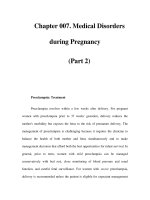
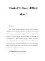
Atopic dermatitis. Lichenified, red plaque with
erosions in the antecubital fossa
PART1.MIF Page 69 Wednesday, October 29, 2003 4:13 PM
70 Atrophie brilliante
Atrophie brilliante
᭤ Confluent and reticulated papillo-
matosis
Atrophoderma of Pasini and
Pierini
Synonym(s)
Idiopathic atrophoderma of Pasini and
Pierini; atrophodermia idiopatica progres-
siva
Definition
Asymptomatic atrophy of the skin charac-
terized by single or multiple, defined,
depressed areas of skin
Pathogenesis
Possibly an end result of morphea; possibly
related to spirochete infection (in Europe)
Clinical manifestation
Presenting as asymptomatic, slightly ery-
thematous plaque or plaques on the trunk;
lesions developing slate-gray to brown pig-
mentation, sharp peripheral border, and
central depression
Differential diagnosis
Morphea; lichen sclerosus; skin atrophy
from steroid injection; anetoderma; post-
inflammatory hyperpigmentation
Therapy
Doxycycline; amoxicillin; hyperpigmenta-
tion component: Q-switched alexandrite
laser
References
Buechner SA, Rufli T (1994) Atrophoderma of
Pasini and Pierini. Clinical and histopathologic
findings and antibodies to Borrelia burgdorferi
in thirty-four patients. Journal of the American
Academy of Dermatology 30(3):441–446
Atrophoderma
pigmentosum
᭤ Xeroderma pigmentosum
Atrophoderma reticulatum
᭤ Keratosis pilaris atrophicans
Atrophoderma
vermiculatum
᭤ Keratosis pilaris atrophicans
᭤ Ulerythema ophryogenes
Atypical fibroxanthoma
Synonym(s)
Paradoxical fibrosarcoma; pseudosarcoma;
pseudosarcomatous reticulohistiocytoma;
pseudosarcomatous dermatofibroma
Definition
Rapidly enlarging tumor, arising in chroni-
cally sun-exposed skin, with histologic fea-
tures suggesting a malignant connective tis-
sue neoplasm, but usually benign clinical
course
Pathogenesis
Solar radiation and prior X-irradiation pos-
sible predisposing factors
Clinical manifestation
Firm, solitary, eroded or ulcerated papule
or nodule on sun-exposed skin, particu-
larly the ear, nose, and cheek; most com-
mon in elderly patients
PART1.MIF Page 70 Wednesday, October 29, 2003 4:13 PM
Atypical mole syndrome 71
A
Differential diagnosis
Squamous cell carcinoma; pyogenic granu-
loma; melanoma; basal cell carcinoma;
Merkel cell carcinoma; cutaneous metasta-
sis; leiomyosarcoma; dermatofibrosarcoma
protuberans
Therapy
Mohs micrographic surgery
ଙ
; elliptical
excision; destruction by electrodesiccation
and curettage
References
Davis JL, Randle HW, Zalla MJ, Roenigk RK,
Brodland DG (1997) A comparison of Mohs mi-
crographic surgery and wide excision for the
treatment of atypical fibroxanthoma. Dermato-
logic Surgery 23(2):105–110
Atypical lipoma
᭤ Liposarcoma
Atypical lipomatous tumors
᭤ Liposarcoma
Atypical melanocytic nevus
᭤ Atypical mole
Atypical mole
Synonym(s)
Active junctional nevus; atypical melano-
cytic nevus; B-K mole, Clark's nevus;
atypical mole syndrome; dysplastic mole;
dysplastic nevus
Definition
Benign melanocytic growth, possibly shar-
ing some of the clinical or microscopic fea-
tures of melanoma, but not a melanoma
Pathogenesis
Genetic component in some patients
(melanoma-prone families; familial atypi-
cal mole syndrome); sunlight exposure pos-
sibly a factor
Clinical manifestation
Va r i a b le features, with some or all of the
following: asymmetrical conformation;
irregular border which can fade impercepti-
bly into the surrounding skin; variable col-
oration, with shades of tan, brown, black;
and red; diameter > 6 mm; elevated center
and feathered, flat border, giving the lesion
the appearance of a fried egg
Differential diagnosis
Melanoma; compound nevus; seborrheic
keratosis; dermatofibroma; wart
Therapy
Avoidance of excessive sun exposure; use of
sunscreen with a sun protective factor of 15
or greater; evaluation of other family mem-
bers for evidence of atypical moles; base-
line photographs of entire skin surface, if
possible
References
Slade J, Marghoob AA, Salopek TG, Rigel DS, Kopf
AW, Bart RS (1995) Atypical mole syndrome:
risk factor for cutaneous malignant melanoma
and implications for management. Journal of
the American Academy of Dermatology
32(3):479–494
Atypical mole syndrome
᭤ Atypical mole
PART1.MIF Page 71 Wednesday, October 29, 2003 4:13 PM
72 Audry’s glands
Audry’s glands
᭤ Fordyce’s disease
Auranofin
Trade name(s)
Ridaura
Generic available
No
Drug class
Anti-rheumatic
Mechanism of action
Inhibition of complement and lysosomal
enzymes; normalization of defective Lang-
erhans cell antigen presentation
Dosage form
3 mg tablet
Dermatologic indications and dosage
See table
Common side effects
Cutaneous: skin eruption, stomatitis, pruri-
tus, glossitis
Gastrointestinal: diarrhea, abdominal pain
Laboratory: anemia, leukopenia, proteinu-
ria
Neurologic: change in taste sensation
Ocular: keratitis
Serious side effects
Bone marrow: agranulocytosis
Neurologic: seizures
Pulmonary: pneumonitis
Renal: renal failure, nephrotic syndrome
Drug interactions
Atovaquone/proguanil
Auranofin. Dermatologic indications and dosage
Disease Adult dosage Child dosage
Cicatricial
pemphigoid
3 mg PO twice daily Initial: 0.1 mg per kg daily in 1–2
divided doses; usual maintenance:
0.15 mg/kg/day in 1–2 divided
doses; maximum: 0.2 mg/kg/day in
1–2 divided doses
Epidermolysis bullosa
acquisita
3 mg PO twice daily Initial: 0.1 mg per kg daily; usual
maintenance: 0.15 mg/kg/day in
1–2 divided doses; maximum:
0.2 mg/kg/day in 1–2 divided doses
Lupus erythematosus 3 mg PO twice daily Initial: 0.1 mg per kg daily in 1–2
divided doses; usual maintenance:
0.15 mg/kg/day in 1–2 divided
doses; maximum: 0.2 mg/kg/day in
1–2 divided doses
Pemphigus vulgaris 3 mg PO twice daily Initial: 0.1 mg per kg daily in 1–2
divided doses; usual maintenance:
0.15 mg/kg/day in 1–2 divided
doses; maximum: 0.2 mg/kg/day in
1–2 divided doses
PART1.MIF Page 72 Wednesday, October 29, 2003 4:13 PM
Aurothioglucose 73
A
Contraindications/precautions
Hypersensitivity to drug class or compo-
nent; pulmonary fibrosis; bone marrow
aplasia; caution with impaired liver or renal
function
References
Papp KA, Shear NH (1991) Systemic gold therapy.
Clinics in Dermatology 9(4):535–551
Auriculotemporal syndrome
Synonym(s)
Frey’s syndrome; Baillarger's syndrome;
Dupuy's syndrome; salivosudoriparous
syndrome; sweating gustatory syndrome;
gustatory sweating
Definition
Gustatory sweating secondary to auriculo-
temporal nerve injury
Pathogenesis
Misdirection of parasympathetic fibers,
which migrate into the postganglionic sym-
pathetic fibers to innervate the sweat glands
Clinical manifestation
Flushing or sweating on one side of the face
when certain foods are eaten
Differential diagnosis
Gustatory sweating from diabetic neuropa-
thy or post-herpetic neuralgia; Horner’s
syndrome; lacrimal sweating; harlequin
syndrome
Therapy
Surgical: tympanic neurectomy for severe
symptoms; perineural alcohol injection
Medical: scopolamine 3–5% cream applied
twice daily; aluminium chloride
᭤ Gustatory sweating
References
Kaddu S, Smolle J, Komericki P, Kerl H (2000) Au-
riculotemporal (Frey) syndrome in late child-
hood: an unusual variant presenting as
gustatory flushing mimicking food allergy.
Pediatric Dermatology 17(2):126–128
Aurothioglucose
Trade name(s)
Solganol
Generic available
No
Drug class
Anti-rheumatic
Mechanism of action
Inhibition of complement and lysosomal
enzymes; normalization of defective Lang-
erhans cell antigen presentation
Dosage form
Intramuscular injection
Dermatologic indications and dosage
See table
Common side effects
Cutaneous: stomatitis, glossitis, skin erup-
tion, pruritus
Gastrointestinal: diarrhea, abdominal pain,
dyspepsia, change in taste sensation
Laboratory: proteinuria, anemia, leukope-
nia
Neurologic: change in taste sensation
Ocular: keratitis
Serious side effects
Laboratory: agranulocytosis
Neurologic: seizures
Pulmonary: pneumonitis
Renal: renal failure, nephrotic syndrome
Drug interactions
Atovaquone/proguanil
PART1.MIF Page 73 Wednesday, October 29, 2003 4:13 PM
74 Auspitz sign
Contraindications/precautions
Hypersensitivity to drug class or compo-
nent; pulmonary fibrosis; bone marrow
aplasia; caution with impaired liver or renal
function
References
Papp KA, Shear NH (1991) Systemic gold therapy.
Clinics in Dermatology 9(4):535–551
Auspitz sign
Definition
Bleeding points appearing when overlying
scale removed physically from a lesion of
psoriasis
References
Bernhard JD (1997) Clinical pearl: auspitz sign in
psoriasis scale. Journal of the American Acade-
my of Dermatology 36(4):621
Autoeczematization
᭤ Id reaction
Autoerythrocyte
sensitization
᭤ Autoerythrocyte sensitization syn-
drome
Aurothioglucose. Dermatologic indications and dosage
Disease Adult dosage Child dosage
Cicatricial
pemphigoid
25–50 mg IM once weekly Initial – 0.25 mg per kg per dose first
week; increment at 0.25 mg per kg
per dose increasing with each
weekly dose; maintenance –
0.75–1 mg per kg per dose weekly,
not to exceed 25 mg per dose
Epidermolysis bullosa
acquisita
25–50 mg IM once weekly Initial – 0.25 mg per kg per dose first
week; increment at 0.25 mg per kg
per dose increasing with each
weekly dose; maintenance –
0.75–1 mg per kg per dose weekly,
not to exceed 25 mg per dose
Lupus erythematosus;
pemphigus vulgaris;
cicatricial
pemphigoid;
epidermolysis bullosa
acquisita
25–50 mg IM once weekly Initial – 0.25 mg per kg per dose first
week; increment at 0.25 mg per kg
per dose increasing with each
weekly dose; maintenance –
0.75–1 mg per kg per dose weekly,
not to exceed 25 mg per dose
Pemphigus vulgaris 25–50 mg IM once weekly Initial – 0.25 mg per kg per dose first
week; increment at 0.25 mg per kg
per dose increasing with each
weekly dose; maintenance –
0.75–1 mg per kg per dose weekly,
not to exceed 25 mg per dose
PART1.MIF Page 74 Wednesday, October 29, 2003 4:13 PM
Azathioprine 75
A
Autoerythrocyte
sensitization syndrome
Synonym(s)
Gardner-Diamond syndrome; autoeryth-
rocyte sensitization; psychogenic purpura;
purpura autoerythrocytica
Definition
Purpuric disorder in women, characterized
by painful ecchymotic patches, unrelated to
vascular or clotting abnormalities
Pathogenesis
Possibly an immune-mediated reaction;
psychological issues in the patients possi-
bly the main causative factor
Clinical manifestation
Painful ecchymoses, often appearing after
minor trauma, usually over the extremities
and trunk; lesions appearing in crops, and
lasting for weeks to months
Differential diagnosis
Anaphylactoid purpura; Ehlers-Danlos syn-
drome; child abuse; factitial purpura; amy-
loidosis; thrombotic thrombocytopenic
purpura; solar purpura; leukemia
Therapy
Medroxyprogesterone acetate 10 mg PO per
day or 150 mg intramuscularly once per
month; prednisone; antihistamines, first
generation
References
Berman DA, Roenigk HH, Green D (1992) Auto-
erythrocyte sensitization syndrome (psycho-
genic purpura). Journal of the American
Academy of Dermatology 27(5 Pt 2):829–832
Autoimmune alopecia
᭤ Alopecia areata
Autoimmune dermatosis of
pregnancy
᭤ Herpes gestationis
Autosensitization
᭤ Id reaction
Autosomal dominant
ichthyosis
᭤ Ichthyosis vulgaris
Autumnal fever
᭤ Leptospirosis
Axillary freckling
Definition
Brown macules in the axillary vault, present
in more than 90 percent of people with neu-
rofibromatosis, type 1
References
Wainer S (2002) A child with axillary freckling
and cafe au lait spots. Canadian Medical Asso-
ciation Journal 167(3):282–283
Azathioprine
Trade name(s)
Imuran
PART1.MIF Page 75 Wednesday, October 29, 2003 4:13 PM
76 Azathioprine
Generic available
Ye s
Drug class
Antimetabolite; immunosuppressant
Mechanism of action
Active metabolite is purine analog, which
inhibits DNA and RNA synthesis and has
immunosuppressive activity
Azathioprine. Dermatologic indications and dosage
Disease Adult dosage Child dosage
Atopic dermatitis 2–3 mg per kg PO daily 2–3 mg per kg PO daily
Behçet’s disease Corticosteroid sparing function;
2–3 mg per kg PO daily
Corticosteroid sparing function;
2–3 mg per kg PO daily
Bullous pemphigoid Corticosteroid sparing function;
2–3 mg per kg PO daily
Corticosteroid sparing function;
2–3 mg per kg PO daily
Chronic actinic
dermatitis
Corticosteroid sparing function;
2–3 mg per kg PO daily
Corticosteroid sparing function;
2–3 mg per kg PO daily
Cicatricial
pemphigoid
Corticosteroid sparing function;
2–3 mg per kg PO daily
Corticosteroid sparing function;
2–3 mg per kg PO daily
Fogo selvagem 2–3 mg per kg PO daily 2–3 mg per kg PO daily
Leukocytoclastic
vasculitis
Corticosteroid sparing function;
2–3 mg per kg PO daily
Corticosteroid sparing function;
2–3 mg per kg PO daily
Lupus erythematosus,
acute
Corticosteroid sparing function;
2–3 mg per kg PO daily
Corticosteroid sparing function;
2–3 mg per kg PO daily
Mixed connective
tissue disease
2–3 mg per kg PO daily 2–3 mg per kg PO daily
Paraneoplastic
pemphigus
Corticosteroid sparing function;
2–3 mg per kg PO daily
Corticosteroid sparing function;
2–3 mg per kg PO daily
Pemphigus foliaceus 2–3 mg per kg PO daily 2–3 mg per kg PO daily
Pemphigus vulgaris Corticosteroid sparing function;
2–3 mg per kg PO daily
Corticosteroid sparing function;
2–3 mg per kg PO daily
Persistent light
reaction
Corticosteroid sparing function;
2–3 mg per kg PO daily
Corticosteroid sparing function;
2–3 mg per kg PO daily
Polyarteritis nodosa Corticosteroid sparing function;
2–3 mg per kg PO daily
Corticosteroid sparing function;
2–3 mg per kg PO daily
Pyoderma
gangrenosum
Corticosteroid sparing function;
2–3 mg per kg PO daily
Corticosteroid sparing function;
2–3 mg per kg PO daily
Relapsing
polychondritis
2–3 mg per kg PO daily 2–3 mg per kg PO daily
Sarcoidosis 2–3 mg per kg PO daily 2–3 mg per kg PO daily
Sulzberger-Garbe
syndrome
2–3 mg per kg PO daily 2–3 mg per kg PO daily
Weber-Christian
disease
2–3 mg per kg PO daily 2–3 mg per kg PO daily
Wegener’s
granulomatosis
Corticosteroid sparing function;
2–3 mg per kg PO daily
Corticosteroid sparing function;
2–3 mg per kg PO daily
PART1.MIF Page 76 Wednesday, October 29, 2003 4:13 PM
Azelaic acid 77
A
Dosage form
50 mg tablet
Dermatologic indications and dosage
See table
Common side effects
Cutaneous: alopecia, skin eruption
Gastrointestinal: nausea and vomiting,
diarrhea, dyspepsia
Laboratory: elevated liver enzymes
Serious side effects
Cutaneous: hypersensitivity reaction
Gastrointestinal: hepatotoxicity, pancreati-
tis
Immune: immunosuppression
Neoplastic: increased risk of neoplasm,
particularly lymphoma
Drug interactions
ACE inhibitors; allopurinol; cisplatin; cyto-
toxic chemotherapeutic agents; interferon
alfa 2a; interferon beta; mycophenolate
mofetil; warfarin; zidovudine
Contraindications/precautions
Hypersensitivity to drug class or compo-
nent; pregnancy; caution if patient has low
levels or lacks thiopurine methyltrans-
ferase (measure enzyme level before start-
ing therapy); caution if impaired liver func-
tion
References
Silvis NG (2001) Antimetabolites and cytotoxic
drugs. Dermatologic Clinics 19(1):105–118
Azelaic acid
Trade name(s)
Azelex; Finacea
Generic available
No
Drug class
Anti-acne; anti-rosacea
Mechanism of action
May be related to antimicrobial effects
Dosage form
15% cream, 20% cream
Dermatologic indications and dosage
See table
Common side effects
Cutaneous: pruritus, burning sensation,
dryness, skin eruption
Serious side effects
None
Drug interactions
None
Contraindications/precautions
Hypersensitivity to drug class or compo-
nent
Azelaic acid. Dermatologic indications and dosage
Disease Adult dosage Child dosage
Acne vulgaris Apply twice daily Apply twice daily
Melasma Apply twice daily Apply twice daily
Postinflammatory
hyperpigmentation
Apply twice daily Apply twice daily
Rosacea Apply twice daily Apply twice daily
PART1.MIF Page 77 Wednesday, October 29, 2003 4:13 PM
78 Azithromycin
References
Nguyen QH, Bui TP (1995) Azelaic acid: pharma-
cokinetic and pharmacodynamic properties
and its therapeutic role in hyperpigmentary
disorders and acne. International Journal of
Dermatology 34(2):75–84
Azithromycin
Trade name(s)
Zithromax
Generic available
No
Drug class
Macrolide antibiotic
Mechanism of action
Inhibits protein synthesis of sensitive bacte-
rial organisms
Dosage form
250 mg, 500 mg tablet; powder for oral sus-
pension
Dermatologic indications and dosage
See table
Common side effects
Cutaneous: skin eruption, vaginitis
Gastrointestinal: nausea, vomiting, abdom-
inal pain, diarrhea, anorexia
Serious side effects
Cutaneous: anaphylaxis, Stevens-Johnson
syndrome, toxic epidermal necrolysis
Gastrointestinal: pseudomembranous coli-
tis, cholestatic jaundice
Drug interactions
Antacids; oral contraceptives; warfarin; dig-
oxin
Azithromycin. Dermatologic indications and dosage
Disease Adult dosage Child dosage
Bacillary angiomatosis 500 mg PO on day 1; 250 mg PO on
days 2–5
Not indicated in those < 45 kg in
weight; 500 mg PO on day 1; 250 mg
PO on days 2–5
Bartonellosis 500 mg PO on day 1; 250 mg PO on
days 2–5
Not indicated in those < 45 kg in
weight; 500 mg PO on day 1; 250 mg
PO on days 2–5
Cellulitis 500 mg PO on day 1; 250 mg PO on
days 2–5
Not indicated in those < 45 kg in
weight; 500 mg PO on day 1; 250 mg
PO on days 2–5
Chancroid 1 gm PO for 1 dose Not indicated in those < 45 kg in
weight; 20 mg per kg PO for 1 dose
Ecthyma 500 mg PO on day 1; 250 mg PO on
days 2–5
Not indicated in those < 45 kg in
weight; 500 mg PO on day 1; 250 mg
PO on days 2–5
Furuncle 500 mg PO on day 1; 250 mg PO on
days 2–5
Not indicated in those < 45 kg in
weight; 500 mg PO on day 1; 250 mg
PO on days 2–5
Impetigo 500 mg PO on day 1; 250 mg PO on
days 2–5
Not indicated in those < 45 kg in
weight; 500 mg PO on day 1; 250 mg
PO on days 2–5
Trench fever 250-500 mg PO for 4 weeks Not indicated in those < 45 kg;
250 mg PO daily for 4 weeks
PART1.MIF Page 78 Wednesday, October 29, 2003 4:13 PM
Azul 79
A
Contraindications/precautions
Hypersensitivity to drug class or compo-
nent; caution in those with impaired liver
function; do not use concomitantly with
terfenadine or astemizole
References
Alvarez-Elcoro S, Enzler MJ (1999) The mac-
rolides: erythromycin, clarithromycin, and azi-
thromycin. Mayo Clinic Proceedings 74(6):613–
634
Azole antifungal agents
Trade name(s)
Generic in parentheses:
Exelderm (sulconazole); Lamisil AT (terbin-
afine); Lotrimin; Mycelex (clotrimazole);
Micatin (miconazole); Nizoral (ketocona-
zole); Oxistat (oxiconazole); Spectazole
(econazole)
Generic available
Ye s
Drug class
Azole antifungal agents
Mechanism of action
Cell wall ergosterol inhibition secondary to
blockade of 14α-demethlyation of lanos-
terol
Dosage form
Cream; solution; lotion
Dermatologic indications and dosage
See table
Common side effects
Cutaneous: skin eruption, pruritus
Serious side effects
None
Drug interactions
None
Contraindications/precautions
Hypersensitivity to drug class or compo-
nent
References
We instein A, Berman B (2002) Topical treatment
of common superficial tinea infections. Ameri-
can Family Physician 65(10):2095–2102
Azul
᭤ Pinta
Azole antifungal agents. Dermatologic indications and dosage
Disease Adult dosage Child dosage
Angular cheilitis Apply twice daily for 2–4 weeks Apply twice daily for 2–4 weeks
Cutaneous candidiasis Apply twice daily for 2–4 weeks Apply twice daily for 2–4 weeks
Majocchi granuloma Apply twice daily for 4–8 weeks Apply twice daily for 4–8 weeks
Onychomycosis Apply twice daily for 2–4 weeks Apply twice daily for 2–4 weeks
Tinea corporis Apply twice daily for 2–4 weeks Apply twice daily for 2–4 weeks
Tinea cruris Apply twice daily for 2–4 weeks Apply twice daily for 2–4 weeks
Tinea faciei Apply twice daily for 2–4 weeks Apply twice daily for 2–4 weeks
Tinea nigra Apply twice daily for 2–4 weeks Apply twice daily for 2–4 weeks
Tinea pedis Apply twice daily for 2–4 weeks Apply twice daily for 2–4 weeks
Tinea versicolor Apply twice daily for 2–4 weeks Apply twice daily for 2–4 weeks
White piedra Apply twice daily for 2–4 weeks Apply twice daily for 2–4 weeks
PART1.MIF Page 79 Wednesday, October 29, 2003 4:13 PM
PART1.MIF Page 80 Wednesday, October 29, 2003 4:13 PM
B
B-K mole
᭤
Atypical mole
Bacillary ailuronosis
᭤
Bacillary angiomatosis
Bacillary angiomatosis
Synonym(s)
Epithelioid angiomatosis
;
bartonellosis
;
bacillary ailuronosis
;
disseminated cat-
scratch disease
Definition
Infection caused by closely related gram-
negative bacteria, Bartonella henselae and
Bartonella quintana, occurring mostly in
immunocompromised patients
Pathogenesis
Gram-negative bacillary infection results
from exposure to flea-infested cats with
B
henselae and the human body louse for
B
quintana
Clinical manifestation
Globular angiomatous papules or nodules
resembling pyogenic granulomas; viola-
ceous nodules resembling Kaposi’s sar-
coma; lichenoid violaceous plaques; subcu-
taneous papules or nodules, with or with-
out ulceration
Differential diagnosis
Kaposi’s sarcoma; glomangioma; verruga
peruana; angiokeratoma; hemangioma;
pyogenic granuloma; gram-positive bacte-
rial abscess; nodal myofibromatosis;
melanoma
Therapy
Erythromycin
ଙ
; azithromycin; clarithromy-
cin; doxycycline
References
Manders SM (1996) Bacillary angiomatosis. Clin-
ics in Dermatology 14(3):295–299
Bacillary peliosis
᭤
Bartonellosis
Bacitracin
Trade name(s)
Bacitracin as single agent: Baciguent; baci-
tracin as one component of a multi-agent
preparation: Betadine antibiotic ointment;
Gold Bond Triple Action; Mycitracin;
Neosporin; Polysporin; Spectrocin Plus
PART2.MIF Page 81 Wednesday, October 29, 2003 4:21 PM
82 Bagdad boil
Generic available
Ye s
Drug class
Antibiotic
Mechanism of action
Inhibits bacterial cell wall synthesis
Dosage form
Cream; ointment
Dermatologic indications and dosage
See table
Common side effects
Cutaneous:
contact dermatitis
Serious side effects
None
Drug interactions
None
Contraindications/precautions
Hypersensitivity to drug class or compo-
nent
References
Bass, JW, Chan DS, Creamer KM, Thompson MW,
Malone FJ, Becker TM, Marks SN (1997) Com-
parison of oral cephalexin, topical mupirocin
and topical bacitracin for treatment of impeti-
go. Pediatric Infectious Disease Journal
16(7):708–710
Bagdad boil
᭤
Leishmaniasis, cutaneous
Baillarger's syndrome
᭤
Auriculotemporal syndrome
Balanitis
Synonym(s)
Balanoposthitis
Definition
Inflammation of the foreskin and head of
the penis
References
Bunker CB (2001) Topics in penile dermatology.
Clinical & Experimental Dermatology
26(6):469–479
Balanitis circumscripta
plasmacellularis
᭤
Zoon balanitis
Balanitis xerotica obliterans
Synonym(s)
Lichen sclerosus of the penis
;
male genital
lichen sclerosus
;
lichen sclerosus et atrophi-
cus of the penis
;
penile lichen sclerosus
Bacitracin. Dermatologic indications and dosage
Disease Adult dosage Child dosage
Impetigo Apply twice per day for 7 days Apply twice per day for 7 days
Postoperative wound
infection prophylaxis
Apply twice per day for 7 days Apply twice per day for 7 days
PART2.MIF Page 82 Wednesday, October 29, 2003 4:21 PM
Bannayan-Riley-Ruvalcaba syndrome 83
B
Definition
Chronic, progressive, sclerosing, inflamma-
tory dermatosis of the penis and prepuce
Pathogenesis
Unknown; minor relationship with autoim-
mune disorders
Clinical manifestation
Presents with soreness, burning sensation,
mild erythema and hypopigmentation; as
disease progresses, single or multiple dis-
crete erythematous papules or macules coa-
lescing into atrophic ivory, white, or purple-
white patches or plaques, which may erode;
possible development of vesiculation; possi-
ble phimosis occurring in uncircumcised
men; occasional signs of lichen sclerosus at
other skin sites
Differential diagnosis
Plasma cell balanitis; candidiasis; lichen
planus; psoriasis; vitiligo; Reiter syndrome;
erythroplasia of Queyrat
Therapy
Surgical therapy: circumcision; laser vapor-
ization
Medical therapy: superpotent topical corti-
costeroids; testosterone propionate 1% oint-
ment applied twice daily; acitretin
᭤
Lichen sclerosus
References
Das S, Tunuguntla HS (2000) Balanitis xerotica
obliterans – A review. World Journal of Urology
18(6):382–387
Balanoposthitis
᭤
Balanitis
Baldness
᭤
Alopecia
Bamboo hair
᭤
Tr ichorrhexis invaginata
Bancroftian filariasis
᭤
Filariasis
Bannayan syndrome
᭤
Bannayan-Riley-Ruvalcaba syn-
drome
Bannayan-Riley-Ruvalcaba
syndrome
Synonym(s)
Bannayan-Zonana syndrome
;
Riley-Smith
syndrome
;
Ruvalcaba-Myhre syndrome
;
Ruvalcaba-Myhre-Smith syndrome
;
Banna-
yan syndrome
;
Cowden/Bannayan-Riley-
Ruvalcaba overlap syndrome
;
PTEN hama-
rtoma tumor syndrome
;
macrocephaly
;
pseudopapilledema
;
multiple hemangiom-
ata syndrome
;
multiple lipomas
Definition
Disease characterized by hamartomatous
polyps of the small and large intestine,
macrocephaly, lipomas, hemangiomas, thy-
roid abnormalities, and freckling of the
penis
Pathogenesis
Autosomal dominant inheritance; muta-
tion in the tumor suppressor gene, PTEN
PART2.MIF Page 83 Wednesday, October 29, 2003 4:21 PM
84 Bannayan-Zonana syndrome
Clinical manifestation
Hamartomatous polyps of the small and
large intestine; macrocephaly; lipomas;
hemangiomas; thyroid abnormalities;
penile freckling; developmental delay;
increased risk for both benign and malig-
nant tumors
Differential diagnosis
Cowden’s syndrome; Gardner’s syndrome;
multiple lentigines syndrome
Therapy
Increased breast, thyroid, and colon cancer
surveillance; surgical excision of lipomas
and hemangiomas for cosmetic purposes
only
References
Fargnoli MC, Orlow SJ, Semel-Concepcion J, Bo-
lognia JL (1996) Clinicopathologic findings in
the Bannayan-Riley-Ruvalcaba syndrome. Ar-
chives of Dermatology 132(10):1214–1218
Bannayan-Zonana
syndrome
᭤
Bannayan-Riley-Ruvalcaba syn-
drome
Barber's itch
᭤
Sycosis barbae
Barber-Say syndrome
Synonym(s)
Say syndrome
Definition
Disease entity consisting of hypertrichosis,
xerosis, cutis laxa, dysmorphic facial fea-
tures, and eye changes
Pathogenesis
Autosomal recessive inheritance
Clinical manifestation
Hypertrichosis over the upper trunk and
face; xerosis; generalized cutis laxa; macros-
tomia; opacification of the corneas; varia-
ble nystagmus
Differential diagnosis
Cone-rod congenital amaurosis; ablepha-
ron-macrostomia syndrome; Turner’s syn-
drome; Brachmann-de Lange syndrome;
Sanfilippo syndrome; Hunter’s syndrome;
leprechaunism
Therapy
No effective therapy
References
Martinez Santana S, Perez Alvarez F, Frias JL,
Martinez-Frias ML (1993) Hypertrichosis,
atrophic skin, ectropion, and macrostomia
(Barber-Say syndrome): report of a new case.
American Journal of Medical Genetics
47(1):20–23
Barlow’s disease
Synonym(s)
Möller-Barlow disease
;
Barlow’s syndrome
;
Cheadle-Möller-Barlow syndrome
;
Moeller's disease
;
infantile scurvy
;
vitamin
C deficiency syndrome
Definition
Vitamin C deficiency disease in children,
manifested by gingival lesions, hemor-
rhage, arthralgia, loss of appetite, and list-
lessness
Pathogenesis
Vitamin C deficiency, after at least 3 months
of severe or total lack of vitamin C, result-
ing in defective collagen synthesis and
defective folic acid and iron utilization
PART2.MIF Page 84 Wednesday, October 29, 2003 4:21 PM
Bartonellosis 85
B
Clinical manifestation
Perifollicular hyperkeratotic papules, sur-
rounded by a hemorrhagic halo; hairs
twisted like corkscrews and possibly frag-
mented; submucosal gingival bleeding, sub-
periosteal hemorrhage, arthralgia; ano-
rexia; listlessness; exophthalmos and con-
junctival hemorrhage; poor wound healing
Differential diagnosis
Vasculitis; child abuse; coagulation abnor-
malities with leukemia; platelet abnormali-
ties, etc.; deep vein thrombosis; throm-
bophlebitis
Therapy
Ascorbic acid 150–300 mg per day for 1
month
References
Ghorbani AJ, Eichler C (1994) Scurvy. Journal of
the American Academy of Dermatology 30(5 Pt
2):881–883
Barlow’s syndrome
᭤
Barlow’s disease
Barraquer-Simons disease
᭤
Progressive lipodystrophy
Barraquer-Simons syndrome
᭤
Progressive lipodystrophy
Bartonellosis
Synonym(s)
Cat scratch disease
;
catscratch disease
;
trench fever
;
urban trench fever
;
bacillary
peliosis
;
Parinaud oculoglandular syn-
drome
;
Parinaud's oculoglandular syn-
drome
;
Oroya fever
;
Carrión disease
;
Carrión's disease
;
verruga peruana; benign
lymphoreticulosis
Definition
Infections caused by species belonging to
the bacterial genus Bartonella
Pathogenesis
Bartonella henselae found in association
with both domestic and feral cats and pre-
sumably passed from cat to human; Bar-
tonella quintana spread via human body
louse
Clinical manifestation
Cat scratch disease: papule or pustule
developing 5–10 days after exposure; fever;
malaise; lymphadenopathy
Oroya fever (verruga peruana): onset of
fever 3–12 weeks after a sand fly bite; crops
of small papules enlarging and healing by
fibrosis over several months
Differential diagnosis
Lymphoma; leukemia; deep fungal infec-
tion; tuberculosis; plague; lymphogranu-
loma venereum; AIDS; syphilis; dengue
fever; malaria; babesiosis
Therapy
Doxycycline; erythromycin; azithromycin;
clarithromycin
᭤ Bacillary angiomatosis
References
Maguina C, Gotuzzo E (2000) Bartonellosis: new
and old. Infectious Disease Clinics of North
America 14(1):1–22
PART2.MIF Page 85 Wednesday, October 29, 2003 4:21 PM
86 Bart’s syndrome
Bart’s syndrome
Synonym(s)
None
Definition
Subtype of dominant dystrophic epidermol-
ysis bullosa with congenital localized
absence of skin, nail abnormalities, and
blistering
Pathogenesis
Mutation of the COLA7A1 gene, resulting in
the production of poorly formed anchoring
fibrils at the skin’s basement membrane
zone
Clinical manifestation
Congenital erosions of the lower extremi-
ties, which heal with hairless scars; trauma-
induced blistering; absent or dystrophic
nails; mucous membrane erosions only in
early life
Differential diagnosis
Aplasia cutis congenita; epidermolysis bul-
losa simplex; junctional epidermolysis bul-
losa; child abuse
Therapy
Hydrocolloid dressings to erosions; petrola-
tum between toes to minimize scarring
References
Amichai B, Metzker A (1994) Bart's syndrome. In-
ternational Journal of Dermatology 33(3):161–
163
Basal cell carcinoma
Synonym(s)
Basal cell epithelioma; basalioma; Jacob’s
ulcer; rodent ulcer
Definition
Cutaneous neoplasm arising from pluripo-
tential cells of the epidermis or its append-
ages
Pathogenesis
Early, intense sun exposure possibly caus-
ing p53 tumor suppressor gene mutations,
allowing unrestricted proliferation
Clinical manifestation
Nodular variant: pearly, translucent papule
with central depression, erosion, or ulcera-
tion; rolled borders; telangiectasia on the
surface
Pigmented variant: flecks of gray or blue
pigment in addition to features described
for nodular variant
Superficial variant: pink-to-brown, scaly
plaque or papule, often with annular con-
figuration
Morpheaform variant: poorly demarcated,
sclerotic plaque or papule
Differential diagnosis
Squamous cell carcinoma; nevus; fibrous
papule; wart; appendage tumor; seborrheic
keratosis; sebaceous gland hyperplasia;
Bowen’s disease
Therapy
Primary tumor in anatomically insensitive
sites: destruction by electrodesiccation and
curettage; elliptical excision; cryotherapy;
orthovoltage radiation therapy; fluorour-
acil cream
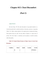
Basal cell carcinoma. Papule with rolled margins
and central erosion on the nasal bridge
PART2.MIF Page 86 Wednesday, October 29, 2003 4:21 PM
Basan syndrome 87
B
Recurrent tumor or tumors in anatomically
sensitive sites: Mohs micrographic sur-
gery
ଙ
References
Thissen MR, Neumann MH, Schouten LJ (1999) A
systematic review of treatment modalities for
primary basal cell carcinomas. Archives of Der-
matology 135(10):1177–1183
Basal cell epithelioma
᭤ Basal cell carcinoma
Basal cell nevus syndrome
Synonym(s)
Nevoid basal cell carcinoma syndrome;
Gorlin syndrome; Gorlin-Goltz syndrome;
bifid-rib basal-cell nevus syndrome
Definition
Inherited group of defects involving the
skin, nervous system, eyes, endocrine
glands, and bones, producing an unusual
facial appearance and a predisposition for
skin cancers
Pathogenesis
Chromosomal mutation of the PTC gene, a
tumor suppressor gene; inactivation of this
gene associated with development of basal
cell carcinoma, other tumors, and develop-
mental errors
Clinical manifestation
Pitting of the palms or soles; multiple basal
cell carcinomas, often early in life; jaw
cysts; cleft palate; coarse facies with milia,
frontal bossing, widened nasal bridge, and
mandibular prognathia; strabismus; dys-
trophic canthorum; ocular hypertelorism;
calcification of the falx cerebri; spine and
rib abnormalities; high arched eyebrows
and palate; kidney anomalies; hypogonad-
ism in males
Differential diagnosis
Non-syndromic basal cell carcinoma; Bazex
syndrome; linear unilateral basal cell nevus
with comedones; Rasmussen syndrome;
Rombo syndrome
Therapy
Medical therapy: fluorouracil cream;
isotretinoin; radiation therapy
Surgical therapy: primary tumor in ana-
tomically insensitive sites – destruction by
electrodesiccation and curettage; elliptical
excision; cryotherapy; fluorouracil cream;
recurrent tumor or those in anatomically
sensitive sites: Mohs micrographic sur-
gery
ଙ
References
Gorlin RJ (1987) Nevoid basal-cell carcinoma syn-
drome. Medicine 66(2):98–113
Basal cell papilloma
᭤ Seborrheic keratosis
Basalioma
᭤ Basal cell carcinoma
Basan syndrome
Synonym(s)
Ectodermal dysplasia absent der-
matoglyphics
Definition
Autosomal dominant syndrome consisting
of ectodermal dysplasia, absent derma-
toglyphic pattern, nail abnormalities, and a
simian crease
Pathogenesis
Inherited; mutation site unknown
PART2.MIF Page 87 Wednesday, October 29, 2003 4:21 PM
88 Bather’s itch
Clinical manifestation
Thin skin; simian crease; multiple dental
caries; absent or decreased eyebrows; nail
dystrophy; sparse or absent scalp hair;
decreased sweating; photophobia; absent
dermatoglyphic pattern
Differential diagnosis
Anhidrotic ectodermal dysplasia; hidrotic
ectodermal dysplasia; focal dermal hypo-
plasia; Down’s syndrome; progeria
Therapy
No effective therapy
References
Masse JF, Perusse R (1994) Ectodermal dysplasia.
Archives of Disease in Childhood 71(1):1–2
Bather’s itch
᭤ Cercarial dermatitis
Bazin’s disease
᭤ Nodular vasculitis
Beals’ arachnodactyly
᭤ Beals-Hecht syndrome
Beals’ syndrome
᭤ Beals-Hecht syndrome
Beals-Hecht syndrome
Synonym(s)
Beals’ arachnodactyly; Beals’ syndrome;
Hecht-Beals syndrome; congenital contrac-
tural arachnodactyly syndrome
Definition
Heritable disorder of connective tissue,
present from birth, combining features of
Marfan’s syndrome with arthrogryposis
Pathogenesis
Unknown; autosomal dominant inheritance
Clinical manifestation
Multiple, congenital, joint contractures;
arachnodactyly; dolichostenomelia; kypho-
scoliosis; changes of the ear muscle, pro-
ducing crumpled-appearing ears
Differential diagnosis
Marfan’s syndrome; Stickler’s syndrome
Therapy
None
References
Jones JL, Lane JE, Logan JJ, Vanegas ME (2002)
Beals-Hecht syndrome. Southern Medical Jour-
nal 95(7):753–755
Bean syndrome
᭤ Blue rubber bleb nevus syndrome
Bean-Walsh angioma
᭤ Ve n o us lake
PART2.MIF Page 88 Wednesday, October 29, 2003 4:21 PM
Beckwith-Wiedemann syndrome 89
B
Beau’s lines
Definition
Transverse grooves or lines seen on finger-
nails following systemic illness, local
trauma, or skin disease involving the fin-
gertips
References
De Berker D (1994) What do Beau's lines mean?
International Journal of Dermatology
33(8):545–546
Becker melanosis
᭤ Becker’s nevus
Becker nevus
᭤ Becker’s nevus
Becker pigmented hairy
nevus
᭤ Becker’s nevus
Becker’s nevus
Synonym(s)
Becker melanosis; Becker nevus; Becker’s
pigmented hairy nevus; Becker pigmented
hairy nevus; nevus spilus tardus; pigmented
hairy epidermal nevus
Definition
Acquired melanosis and hypertrichosis in a
unilateral distribution
Pathogenesis
Androgens possibly a factor in growth of
the lesion
Clinical manifestation
Asymptomatic, irregular, tan-to-brown
patch, most commonly located over the
chest, shoulder, or back; often at the time of
puberty; thick, brown-to-black hairs devel-
ops both within and in close proximity to
the patch; possibly associated with underly-
ing smooth muscle hamartoma
Differential diagnosis
Melanoma; café au lait macule; Albright’s
syndrome; congenital melanocytic nevus;
nevus spilus; postinflammatory hyperpig-
mentation
Therapy
Treat ment for cosmetic reasons only – sur-
gical excision; Q-switched ruby laser abla-
tion; Q-switched neodymium: yttrium-alu-
minium-garnet (YAG) laser
References
Goldman MP, Fitzpatrick RE (1994) Treatment of
benign pigmented cutaneous lesions. Cutane-
ous Laser Surgery 106–141
Becker’s pigmented hairy
nevus
᭤ Becker’s nevus
Beckwith-Wiedemann
syndrome
Synonym(s)
None
Definition
Disorder consisting of macroglossia, viscer-
omegaly, large body size, umbilical hernia
or omphalocele, neonatal hypoglycemia
PART2.MIF Page 89 Wednesday, October 29, 2003 4:21 PM