Diseases of the Gallbladder and Bile Ducts - part 4 pot
Bạn đang xem bản rút gọn của tài liệu. Xem và tải ngay bản đầy đủ của tài liệu tại đây (1.36 MB, 44 trang )
124 Section 2: Diagnostic and therapeutic approaches for the biliary tree and gallbladder
The primary differential diagnosis of PSC is diffuse scleros-
ing carcinoma of the bile ducts, which represents less than
10% of bile duct carcinomas [11]. Diffuse metastatic disease
to the liver can cause multiple strictures of the intrahepatic
ducts without biliary dilatation.
Primary biliary cirrhosis can mimic PSC and is seen in
middle-aged women. Recurrent biliary infections related to
gallstones or surgical stricture produce similar findings.
Sclerosing cholangitis can also be iatrogenic, occurring after
infusion of chemotherapeutic agents through the hepatic
artery [12].
Cystic biliary disease
The etiology of cystic biliary disease is unclear (see Chapter
17). The disorder may be related to anomalous drainage of
the pancreatic and biliary ducts and loss of the distal sphinc-
ter mechanism [13]. The most commonly used classifi cation
system for biliary cystic disease is the Todani modifi cation of
the Alonso–Lej classifi cation. This system describes five
types of cysts [13]. Type I is the most common (80 to 90%)
and is a single cystic dilatation of the common hepatic duct,
common bile duct, or both. Type II is a diverticulum of the
common bile duct and accounts for 3%. Type III is a cystic di-
latation of the common bile duct in the wall of the duode-
num, accounting for 5%. Type IV is made up of multiple cysts
involving the extrahepatic and/or intrahepatic ducts and
accounts for 10% of cases. Type V is the variant known as
Caroli’s disease. Type V is commonly associated with con-
genital fibrosis and cysts outside of the liver.
Complications include biliary obstruction, hepatic abscess,
cholangitis, and bile duct cancer. The risk of bile duct cancer
is increased 20 fold in this patient group. It is unusual for gall-
stones to be found in association with biliary cystic disease.
Choledocholithiasis
Stones in the bile ducts either form there primarily or migrate
there from the gallbladder. Primary bile duct stones are com-
posed mainly of calcium bilirubinate. Bile stasis, dietary fac-
tors, and bacterial or parasitic infection contribute to their
formation, although their precise pathogenesis is unknown
[14].
Single or multiple filling defects in the biliary tree charac-
terize the presence of gallstones (Figs 6.4 and 6.5). Because
contrast may obscure gallstones in the biliary tree, the con-
trast should be diluted with normal saline for optimal visual-
ization. Air bubbles or blood clots can obscure or mimic
gallstones. Changing patient positioning while observing
the filling defects under fluoroscopy helps to differentiate air
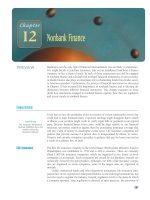
Figure 6.3 Cholangiogram in a patient with primary sclerosing
cholangitis demonstrates multiple long strictures (arrows) of the right
hepatic ducts with areas of focal dilatation between the strictures.
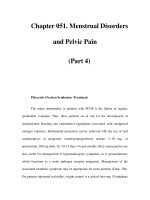
Figure 6.4 Multifaceted gallstones (arrows) appear as filling defects
throughout the gallbladder and bile ducts.
Chapter 6: Percutaneous biliary imaging and intervention 125
bubbles from stones. The air bubbles seek ananterior location
and coalesce with one another.
Blood clots are more diffi cult to differentiate from stones.
Blood often enters the biliary tree during the puncture by the
PTC needle. The suspected presence of blood clots requires
repeating the cholangiogram in several days. The lytic prop-
erties of bile and the passing of clots through the drainage
catheter will have cleared blood clots from the biliary tree
during that time.
Gallstones sometimes become impacted within the bile
ducts. In this form, they can be mistaken for a polypoid tumor
[11]. Manipulation with a stone extraction basket or balloon
may help differentiate between the two entities.
Mirizzi’s syndrome occurs when a gallstone lodges in the
cystic duct or gallbladder neck and causes extrinsic compres-
sion of the common bile duct. The compression usually
occurs at the lateral aspect of the common bile duct [15]. The
patient develops jaundice because of common bile duct
obstruction.
Benign biliary strictures
More than 90% of benign biliary strictures are the result of
surgical trauma, most commonly cholecystectomy (see
Chapter 10) [16]. Surgical strictures may be caused by duct li-
gation or clipping, as is seen with emergency maneuvers to
control massive bleeding. They can also result from thermal
injury or injury to the small arteries that run within the com-
mon bile duct wall [16]. Transection of the duct interrupts
the delicate arterial blood supply to the ducts. This may be the
reason for ischemia and stenosis sometimes seen with bili-
ary–enteric bypass operations (Fig. 6.6). Torsion of the bile
duct may also occur following choledochojejunostomy
(Fig. 6.7).
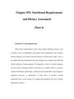
Figure 6.5 Multifaceted gallstones appear as filling defects above a
benign anastomotic stricture (arrow) which developed in a patient who
underwent biliary-enteric bypass for a laparoscopic cholecystectomy
bile duct injury.
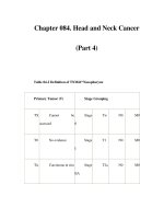
Figure 6.6 A benign focal anastomotic stricture
(arrow) is present in a patient who underwent
biliary–enteric bypass for pancreatic cancer.
126 Section 2: Diagnostic and therapeutic approaches for the biliary tree and gallbladder
Benign biliary strictures are a common problem following
orthotopic liver transplantation and occur in 3 to 22% of the
patients (see Chapter) [17]. The etiology of anastomotic stric-
tures in this group is not well understood. Postoperative
fibrosis and possibly ischemia are felt to be the causes. Pro-
longed cold ischemic time, hepatic artery thrombosis, surgi-
cal interruption of the peribiliary arterial plexus, and chronic
rejection are potential causes of nonanastomotic biliary stric-
tures in the transplanted liver [17] (Figs 6.8 and 6.9).
Postoperative benign strictures are usually short and have
an abrupt change in caliber at the site of abnormality. There is
ductal dilatation above the stricture. Intrahepatic abscesses
may be present. A longer stricture should raise the suspicion
of malignancy [6].
Nonsurgical causes of benign biliary obstruction include
gallstone erosion into the main bile duct, pericholedochal
abscess, blunt trauma, compression by pseudoaneurysm or
pseudocyst, and pancreatitis (Figs 6.10 and 6.11).
Malignant biliary strictures
Distinguishing between malignant and benign strictures is
difficult. Although certain cholangiographic features de-
scribed in this section may suggest the presence of a malig-
nant stricture, these features are not specific. Clinical
information and results of noninvasive radiologic tests, such
as CT, MRI, and ultrasound, may help to confirm a diagnosis
of malignancy. CT, MRI, and ultrasound provide informa-
tion about liver tissue surrounding the intrahepatic ducts
and organs that surround the extrahepatic ducts. Results of
these imaging modalities may be inconclusive, in which case
a biliary biopsy may be helpful.
Cholangiocarcinoma is a slowly growing tumor that usu-
ally presents in the sixth decade of life (see Chapter 20). Pa-
tients present at a younger age if the tumor is found in
association with other diseases that predispose to cholangio-
carcinoma, suchas primary sclerosing cholangitis and chole-
dochal cyst disease.
Figure 6.7 (A) Postoperative cholangiogram following biliary–enteric
anastomosis in a patient who underwent hepatic trisegmentectomy for
metastatic colon cancer. Torsion has occurred at the anastomosis causing
obstruction (arrow) of the bile duct. (B)The biliary-enteric anastomosis
(arrow) is widely patent following revision of the anastomosis.
(A)
(B)
Chapter 6: Percutaneous biliary imaging and intervention 127
Figure 6.8 Multiple focal ischemic strictures following orthotopic liver
transplantation.
Figure 6.9 Ischemic stricture (arrowhead) involving a branch of the
right hepatic duct following orthotopic liver transplantation for primary
sclerosing cholangitis. There is gross dilatation of the bile ducts above
the stricture and a large amount of debris within the ducts.
Figure 6.10 Obstruction of the common bile duct
(arrow) secondary to chronic pancreatitis.
128 Section 2: Diagnostic and therapeutic approaches for the biliary tree and gallbladder
Cholangiocarcinoma presents as long or focal bile duct
strictures. It spreads through local extension along the bile
ducts or into the liver substance [6]. The distal left or right
main bile ducts and the common hepatic duct are the most
common sites of involvement (Fig. 6.12). The tumor occurs at
the junction of the left and right main hepatic ducts in 20.5 to
45.5%, the common bile duct i n 33 to 40.5% , and the cystic
duct in 6% [6]. The differential diagnosis for intrahepatic
ductal involvement of cholangiocarcinoma includes PSC and
liver metastases (Fig. 6.13). Pancreatic carcinoma, ampulla-
ry carcinoma, and chronic pancreatitis should be considered
when the disease is confined to the distal common bile duct.
Gallbladder carcinoma occurs more frequently in females
and usually presents in the sixth and seventh decades of life
(see Chapter 15). Choledocholithiasis is found in 80% of the
patients [6]. Direct extension of the tumor is common and
sometimes causes jaundice by obstructing the common he-
patic duct (Fig. 6.14). The other common form of tumor
spread is lymphangitic.
Pancreatic carcinoma is the fourth leading cause of cancer
death in the United States. It is the most common cause of
malignant biliary obstruction in patients in their sixth de-
cade of life or older. Pancreatic cancer causes compression
and obstruction of the mid to distal common bile duct (Fig.
6.15). The contrast column passing through the tumor is typ-
ically irregular, with a “rat tail” appearance. Narrowing is
usually concentric. The site of obstruction may have a nipple-
like appearance [6]. The proximal bile ducts are usually
dilated.
Most patients with ampullary carcinoma present in the
sixth and seventh decade of life. Ampullary carcinoma on
cholangiography appears as an irregular filling defect located
in the distal most portion of the common bile duct.
Metastatic disease from other organs causes biliary ob-
struction when it involves the hepatic hilum, periportal
lymph nodes, or peripancreatic lymph nodes (Figs 6.16, 6.17,
and 6.18). Direct extension of tumor from adjacent organs,
such as the stomach, may also cause biliary obstruction (Fig.
6.19). Tumor encasement can cause irregularity and dis-
placement of the contrast column on cholangiography. Por-
tal lymph nodes replaced by tumor may produce extrinsic
compression of the contrast column.
Bile leaks
Most bile leaks are iatrogenic and occur following cholecys-
tectomy, partial liver resection, or orthotopic liver transplan-
tation. Uncomplicated bile leaks, such as cystic duct leak and
Duct of Lushka leak following cholecystectomy, usually
respond to biliary decompression with an endoscopic stent
[18]. More extensive bile duct injuries require surgical
Figure 6.11 A pancreatic pseudocyst causes obstruction (arrow) of the
com m o n b i l e d u c t by ex t r in si c co mp re s s i o n.
Figure 6.12 A cholangiocarcinoma causes a malignant stricture
(arrows) of the common hepatic duct, left main hepatic duct, and the
first two divisions of the right hepatic duct.
Chapter 6: Percutaneous biliary imaging and intervention 129
Figure 6.13 Diffuse cholangiocarcinoma causes multiple strictures of the
right intrahepatic ducts (arrows), left hepatic duct, and common hepatic
duct.
Figure 6.14 (A) Metastatic adenosquamous
carcinoma of the gallbladder following biliary-
enteric bypass causes obstruction (arrow) of the
common hepatic duct. (B)A small amount of
contrast passes through the biliary–enteric
anastomosis showing marked thickening of the
jejunal folds caused by tumor invasion (arrowheads).
(A)
(B)
130 Section 2: Diagnostic and therapeutic approaches for the biliary tree and gallbladder
biliary enteric reconstruction in the form of a Roux-en-Y
anastomosis. Percutaneous methods can sometimes be used
to treat a complex bile duct injury without surgery. More
often, percutaneous interventions are performed prior to
surgical repair to aid in identifi cation of the bile ducts
intraoperatively.
ERCP will demonstrate the abnormal bile duct in most
cases of bile leak. PTC becomes necessary when a bile leak is
occurring above a clipped or ligated common bile duct or
when a common bile duct is transected and the intrahepatic
ducts cannot be opacified in a retrograde fashion.
Patients with a bile leak will usually have the biloma
drained percutaneously first under CT or sonographic guid-
ance. Successful repair of the bile leak requires careful re-
view of all intraoperative and postoperative cholangiograms
and knowledge of normal and variant bile duct anatomy. This
is especially important in cases where an aberrant bile duct
has been inadvertently divided and no longer communicates
with the remainder of the biliary tree [19]. Cholangiography
of the main biliary tree in such a case may lead the observer to
believe that the entire biliary tree is intact.
PTC is difficult in cases of bile duct leak because of the small
caliber of the decompressed bile ducts. Successful needle ac-
cess to the decompressed bile ducts may require many needle
passes, increasing the risk of vascular injury. The decom-
pressed bile ducts can more easily be found by injecting the
biloma drain with contrast and observing for retrograde flow
of contrast into the torn bile duct. Once a peripheral branch of
the torn bile duct is identified, the duct can be accessed with a
needle for subsequent catheterization and diagnostic cholan-
giography. If this method of duct opacifi cation fails, ultra-
sound can be used to direct needle passes into the portal
region, increasing the chances of successful needle access to a
decompressed bile duct [19]. The opacified biliary tree must
be examined in multiple projections to be certain that all
ducts are accounted for. Occluding the torn bile duct with a
balloon occlusion catheter during contrast injection prevents
rapid egress of contrast into the biloma, allowing maximal
duct opacifi cation. The length of intact bile duct above the
tear must be demonstrated if biliary enteric reconstruction is
planned. Partial tears of large ducts or complete tears of small
Figure 6.15 Pancreatic carcinoma has caused complete obstruction
(arrow) of the distal common bile duct. An occluded endoscopically
placed stent (arrowheads) is present.
Figure 6.16 Pancreatic carcinoma metastasis to a portal lymph node
(arrows) causes obstruction of the common hepatic duct.
Chapter 6: Percutaneous biliary imaging and intervention 131
ducts may respond to biliary diversion techniques, either
percutaneous or endoscopic.
Percutaneous interventions in the
biliary tree
Introduction
Image-guided instrumentation for percutaneous interven-
tions in the biliary tree has improved greatly since the earliest
interventions were first performed in the 1950s. Current in-
dications for percutaneous biliary access include: (1) percu-
taneous biliary drainage or stent placement for biliary
obstruction; (2) biliary diversion as a definitive treatment for
bile leakage or as a step to operative treatment; (3) gallblad-
der drainage for the nonoperative candidate with cholecysti-
tis; (4) percutaneous gallstone extraction or gallstone contact
lithotripsy; (5) percutaneous access for brachytherapy for
malignant bile duct obstruction; (6) percutaneous biliary bi-
opsy; (7) transhepatic enteric access for jejunal feeding tube
placement in the patient with a percutaneous biliary drain
already in place [20]; (8) percutaneous choledochocholedo-
chostomy in the patient with intrahepatic benign bile duct
obstruction [21]; and (9) percutaneous choledochojejunos-
tomy in the post-operative patient with an excluded aberrant
bile duct and an existing Roux-en-Y limb [22].
Figure 6.17 Obstruction of the common bile duct (arrows) secondary
to pancreatic carcinoma.
Figure 6.18 Recurrent pancreatic cancer causes stricturing of the
biliary bifurcation (arrows) following Roux-en-Y biliary–enteric
anastomosis.
Figure 6.19 Local recurrence of gastric cancer involves the common
bile duct and duodenum. There is complete occlusion of the distal
common bile duct (open arrow) and duodenum (closed arrows).
132 Section 2: Diagnostic and therapeutic approaches for the biliary tree and gallbladder
Percutaneous access of the biliary tree for
biliary interventions
Percutaneous access to the biliary tree becomes necessary
when a biliary obstruction or leak: (1) fails to respond to en-
doscopic treatment; (2) is located at or above the biliary bifur-
cation where endoscopic therapy may be ineffective; (3) will
be treated surgically and percutaneous biliary drainage cath-
eters must be in place at the time of surgery to facilitate iden-
tifi cation of the bile ducts; or (4) was in a favorable location to
be treated endoscopically but ERCP was technically unsuc-
cessful. ERCP (see Chapter 5) can be unsuccessful when the
endoscopist fails to: (1) cannulate the ampulla because of un-
favorable anatomy or tumor; (2) cross an obstruction or tear
in the extrahepatic bile duct; or (3) pass the endoscope
through the efferent limb of a biliary enteric bypass. When
this occurs, the endoscopist attempts placing a nasobiliary
tube before removing the endoscope. The patient is then
transferred to the interventional radiologist for percutane-
ous cholangiography and biliary drainage. The radiologist
injects contrast into the nasobiliary drain to opacify the bili-
ary tree (Fig. 6.20). This greatly simplifies percutaneous nee-
dle access to the biliary tree for diagnosis and possibly
intervention [4]. Lower procedure time reduces risk and ra-
diation dose for the patient.
The risk of vascular injury during percutaneous biliary in-
terventions is greatest in the central portion of the liver,
where the vascular structures are the greatest in caliber.
Therefore, the biliary tree is best entered through a peripher-
Figure 6.20 (A) The tip of a nasobiliary drainage
catheter (arrows) was passed into the right biliary
tree in a patient with cholangiocarcinoma involving
the biliary bifurcation. (B) The nasobiliary drain was
used to opacify the biliary tree for facilitation of right-
sided percutaneous biliary drainage (arrows). The
noncommunicating left biliary system (curved
arrow) was accessed under sonographic guidance.
Multiple radiopaque gallstones fill the gallbladder.
(A)
(B)
Chapter 6: Percutaneous biliary imaging and intervention 133
al bile duct. In this manner, all central instrumentation will
be done within the confines of the biliary tree.
Once a peripheral bile duct is accessed with a 22-gauge
needle, the needle is replaced with a temporary 3 French
drainage catheter. Most of the bile is aspirated from the bili-
ary tree. This avoids over-distension of the infected biliary
tree when injecting contrast into the biliary tree for cholan-
g i og r ap h y. A s p i r a t io n o f b i l e a l s o pr e ve nt s s pi l l a g e o f bi l e i n to
the peritoneal cavity during catheter exchanges and tract dil-
atation. Once the bile ducts are opacified, the cholangiogram
is analyzed for the presence of any bile duct abnormalities.
The percutaneous tract is then evaluated using a pullback
contrast injection technique to see if a major vascular struc-
ture has been transgressed [23,24]. A guidewire is left in
place during this maneuver so as not to lose access to the bili-
ary tree. The access is not used for biliary intervention if a
major vessel has been transgressed. Once a favorable tran-
shepatic tract is obtained, a curved tip catheter and guide-
wire are negotiated through sites of leak or obstruction.
W h e n t h e c a t h e t e r r e a c h e s t h e i n t e s t i n e , i t i s r e p l a c e d w i t h a n
8 French percutaneous biliary drainage catheter. If an ob-
struction or tear cannot be passed, a straight or pigtail drain-
age catheter is placed above the abnormal site.
Left biliary drainage is necessary when a bifurcational oc-
clusion prevents communication between the two ductal
systems. A left-sided biliary drainage catheter is easier for the
patient to care for by himself or herself because of ease of ac-
cess. It is also associated with less leakage of ascites around
the catheter. Left biliary access is best performed under sono-
graphic guidance.
The gallbladder can also be used as a portal of entry for in-
terventions involving the common bile duct. Although the
cystic duct is difficult to navigate, it may be used as an avenue
for placement of an internal–external biliary drainage cathe-
ter [25]. This method requires the obstructing lesion to be
below the level of the cystic duct origin.
Once cholangiography is performed and an abnormality is
identified, a drainage catheter is often placed. Aggressive in-
terventions, including balloon dilatation and biopsy, are
avoided during the first patient encounter to avoid biliary
sepsis. There are three types of drainage catheters available
for draining the biliary tree. An external drainage catheter is
placed above an obstruction, draining bile externally into a
bag. An internal–external drainage catheter lies within the
biliary tree and intestine and traverses the obstruction. Bile
can drain externally into a bag or internally into the bowel or
both. An internal drain is more often referred to as a biliary
endoprosthesis or stent. It has no external component. The
biliary stent crosses the obstruction and drains bile inter-
nally only. It is usually placed endoscopically. Plastic, remov-
able stents must be exchanged periodically, usually every 3
months. This avoids occlusion from bile salts and bacterial
colonization. Metallic stents are permanent devices. They
are used almost exclusively for unresectable malignant oc-
clusions and usually remain patent throughout the patient’s
life span. Ingrowth of tumor will occasionally occlude the
stent, requiring coaxial placement of another stent. Metallic
endoprostheses can be placed either percutaneously or
endoscopically.
The right internal jugular vein is an important portal of
entry to the biliary tree in the patient with ascites and a ma-
lignant biliary occlusion [26] (Fig. 6.21). The curved needle
is directed from the inferior vena cava into the middle hepatic
vein, across liver parenchyma and into the dilated biliary
tree. A metallic stent is then placed across the malignant
occlusion through the access and the jugular venous catheter
is removed.
Patients with ascites are at risk for ascites leakage around
the percutaneous biliary drainage catheter. This is less of a
problem with a drainage catheter placed via the left hepatic
lobe rather than the right, possibly because of the right access
being more dependent in the recumbent position. An ostomy
bag can be placed temporarily around the catheter insertion
site to collect ascitic fl uid and prevent skin breakdown. To
stop the leakage of ascites around the catheter, a T-fastener
set can be used to retract the liver surface against the abdomi-
nal wall and seal off the tract from leaking [27]. For patients
in whom percutaneous access is given up after a biliary stent
is placed, cyanoacrylate glue can be injected into a transhe-
patic tract to prevent leakage of ascites and bile at the end of
the procedure [28].
Draining the isolated biliary system
Occasionally, tumor, stricture or surgical clip prevents pas-
sage of a percutaneous biliary drainage catheter from the left
or right bile ducts into the intestine (Fig. 6.22). It then be-
comes necessary to divert bile externally from the isolated
biliary tree. Long-term external drainage of bile complicates
medical management with fl uid and electrolyte loss. The bile
can be rerouted back into bowel by connecting the drainage
catheter externally to a T-tube [29], an internal–external
PBD in the contralateral bile ducts [30,31] or a gastrostomy
feeding tube [32]. A communication between the isolated
bile ducts and the internally draining ducts may also be
created using a sharpened guidewire. This results in an intra-
hepatic choledochocholedochostomy [21].
An isolated left biliary tree can be drained directly into the
stomach. This is done by transhepatic perforation of the left
lobe of the liver into the lesser curvature of the stomach using
fluoroscopic, endoscopic, and laparoscopic guidance. In a
study of 35 patients who underwent hepaticogastrostomy,
the mean patency rate was reported to be 234 days ± 252 [33].
The reintervention rate was 14%. Complications included
cholangitis (20%) and gastritis (12%).
Percutaneous treatment of bile duct fistulas
Bile leaks following cholecystectomy are usually minor and
arise from either the cystic duct stump or a transected bile
134 Section 2: Diagnostic and therapeutic approaches for the biliary tree and gallbladder
Figure 6.21 (A) Transjugular access to the liver was used to avoid ascites
complications in a patient with metastatic colon cancer with common bile
duct obstruction and malignant ascites. A curved needle (arrowheads) was
passed into the middle hepatic vein (arrows) through a vascular sheath
placed in the right internal jugular vein. (B) A pigtail catheter (arrows) was
advanced from the hepatic vein into the biliary tree after a communication
between the two structures was created with the curved needle. There is
complete obstruction of the common bile duct by tumor. (C) A biliary
Wallstent (arrows) was placed across the malignant obstruction and into
the duodenum. The venous access was then removed.
(A)
(C)
(B)
Chapter 6: Percutaneous biliary imaging and intervention 135
duct in the gallbladder fossa (see Chapter 10). Simple drain-
age of the bile collection usually causes the bile duct leak to
seal spontaneously. Bile duct fistulas that traverse the dia-
phragm are rare but have been reported to resolve following
drainage of the bilious pleural effusion [34].
Bile leaks that persist despite percutaneous drainage of the
biloma usually seal following decompression of the biliary
tree with an endoscopic stent or percutaneous biliary drain-
age catheter. If the bile leak does not respond to biliary
decompression, the presence of a transected, noncommuni-
cating aberrant bile duct should be suspected and sought out.
Aberrant bile ducts, when present, are usually found in the
right hepatic lobe. They usually drain into the extrahepatic
ductal system within 30 mm of the cystic duct origin [35]. A
percutaneous biliary drainage catheter is placed in leaking
aberrant bile and plans are made to treat the leak surgically
with a biliary–enteric anastomosis. The presence of a percu-
taneous biliary drainage catheter in the aberrant duct facili-
tates intraoperative identifi cation of the aberrant bile duct by
both palpation and visualization of the catheter. Transected
aberrant bile ducts can sometimes be treated percutaneously
(Fig. 6.23). Percutaneous creation of a choledochojejunosto-
my has been described in a patient with a transected aberrant
bile duct that was excluded from a Roux-en-Y choledochoje-
junostomy at the time of operation [22] (Fig. 6.24).
If bile continues to leak from a peripheral branch of a nor-
mal biliary tree following biliary decompression, the biliary
cutaneous fistula can be sealed percutaneously. This can be
done by injecting either a viscous preparation of 60% etha-
nol (Ethibloc) or isobutyl-2-cyanoacrylate (IBCA) [36] into
the fistula tract. Transhepatic tracts have also been success-
fully closed with N-butyl-2-cyanoacrylate [28]. Thompson
et al. reported the successful use of a polytetrafluoroethyl-
ene–fluorinated ethylene proplylene (ePTFE-FEP) covered
Figure 6.22 (A) A vascular clip was placed on the
common hepatic duct during laparoscopic
cholecystectomy, causing complete obstruction of
the duct (arrowhead). (B)A percutaneous biliary
drainage catheter (arrowheads) was placed in the
biliary tree to facilitate intraoperative identification
of the biliary bifurcation for biliary enteric
anastomosis.
(B)
(A)
136 Section 2: Diagnostic and therapeutic approaches for the biliary tree and gallbladder
Figure 6.23 (A) Cholangiogram following laparoscopic
cholecystectomy shows several vascular clips obstructing the common
hepatic duct (arrow). Note the low lying aberrant right hepatic bile duct
(arrowheads) entering the common hepatic duct near the clipping
injury. (B) A subhepatic biloma developed following repair with biliary–
enteric bypass. Contrast injection of the biloma drain demonstrated
retrograde filling of the aberrant right hepatic duct (arrows) that had
been divided and was excluded from the anastomosis. A needle
(arrowheads) was passed percutaneously into the opacified aberrant
duct for access. A percutaneous biliary drainage catheter lies in the
intact portion of the biliary tree that is unopacified. (C) Contrast
injection of the intact biliary tree shows exravasation of contrast from
the stump of the aberrant right hepatic duct (arrow) into the biloma
(arrowheads). (D) A rendezvous procedure was performed from both
sides of the aberrant bile duct tear to connect the duct remnants. A wire
(arrowheads) was passed percutaneously from the aberrant right
hepatic duct into the biloma. A snare (arrows) was passed from the
intact biliary tree into the stump of the aberrant bile duct and into the
biloma. Inside the biloma, the snare (white open arrow) was used to pull
the wire from the aberrant right hepatic bile duct into the main biliary
tree. A large biloma drain (curved arrow) is present.
(A)
(B)
(C)
(D)
Chapter 6: Percutaneous biliary imaging and intervention 137
nitinol stent to treat a bile duct leak that resulted from radio-
frequency ablation of colorectal liver metastases [37].
A persistent biliary–cutaneous fistula is a common biliary
complication following orthotopic liver transplantation. It is
seen in 7 to 35% of patients following T-tube removal [38].
Goodwin et al. demonstrated a signifi cantly decreased inci-
dence of bile peritonitis following a modifi cation of the T-
tube removal technique [38]. They replaced the tube with a
small-caliber, multiple-side-hole catheter under fluoroscopic
guidance. The catheter was gradually retracted over a 2 to 3-
day period while bile drained externally into a bag. In a group
of 363 patients, bile peritonitis was seen in 8.6% of the pa-
tients who had their T-tube removed with the modified tech-
nique. Bile peritonitis was seen in 19.5% of the control
patients who had the T-tube removed in a conventional
manner.
Use of a metallic endoprosthesis for the treatment
of biliary strictures
Over the last decade, the metallic stent has become a fre-
quently used, permanent endoprosthesis for the treatment of
unresectable malignant biliary obstruction. The Wallstent is
the most commonly used stent for this purpose. Other stents
include the self-expanding Z stent and the AVE stent. Patients
prefer a metallic stent because it eliminates the need for peri-
odic replacement of an internal–external biliary drainage
catheter. The stent also eliminates the discomfort and cos-
metic problems associated with a drainage catheter that exits
the skin.
Metallic stents that are 8 or 10 mm in diameter are used
for treatment of biliary obstruction. The struts of the stent
become incorporated into the bile duct epithelium.
Metallic stents can be placed across a malignant biliary
Figure 6.23 (Continued) (E) Following tract
dilatation, an 8 French biliary drainage catheter
(arrows) was passed from the aberrant right hepatic
bile duct, across the tear, into the main biliary tree
and jejunum. A large biloma drain (curved arrow)
and biliary drainage catheter (arrowheads) in the
intact biliary tree are also seen. (F) Twelve weeks
later, contrast injection through a catheter in the
aberrant right hepatic duct demonstrates no
extravasation from the previous site of tear. There is
prompt flow of contrast from the bile duct into the
jejunum.
(E)
(F)
138 Section 2: Diagnostic and therapeutic approaches for the biliary tree and gallbladder
Figure 6.24 (A) Contrast was injected into a biloma drain in a patient
who had undergone biliary–enteric anastomosis for laparoscopic
cholecystectomy bile duct injury. Contrast from the biloma fills an
aberrant right hepatic duct (arrowheads) that was excluded from the
anastomosis. Two percutaneous biliary drainage catheters (arrows) lie
within the intact left and right hepatic ducts that are not opacified.
(B) A helical stone extraction basket (arrows) was passed into the
jejunum through the intact right hepatic duct. A sharpened guidewire
(arrowheads) was passed from the aberrant bile duct through the wall of
the jejunum, using the basket as a target. (C) Following tract dilatation,
an 8 French catheter (closed arrows) was placed, passing from the
aberrant right hepatic duct into the jejunum. This catheter was placed to
external drainage for 6 weeks, allowing healing of the percutaneous
choledochojejunostomy to take place. Bilateral biliary drainage
catheters (open arrows) are present in the intact biliary tree.
(A)
(B) (C)
Chapter 6: Percutaneous biliary imaging and intervention 139
obstruction either endoscopically or percutaneously (Fig.
6.25). When placed percutaneously, the biliary system is ac-
cessed in the manner described above. Once the malignant
biliary occlusion is traversed with a catheter and guidewire,
an 8 French vascular sheath is placed at the skin access site.
An angioplasty balloon catheter is used to balloon dilate the
malignant obstruction to 8 to 12 mm in diameter. A stent is
then deployed across the obstruction under fluoroscopic
guidance. The Wallstent is kept above the ampulla whenever
possible, to avoid refl ux of intestinal contents into the biliary
tree. Balloon dilation of the stent is usually necessary to fully
expand it. Bilateral biliary Wallstent placement is required
for tumors at or near the biliary bifurcation (Fig. 6.26).
Sludge or tumor ingrowth may cause early stent occlusion
(Fig. 6.27). Lammer et al. reported a 272-day median stent
patency in 52 patients who had a Wallstent placed for malig-
nant biliary obstruction [39]. Mean follow-up was 217 days
(range 3 to 1321 days). The reocclusion rate was 19%, requir-
ing repeat stent placement. These results were favorable
when compared to 49 patients in the same study who had a 12
French plastic stent with a 2.5-mm diameter lumen placed
for malignant biliary obstruction. Median stent patency for
that group was 96 days, with a 27% reocclusion rate. The 30-
day mortality rate was signifi cantly lower (10%) in the group
with metallic stents compared to the group with plastic stents
(24%).
There has been an interest in treating malignant occlu-
sions of the biliary tree with covered stents. Schoder et al.
placed ePTFE-FEP covered nitinol stents in 42 patients with
malignant obstruction of the common bile duct, common
hepatic duct, and hilar confluence [40]. Primary patency
rates at 3, 6, and 12 months were 90, 76, and 76%, repective-
ly. The median period of stent patency was 138 days.
Because of the high reocclusion and reintervention rate for
stents used in the biliary tree, stents are not widely used for
treatment of benign strictures. Hausegger et al. reported the
results of Wallstent placement in 20 patients with benign bil-
iary strictures [41]. Median primary patency was 32 months
Figure 6.25 (A) Th e co m m on h e pa ti c d uc t a n d co m m on b i le d u c t ar e co mp l ete l y o bs t r uc t e d b y p or tal l y mp h n o de m e t a s t as e s (a r ro wh ea ds ) f ro m
gastric cancer. (B) A 10-mm diameter biliary Wallstent was placed across the tumor between the proximal portion of the common hepatic duct and
duodenum.
(A)
(B)
140 Section 2: Diagnostic and therapeutic approaches for the biliary tree and gallbladder
±8.7 during a mean 31.2-month follow-up (range 3 to 78
months). Wallstents coated with polyurethane have not
yielded better patency rates than uncovered stents. In a sepa-
rate study, Hausegger reported the placement of polyure-
thane-coated Wallstents [42]. During the mean follow-up
per iod of 5 mo nt h s (r a nge 15 d ays t o 2 4 mo nt h s) , th ere w a s a
37% reocclusion rate. The patency rate of self-expanding Z
stents in benign strictures is more favorable. Maccioni et al.
reported a patency rate of 68% in a group of 17 patients who
received Z stents, with a mean follow-up of 37 months [43].
Biliary biopsy
It is not always possible to differentiate between benign and
malignant biliary strictures on cholangiography. Several bil-
iary biopsy techniques have been developed to obtain cells
from the stricture for cytologic analysis. Kurzawinski et al.
performed a review analysis of the sensitivity and specificity
of these techniques for the diagnosis of biliary tract stricture
[44]. They reported sensitivities and specificities of 50 to
66% and 93 to 100% for brush cytology, 42 to 67% and 100%
for fine-needle aspiration cytology, 30 to 73% and 100% for
bile cytology, and 30 to 100% and 100% for endobiliary
biopsy forceps.
The Simpson atherectomy catheter is a percutaneous tool
that can be used when repeated biopsy attempts yield nega-
tive results. The device was originally designed for percuta-
neous removal of peripheral arterial atheroma. It has a
cylindrical blade that shaves off layers of cells and compacts
the specimen in a canister for easy removal. Schechter et al.
reported the results of 19 Simpson atherectomy catheter
shave biopsies in 18 patients who had previous negative brush
biopsies (n = 18) [45]. Seven of the patients also had negative
percutaneous needle biopsies. A histologic diagnosis was ob-
tained in 15 of the 19 biopsies (sensitivity 79%) and included
cholangiocarcinoma (n = 7), pancreatic carcinoma (n = 5),
metastatic carcinoma (n = 2), and primary sclerosing cholan-
gitis (n = 1). Two transient but signifi cant hemorrhages oc-
curred, one of which required transfusion.
Gallbladder interventions
Percutaneous drainage of the gallbladder is indicated for the
patient who is unable to undergo emergent operation for
acute cholecystitis because of serious comorbidities or being
hemodynamically unstable (see Chapter 8). Aspiration of
bile from the gallbladder for diagnosing infection is useful
only if the results are positive [3]. False negative results of
Figure 6.26 (A) Th e bi l ia r y b i f ur ca ti o n, co mm o n he p at ic d u c t a n d co m m o n b i l e d u c t a re o b s t r u c te d by m et a s t at ic p a nc re at ic ca nc er ( ar r ow he ad s) .
(B) Bilateral 10-mm biliary Wallstents were simultaneously placed side by side in the common hepatic and common bile duct, extending from the
duodenum into the left and right main hepatic ducts.
(A) (B)
Chapter 6: Percutaneous biliary imaging and intervention 141
bile aspiration are common [46]. Therefore, a gallbladder
drain is often empirically placed in patients in whom all other
sources of infection have been ruled out. This typically
occurs in the intensive care patient who has acalculous
cholecystitis and whose radiologic imaging findings are non-
specific. Lee et al. reported a series of 24 patients who had
persistent unexplained sepsis and nonspecific findings on
gallbladder sonography [47]. Fourteen patients (58%) re-
sponded to percutaneous cholecystostomy. Their white blood
cell count decreased and they were weaned off vasopressors.
Gallbladder drainage is also an effective treatment for spon-
taneous gallbladder perforation and iatrogenic bile leak [47].
Prior to gallbladder instrumentation, the patient’s radio-
logic images are reviewed. The gallbladder size and any inter-
posed bowel loops in the needle path are noted. The distance
from the gallbladder to the anterior skin surface is usually
5.0 cm [48]. The shortest route from the skin to gallbladder
usually requires passage of the catheter through 1 cm of liver
tissue. Passing the needle through the window of liver tissue
also stabilizes the guidewire during tract dilatation and lim-
its the amount of bile leakage around the catheter into the
peritoneal cavity.
Gallbladder drainage is most easily performed under sono-
graphic guidance with a needle guide. The procedure is per-
formed in the interventional radiology suite. Coagulation
abnormalities are first corrected and antibiotics are adminis-
tered. An 18-gauge needle is advanced into the gallbladder
below the costal margin during quiet respiration. Once the
needle tip is confirmed to be inside the gallbladder, fluoro-
scopic guidance is used to advance a guidewire into the gall-
bladder. The needle is removed and the tract is dilated to 8
French. An 8 French pigtail drainage catheter is placed. The
catheter is placed to Jackson Pratt bulb drainage. Diagnostic
cholecystography is performed after the gallbladder has been
drained for 24 to 48 hours (Fig. 6.28). This delay avoids bac-
teremia caused by tube injection with contrast.
Gallbladder drainage is sometimes performed in the inten-
sive care unit for the patient who is too unstable to be trans-
ported to the radiology department. A portable ultrasound
unit is used for this life saving procedure. Portable fluoros-
copy, when available, helps insure safe drainage tube
placement.
Complications of gallbladder interventions include vagal
reactions, hypotension, bile peritonitis, secondary infection,
and catheter dislodgment [3]. The procedure is safer than
surgery for controlling gallbladder sepsis in the acutely ill
high-risk patient. There were no procedure-related deaths in
a series of 322 patients who underwent gallbladder drainage
[49].
After gallbladder drainage, the catheter is left in place until
cholecystectomy is performed. If stones are present and the
patient will never be an operative candidate, the tube re-
mains in place for the life of the patient. It is exchanged every
3 months. In patients with acalculous cholecystitis, the tube
may be removed after contrast injection confirms patency of
the cystic duct and common bile duct. The tract should be
allowed to mature for 6 weeks before removing the cathe-
ter. This prevents leakage of bile into the peritoneal cavity
following tube removal.
Although there had been much interest in percutaneous
removal of gallstones from the gallbladder and radiologic
gallbladder ablation, this interest has waned. Gallstones have
been removed from the gallbladder by extraction with a bas-
ket, methyl tert-butyl ether [50], and extracorporeal shock
wave lithotripsy [51]. However, a 50% recurrence rate of
gallstones over 5 years was reported in patients whose gall-
bladder was preserved following stone removal [52].
Several investigators have reported ablating the gallblad-
der mucosa using liquid sclerosing agents such as ethanol,
tetracycline, hot contrast material, morrhuate sodium, cya-
Figure 6.27 Tumor ingrowth (arrowheads) has caused occlusion of a
metallic biliary stent in a patient with cholangiocarcinoma.
142 Section 2: Diagnostic and therapeutic approaches for the biliary tree and gallbladder
noacrylate-nitrocellulose and trifluoroacetic acid [53–55].
These agents cause either complete or partial obliteration of
the gallbladder mucosa and lumen. It is essential that the
cystic duct be occluded when using these agents to prevent
injury to the biliary tree. Becker et al. reported the use of per-
cutaneous bipolar radiofrequency electrocoagulation to ab-
late the cystic duct prior to gallbladder ablation [56].
While gallbladder ablation may seem to be an attractive al-
ternative to surgical removal of the gallbladder, it is not clear
what becomes of the gallbladder mucosal remnant over time.
Some authors have suggested that there could be an increased
risk of gallbladder carcinoma after these procedures [57].
The technique of chemical gallbladder ablation has not
gained widespread clinical use.
Percutaneous management of benign
biliary strictures
The treatment of choice for primary benign biliary strictures
is surgical repair, which has a success rate of 78 to 88% [16].
The most successful surgical repair is a Roux-en-Y choledo-
chojejunostomy (see Chapter 8). Secondary surgical repairs
have a success rate of 61% because of periductal scarring and
progressive shortening of the bile duct [16].
Percutaneous balloon dilatation of the stricture provides a
safe alternative to repeat surgery in patients who develop a
recurrent benign biliary stricture at the surgical anastomosis
(Fig. 6.29). The biliary tree is accessed transhepatically as de-
scribed in Chapter 5. Intrahepatic and extrahepatic duct
strictures are balloon dilated to 8 to 10 mm diameter. Stric-
tures at the biliary enteric anastomosis are dilated to 10 to
12 mm diameter. An 8 French drainage catheter is left in
place across the dilated stricture for 2 to 4 weeks. Rossi et al.
reported a success rate of 68% for percutaneous balloon dila-
tation of strictures in 47 patients with a mean 23 months’
follow-up [16].
Focal biliary strictures have the lowest recurrence rate fol-
lowing balloon dilatation. Longer or multifocal strictures
may not respond to balloon dilatation. They usually require
chronic indwelling biliary drainage catheters. As mentioned
earlier, metallic stents are not widely used to treat benign bil-
iary strictures because of a high reocclusion rate. However,
the stent may have a role in the treatment of a benign stric-
ture in the transplanted liver. In liver recipients who are not
candidates for surgical treatment of a stricture, placement
of a stent allows the percutaneous drainage catheter, a source
of infection in this group of patients, to be removed (see
Chapter 18). Petersen et al. used the Z stent to treat 12 stric-
tures that developed in eight patients following orthotopic
liver transplantation [17]. Four of the eight patients did
not require reintervention at mean 31 months’ follow-up.
The other four patients required repeat percutaneous or
endoscopic interventions to maintain stent patency during
follow-up.
Management of hemobilia related to
biliary interventions
Hemorrhagic complications during biliary interventions are
avoided by accessing only peripheral bile ducts. A fourth-
order branch duct above the common hepatic duct is a desir-
able target [58]. This avoids injury to large central branches
of the portal vein and hepatic artery. Most bleeding associa-
ted with a biliary intervention is venous. It occurs when the
PBD passes through a portal or hepatic vein. Bleeding occurs
around or through the tube. This problem is corrected by
proper positioning of the PBD so that the sideholes are located
completely within the biliary tree and not in surrounding
veins. If a venous bleed cannot be treated in this manner, the
PBD is removed and the parenchymal tract is embolized with
gelfoam pledgets. A new PBD is then placed using a new
access site.
Figure 6.28 A percutaneous drainage catheter
(black arrow) was placed in an infected gallbladder
containing a large gallstone (white arrows).
Contrast flows through the cystic duct into the
common bile duct, which is free of stones.
Chapter 6: Percutaneous biliary imaging and intervention 143
Figure 6.29 (A) Biliary strictures (arrow)
developed in the left and right main hepatic ducts
following biliary–enteric anastomosis performed for
a bi l e du c t i nj ur y th a t o ccu r re d d ur i ng l ap a r os co pi c
cholecystectomy. (B) Th e le f t main bi le du c t w a s
balloon (arrow) dilated to 10 mm diameter. (C)
There is improvement in the appearance of the left
main bile duct (arrow) following balloon dilatation.
(A)
(B)
(C)
144 Section 2: Diagnostic and therapeutic approaches for the biliary tree and gallbladder
Arterial bleeding is a more worrisome and potentially fatal
complication. This occurs when a hepatic artery branch is
punctured during PBD. An arterial transection, pseudoan-
eurysm or arteriovenous fistula can be created. As with ve-
nous complications, bleeding is usually seen through or
around the PBD with arterial injuries. An intrahepatic he-
matoma may also develop. Untreated arterioportal venous
fistulas may result in portal venous hypertension. Untreated
arteriohepatic venous fistulas may cause high-output cardiac
failure.
Contrast injection of a PBD is usually unrevealing when an
arterial injury has occurred. Emergent angiography should
be performed and the radiologist should plan to embolize the
injured vessel when found. An arterial branch is usually in-
volved, in which case the main hepatic arteries can be spared
from embolization. If the arteriogram is normal, the PBD
should be removed over a guidewire and the arteriogram re-
peated. This maneuver releases the tamponade effect of the
drainage catheter on the injured vessel and usually discloses
the bleeding site. When an abnormal site is seen, the catheter
is advanced into the artery and coils are deployed to bridge
the site of injury. Acute hepatic failure because of hepatic ar-
tery embolization is rare but may occur in the presence of
advanced cirrhosis or portal vein occlusion.
Occasionally, the radiologist will tear the intercostal artery
during a biliary intervention. Bleeding into the pleural space
or the chest wall results. When this occurs, emergent angiog-
raphy is performed and the catheter is advanced from the
aorta into the injured intercostal artery. Coils are deposited
on each side of the injury to avoid continued bleeding from
both the aortic and internal mammary supplies to the inter-
costal artery.
Questions
1. A potential complication of percutaneous cholangiography is
a. hemorrhage
b. sepsis
c. leakage of bile into peritoneal cavity
d. all of the above
2. A potential vascular complication occurring during
percutaneous cholangiography and biliary drainage is
a. hepatic artery pseudoaneurysm
b. arteriovenous fistula formation
c. a and b
3. An arterial injury occurring during percutaneous
cholangiography is managed by
a. conservative treatment
b. intraoperative repair
c. transcatheter embolization
4. Regarding cystic biliary disease:
a. type I is the most common
b. type II is a cystic dilatation of the common bile duct in the wall
of the duodenum
c. gallstones are usually found in association with this disorder
d. all of the above
5. Which of the following contributes to the formation of stones in
the biliary tree?
a. bile stasis
b. dietary factors
c. parasitic infections
d. bacterial infections
e. all of the above
6. Mirizzi’s syndrome is characterized by
a. obstruction of the cystic duct but not the common bile duct
b. a filling defect seen within the common hepatic duct
c. compression at the lateral aspect of the common bile duct
d. malignant obstruction of the common bile duct
7. Benign biliary anastomotic strictures
a. are best treated with a metallic stent
b. can only be treated with an operative revision of the
choledochoenterostomy
c. may respond to balloon dilatation if focal
8. Gallbladder carcinoma
a. occurs more frequently in women
b. usually presents in the 6th and 7th decades of life
c. is associated with gallstones in 80% of cases
d. all of the above
9. Postoperative bile leaks
a. may respond to endoscopic decompression with a biliary stent
if uncomplicated
b. never require surgical repair with a choledochoenterostomy
c. are usually first drained under CT or ultrasound guidance
d. are usually associated with intrahepatic ductal dilatation
e. a and c
10. Metallic biliary stents
a. are used to treat malignant or benign biliary strictures
b. can only be placed endoscopically
c. should be placed above the ampulla if possible
d. cannot be used for strictures located at the hilum
References
1. Burke D, Lewis C, JF C, Citron S, et al. Quality improvement
guidelines for percutaneous transhepatic cholangiography and
biliary drainage. J Vasc Interv Radiol 1997;8:677–81.
Chapter 6: Percutaneous biliary imaging and intervention 145
2. Therasse E, Choiniere M, Soulez G, et al. Percutaneous biliary
drainage: clinical trial of analgesia with interpleural block. Ra-
diology 1997;205:663–8.
3. vanSonnenberg E, D’Agostino H, Casola G. Interventional gall-
bladder procedures. Radiol Clin North Am 1990;28:1185–90.
4. Mergener K, Suhocki P, Enns R, et al. Endoscopic nasobiliary
drain placement facilitates subsequent percutaneous transhe-
patic cholangiography. Gastrointest Endosc 1999;49:240–2.
5. McPherson S, Gibson N, Collier N, et al. Percutaneous transjeju-
nal biliary intervention: 10 year experience with access via
Roux-en-Y loops. Radiology 1998;206:665–72.
6. Kadir S. Diagnostic angiography. Philadelphia: W.B. Saunders,
1986.
7. Gazelle G, Lee M, Mueller P. Cholangiographic segmental anat-
omy of the liver. Radiographics 1994;14:1005–13.
8. Teplick S, Flick P, Brandon J. Transhepatic cholangiography in
patients with suspected biliary disease and nondilated intrahe-
patic bile ducts. Gastrointest Radiol 1991;16:193–7.
9. MacCarty R, LaRusso N, Wiesner R, Ludwig J. Primary scleros-
ing cholangitis: findings on cholangiography and pancreatog-
raphy. Radiology 1983;149:39–44.
10. MacCarty R, LaRusso N, May G, et al. Cholangiocarcinoma
complicating primary sclerosing cholangitis: cholangiography
appearances. Radiology 1985;156:43–6.
11. Shlansky-Goldberg R, Weintraub J. Cholangiography. Semin
Roentgenol 1997;32:150–60.
12. Pien E, Zeman R, Benjamin S, et al. Iatrogenic sclerosing chol-
angitis following hepatic arterial chemotherapy infusion. Radi-
ology 1985;156:329–30.
13. Meyers W, Jones R. Textbook of liver and biliary surgery. Phila-
delphia: J.B. Lippincott, 1990.
14. Roston A, Lichtenstein D, Brooks D, Carr-Locke D. Hepatobili-
ary and pancreatic disease. Boston: Little, Brown and Company,
1995.
15. Clement A, Lowman R. The roentgen features of the Mirizzi
syndrome. Am J Roentgenol 1965;94:480–3.
16. Rossi P, Salvatori F, Bezzi M, et al. Percutaneous management of
benign biliary strictures with balloon dilation and self-expand-
ing metallic stents. Cardiovasc Intervent Radiol 1990;13:
231–9.
17. Petersen B, Maxfield S, Ivancev K, et al. Biliary strictures in he-
patic transplantation: treatment with self-expanding Z stents. J
Vasc Interv Radiol 1996;7:221–8.
18. Neidich R, Soper N, Edmundowicz S, et al. Endoscopic manage-
ment of bile duct leaks after attempted laparoscopic cholecyste-
comy. Surg Laparosc Endosc 1996;6:348–54.
19. Suhocki P, Meyers W. Injury to aberrant bile ducts during chole-
cystectomy: a common cause of diagnostic error and treatment
delay. Am J Roentgenol 1999;172:955–9.
20. Suhocki P, Matsumoto A, Potter J, Barth K. Percutaneous tran-
shepatic feeding tube: an alternate method for enteral alimen-
tation. J Interv Radiol 1990;5:65–7.
21. Workman M, Suhocki P, Meyers W, Branch M. Percutaneous
transhepatic choledochocholedochostomy in the management
of the post-operative patient. J Vasc Interv Radiol 1998;9:
359– 62.
22. Suhocki P, Clavien P. Percutaneous creation of a choledochoje-
junostomy. Am J Roentgenol 1999;172:655–7.
23. Goodwin S, Stainken B, McNamara T, Yoon H. Prevention of
signifi cant hemobilia during placement of transhepatic biliary
drainage catheters: technique modifi cation and initial results. J
Vasc Interv Radiol 1995;6:229–32.
24. Goodwin S, Bansal V, Greaser L, et al. Prevention of hemobilia
during percutaneous biliary drainage: long-term follow-up. J
Vasc Interv Radiol 1997;8:881–3.
25. Vingan H, Wohlgemuth S, Bell J. Percutaneous cholecystosto-
my drainage for the treatment of acute emphysematous chole-
cystitis. Am J Roentgenol 1990;155:1013–4.
26. Ring E, Gordon R, LaBerge J, Shapiro H. Malignant biliary ob-
struction complicated by ascites: transjugular insertion of an
expandable metallic endoprothesis. Radiology 1991;180:
580–1.
27. Hayashi N, Sakai T, Kitagawa M, et al. Application of gastroin-
testinal suture anchor to prevent pericatheter fluid leakage in
percutaneous biliary drainage. J Vasc Interv Radiol 1996;7:
555–6.
28. Cekirge S, Akhan O, Ozmen M, et al. Malignant biliary
obstruction complicated by ascites: closure of the tran-
shepatic tract with cyanoacrylate glue after placement of an
endoprosthesis. Cardiovasc Intervent Radiol 1997;20:228–
31.
29. Kaude J, Weidenmier C, Agee O. Decompression of bile ducts
with the percutaneous transhepatic technique. Radiology
1969;93:69–71.
30. Hoevels J, Lundeerquist A, Ihse I. Percutaneous transhepatic
intubation of bile ducts for combined internal-external drain-
age in preoperative and palliative treatment of obstructive jaun-
dice. Gastrointest Radiol 1978;3:23–31.
31. Becker C, Fache J, Gibney R, Burhenne H. External-internal
cross connection for bilateral percutaneous biliary drainage.
Am J Roentgenol 1987;149:91–2.
32. Morita S, Matsumoto S, Soejima T, et al. Biliary drainage: con-
version of external to internal drainage. Radiology 1988;167:
267–8.
33. Soulez G, Therasse E, Oliva V, et al. Left hepaticogastrostomy for
biliary obstruction: long-term results. Radiology 1997;204:
780–6.
34. Feld R, Wechsler R, Bonn J. Biliary-pleural fi stulas without bili-
ary obstruction: percutaneous catheter management. Am J
Roentgenol 1997;169:381–3.
35. Moosman D, Coller F. Prevention of traumatic injury to the bile
ducts. Am J Surg 1951;82:132–43.
36. Gorich J, Rilinger N, Sokiranski R, et al. Percutaneous transhe-
patic embolization of bile duct fistulas. J Vasc Interv Radiol
1996;7:435–8.
146 Section 2: Diagnostic and therapeutic approaches for the biliary tree and gallbladder
37. Thompson P, H are C, Lees W. Bile duct disruption followi ng ra-
diofrequency ablation: successful repair using a covered stent.
Cardiovasc Intervent Radiol 2004;27:383–5.
38. Goodwin S, Bittner C, Patel M, et al. Technique for reduction of
bile peritonitis afterT-tube removal in liver transplant patients.
J Vasc Interv Radiol 1998;9:986–90.
39. Lammer J, Hausegger K, Fluckiger F, et al. Common bile duct
obstruction due to malignancy: treatment with plastic versus
metal stents. Radiology 1996;201:167–72.
40. Schoder M, Rossi P, Uflacker R, et al. Malignant biliary obstruc-
tion: treatment with ePTFE-FEP-covered endoprotheses
—
ini-
tial technical and clinical experiences in a multicenter trial.
Radiology 2002;225:35–42.
41. Hausegger K, Kugler C, Uggowitzer M, et al. Benign biliary ob-
struction: is treatment with the Wallstent advisable? Radiology
1996;200:437– 41.
42. Hausegger K, Thumher S, Bodendorfer G, et al. Treatment of
malignant biliary obstruction with polyurethane-covered
Wallstents. Am J Roentgenol 1998;170:403–8.
43. Maccioni F, Rossi M, Salvatori F, et al. Metallic stents in benign
biliary strictures: three-year follow-up. Cardiovasc Intervent
Radiol 1992;15:360–6.
44. Kurzawinski T, Deery A, Davidson B. Diagnostic value of cytol-
ogy for biliary stricture. Br J Surg 1993;80:414–21.
45. Schechter M, Doemeny J, Johnson J. Biliary ductal shave biopsy
with use of the Simpson atherectomy catheter. J Vasc Interv Ra-
diol 1993;4:819–24.
46. McGahan J, Walter J. Diagnostic percutaneousaspiration of the
gallbladder. Radiology 1985;155:619–22.
47. Lee M, Saini S, Brink J, et al. Treatment of critically ill patients
with sepsis of unknown cause: value of percutaneous cholecys-
tostomy. Am J Roentgenol 1991;156:1163–6.
48. Warren L, Kadir S, Dunnick N. Percutaneous cholecystectomy:
anatomic considerations. Radiology 1988;168:615–6.
49. Malone D, Burhenne H. Advantages and disadvantages of the
newer “interventional” procedures for the treatment of chole-
cystolithiasis. Hepatogastroenterology 1989;36:317–26.
50. Allen M, Borody T, Bugliosi T, et al. Rapid dissolution of gall-
stones by methyl tert-butyl ether: preliminary observations. N
Engl J Med 1985;312:217–20.
51. Ferruucci J. Biliary lithotripsy. Am J Roentgenol 1989;153:
15–22.
52. Lanzini A, Jazrawi R, Kupfer R, et al. Gallstone recurrence after
medical dissolution: an overestimated threat? J Hepatol
1986 ;3:241–6.
53. Gerajdman G, O’Toole K, Logerfo P, et al. Transcatheter sclero-
sis of the gallbladder in rabbits: a preliminary study. Invest Ra-
diol 1985;20:393–8.
54. Remley K, Cubberley D, Watanabe A, et al. Systemic absorption
of gallbladder sclerosing agents in the rabbit: a preliminary
study. Invest Radiol 1986;21:396–9.
55. Salomonowitz E, Frick M, Simmons R, et al. Obliteration of the
gallbladder without formal cholecystectomy. Arch Surg
1984;119:725–9.
56. Becker C, Burhenne H. Percutaneous ablation of the cystic duct
and gallbladder: experimental and early clinical results. Radiol
Clin North Am 1990;28:1277–87.
57. Blenkinsopp W. Comparison of tetradecyl sulfate of sodium
with other sclerosants in rats. Br J Exp Pathol 1986;49:
197–201.
58. Harris V, Kopecky K, Harman J, Crist D. Percutaneous transhe-
patic drainage of the nondilated biliary system. J Vasc Interv
Radiol 1993;4:591–5.
CHAPTER 7
Radiation therapy for disease of the
biliary tree and gallbladder
Brian G. Czito and Mitchell S. Anscher
7
OBJECTIVES
• Review the history of radiation therapy with regard to its use in biliary carcinomas
• Review normal tissue tolerance to radiation in the upper abdomen
• Review patterns of spread and modes of failure in biliary tract malignancies
• Discuss the rationale and data supporting adjuvant and “definitive” radiation with chemotherapy in resectable and
unresectable biliary malignancies
• Discuss various radiation techniques, including external beam irradiation, intensity modulated radiation therapy,
brachytherapy, and intraoperative irradiation
• Make radiation treatment recommendations for various biliary malignancies
• Discuss future possibilities in biliary cancers as they relate to radiation
History of radiation therapy
The X-ray was discovered by Wilhelm Conrad Roentgen of
Germany, at the Institute of Physics in the University of
Wurzburg, in 1895. The following year, Antonine Henri
Becquerel of France discovered natural radioactivity when
working with uranium. This was followed by the discovery of
radium, another radionuclide, by Marie Curie of France, in
1898. During this same period, in the United States, Thomas
A. Edison of New Jersey, W.F. Magie of Princeton, and E.P.
Thompson of New York investigated the ability of the newly
discovered roentgen ray to induce fluorescence in various
combinations of substances and explored its potential clini-
cal use [1].
Following the discovery of the roentgen ray (later called
the X-ray), various researchers investigated its possible
therapeutic effects. It is believed that E.H. Grubbe of Chicago
first delivered roentgen rays to a patient with carcinoma
of the breast on January 29, 1896, and to a case of lupus
vulgaris the next day [1]. In the same time period, a Euro-
pean report described a case of a patient with nasopharyn-
geal carcinoma whose pain was relieved by treatment with
roentgen rays. V. Despeignes of France was credited with
having been the first to publish on the therapeutic applica-
tion of roentgen rays to a patient with gastric carcinoma, in
July 1896 [2].
147
Thereafter, investigators in the United States and Europe
began using X-rays to treat various malignant and nonmalig-
nant disease. Because of the low energy and limited penetra-
tion of X-rays used during this period, only cutaneous lesions
and other superficial malignancies were amenable to treat-
ment. When investigators attempted to administer X-ray
treatments to deeply situated tumors, signifi cant side-effects
were reported, notably radiation-induced “dermatitis.” In
1903, C.E. Skinner reported “The X-ray treatment of intra-
abdominal and other deeply located malignant disease” [3].
In this he described 33 cases of such “deep cancer.” Other
investigators proposed direct implantation of radioactive
sources into deep seated tumors (referred to as brachythera-
py), an approach not used in the treatment of biliary malig-
nancies until later in the 20th century [1].
Initially, the effects of radiation delivered to the hepato-
biliary region were largely unknown. Early reports of hepa-
tobiliary irradiation in various animal models described
infl ammation, congestion, edema, hemorrhage, and epithe-
lial necrosis [4]. Wetzel published a report of hepatic necrosis
following X-ray therapy for gastric cancer in 1921; Case and
Warthin reported on three cases in which X-ray therapy was
used for treating malignancies in the upper gastrointestinal
region in 1924 [4,5]. From gross examination and histopath-
ologic studies at autopsy, they concluded that the epithelium
of the biliary tract, especially smaller ducts, could be injured
Diseases of the Gallbladder and Bile Ducts: Diagnosis and Treatment, Second Edition
Edited By Pierre-Alain Clavien, John Baillie
Copyright © 2006 by Blackwell Publishing Ltd
148 Section 2: Diagnostic and therapeutic approaches for the biliary tree and gallbladder
by irradiation. They further characterized the injury as “vac-
uolation, swelling, and necrosis of the epithelial cells of the
ducts, and by a slow and atypical regeneration” eventually
resulting in “biostasis and hemorrhage.” Warren later de-
scribed the effects of radiation on normal tissue, including
the gallbladder and biliary region, with similar findings [6].
During the 1950s radiotherapy became more commonly
used in the treatment of gallbladder and biliary tract malig-
nancies (collectively called the extrahepatic bile duct (EHBD)
system) as a result of implementation of higher energy pho-
tons, initially with cobalt 60 and later linear accelerators [7].
These high-energy X-ray sources permitted the delivery of
therapeutic radiation doses at depth, while sparing the
superficial structures.
Early reports of radiotherapy for gallbladder and bile duct
cancer focused on its use in the palliative setting, with an oc-
casional cure reported. In 1972, a report of over 1800 cases of
gallbladder and EHBD carcinoma from the California tumor
registry reported 24% of patients had received radiotherapy
during the course of their disease [8]. Green et al. and
Hudgins et al. reported on the palliative benefit of radiation
therapy in treating bile duct carcinoma, as evidenced by the
relief of jaundice, tumor shrinkage, and, rarely, tumor dis-
appearance [9,10]. Kopelson and colleagues also described
similar success with radiotherapy, with 92% of treated pa-
tients receiving signifi cant palliation by irradiation [11].
Pilepich and Lambert also reported occasional long-term
disease-free survival following external beam irradiation
and suggested that radiotherapy may contribute to the cure
of EHBD carcinoma [12].
Tolerance of the hepatobiliary tree, liver,
and surrounding structures to radiation
When using radiotherapy in the treatment of various malig-
nancies, normal tissue tolerance of surrounding organs and
structures must be respected. Tolerance depends upon many
factors, including volume of tissue irradiated, dose per frac-
tion, the use of concurrent chemotherapy, and other coexist-
ing medical conditions. Potential dose-limiting organs
adjacent to the hepatobiliary tree include liver, adjacent bile
ducts, kidneys, small bowel, stomach, distal esophagus, and
spinal cord. Rubin and colleagues estimated the risk of radia-
tion related complications, based upon dose per fraction,
treatment volume, and the cumulative radiation dose [13].
These data were derived empirically and not based on formal
dose escalation studies. The tolerance dose defined as TD 5/5
represents the radiation dose that would result in a 5% risk of
severe complications within 5 years after irradiation. TD
50/5 is defined as the radiation dose that would result in a
50% probability of developing severe complications within 5
years after treatment. Table 7.1 summarizes the radiation
tolerance of various organs [14].
There is general agreement that whole liver tolerance is
approximately 30 Gy. Whole liver doses beyond this result in
an increasing incidence of radiation-induced liver disease
(RILD) which is characterized by hepatomegaly, ascites, and
elevated liver function tests, generally 2 weeks to 4 months
following radiation completion. This condition may lead to
progressive liver failure and death. The pathogenesis of RILD
is thought to be secondary to small vessel veno-occlusive
disease. Until recently, relatively little data existed regarding
partial liver irradiation. Early estimates gauged the TD 5/5
and TD 50/5 for treatment of one-third of the hepatic volume
at 50 and 55 Gy, respectively. However, with the advent of
three-dimensional planning and computer-aided volumet-
ric analysis, the TD 5/5 and TD 50/5 have been estimated to
be as high as 90 Gy and beyond, depending on the volume
irradiated. Additionally, recent literature suggests that TD
5/5 and TD 50/5 for two-thirds of the liver volume range from
43 to 52 Gy and 61 to 75 Gy, respectively [15]. With the ad-
ministration of concurrent chemotherapy, these numbers
would be less. Bile duct tolerance is thought to be approxi-
mately 60 Gy using conventional fractionation, including in-
trahepatic ducts, when treated to small volume. Reported
complications include biliary fibrosis [16].
When kidneys are included in radiation treatment fields, a
minimum of two-thirds of one functional kidney should be
excluded to reduce the risk of irreversible renal complica-
tions. Unilateral renal irradiation results in minimal long-
term clinical sequelae, assuming baseline renal function in
the contralateral kidney is normal [17]. Strictly speaking,
renal dose is often limited to 15 to 18 Gy using standard dose-
fractionation (1.8 to 2 Gy) to avoid irreversible damage,
which is lower than the estimated TD 5/5. Tolerance of the
spinal cord, small intestine, and stomach is 45 to 50 Gy of
external beam radiotherapy (1.8 to 2.0 Gy per fraction), de-
Table 7.1 Normal tissue tolerance to external irradiation (with
fractionated dose of 1.8 Gy/day).
Structure TD 5/5
a
(Gy) TD 50/5
a
(Gy)
Whole liver 30 40
Partial liver see text see text
Bile duct 60 –
Duodenum 50 60
Small bowel 50 60
Esophagus 60 75
Stomach 50 55
Kidney 23 28
Spinal cord 50 60
a
TD 5/5 represents the radiation dose that results in a 5% severe
complication rate within 5 years after irradiation. TD 50/5 represents the
radiation dose that results in a 50% severe complication rate within
5 years after irradiation.