JUST THE FACTS IN EMERGENCY MEDICINE - PART 9 doc
Bạn đang xem bản rút gọn của tài liệu. Xem và tải ngay bản đầy đủ của tài liệu tại đây (764.54 KB, 62 trang )
474 SECTION 20
•
TRAUMA
underlying injury. The most common injury is pul-
monary contusion, which may not be visible on
the initial chest radiograph.
8
• Elevated amylase levels are associated with injur-
ies of both the pancreas and bowel.
9
• The spleen, followed by the liver, is the most com-
monly injured abdominal organ in children. Han-
dlebar injuries often cause isolated pancreatic
trauma.
10
• Pelvic fractures, particularly anterior ring frac-
tures, are associated with urethral and bladder
injury.
• The degree of hematuria correlates with the sever-
ity of injury in genitourinary trauma, although dis-
ruption of the renal pedicle may not be associated
with hematuria.
11
DIAGNOSIS AND DIFFERENTIAL
• The process of evaluating victims of trauma is the
same for both children and adults; the primary
and secondary surveys are completed in a system-
atic fashion.
• The imaging modality of choice for the evaluation
of head injury is the computed tomography (CT)
scan; indications for ordering this test include sig-
nificant loss of consciousness, deteriorating level
of consciousness, neurologic deficits, apparent
skull fracture on physical examination, persistent
nausea and vomiting, and seizure.
• A high clinical suspicion must be maintained for
SCIWORA and high cervical spine injury in the
younger child. Physical examination findings con-
sistent with spinal cord injury or abnormalities
on spine radiographs are strong indications for
CT scanning.
• In the evaluation of abdominal injury in the pedi-
atric patient, the physical examination has both a
high false-positive and relatively high false-nega-
tive rate. Therefore, either CT scanning or diag-
nostic peritoneal lavage (primarily for hemody-
namically unstable patients) is utilized frequently.
CT scan is also indicated for patients with genito-
urinary trauma demonstrating as few as 20 red
blood cells per high-power field.
• Cystourethrography is required for all patients
with suspected injuries of the lower urinary tract.
EMERGENCY DEPARTMENT CARE
AND DISPOSITION
• Airway management in children can be particu-
larly challenging. Anatomic differences responsi-
ble for this include a relatively larger tongue and
more cephalad location of the larynx.
• All patients should initially be administered
100% oxygen. Suctioning, jaw thrust or chin lift
maneuvers, and placement of either a nasal or an
oral airway are other measures to be considered.
• The indications for endotracheal intubation are
essentially the same as those for adults. The oral
route for intubation is preferred; nasotra-
cheal intubation should be avoided due to the
cephalad location of the glottis and the pro-
pensity to traumatize the upper airway with this
approach.
• In children less than 8 years of age, the narrowest
portion of the airway is subglottic and a tube that
fits through the vocal cords may not pass through
this region. An endotracheal tube of appropriate
size is selected by using the following formula:
Internal diameter (in mm)
ϭ (16 ϩ age of patient in years)/4
Patients in this age range should have an uncuffed
endotracheal tube placed.
• Rapid-sequence intubation is performed, using
pretreatment with 100% oxygen, lidocaine at 1.0
mg/kg IV, and appropriate sedation (e.g., midazo-
lam 0.1 mg/kg IV). Pharmacologic paralysis may
be achieved by using either succinylcholine 1.0 to
1.5 mg/kg IV or a nondepolarizing paralytic agent
(e.g., rocuronium at a dose of 1 mg/kg IV). Secur-
ing an airway in the setting of severe facial trauma
may be achieved by transtracheal catheter ventila-
tion. Cricothyrotomy is not recommended in chil-
dren less than 5 years since identification of the
cricothyroid membrane can be difficult and the
cricoid cartilage is easily damaged.
• Prior to intubation, atropine at 0.02 mg/kg IV
(minimum dose 0.1 mg, maximum dose 1.0 mg)
should be administered to children younger than
6 years of age if succinylcholine will be used as
the paralyzing agent.
• If IV access is not readily obtained, early place-
ment of an intraosseous line should be performed.
The femoral vein is the next easiest site because
of the identifiable landmarks and the relative ease
of this procedure compared with the placement
of other central venous lines in children.
• Resuscitative fluids should be administered in 20-
mL/kg boluses of crystalloid; if there is no im-
provement or deterioration occurs after an initial
response, 10-mL/kg boluses of packed red blood
cells or whole blood are indicated.
• Fluids should be warmed and used in conjunction
with warming lights to prevent hypothermia.
CHAPTER 154
•
PEDIATRIC TRAUMA 475
• Burn patients should be resuscitated according to
a standard burn formula such as the Parkland
formula.
• Children tend to recover better from head injury
than adults, but aggressive treatment of hypoxia
and hypotension is important to facilitate a good
outcome. Severe head injury should be treated
with tracheal intubation, elevation of the head of
the bed to 30 degrees, and maintaining the head
and neck in neutral position. Intravenous mannitol
at 0.5 to 1.0 g/kg and furosemide at 1.0 mg/kg
may be useful in treating cerebral edema.
• Aggressive hyperventilation in head-injured chil-
dren has been associated with worsened cerebral
ischemia as compared with more moderate hyper-
ventilation.
12
Aggressive hyperventilation should
be reserved for children with signs of impending
herniation.
• Prophylactic anticonvulsant therapy should be
strongly considered in a head-injured child with
a Glasgow Coma Scale score under 8, even if no
seizures have yet occurred, because the risk of
developing acute posttraumatic seizures is high
and many of these children already have a high
intracranial pressure that will increase further with
a seizure.
13
• In massive hemothorax, operative thoracotomy
should be considered if the initial drainage is
greater than 15 mL/kg or the chest tube output
exceeds 4 mL/kg/h.
• Children with abdominal pain and an elevated
serum amylase require an abdominal CT scan and
should be hospitalized for observation even if the
CT scan findings are normal.
14
• Pediatric patients should be admitted to the hospi-
tal if they have sustained skull fractures or evi-
dence of intracranial injury on CT scan, spinal
trauma, significant chest trauma, abdominal
trauma with evidence of internal organ injury, or
significant burns.
TABLE 154-1 Indications for Transfer to a Pediatric
Trauma Center
Mechanism of injury Ejected from a motor vehicle
Prolonged extrication
Death of other occupant in motor vehicle
Fall from greater distance than three
times the child’s height
Anatomic injury Multiple severe trauma
More than three long-bone fractures
Spinal fractures or spinal cord injury
Amputations
Severe head or facial trauma
Penetrating head, chest, or abdominal
trauma
• Table 154-1 reviews the indications for transfer to
a pediatric trauma center.
R
EFERENCES
1. National Safety Council (NSC): National Safety Council
Accident Facts. Chicago, NSC, 1987.
2. Rhodes M, Smith S, Boorse D: Pediatric trauma pa-
tients in an ‘‘adult’’ trauma center. J Trauma 35:384,
1993.
3. Rosenberg ML, Rodriguez JR, Chorba TL: Childhood
injuries: Where we are. Pediatrics 86:1084, 1997.
4. Fingerhut LA, Warner M: Injury Chartbook, Health,
United States, 1996–97. Hyattsville, MD, NationalCenter
for Health Statistics, 1997.
5. Hadley MN, Zabramski JM, Browner CM, et al:
Pediatric spinal trauma: Review of 122 cases of spinal
cord and vertebral column injuries. J Neurosurg 68:18,
1998.
6. Pang D, Wilberger JE: Spinal cord injury without radio-
graphic abnormalities in children. J Neurosurg 57:114,
1982.
7. Schutzman SA, Barnes PD, Mantello M, et al: Epi-
dural hematomas in children. Ann Emerg Med 22:31,
1993.
8. Peclet MH, Newman KD, Eichelberger MR, et al: Tho-
racic trauma in children: An indicator of increased mor-
tality. J Pediatr Surg 25:961, 1990.
9. McAnena OJ, Marx JA, Moore EE: Peritoneal lavage
enzyme determinations following blunt and penetrating
abdominal trauma. J Trauma 31:1161, 1991.
10. Arkovitz MS, Johnson N, Garcia VF: Pancreatic trauma
in children: Mechanisms of injury. J Trauma 42:49,
1997.
11. Abou-Jaoude WA, Sugarman JM, Fallat ME, et al: Indi-
cators of genitourinary tract injury or anomaly in
cases of pediatric blunt trauma. J Pediatr Surg 31:86,
1996.
12. Harris BH, Barlow BA, Ballantine TV, et al: American
Pediatric Surgical Association principles of pediatric
trauma care. J Pediatr Surg 27:423, 1992.
13. Lewis RJ, Lee L, Inkelis SH, et al: Clinical predictors
of post-traumatic seizures in children with head trauma.
Ann Emerg Med 22:1114, 1993.
14. Katz S, Lazar L, Rathaus V, et al: Can ultrasonography
replace computed tomography in the initial assessment
of children with blunt abdominal trauma? J Pediatr Surg
31:649, 1996.
For further reading in Emergency Medicine: A Com-
prehensive Study Guide, 5th ed., see Chap. 244,
‘‘Pediatric Trauma,’’ by William E. Hauda II.
476 SECTION 20
•
TRAUMA
155 GERIATRIC TRAUMA
O. John Ma
EPIDEMIOLOGY
• While persons over 65 years of age represent 12
percent of the population, they account for 36
percent of all ambulance transports, 25 percent of
hospitalizations, and 25 percent of total trauma
costs.
1
• Approximately 28 percent of deaths due to acci-
dental causes involve persons 65 years and older.
The elderly have the highest population-based
mortality rate of any age group.
1
PATHOPHYSIOLOGY
• Chronologic age is the actual number of years an
individual has lived. Physiologic age describes the
actual functional capacity of a patient’s organ sys-
tems in a physiologic sense.
• Comorbid disease states such as diabetes mellitus,
coronary artery disease, renal disease, arthritis,
and pulmonary disease can decrease the physio-
logic reserve of certain patients, which makes it
more difficult for them to recover from a trau-
matic injury.
2,3
• Physiologic reserve describes the various levels of
functioning of patients’ organ systems that allow
them to compensate for traumatic derangement.
1
CLINICAL FEATURES
• Falls are the most common accidental injury in
patients over 75 years of age and the second most
common injury in the 65 to 74 age group.
1
Falls
are reported as the underlying cause of 9500
deaths each year in patients over the age of 65
years. In the Ͼ85-year-old age group, 20 percent
of fatal falls occur in nursing homes.
4
• Motor vehicle–related injuries rank as the leading
mechanism of injury that brings elderly patients
to a trauma center in the United States. Motor
vehicle crashes are the most common mechanism
for fatal incidents in elderly persons through 80
years of age.
1
• The clinician should not be led into a false sense
of security by ‘‘normal’’ vital signs. In one study
of 15 patients initially considered to be hemody-
namically ‘‘stable,’’ 8 had cardiac outputs less than
3.5 L/min and none had an adequate response to
volume loading. Of 7 patients with a normal car-
diac output, 5 had inadequate oxygen delivery.
5
• There is progressive stiffening of the myocardium
with age, which results in a decreased effectiveness
of the pumping mechanism. A normal tachycardic
response to pain, hypovolemia, or anxiety may be
absent or blunted in the elderly trauma patient.
6
Medications such as beta blockers may mask
tachycardia and hinder the evaluation of the el-
derly patient.
• Elderly persons suffer a much lower incidence of
epidural hematomas than the general population.
There is a higher incidence of subdural hemato-
mas in elderly patients. As the brain mass de-
creases with advancing age, there is greater
stretching and tension of the bridging veins that
pass from the brain to the dural sinuses.
7
• Severe thoracic injuries, such as hemopneumotho-
rax, pulmonary contusion, flail chest, and cardiac
contusion, can quickly lead to decompensation in
elderly individuals whose baseline oxygenation
status may already be diminished.
• Reduction in pulmonary compliance, total lung
surface area, and mucociliary clearance of foreign
material and bacteria result in an increased risk
for elderly patients to develop nosocomial gram-
negative pneumonia.
6
• Hip fracture is the single most common diagnosis
that leads to hospitalization in all age groups in
the United States. Hip fractures occur primarily
in four areas: intertrochanteric, transcervical, sub-
capital, and subtrochanteric. Intertrochanteric
fractures are the most common, followed by trans-
cervical fractures.
6
Emergency physicians must be
aware that pelvic and long bone fractures are not
infrequently the sole etiology for hypovolemia in
elderly patients.
• The incidence of humeral head and surgical neck
fractures in elderly patients are increased by falls
on the outstretched hand or elbow.
DIAGNOSIS AND DIFFERENTIAL
• For older patients, the adhesions associated with
previous abdominal surgical procedures may in-
crease the risk of performing diagnostic peritoneal
lavage in the emergency department.
1
For com-
puted tomography (CT) scanning, it is important
to ensure adequate hydration and baseline assess-
ment of renal function prior to the contrast load
for the CT scan. Some patients may be volume
depleted due to medications, such as diuretics.
This hypovolemia coupled with contrast adminis-
CHAPTER 156
•
TRAUMA IN PREGNANCY 477
tration may exacerbate any underlying renal pa-
thology.
1
• For unstable patients, and especially those with
multiple scars on the abdominal wall from previ-
ous procedures, the trauma ultrasound examina-
tion is the ideal diagnostic study to detect free
intraperitoneal fluid.
8
EMERGENCY DEPARTMENT CARE
AND DISPOSITION
• Prompt tracheal intubation and use of mechanical
ventilation should be considered in patients with
more severe injuries, respiratory rates Ͼ40 breaths
per minute, or when the PaO
2
is Ͻ60 mmHg or
PaCO
2
Ͼ50 mmHg.
9
• Early invasive monitoring has been advocated to
help physicians assess the hemodynamic status of
the elderly. One study demonstrated that by re-
ducing the time to invasive monitoring in elderly
trauma patients from 5.5 h to 2.2 h, and thus recog-
nizing and appropriately treating occult shock, the
survival rate of their patients increased from 7
to 53 percent. Survival was improved because of
enhanced oxygen delivery through the use of ade-
quate volume loading and inotropic support.
5
• During the initial resuscitative phase, crystalloid,
while the primary option, should be administered
judiciously since elderly patients with diminished
cardiac compliance are more susceptible to vol-
ume overload. Strong consideration should be
made for early and more liberal use of red blood
cell transfusion.
• Among geriatric trauma patients who are hospital-
ized, the mortality rate has been reported to be
between 15 and 30 percent. These figures far ex-
ceed the mortality rate of 4 to 8 percent found
in younger patients.
1
In general, multiple organ
failure and sepsis cause more deaths in elderly
patients than they do in younger trauma victims.
10
• Several markers for poor outcome in elderly
trauma victims have been determined. Age Ͼ75
years, Glasgow Coma Scale score Յ7, presence of
shock upon admission, severe head injury, and
development of sepsis are associated with poor
outcome and high mortality figures.
11
• One study demonstrated that immediately after
discharge, one-third of trauma survivors return to
independent living, one-third return to dependent
status but live at home, and one-third require nurs-
ing home facilities. Altogether, at long-term fol-
low-up, 89 percent returned home after trauma
and 57 percent returned to independent living.
12
R
EFERENCES
1. Schwab CW, Kaunder DR: Trauma in the geriatric pa-
tient. Arch Surg 127:701, 1992.
2. MacKenzie EJ, Morris JA, Edelstein SL: Effect of pre-
existing disease on length of hospital stay in trauma
patients. J Trauma 29:757, 1989.
3. Morris JA, MacKenzie EJ, Edelstein SL: The effect of
pre-existing conditions on mortality in trauma patients.
JAMA 263:1942, 1990.
4. Tinetti ME, Speechley M: Prevention of falls among the
elderly. N Engl J Med 320:1055, 1989.
5. Scalea TM, Simon HM, Duncan AO, et al: Geriatric
blunt trauma: Improved survival with early invasive
monitoring. J Trauma 30:129, 1990.
6. Demarest GB, Osler TM, Clevenger FW: Injuries in
the elderly: Evaluation and initial response. Geriatrics
45:36, 1990.
7. Kirkpatrick JB, Pearson J: Fatal cerebral injury in the
elderly. J Am Geriatr Soc 26:489, 1978.
8. Ma OJ, Mateer JR, Ogata M, et al: Prospective analysis
of a rapid trauma ultrasound examination performed by
emergency physicians. J Trauma 38:879, 1995.
9. Allen JE, Schwab CW: Blunt chest trauma in the elderly.
Am Surgn 51:697, 1985.
10. Horst HM, Obeid FN, Sorensen VJ, et al: Factors influ-
encing survival of elderly trauma patients. Crit Care Med
14:681, 1986.
11. van Aalst JA, Morris JA, Yates HK, et al: Severely
injured geriatric patients return to independent living: A
study of factors influencing function and independence. J
Trauma 31:1096, 1991.
12. DeMaria EJ, Kenney PR, Merriam MA, et al: Survival
after trauma in geriatric patients. Ann Surg 206:738,
1987.
For further reading in Emergency Medicine: A Com-
prehensive Study Guide, 5th ed., see Chap. 245,
‘‘Geriatric Trauma,’’ by O. John Ma and Daniel
J. DeBehnke.
156 TRAUMA IN PREGNANCY
Stefanie R. Seaman
PHYSIOLOGIC CHANGES OF PREGNANCY
AND PATHOPHYSIOLOGY
• By week 10, blood volume increases and red cell
mass remains unchanged, leading to a physiologic
anemia. Cardiac output and heart rate increase
478 SECTION 20
•
TRAUMA
in the second trimester. There is a subsequent
decrease in blood pressure by 10 to 15 mmHg.
With these changes, a pregnant woman may lose
up to 30 to 35% of her circulating blood volume
to demonstrate physiologic changes of shock.
• After 12 weeks, the uterus and bladder become
intraabdominal organs, making both susceptible
to injury.
• At 20 weeks, the expanding uterus begins to com-
press the inferior vena cava. This may cause de-
creased venous return and decreased cardiac out-
put, leading to hypotension while the patient is in
the supine position. The enlarged uterus may also
cause engorgement of lower extremities and in-
traabdominal vessels, making the patient suscepti-
ble to retroperitoneal hemorrhage.
• After 20 weeks, tidal volume increases and resid-
ual volume and functional residual capacity de-
crease. Compensation to these changes results in
respiratory alkalosis. Delayed gastric emptying in-
creases the risk for potential aspiration.
1,2
CLINICAL FEATURES
• Trauma during pregnancy is associated with risk of
preterm labor, placental abruption, fetal-maternal
hemorrhage, and pregnancy loss.
• Splenic injury is the leading cause of intraabdomi-
nal hemorrhage.
• Lower abdominal viscera are protected by the en-
larging uterus. However, uterine irritability and
preterm labor can develop.
• Upward displacement of intestines may result in
complex injuries in penetrating trauma to the up-
per abdomen.
• Uterine rupture, most commonly seen during the
second and third trimesters, is uncommon. It is
diagnosed by loss of palpable uterine contour,
ease of palpation of fetal parts, or radiologic evi-
dence of abnormal fetal location. Uterine rupture
is more likely to occur in the second and third
trimesters. Fetal mortality is nearly 100 percent,
while maternal mortality is less than 10 percent.
• Maternal death is the leading cause of fetal death.
• The second leading cause of fetal death is placen-
tal abruption, which presents with abdominal pain,
vaginal bleeding, and uterine contractions. It may
also lead to disseminated intravascular coagula-
tion due to the introduction of placental products
into the maternal circulation.
• Up to 12 weeks’ gestation, the fetus is protected
by the bony pelvis, making injury uncommon.
Later in pregnancy, fetal injuries tend to involve
the head.
• Fetal-maternal hemorrhage occurs in over 30 per-
cent of cases of significant trauma and may result
in Rh-isoimmunization of Rh-negative women. As
little as 0.1 to 0.3 mL of fetal cells is needed to
sensitize an Rh-negative woman. Fetal hemor-
rhage may also cause fetal hypovolemia, distress,
and death.
3–5
DIAGNOSIS AND DIFFERENTIAL
• Appropriate laboratory evaluation includes the
complete blood cell count, blood type and Rh
determination, and coagulation studies. The Klei-
hauer-Betke test on maternal blood is useful to
quantify the degree of fetal-maternal hemorrhage.
• Intraabdominal injury may be detected using com-
puted tomography (CT) of the abdomen, the
trauma ultrasound exam, or diagnostic peritoneal
lavage, which is performed using a supraumbili-
cal approach.
• The indications for emergent laparotomy re-
main unchanged.
• While efforts should be made to limit radiographic
studies to those that are clinically mandatory, stud-
ies should not be withheld out of concern for the
fetus. Adverse fetal effects from radiation are
greatest during the first 8 weeks of gestation and
are negligible from doses less than 10 rad. Abdom-
inal CT scan delivers between 2 and 5 rads. This
can be reduced by decreasing the number of slices
obtained. Standard trauma radiographs deliver
substantially less than 1 rad.
• Fetal radiation exposure can be further limited by
judicious shielding of the uterus. Magnetic reso-
nance imaging and ventilation/perfusion scanning
have not been associated with adverse fetal
outcome.
6,7
EMERGENCY DEPARTMENT CARE
AND DISPOSITION
• The best care for the fetus is proper resuscitation
of the mother. Establishment of a patent airway,
adequate ventilation, and large-bore vascular ac-
cess are paramount.
• The airway should be secured and supplemental
oxygen administered. Early passage of a nasogas-
tric or orogastric tube decreases the risk of aspi-
ration.
• Crystalloid IV fluids should be administered to
treat hypovolemia. Vasopressors impair uterine
blood flow and should be considered only after
aggressive fluid resuscitation.
CHAPTER 157
•
HEAD INJURY 479
• The patient should be kept in the left lateral de-
cubitus position, where feasible, to minimize hy-
potension due to compression of the inferior vena
cava by the gravid uterus.
• Rh immune globulin (RhoGAM), 300 Ȑg IM,
should be administered to all Rh-negative patients
beyond 12 weeks’ gestation with abdominal
trauma. One dose protects against 30 mL of fetal
blood. The Kleihauer-Betke test an be used to
determine the need for additional doses.
• Tetanus prophylaxis is safe to administer as
needed.
• The use of tocolytic agents for increased uterine
contractility should be individualized, as these
drugs may interfere with the diagnosis of maternal
and fetal injuries.
• The uterus should be assessed for tenderness or
contractions and a sterile pelvic exam performed,
inspecting for injuries or vaginal bleeding. Rup-
ture of amnionic membranes is indicated by the
presence of clear fluid of pH 7 in the vaginal canal
that produces ‘‘ferning’’ when dried on a micro-
scope slide.
• Fetal assessment starts with determination of the
fetal heart rate. Fetal viability is directly related
to the presence of fetal heart sounds. When these
sounds are absent on patient arrival, resuscitation
should be directed solely at the mother.
• The normal fetal heart rate is 120 to 160 per mi-
nute. Bradycardia suggests hypoxia, often due to
maternal hypotension, hypothermia, respiratory
compromise, or abruption. Tachycardia may re-
sult from hypoxia or hypovolemia. Bedside ultra-
sound can be used to determine fetal heart rate
as well as gestational age, fetal activity, placental
location, and amnionic fluid volume. Ultrasound
has not been shown useful in diagnosing placental
abruption or uterine rupture.
• External fetal monitoring should be initiated
early. A minimum of 4 h of monitoring is pre-
dictive of immediate adverse outcome. After 20
weeks’ gestation, the presence of more than eight
contractions per hour is predictive of placental
abruption. Beyond the viable gestational age of
23 weeks, fetal tachycardia, late decelerations, or
lack of beat-to-beat variability may be indications
for emergent cesarean section.
• Should the pregnant trauma patient die, perimor-
tem cesarean section may be considered if fetal
heart tones are detected on patient arrival and the
gestation is determined to be beyond 23 weeks.
Resuscitation of the mother should be continued
during the procedure. Infant outcome is excellent
when this operation is performed within 5 min of
maternal death.
• Patients who display evidence of fetal distress or
increased uterine irritability during the initial ob-
servation should be admitted.
R
EFERENCES
1. Pearlman MD, Tintinalli JE, Lorenz RP: A prospective
controlled study of outcome after trauma during preg-
nancy. Am J Obstet Gynecol 162:1502, 1990.
2. Scorpio RJ, Esposito TJ, Smith LG, et al: Blunt trauma
during pregnancy: Factors affecting fetal outcome. J
Trauma 32:2133, 1992.
3. Morris JA, Rosenbower TJ, Jurkovich GJ, et al: Infant
survival after cesarean section for trauma. Ann Surg
223:481, 1996.
4. Pearlman MD, Tintinalli JE: Evaluation and treatment of
the gravida and fetus following trauma during pregnancy.
Obstet Gynecol Clin North Am 18:371, 1991.
5. Esposito TJ, Gens DR, Smith LG, et al: Trauma during
pregnancy: A review of 79 cases. Arch Surg 126:1073,
1991.
6. Ma OJ, Mateer JR, DeBehnke DJ: Use of ultrasonogra-
phy for the evaluation of pregnant trauma patients. J
Trauma 40:665, 1996.
7. Dahmus MA, Sibai BM: Blunt abdominal trauma: Are
there any predictive factors for abruptio placentae or ma-
ternal-fetal distress? Am J Obstet Gynecol 169:1054,
1993.
For further reading in Emergency Medicine: A Com-
prehensive Study Guide, 5th ed., see Chap. 246,
‘‘Trauma in Pregnancy,’’ by Nelson Tang.
157 HEAD INJURY
Mark E. Hoffmann
EPIDEMIOLOGY
• Approximately 1.5 million people per year sustain
a nonfatal traumatic brain injury (TBI)
1
and TBI
accounts for 50 percent of all trauma-related
deaths.
• Young men, the elderly, children, and alcoholics
are at greater risk for TBI.
2,3
480 SECTION 20
•
TRAUMA
PATHOPHYSIOLOGY
• Direct injury is caused immediately by the forces
of an object striking the head or by a penetrat-
ing injury.
• Indirect injuries are from acceleration/decelera-
tion forces that result in the movement of the
brain inside the skull.
• Secondary injury occurs minutes to days after the
event and may result in intracranial hemorrhage,
cerebral edema, mass lesions, and increased intra-
cranial pressure (ICP). Further brain injury may
be prevented by treating hypoxia, anemia, hypo-
tension, hyperglycemia, and hyperthermia.
4
• Cerebral perfusion pressure (CPP) is the differ-
ence between the mean arterial pressure (MAP)
and the ICP.
5
The elevation of the ICP and/or
hypotension results in a depressed CPP and leads
to further brain injury.
• Rapid rises in the ICP can lead to the ‘‘Cushing
reflex,’’ characterized by hypertension, bradycar-
dia, and respiratory irregularities. The Cushing
reflex is seen uncommonly and usually in children.
CLINICAL FEATURES
• Out-of-hospital medical personnel often provide
critical aspects of the history, including mechanism
and time of injury, presence and length of uncon-
sciousness, initial mental status, seizure activity,
vomiting, verbalization, and movements of ex-
tremities.
TABLE 157-1 The Glasgow Coma Scale for All Age Groups*
4 YEARS TO ADULT CHILD Ͻ4 YEARS INFANT
EYE OPENING
4 Spontaneous Spontaneous Spontaneous
3 To speech To speech To speech
2 To pain To pain To pain
1 No response No response No response
VERBAL RESPONSE
5 Alert and oriented Oriented, social, speaks, interacts Coos, babbles
4 Disoriented conversation Confused speech, disoriented, consolable, aware Irritable cry
3 Speaking but nonsensical Inappropriate words, inconsolable, unaware Cries to pain
2 Moans or unintelligible sounds Incomprehensible, agitated, restless, unaware Moans to pain
1 No response No response No response
MOTOR RESPONSE
6 Follows commands Normal, spontaneous movements Normal, spontaneous movements
5 Localizes pain Localizes pain Withdraws to touch
4 Movement or withdrawal to pain Withdraws to pain Withdraws to pain
3 Decorticate flexion Decorticate flexion Decorticate flexion
2 Decerebrate extension Decerebrate extension Decerebrate extension
1 No response No response No response
3–15
* GCS reporting should be modified for intubated and paralyzed patients.
• The Glasgow Coma Scale (GCS, Table 157-1), a
numeric rating of the best eye/verbal/motor re-
sponse, can be used to classify TBI as mild (GSC
Ͼ13), moderate (GCS between 13 and 9), and
severe (GCS Ͻ9) in the nonintubated and nonse-
dated patient.
6
• The neurologic exam should note the patient’s
mental status, GCS, pupil size and reactivity, ani-
socoria, cranial nerve function, motor/sensory/
brainstem function, deep tendon reflexes, and any
decorticate or decerebrate posturing.
• Skull fractures that are linear and nondepressed
with an intact scalp are common and do not re-
quire treatment; however, a computed tomogra-
phy (CT) scan may be warranted if the fracture
line crosses the middle meningeal artery or a ma-
jor dural sinus. Depressed skull fractures should
be elevated surgically. Basilar skull fractures may
present with hemotympanum, periorbital ecchy-
mosis (raccoon eyes), rhinorrhea, or retroauricu-
lar ecchymosis (Battle’s sign).
• Concussion is a diffuse head injury, usually associ-
ated with transient loss of consciousness, that oc-
curs immediately following blunt head trauma.
Symptoms of amnesia and confusion are clinical
hallmarks.
• Contusions and intracerebral hemorrhages are
common in the frontal poles, the subfrontal cortex,
and the anterior temporal lobes. Contusions may
occur directly under the site of impact (coup le-
sion) or on the contralateral side (contrecoup le-
sion). Patients may demonstrate significant mental
status changes or focal neurologic deficits. These
CHAPTER 157
•
HEAD INJURY 481
lesions may exert a mass effect that can result in
the elevation of ICP and an increased risk of a
herniation syndrome.
• Epidural hematomas are convex areas of ex-
traaxial arterial bleeding between the dura and
the skull. Approximately 80 percent of cases are
associated with a skull fracture and a laceration
of a meningeal artery, most commonly the middle
meningeal artery. Patients may experience a ‘‘lu-
cid interval’’ prior to deterioration.
• A subdural hematoma is a concave collection of
venous blood between the dura and the arachnoid
resulting from tears of the bridging veins that ex-
tend from the subarachnoid space to the dural
venous sinuses. Patients with cortical atrophy,
such as alcoholics and the elderly, are more sus-
ceptible to subdural hematoma formation when
undergoing acceleration-deceleration forces dur-
ing head trauma. After 2 weeks, patients are de-
fined as having a chronic subdural hematoma,
which appear hypodense on a CT scan.
• Subarachnoid hemorrhage results from the dis-
ruption of subarachnoid vessels and presents with
blood in the cerebrospinal fluid. Patients may com-
plain of headache, photophobia, and have mild
meningeal signs.
• Diffuse or focally increased ICP can result in her-
niation of the brain at several locations.
• Transtentorial (uncal) herniation occurs when the
uncus of the temporal lobe is forced through the
tentorial hiatus causing compression of the ipsilat-
eral third cranial nerve and the cerebral peduncle.
This leads to a dilated ipsilateral pupil and contra-
lateral hemiparesis.
• Cerebellotonsillar herniation through the fora-
men magnum occurs much less frequently. Medul-
lary compression causes bradycardia, apnea, and
death.
• Cingulate or subfalcial herniation occurs when
part of the cerebral cortex is displaced underneath
the falx cerebri into the opposite supratentorial
space.
• Penetrating injury to the brain results from gun-
shot wounds and penetrating sharp objects. The
degree of neurologic injury depends on the energy
of the missile, whether the trajectory involves a
single or multiple lobes or hemispheres of the
brain, the amount of scatter of bone and metallic
fragments, and whether a mass lesion is present.
DIAGNOSIS AND DIFFERENTIAL
• Approximately 5 percent of patients suffering a
severe TBI have an associated cervical spine frac-
ture. Cervical spine radiographs should be ob-
tained on all patients with TBI who present with
altered mental status, neck pain, intoxication, neu-
rologic deficit, severe distracting injury, or mecha-
nism of injury capable of producing cervical
spine injury.
• All patients with moderate to severe TBI should
undergo a CT scan of the head without contrast.
Other indications for CT scan include mild TBI
with failure to improve or deterioration, amnesia,
loss of consciousness, vomiting, intoxication with
failure to improve, posttraumatic seizures, coagu-
lopathy, focal neurologic deficit, or suspected skull
fracture over the meningeal artery or dural si-
nuses.
7
• Skull radiographs are indicated for penetrating
trauma to help localized foreign bodies or assess
the degree of bone depression.
• Laboratory work for significant head injury pa-
tients should include type and cross-matching,
complete blood cell count, electrolytes, glucose,
arterial blood gas, directed toxicologic studies,
prothrombin time, partial thromboplastin time,
platelets, and disseminated intravascular coagula-
tion panel.
• Occult trauma should be addressed by the history
and physical examination. Approximately 60 per-
cent of patients with TBI have associated major
injuries. Further imaging and intervention should
proceed when appropriate.
EMERGENCY DEPARTMENT CARE
AND DISPOSITION
• Oxygen, cardiac monitoring, and two intravenous
(IV) lines should be secured. For patients with
severe TBI, endotracheal intubation to protect the
airway and prevent hypoxemia is the top priority.
Orotracheal rapid sequence intubation should be
utilized. When properly performed, it assists in
preventing increased ICP and has a low complica-
tion rate. When performing rapid sequence intu-
bation, it is imperative to provide adequate cervi-
cal spine immobilization and to use a sedation/
induction agent.
• Hypotension can lead to depressed CPP. Restora-
tion of adequate blood pressure is initially main-
tained by IV crystalloid fluid. Intravenous fluids
should be administered cautiously to avoid cere-
bral edema. Hypotonic and glucose-containing so-
lutions should be avoided. Hypotension is usually
caused by the associated injuries, not the TBI.
• Initial management of increased ICP includes ele-
vating the head of the patient’s bed to 30Њ, provid-
482 SECTION 20
•
TRAUMA
ing adequate resuscitation to maintain a MAP of
90 mmHg, and maintaining adequate arterial oxy-
genation.
8
Administration of mannitol 0.25 to 1.0
g/kg IV should be considered. Hypoventilation
should be avoided. Use of hyperventilation is con-
troversial; it should be reserved as a last resort
for decreasing the ICP. If used, hyperventilation
should be implemented as a temporary mea-
sure, aiming to maintain a pCO
2
between 30
to 35 mmHg. The pCO
2
should be monitored
closely.
9
• For posttraumatic seizures, IV lorazepam or diaz-
epam should be administered. Phenytoin at a load-
ing dose of 18 mg/kg IV should be infused no
faster than 50 mg/min.
• Patients with an initial GCS of 15 that is main-
tained, normal serial neurologic exams, and a nor-
mal CT scan may be discharged home. Those with
a positive CT scan require neurosurgical consulta-
tion and admission. All patients who experience
a head injury should be discharged home with a
reliable companion who can observe the patient
for at least 24 h, carry out appropriate discharge
instructions, and follow the head injury sheet in-
structions.
R
EFERENCES
1. Sosin DM, Sniezek JE, Waxweiler RJ: Trends in deaths
associated with traumatic brain injury, 1979–1992. JAMA
273(22):1778, 1995.
2. Honkanen R, Smith G: Impact of acute alcohol intoxica-
tion on patterns of non-fatal trauma: Cause-specific analy-
sis of head injury effect. Injury 22:225, 1991.
3. Max W, McKenzie EJ, Rice DP: Head injuries: Costs and
consequences. J Head Trauma Rehab 6:76, 1991.
4. Chestnut RM, Marshall LF, Klauber MR, et al: The role
of secondary brain injury: Determining outcome from
severe head injury. J Trauma 34:216, 1993.
5. Chestnut RM: The management of severe traumatic brain
injury. Emerg Med Clin North Am 15:581, 1997.
6. Teasdale G, Jennett B: Assessment of coma and impaired
consciousness: A practical scale. Lancet 2:81, 1974.
7. Arienta C, Caroli M, Balbi S: Management of head-in-
jured patients in the emergency department: A practical
protocol. Surg Neurol 48:213, 1997.
8. Bullock R, Chestnut R, Clifton G, et al: Guidelines for
Management of Severe Head Injury. New York, Brain
Trauma Foundation, 1996.
9. Chestnut RM: Guidelines for the management of severe
head injury: What we know and what we think we know.
J Trauma 42:S19, 1997.
For further reading in Emergency Medicine: A Com-
prehensive Study Guide, 5th ed., see Chap. 247,
‘‘Head Injury,’’ by Thomas Kirsch, Salvatore
Migliore, and Teresita Hogan.
158 SPINAL INJURIES
Mark E. Hoffmann
EPIDEMIOLOGY
• The incidence of traumatic spinal cord injuries
(SCI) in the United States has been estimated at
30 cases per million population at risk.
• The mean age has been reported as 33.5 years,
with a male-to-female predominance of 4 to 1.
1
• Ninety percent of SCI are related to motor vehi-
cle crashes.
PATHOPHYSIOLOGY
• The vertebral column serves as the central sup-
porting structure for the head and trunk and pro-
vides protection for the spinal cord with 33 ver-
tebrae.
• The vertebrae of the cervical, thoracic, and lumbar
spine are stacked atop each other and are sepa-
rated by intervertebral disks that cushion axial
loads.
• There are 3 vertical columns that provide stability
to the spine: the anterior column (anterior longitu-
dinal ligament and the anterior half of the verte-
bral body), the middle column (posterior longitu-
dinal ligament and the posterior half of the
vertebral body), and the posterior column (the
pedicles, lamina, spinous processes, and the poste-
rior ligament complex).
2
• Failure of 2 or more columns results in an unstable
injury (radiographs may be without fractures in a
pure ligamentous injury).
• The spinal cord is composed of three major tracts:
the posterior columns (ipsilateral sensation and
proprioception), the corticospinal tracts (ipsilat-
eral motor fibers), and the spinothalamic tracts
(contralateral pain and temperature).
• The lower nerve roots, inferior to the conus me-
dullaris, form an array of nerves around the filum
terminale; this is called the cauda equina.
• Various fractures, dislocations, blunt and pene-
trating injury patterns, and disk herniations may
lead to SCI or nerve root impingement syndromes.
CHAPTER 158
•
SPINAL INJURIES 483
CLINICAL FEATURES
• Unstable bony injury may exist without actual SCI
or nerve root trauma.
• Vertebral fractures may have localized pain on
palpation of the injured spine, muscle spasms,
splinting, and resistance to movement. Palpable
crepitus, deformity, and step-off may also be pres-
ent on examination of the midline.
• Paresthesias, dysesthesias, sensory disturbances,
motor deficits, reflex abnormalities, and spinal
shock may be present with bony fractures and SCI.
• Injury to the corticospinal tract produces an ipsi-
lateral upper motor neuron lesion that results in
increased deep tendon reflexes, spasticity, weak-
ness, and a Babinski sign.
• Injury to the dorsal column, located in the poste-
rior aspect of the spinal cord, results in loss of
ipsilateral light touch sensation and proprio-
ception.
• Injury to the spinothalamic tracts results in contra-
lateral pain and temperature sensory losses. These
fibers decussate in the anterior aspect of the spinal
cord at the vertebral level.
• Injury to the nerve roots produces an ipsilateral
lower motor neuron lesion and a radiculopathy
that may result in decreased deep tendon reflexes,
weakness, and sensory loss in that nerve distri-
bution.
• Spinal shock is characterized by warm, pink, dry
skin; adequate urine output; and relative brady-
cardia. Other signs of autonomic dysfunction may
accompany spinal shock, such as ileus, urinary re-
tention, fecal incontinence, and priapism.
DIAGNOSIS AND DIFFERENTIAL
• The history is useful in defining the mechanism
of SCI, thus allowing the clinician to anticipate
specific potential injury patterns.
• The physical examination should focus on com-
plete palpation of the spine, testing the symmetry
of reflexes, motor strength, pain sensation, and
light touch and proprioception in each extremity.
• Rectal tone, perianal sensation and wink, and bul-
bocavernosus reflexes should be assessed.
• Plain film radiography of the traumatized portion
of the spine is required when the following are
present: (a) midline pain or bony tenderness, crep-
itus, or step-off; (b) neurologic deficit; (c) presence
of distracting injuries; (d) altered mental status;
(e) complaint of paresthesia or numbness.
3
• Cervical spine radiographs require an anteropost-
erior view, a lateral view, and an odontoid view.
• A computed tomography (CT) scan with or with-
out myelography or a magnetic resonance imaging
(MRI) scan may be required to further evaluate
the extent of the spinal injury.
• Once a bony abnormality is identified, a key com-
ponent of the differential is the degree of stability
associated with that particular type of injury.
• Fractures of the odontoid with rupture of the
transverse atlantal ligament are extremely un-
stable.
• A Hangman’s fracture is an unstable fracture of
the pedicles of the posterior arch of C2 caused by
extension and distraction injury.
• A Jefferson fracture is an axial load compression
fracture of the anterior and posterior arches of
C1 and is an unstable fracture.
• Extension ‘‘teardrop’’ fractures are unstable frac-
tures where the anterior longitudinal ligament
avulses the anterior-inferior corner of the verte-
bral body.
• Wedge or compression fractures may be unstable
if there is a loss of greater than 50 percent of
vertebral body height and failure of the poste-
rior ligaments.
• Burst fractures result from axial loading and may
be responsible for retropulsion of fragments caus-
ing spinal cord compression.
• Distraction fractures are associated with motor
vehicle crashes; a severe and unstable variant is
the Chance fracture with horizontal fracture
from the spinous process through the vertebral
body.
• Thoracolumbar fracture-dislocations are grossly
unstable and have a significant incidence of associ-
ated SCI.
• For patients with obvious SCI, the differential in-
cludes complete lesions and a number of incom-
plete lesions and syndromes.
• Anterior cord syndromes involve the loss of motor
function and pain and temperature sensation dis-
tal to the level of injury with preservation of light
touch, vibration, and proprioception.
4
• A central cord syndrome, associated with hyper-
extension injuries, presents with motor weakness
more prominent in the arms than in the legs and
with variable sensory loss.
5
• The Brown-Sequard syndrome most often results
from penetrating trauma and is caused by a hemi-
section of the spinal cord. There is loss of ipsilat-
eral motor function, proprioception, light touch
sensation, and loss of contralateral pain and tem-
perature sensation.
• The cauda equina syndrome is less of a spinal cord
lesion than it is a peripheral nerve injury, and it
presents with variable motor and sensory loss in
484 SECTION 20
•
TRAUMA
the lower extremities, sciatica, bowel or bladder
dysfunction, and ‘‘saddle anesthesia.’’
• In the pediatric patient, injuries due to child abuse
and spinal cord injury without radiographic abnor-
mality (SCIWORA) may be encountered.
EMERGENCY DEPARTMENT CARE
AND DISPOSITION
• Airway, breathing, and circulation should be stabi-
lized.
• Cervical and complete spinal immobilization with
long spine board and a hard cervical collar should
be in place.
6
Patients should be placed on 100%
oxygen, a cardiac monitor, pulse oximetry, and
blood pressure monitoring, and have 2 large-bore
intravenous (IV) lines established.
• If rapid sequence intubation is performed, then
careful in-line cervical stabilization (not traction)
should be applied.
• Strong consideration should be given to CT, ultra-
sound, or diagnostic peritoneal lavage to exclude
the possibility of intraabdominal injury.
7
• Hypotension resulting from spinal shock should
be treated with IV crystalloid fluid and low dose
dopamine at 5 to 10 Ȑg/kg/min. A Foley catheter
should be inserted to monitor urinary output.
• Closed SCIs should be treated with high-dose
methylprednisolone, with a loading dose of 30 mg/
kg over 15 min, followed 45 min later by an IV
drip at 5.4 mg/kg/h for the next 23 h.
8
• Removal of the patient from the long spine board
within 2 h, with full spine precautions, is recom-
mended to prevent skin breakdown and pres-
sure sores.
• Stable patients may be further imaged with spe-
cific spinal radiographs, CT scans, or MRI.
• Neurosurgical or orthopedic consultation is re-
quired for clinically significant spinal fractures
or SCI.
• Any patient with an unstable spine, nerve root
compression, uncontrollable pain, or intestinal il-
eus should be admitted to the hospital.
• Patients with significant vertebral or spinal cord
trauma should be managed at a regional trauma
or spinal cord injury center.
R
EFERENCES
1. Burney RE, Maio RF, Maynard F, et al: Incidence,
characteristics, and outcome of spinal cord injury at
trauma centers in North America. Arch Surg 128:596,
1993.
2. Denis F: The three column spine and its significance in
the classification of acute thoracolumbar spinal injuries.
Spine 8:817, 1983.
3. Bachulis BL, Long WB, Hynes GD, et al: Clinical indica-
tions for cervical spine radiographs in the traumatized
patient. Am J Surg 153:473, 1987.
4. Schneider RC: The syndrome of acute anterior cervical
spinal cord injury. J Neurosurg 12:95, 1995.
5. Schneider RC, Cherry G, Pantek H: The syndrome of
acute central cervical spinal cord injury with special refer-
ence to the mechanisms involved in hyperextension injur-
ies of the cervical spine. J Neurosurg 11:546, 1994.
6. Benzel EC (ed): Biomechanics of Spine Stabilization. New
York, McGraw-Hill, 1995, pp 247–262.
7. Soderstrom C, McArdle DQ, Ducker TB, Militello PR:
The diagnosis of intra-abdominal injury in patients with
cervical cord trauma. J Trauma 23:1061, 1983.
8. Hall ED: The neuroprotective pharmacology of methyl-
prednisolone. J Neurosurg 76:13, 1992.
For further reading in Emergency Medicine: A Com-
prehensive Study Guide, 5th ed., see Chap. 248,
‘‘Spinal Cord Injuries,’’ by Bonny Baron and
Thomas Scalea.
159 MAXILLOFACIAL TRAUMA
M. Chris Decker
EPIDEMIOLOGY
• The most common etiologies for facial fractures
in the urban setting are assault and penetrating
trauma.
• The most common etiologies for facial fractures
in the community setting are motor vehicle crashes
and sporting and recreational injuries.
• Approximately 30 percent of maxillofacial frac-
tures in women are associated with sexual or do-
mestic violence.
1
• There is a strong association with facial trauma
and domestic violence in the elderly.
• More than 50 percent of abused children sustain
injuries to the head, face, mouth, or neck.
2
PATHOPHYSIOLOGY
• The facial buttresses and bony arches are joined
by suture lines that provide vertical and horizontal
support for the face.
CHAPTER 159
•
MAXILLOFACIAL TRAUMA 485
• Sutures linking the facial bones rupture in a pre-
dictable fashion during trauma.
• The most complex aspect of facial anatomy is the
orbit, an elaborate structure comprising seven dif-
ferent bones: maxilla, zygoma, frontal, sphenoid,
palatine, ethmoid, and lacrimal.
• The orbital foramina contains cranial nerves II,
III, VI, and the branches of V.
CLINICAL FEATURES
• The mechanism of injury, any history of loss of
consciousness, visual changes, diplopia, paresthe-
sias, and malocclusion are essential components
of the history.
• The physical examination should include the in-
spection and palpation of the following: the scalp,
ears, auditory canals, tympanic membranes, mas-
toids, orbits, eyes, zygomas, maxilla, teeth, tongue,
lips, mandible, and neck. The examination should
include a complete sensorimotor evaluation of the
face. Any facial tenderness, crepitus, and subcuta-
neous air should be noted.
• Approximately 90 percent of facial fractures are
detected by palpation.
3
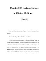
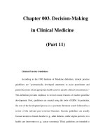
• The degree of facial instability associated with the
LeFort fractures should be assessed by grasping
the maxillary arch (above the incisors) with one
hand while stabilizing the forehead with the other
(Fig. 159-1).
• The LeFort I is a transverse fracture through the
maxilla, pterygoid plate, and nasal septum, re-
sulting in a floating maxilla. Clinically, the hard
palate and upper teeth move with stressing.
• The LeFort II is a pyramidal fracture of the central
maxilla across the bridge of the nose. The nose,
hard palate, and upper teeth move as a unit dis-
joined from the zygomas with stressing.
• The LeFort III, or craniofacial disjunction, in-
volves the maxilla, nasal bones, ethmoid, and zy-
goma. The entire face moves with stressing.
• The eye examination should document visual acu-
ity, pupil shape/size, alignment, and reactivity. A
Marcus Gunn pupil (initial dilation with the swing-
ing light test) suggests retinal or optic nerve injury.
A teardrop pupil suggests globe rupture. Monocu-
lar diplopia may represent lens dislocation; binoc-
ular diplopia may represent entrapment of the
inferior rectus or cranial nerve injury. The anterior
chamber should be evaluated for the presence of
a hyphema, and the cornea should be stained with
fluorescein to identify abrasions.
• Facial sensation should be tested for anesthesia
of the upper lip, nasal mucosa, lower lid, and max-
FIG. 159-1 Schematic of midfacial fracture lines: Le Fort I,
II, and III. (Reprinted with permission from Dingman RO,
Nativg P: Surgery of Facial Fractures. Philadelphia, Saunders,
1964, p 248.)
illary teeth. Positive findings suggest infraorbital
nerve injury.
• The mandible should be palpated for step-off, ten-
derness, crepitus, and instability.
• The mouth should be examined for lacerations,
tooth fractures, malocclusion, tenderness, or anes-
thesia. Anesthesia to the dentition, lower lip, or
chin may represent a mandibular fracture.
• Mastoid ecchymosis (Battle’s sign), hemotympa-
num, periorbital ecchymosis (‘‘raccoon eyes’’),
and cerebrospinal fluid (CSF) otorrhea are clinical
signs of a basilar skull fracture.
• The nose should be assessed for septal hematoma
and CSF rhinorrhea (halo/double-ring sign).
• The ear should be inspected for subperichon-
dral hematoma.
DIAGNOSIS AND DIFFERENTIAL
• The diagnosis of specific maxillofacial injuries is
based on clinical findings, facial radiographs, and
facial computed tomography (CT). Patient stabil-
ity will dictate the timing and order of these im-
aging modalities.
• The following radiographs may be useful. The Wa-
ters view (occipital mental view) is the most valu-
486 SECTION 20
•
TRAUMA
able for midface fractures. The posteroanterior
(PA or Caldwell) view best details the upper facial
bones. The ‘‘jug-handle’’ (submental vertex) view
is the best for evaluating the zygomatic arches.
The Townes view is useful for mandibular rami
and basilar skull fractures. Lateral radiographs can
assess air-fluid levels in the ethmoid and sphe-
noid sinuses.
EMERGENCY DEPARTMENT CARE
AND DISPOSITION
• The major focus in prehospital care is airway man-
agement and spinal immobilization. Airway man-
agement and hemorrhage control are paramount
in the emergency department (ED). Chin lift, jaw
thrust, and oral suctioning without neck extension
often restore patency.
• Severe mandibular fractures often cause posterior
displacement of the tongue. The tongue should
be pulled forward with a gauze pad, towel clip, or
large suture to relieve any obstruction.
4
• For endotracheal intubation, the oral route is pre-
ferred because of the risk of nasocranial intuba-
tion or severe epistaxis associated with nasotra-
cheal intubation attempts. The use of
neuromuscular blocking agents should be avoided
if at all possible. Fiberoptic intubation, a Bullard
intubating blade, and the laryngeal mask airway
may be useful adjuncts with a difficult airway. If
neuromuscular blocking agents are used, equip-
ment for emergent cricothyrotomy should be at
the bedside.
• The cervical spine should be cleared, either clini-
cally or radiographically.
• Hemorrhage should be controlled with direct
pressure; blind clamping should be avoided. Pha-
ryngeal bleeding can be controlled with packing
around the endotracheal tube. Severe epistaxis
may be controlled with direct pressure and poste-
rior nasal packing.
• Management of the airway, proper fluid resuscita-
tion, and evaluation of associated head, chest, ab-
dominal and spinal trauma should take prece-
dence over facial radiographs.
• For reliable patients who have been cleared of
serious injuries, radiographic evaluation may be
performed on an outpatient basis.
CARE OF SPECIFIC FRACTURES
• Antibiotics—such as amoxicillin-clavulanate, tri-
methoprim-sulfamethoxazole, or a first-genera-
tion cephalosporin—should be administered to
patients with sinus fractures or with nasal packing.
Isolated sinus and frontal bone fractures can be
managed on an outpatient basis. Inpatient man-
agement is warranted for sinus fractures of the
posterior wall, depressed fractures, or intracran-
ial injury.
• Orbital blowout fractures are the most common
type of orbital fracture. A CT scan should be ob-
tained to determine the surface area of injury.
Indications for surgery include enophthalmos,
persistent diplopia, and entrapment of the extra-
ocular muscles.
5
• Septal hematomas should be drained under local
anesthesia, using a no. 11 blade, by incising along
the inferior border of the hematoma. The nostril
and septum should be packed and appropriate
antibiotics prescribed.
• Zygomatic fractures: Patients with tripod frac-
tures, which involve the infraorbital rim, a diasta-
sis of the zygomatic-frontal suture, and disruption
of the zygomatic-temporal junction at the arch
require admission for open reduction and internal
fixation. Those with fractures of the zygomatic
arch can have elective outpatient elevation and
repair.
• Open mandibular fractures require admission and
IV antibiotics.
• Temporomandibular joint (TMJ) dislocation
should be reduced with the physician standing be-
hind the seated patient and pushing downward
and backward on the posterior molar. Sedation
and local anesthesia to the TMJ, lateral pterygoid,
and masseter muscles may be necessary. A Barton
bandage should be applied after reduction.
• Children under 6 years of age are more likely to
have injury to the frontal bone, given its promi-
nence. Maxillary fractures are uncommon in chil-
dren due to the lack of maxillary sinus develop-
ment. By age 12, the child’s fracture pattern is the
same as that of the adult.
• Early follow-up is important for pediatric facial
fractures, given the rapid healing rates in children
and the potential for asymmetric facial growth.
R
EFERENCES
1. Hartzell KN, Botek AA, Goldberg SH: Orbital fractures
in women due to sexual assault and domestic violence.
Ophthalmology 103:953, 1996.
2. Jessee SA: Physical manifestations of child abuse to the
head, face and mouth: A hospital survey. ASDC J Dent
Child 62:245, 1995.
CHAPTER 160
•
NECK TRAUMA 487
3. Thai KN, Hummel RP III, Kitzmiller WJ, Luchette FA:
The role of computed tomographic scanning in the man-
agement of facial trauma. J Trauma 43:214, 1997.
4. Bavits JB, Collicott PE: Bilateral mandibular subcondylar
fractures contributing to airway obstruction. Int J Oral
Maxillofac Surg 24:273, 1998.
5. Bhattacharya J, Moseley IF, Fells P: The role of plain
radiography in the management of suspected orbital blow-
out fractures. Br J Radiol 70:29, 1997.
For further reading in Emergency Medicine: A Com-
prehensive Study Guide, 5th ed., see Chap. 249,
‘‘Maxillofacial Trauma,’’ by Stephen Colucciello.
160 NECK TRAUMA
M. Chris Decker
EPIDEMIOLOGY
• The demographics of neck trauma patients are
expected to mirror those of other trauma victims.
• Multiple injuries occur 44 to 52 percent of the
time with penetrating trauma.
1–5
PATHOPHYSIOLOGY
• The neck contains a high concentration of vascu-
lar, aerodigestive, and spinal structures in a rela-
tively confined space.
• The Roon and Christensen anatomic classification
divides the neck into three zones (Table 160-1).
The at-risk structures located in zone 1 are the
vertebral and proximal carotid arteries, major tho-
racic vessels, superior mediastinum, lungs, esopha-
gus, trachea, thoracic duct, and spinal cord. The
at-risk structures located in zone 2 are the carotid
and vertebral arteries, jugular vein, esophagus,
trachea, larynx, and the spinal cord. The at-risk
structures located in zone 3 are the distal carotid
and vertebral arteries, pharynx, and the spinal
cord.
• The platysma is the most superficial structure be-
neath the skin and serves as an important planar
TABLE 160-1 Zones of the Neck
Zone I Base of the neck to the cricoid cartilage
Zone II Cricoid cartilage to the angle of the mandible
Zone III Angle of the mandible to the base of the skull
landmark in evaluating penetrating neck injuries.
Beneath the platysma is the deep cervical fascia
and the fascial compartments that support the
muscles, vessels, and viscera of the neck. The tight
fascial compartments offer a tamponade effect,
which helps limit potential for external bleeding
from vascular injuries; however, bleeding within
this confined space can result in extrinsic compres-
sion and airway compromise.
CLINICAL FEATURES
• Presentations of neck injuries involve manifesta-
tions of vascular, aerodigestive, and neurologic
symptoms and signs. All signs require diagnostic
evaluation but hard signs are more often associ-
ated with significant injury (Table 160-2).
• Both blunt and penetrating laryngeal or pharyn-
geal trauma can cause dysphonia, stridor, hemop-
tysis, hematemesis, dysphagia, neck emphysema,
and dyspnea progressing to respiratory arrest.
• Acute hemorrhage may be visible externally or
can occur internally, leading to hematoma forma-
tion with tracheal deviation or bleeding into the
pharynx. In both situations, tachycardia, hypoten-
sion, and other signs of shock indicate significant
blood loss; airway compromise may result from
the mass effect of an expanding hematoma.
• Neurologic symptoms and signs range from com-
plaints of pain or paresthesias to hemiplegia, quad-
riplegia, and coma.
• Gastrointestinal injury initially may be asymptom-
atic, though patients may complain of dysphagia
and hematemesis may be observed.
• Strangulation may cause petechiae of the skin
TABLE 160-2 Signs and Symptoms of Neck Injury
HARD SIGNS SOFT SIGNS
Hypotension in Emergency Hypotension in field
Department
Active arterial bleeding History of arterial bleeding
Diminished carotid pulse Tracheal deviation
Expanding hematoma Nonexpanding large hematoma
Thrill/bruit Apical capping on chest x-ray
Lateralizing signs Stridor
Hemothorax Ͼ1000 cc Hoarseness
Air or bubbling in wound Vocal cord paralysis
Hemoptysis Subcutaneous emphysema
Hematemesis Seventh cranial nerve injury
Unexplained bradycardia (with-
out CNS injury)
488 SECTION 20
•
TRAUMA
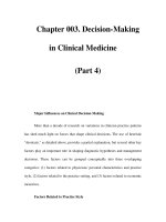
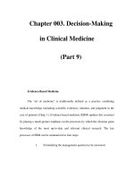
FIG. 160-1 Management of penetrating neck injury.
above the site of ligature and in the subconjunc-
tivae.
6–9
DIAGNOSIS AND DIFFERENTIAL
• Penetrating wounds are classified by the zone of
injury and evaluated for possible violation of the
platysma muscle. No further probing of deep
wounds is warranted in the emergency department
(ED); full exploration awaits surgical consultation
and the capacity for proximal and distal vascular
control in the operating room.
• Plain radiographs can identify cervical spine in-
jury, the presence of any penetrating foreign body,
air in the soft tissues, and soft tissue swelling. A
chest radiograph is warranted for any suspected
thoracic cavity penetration.
• Additional diagnostic procedures to be consid-
ered, in conjunction with surgical consultation, in-
CHAPTER 160
•
NECK TRAUMA 489
clude arteriography or duplex sonography for sus-
pected arterial injury, computed tomography (CT)
scanning of the larynx or cervical spine, endoscopy
of the airway and esophagus, or contrast studies
of the esophagus.
• The differential diagnosis relates to the various
structures at risk for injury. Airway injury may be
encountered in cases involving blunt trauma as
well as penetrating mechanisms of injury. Vascular
injury is most common with penetrating trauma,
although major vessel injury can occur due to
blunt trauma and may simulate an acute stroke.
Neurologic injuries include generalized brain isch-
emia (seen primarily with strangulation), spinal
cord trauma, nerve root damage, and peripheral
nerve damage. Cervical spine injury initially may
present without neurologic deficit, but the spine
can be cleared clinically in selected blunt trauma
and gunshot wound victims. Gastrointestinal in-
juries are often occult and generally require evalu-
ation by endoscopy or contrast radiography.
EMERGENCY DEPARTMENT CARE
AND DISPOSITION
• Hemodynamic and cardiac monitoring, IV access,
and 100% oxygen with pulse oximetry are required
initially for all patients.
• Airway management is made critical by the poten-
tial for direct injury and resulting potential for
airway compromise. Tracheal intubation is indi-
cated for patients unable to maintain airway pat-
ency secondary to structural disruption, edema,
secretions, bleeding, enlarging hematoma, or im-
pending respiratory arrest. In cases where oral or
nasal intubation is not possible or is contraindi-
cated, cricothyrotomy or transtracheal jet insuf-
flation may be performed.
• The chest must be evaluated for pneumothorax
and hemothorax secondary to vascular injury, pri-
marily in the setting of penetrating trauma.
• External hemorrhage is controlled with direct
pressure; blind clamping of bleeding vessels is con-
traindicated due to the complex vital anatomy
compressed into a relatively small space and the
danger of causing further injury with a misguided
surgical instrument.
• Fluid resuscitation should begin with crystalloid,
followed by blood products if needed.
• The cervical spine is secured and cleared clinically
or radiographically, as appropriate.
• Penetrating wounds that do not violate the
platysma muscle require only standard meticulous
wound care and closure. After a period of observa-
tion, asymptomatic patients with these injuries can
often be discharged home with close follow-up,
presuming their medical condition otherwise
makes this feasible.
• Wounds that violate the platysma muscle mandate
surgical consultation. These patients are admitted
for surgical exploration or for further diagnostic
evaluation of any significant deep structure injury
(Fig. 160-1).
• Patients with blunt neck trauma initially may pres-
ent with subtle signs of injury and may develop
significant symptoms on a delayed basis, particu-
larly those with a strangulation mechanism. After
a period of observation, asymptomatic patients
may be discharged with close follow-up, although
a low threshold for admission should be main-
tained.
• With blunt trauma, hoarseness, dysphagia, and
dyspnea are indications for more extensive evalua-
tion. Any initial symptoms of airway, vascular, or
neurologic injury demand evaluation and stabili-
zation along with urgent surgical consultation
and admission.
10
R
EFERENCES
1. Irish JC, Hekkenberg R, Gullane PJ, et al: Penetrating
and blunt neck trauma: 10 year review of a Canadian
experience. Can J Surg 40:33, 1997.
2. Roon AJ, Christensen N: Evaluation and treatment of
penetrating cervical injuries. J Trauma 19:391, 1979.
3. Shearer VE, Giesecke AH: Airway management for pa-
tients with penetrating neck trauma: A retrospective
study. Anesth Analg 77:1135, 1993.
4. Baron BJ, Sinert RH, Kohl L, et al: The value of physical
examination in penetrating neck trauma. Acad Emerg
Med 4:347, 1997.
5. Sclafani SJA, Cavaliere G, Atweh N, et al: The role
of angiography in penetrating neck trauma. J Trauma
31:557, 1991.
6. Fuhrman GM, Stieg FH, Buerk CA: Blunt laryngeal
trauma: Classification and management protocol. J
Trauma 30:87, 1990.
7. Li MS, Smith BM, Espinosa J, et al: Nonpenetrating
trauma to the carotid artery. Seven cases and a literature
review. J Trauma 36:265, 1994.
8. Watridge CB, Muhlbauer MS, Lowery RD: Traumatic
carotid artery dissection: Diagnosis and treatment. J
Neurosurg 71:854, 1989.
9. Fabian TC, Patton JH, Croce MA, et al: Blunt carotid
injury, importance of early diagnosis and anticoagulant
therapy. Ann Surg 223:513, 1996.
10. Iserson KV: Strangulation: A review of ligature, manual,
and postural neck compression injuries. Ann Emerg Med
13:179, 1984.
490 SECTION 20
•
TRAUMA
For further reading in Emergency Medicine: A Com-
prehensive Study Guide, 5th ed., see Chap. 217,
‘‘Penetrating and Blunt Neck Trauma,’’ by Bon-
nie J. Baron.
161 THORACIC TRAUMA
Kent N. Hall
EPIDEMIOLOGY
• Thoracic trauma is directly responsible for 25 per-
cent of trauma deaths.
• Patients with isolated chest trauma have a rela-
tively low mortality of 5 percent.
• Only 5 to 15 percent of patients with chest trauma
will require a thoracotomy.
PATHOPHYSIOLOGY
• Penetrating injuries routinely result in pneumo-
thorax or hemothorax.
• Blunt trauma to the chest causes organ damage
by compression, direct trauma, or acceleration/
deceleration forces.
GENERAL PRINCIPLES
AND CONDITIONS
• The initial step is to evaluate the patient’s effort
to breathe. No effort indicates a possible central
nervous system problem, such as head trauma,
drugs, or spinal cord injury.
• Significant effort signals an airway obstruction,
most commonly a foreign body (including the
tongue) in the hypopharynx, larynx, or trachea.
• If the patient is attempting to breath and the air-
way is clear, thoracic injuries (flail chest, hemo-
pneumothorax, diaphragmatic injury or parenchy-
mal lung damage) should be considered.
• In all cases of significant respiratory distress, the
airway should be secured and adequate oxygen-
ation and ventilation provided. Indications for
ventilatory support are listed in Table 161-1.
• The most frequent symptoms associated with tho-
racic trauma are chest pain and shortness of
breath. Physical examination begins with inspec-
tion of the chest wall, looking for open (‘‘sucking’’)
chest wounds, flail segments, and contusions.
TABLE 161-1 Indications for Ventilatory Support
Impaired ventilation in spite of an open airway
Shock
Multiple injuries
Coma
Flail chest
Hypoxia (P
O
2
Ͻ50 mmHg on room air)
Drainage of hemopneumothorax
Preexisting pulmonary disease
Respiratory rate Ͼ30 breaths per minute
Relief of chest wall pain
Multiple transfusions required
Elderly
• The neck is examined for the presence of dis-
tended neck veins, which are associated with peri-
cardial tamponade, tension pneumothorax, air
embolus, and cardiac failure. Swelling and cyano-
sis of the face and neck often signal a superior
mediastinal injury resulting in superior vena
cava blockage.
• Subcutaneous emphysema from a bronchial injury
or pulmonary laceration can result in severe swell-
ing of the face and neck. Palpation of the trachea
to determine its normal position, of the chest to
localize areas of tenderness or crepitation, and
of the abdomen for the position of abdominal
contents is important.
• Auscultation of the chest should be done system-
atically and thoroughly. The quality and equality
of breath sounds should be documented. The pres-
ence of bowel sounds in the chest may be the first
indication of a diaphragmatic injury. Inequality of
breath sounds may suggest a pneumothorax, a
hemothorax, or an improperly inserted endotra-
cheal tube.
• Conditions that should be recognized and treated
during the initial survey include tension pneumo-
thorax, cardiac tamponade, massive hemothorax,
open pneumothorax, and flail chest.
CHEST WALL INJURIES
CLINICAL FEATURES, DIAGNOSIS,
AND DIFFERENTIAL
• Simple rib fractures should be suspected in the
patient with point tenderness over a rib. The goal
of evaluating these injuries is to look for complica-
tions, such as pneumothorax, pulmonary contu-
sion, or major vascular injury.
• Suspicion of a pneumothorax that is not corrobo-
rated by chest x-ray might require inspiratory and
expiratory radiographic views for detection.
CHAPTER 161
•
THORACIC TRAUMA 491
• Pain from rib fractures can decrease ventilation,
possibly resulting in atelectasis or pneumonia.
• Fractures of the first and second ribs not due to
direct trauma may be associated with significant
underlying injuries, including myocardial contu-
sions, pulmonary contusions, bronchial tears, and
major vascular injuries.
• Multiple rib fractures, especially the 9th, 10th and
11th, may be associated with intraabdominal injur-
ies. Hypotension may indicate the presence of ten-
sion pneumothorax or hemothorax.
• Segmental fractures of three or more adjacent ribs
produce a flail segment of the chest, which can
increase the work of breathing.
• Flail chest is recognized by paradoxical movement
of the segment during the respiratory cycle (out-
ward during expiration, inward during inspi-
ration).
• In the case of an open pneumothorax (‘‘sucking
chest wound’’), the wound is often obvious. Open
chest wounds indicate invasion into the pleural
space and can act as one-way valves, potentially
creating a tension pneumothorax.
• Traumatic asphyxia, caused by an inability to
breathe due to added weight on the chest wall,
results in subconjunctival hemorrhage or pete-
chiae and vascular engorgement, edema, and cya-
nosis of the head, neck, and upper extremities.
EMERGENCY DEPARTMENT CARE
AND DISPOSITION
• Bleeding from chest wall injuries is best controlled
by direct pressure. Probing of these wounds is not
recommended.
• When subcutaneous emphysema is present, an un-
derlying pneumothorax should be presumed. If
the patient is to be intubated for any reason, a
chest tube should be inserted.
• For rib fractures, adequate analgesia and pulmo-
nary toilet are the mainstays of therapy. Patients
with multiple rib fractures should be admitted for
24 to 48 h if they cannot cough and clear secre-
tions, are elderly, or have preexisting pulmonary
disease.
• Sternal fractures should alert the physician to the
possible presence of underlying soft tissue injuries,
especially of the heart and great vessels. Therapy
for these fractures includes adequate analgesia
and pulmonary toilet. Admission based solely on
the presence of sternal fractures is controversial.
• For flail chest, management consists of stabilizing
the flail segment, either externally by using sand-
bags or internally by endotracheal intubation and
TABLE 161-2 Indications for Endotracheal Intubation
in the Presence of a Flail Segment
Presence of shock
Three or more associated injuries
Severe head injury
Comorbid pulmonary disease
Fracture of eight or more ribs
Age Ͼ65
mechanical ventilation. Nonventilatory manage-
ment includes adequate analgesia, chest physio-
therapy, and restriction of IV fluids.
• Indications for ventilatory support of a patient
with a flail chest are listed in Table 161-2. The
patient with a flail chest should be suspected of
having an underlying pulmonary contusion.
• Open (sucking) chest wounds should be covered
with a sterile occlusive dressing while a chest tube
is inserted simultaneously at a separate site. If
a tension pneumothorax develops, the occlusive
dressing should be removed until the chest tube
is inserted.
LUNG INJURIES
CLINICAL FEATURES, DIAGNOSIS,
AND DIFFERENTIAL
• Pulmonary contusions are usually seen as opacifi-
cations of the lung on chest radiograph within 6 h
of injury.
• Pneumothorax is a collection of air in the pleural
space. It does not usually cause significant symp-
toms unless a tension pneumothorax develops, the
pneumothorax occupies more than 40 percent of
a hemithorax, or the patient has preexisting shock
or cardiopulmonary disease.
• A pneumothorax is readily seen on expiratory
chest radiograph.
• Clinical signs and symptoms of a tension pneumo-
thorax include dyspnea, hypoperfusion, distended
neck veins, deviated trachea, and decreased or
absent breath sounds on the affected side.
• Hemothorax should be considered in the severely
traumatized patient with unilateral decreased
breath sounds and dullness to percussion. Vol-
umes of blood as low as 200 to 300 mL are usually
visualized on upright chest radiograph. However,
volumes in excess of 1 L of blood may be missed
on supine chest radiograph because of its appear-
ance as diffuse haziness without a distinct air-
fluid level.
• Subcutaneous emphysema in the neck and a
‘‘crunching’’ sound during systole (Hamman sign)
492 SECTION 20
•
TRAUMA
should make the clinician suspect pneumomedias-
tinum. The major significance of pneumomediasti-
num is the possibility of associated injuries to the
larynx, trachea, major bronchi, pharynx, or
esophagus.
EMERGENCY DEPARTMENT CARE
AND DISPOSITION
• Treatment of pulmonary contusions involves
maintenance of adequate ventilation, with the use
of mechanical ventilation and positive end-expir-
atory pressure (PEEP) to optimize ventilation/
perfusion matching.
• Mechanical ventilation is often required if more
than 28 percent of lung volume (estimated by chest
radiograph) is involved. With severe unilateral
lung injury, synchronous independent lung venti-
lation through a double-lumen endobronchial
catheter prevents overinflation of the normal lung
and provides better overall oxygenation.
• If a tension pneumothorax is suspected, a large
bore needle or IV catheter (14 gauge) should be
inserted in the second intercostal space at the mid-
clavicular line for needle decompression. Emer-
gent management should not be delayed while a
chest radiograph is obtained. A chest tube can be
inserted for definitive treatment later.
• If a hemothorax or nontension pneumothorax is
suspected in a patient with severe respiratory dis-
tress, a chest tube should be inserted prior to ob-
taining a chest radiograph.
• Small pneumothoraces that have not expanded on
serial chest radiographs taken 6 to 12 h apart do
not usually require chest tube insertion. Admis-
sion of these patients for observation and serial
examinations is important.
• An ‘‘occult pneumothorax’’ [one seen on com-
puted tomography (CT) but not on plain radio-
graph] does not require chest tube insertion unless
the patient is on a ventilator. Insertion of a small
(24 or 28 Fr) chest tube is adequate if no hemotho-
rax is present.
• A chest radiograph should be obtained in all pa-
tients after insertion of a chest tube. Persistent air
leakage and failure of the lung to expand com-
pletely is an indication for thoracotomy.
• With a massive hemothorax, the blood should be
evacuated with a large-bore (38 Fr or larger) chest
tube. Prophylactic antibiotics, while controversial,
have led to a clear reduction in pneumonia or em-
pyema.
• Serial examinations of the chest, including chest
radiographs, and monitoring of ongoing blood loss
through the chest tube are important.
• The decision to perform a thoracotomy should be
based on multiple factors. A conservative ap-
proach is to perform a thoracotomy if ongoing
blood loss from the chest tube exceeds 600 mL/6 h.
Also, if vital signs deteriorate as a large amount
of blood is being evacuated from a chest tube,
the chest tube should be clamped and the patient
should undergo thoracotomy.
• While adequate ventilation is being ensured, res-
toration of adequate tissue perfusion should be
achieved. Management of patients in shock in-
cludes the insertion of two large-bore IV catheters
with rapid infusion of large volumes of crystalloid
or blood.
TRACHEOBRONCHIAL INJURY
• Injuries to the lower trachea and bronchi are usu-
ally caused by severe deceleration forces.
• Common presenting signs and symptoms include
dyspnea, hemoptysis, subcutaneous emphysema,
Hamman’s sign, and sternal tenderness.
• On chest radiograph, a large pneumothorax, pneu-
momediastinum, deep cervical emphysema, or en-
dotracheal tube balloon that appears round all
suggest tracheobronchial injury.
• Management includes assuring adequate ventila-
tion and referral for immediate bronchoscopy to
fully evaluate and treat the injury. Intrathoracic
tracheal injury is usually associated with other in-
trathoracic injuries and is almost invariably fatal.
• Injuries of the cervical trachea usually occur at
the junction of the trachea and cricoid cartilage
and are caused by direct trauma, as from a steering
wheel. Inspiratory stridor is common in these pa-
tients and indicates a 70 to 80 percent obstruction.
• Oral intubation, preferably over a bronchoscope,
should be attempted.
DIAPHRAGMATIC INJURY
• The majority of diaphragmatic injuries are caused
by penetrating trauma. Most series report that
diaphragmatic injury associated with blunt injury
is usually left-sided.
• An entrance wound in the abdomen with the mis-
sile located in the chest cavity should alert the
physician of a probable injury to the diaphragm.
• In the setting of blunt trauma, any abnormality
of the diaphragm or lower lung fields on chest
CHAPTER 161
•
THORACIC TRAUMA 493
radiograph should make the clinician consider the
possibility of diaphragmatic injury.
PENETRATING INJURY TO
THE HEART
CLINICAL FEATURES, DIAGNOSIS,
AND DIFFERENTIAL
• All patients with hypotension and penetrating
chest injury anywhere near the heart should be
considered as having a cardiac injury until proven
otherwise. Patients without signs of life in the field
are not considered candidates for resuscitation.
• Beck’s triad (distended neck veins, hypotension,
and muffled heart tones) suggests a pericardial
tamponade, although most patients with this type
of injury do not have distended neck veins until
volume resuscitation has occurred.
• Chest radiographs are rarely helpful in diagnosing
acute cardiac injury, and changes on electrocardi-
ography (ECG) are usually nonspecific. Transtho-
racic echocardiography is a sensitive test for the
detection of pericardial fluid. Transesophageal
echocardiography (TEE) is a sensitive diagnostic
tool, especially if the patient is already intubated.
• Pericardiocentesis has limited value in the evalua-
tion of the patient with possible cardiac injury due
to the high incidence of false-positive and false-
negative aspirates. In the hemodynamically stable
patient, when echocardiography is not available, a
subxiphoid pericardial window can be performed.
EMERGENCY DEPARTMENT CARE
AND DISPOSITION
• Management of the patient with cardiac injury
includes attention to the airway, assurance of
breathing, and adequate fluid resuscitation. Two
large-bore IV lines should be placed, with one
flowing into the venous system draining into the
inferior vena cava.
• Patients in shock who do not respond to adequate
fluid resuscitation and who are suspected of having
a cardiac injury should undergo emergent thora-
cotomy.
• The patient with penetrating thoracic trauma who
loses vital signs just prior to arrival or in the ED
may require emergent pericardiocentesis or ED
thoracotomy.
• For ED thoracotomy, an incision is made at the
fifth intercostal space on the affected side. The
pericardium is opened vertically, with care to
avoid the phrenic nerve. The heart, lung hilum,
and aorta are inspected for injuries that can be
repaired primarily.
• Patients with blunt traumatic arrest, penetrating
abdominal or head injuries, or prolonged arrest
times receive little if any benefit from ED thora-
cotomy.
BLUNT INJURY TO THE HEART
CLINICAL FEATURES, DIAGNOSIS,
AND DIFFERENTIAL
• The most common mechanism of injury causing
cardiac trauma is a deceleration injury, as with a
high-speed motor vehicle crash. In addition, com-
pression between the sternum and vertebrae, a
sudden increase in intrathoracic pressure, ab-
dominal compression forcing abdominal contents
against the heart, or strenuous cardiac massage
can all cause cardiac injury.
• Blunt trauma to the heart can result in multiple
types of injuries, including rupture of an outer
chamber wall, septal rupture, valvular injuries, di-
rect myocardial injury, laceration of a coronary
artery, or pericardial injury.
• Blunt myocardial injury (BMI) is the term cur-
rently in use to describe injuries previously termed
myocardial concussions and myocardial contu-
sions. The most common clinical features associ-
ated with a significant BMI are tachycardia out of
proportion to blood loss, arrhythmias (especially
premature ventricular contractions and atrial fi-
brillation), and conduction defects.
• Cardiac enzymes, including CPK-MB and the tro-
ponins, have been found to be nonspecific in mak-
ing the diagnosis of significant BMI. Echocardiog-
raphy does not seem to be very useful in evaluating
the patient with suspected BMI, although it is the
most widely used modality.
• BMI causes death very rarely, and the incidence
of clinically significant dysrhythmias and other
cardiac complications is low. Management of the
patient with a significant BMI calls for the admin-
istration of supplemental oxygen and analgesics,
treatment of significant cardiac dysrhythmias, and
the administration of fluids or inotropic agents
for hypotension.
• Cardiac rupture results in immediate death in 80
to 90 percent of cases. Patients with cardiac rup-
ture who arrive at the hospital alive usually have
a right atrial tear.
• Shock that is out of proportion to the degree of
recognized injury and shock that persists despite
494 SECTION 20
•
TRAUMA
control of hemorrhage elsewhere as well as vol-
ume expansion should make one consider the pos-
sibility of cardiac rupture.
• Immediate left anterior thoracotomy may be life-
saving in these cases. Septal defects and valve
injuries are rare after blunt trauma but should be
considered if a murmur exists in the setting of
possible cardiac damage.
• Signs of a ventricular septal defect include severe
early hypoxemia with a relatively normal chest
radiograph, heart murmur, and an injury pattern
on ECG.
• Rupture of the aortic valve is the most common
valvular lesion, followed by rupture of the papil-
lary muscle or chordae tendineae of the mitral
valve.
PERICARDIAL INJURY
• A pericardial effusion may develop acutely or over
time. The rate of fluid collection influences the
onset and severity of symptoms.
• Evidence of acute pericardial injury is usually seen
on the ECG as diffuse ST-segment elevation.
• Most patients are asymptomatic, and no specific
therapy is required. A tear of the parietal pericar-
dium at the apex of the heart may result in sudden
severe shock and cardiac arrest if the heart herni-
ates through the hole.
POSTPERICARDIOTOMY SYNDROME
• This is seen in patients 2 to 4 weeks after heart
surgery or trauma. Classically, patients will have
chest pain, fever, and pleural or pericardial effu-
sions. Friction rubs, arthralgia, and pulmonary in-
filtrates may also be seen.
• The ECG will often show diffuse ST–T-wave
changes consistent with pericarditis.
• Management is symptomatic, with salicylates and
rest often the only therapy required. Occasionally
glucocorticoids are needed.
PENETRATING TRAUMA TO THE
GREAT VESSELS OF THE CHEST
CLINICAL FEATURES, DIAGNOSIS,
AND DIFFERENTIAL
• When a stab wound causes injury to the great
vessels of the chest, survival is generally much
higher than when such an injury is caused by a
gunshot wound. Simple lacerations of the great
vessels can lead to exsanguination, tamponade,
hemothorax, air embolism, or development of an
arteriovenous (AV) fistula or false aneurysm.
• In general, these wounds should not be probed.
Assessment of bilateral upper extremity pulses
for equality is important, as a large mediastinal
hematoma may compress the subclavian vessels.
The entire chest should be auscultated for bruits
that may indicate a false aneurysm or AV fistula.
• Radiographic evaluation starts with a chest radio-
graph. In addition to evaluation for pneumothora-
ces, widening of the upper mediastinum may indi-
cate injury to brachiocephalic vessels. CT scans
are rarely performed immediately for penetrating
wounds of the chest. However, in the stable pa-
tient, a CT scan can help localize hematomas adja-
cent to great vessels. The use of IV contrast helps
further evaluate these structures and may demon-
strate a vascular defect or false aneurysm. The
major role of CT is as a screen for great vessel
injury.
• Arteriograms are most helpful in identifying ma-
jor intrathoracic vascular injuries within hemato-
mas, especially those resulting from penetrating
injury to the lower neck.
• Contrast swallow using meglumine diatrizoate
(Gastrografin) may be performed on stable pa-
tients to evaluate the integrity of the esophagus.
Endoscopy is sometimes used in hemodynamically
stable patients with penetrating wounds of the
chest or lower neck.
• Recently, use of TEE has been advocated, espe-
cially when the CT scan or aortogram are equivo-
cal for injury to the aorta.
EMERGENCY DEPARTMENT CARE
AND DISPOSITION
• Early endotracheal intubation should be per-
formed in patients with penetrating injuries to the
thoracic inlet to avoid the problems associated
with expanding hematomas distorting the airway.
• The patient in severe shock (systolic BP Ͻ60
mmHg) should have immediate surgery, with ag-
gressive fluid resuscitation waiting until after ma-
jor bleeding sites are controlled. If the systolic
blood pressure (BP) is 60 to 90 mmHg, 2 to3Lof
crystalloid should be given rapidly. If the patient
remains hypotensive, immediate surgery is re-
quired.
• If the patient did not have ‘‘signs of life’’ in the
field, no resuscitative efforts are warranted. How-
ever, if the patient ‘‘lost vital signs’’ immediately
CHAPTER 161
•
THORACIC TRAUMA 495
prior to arriving or in the ED, emergent ED thora-
cotomy is indicated.
• Bullets that enter great vessels can embolize to
distant sites and should be sought by using multi-
ple radiographs or fluoroscopy.
1–4
BLUNT TRAUMA TO THE GREAT
VESSELS OF THE CHEST
CLINICAL FEATURES, DIAGNOSIS,
AND DIFFERENTIAL
• Some 90 percent of patients with injury to great
vessels from blunt trauma who arrive at the hospi-
tal alive have an injury at the isthmus of the aorta
(between the left subclavian artery and liga-
mentum arteriosum). Other common sites of in-
jury are the innominate or left subclavian artery
at their origins or a subclavian artery over the
first rib.
• This injury can occur even when no external signs
of trauma exist. Therefore, it should be suspected
in any patient with a high-speed deceleration
mechanism of injury or high-speed impact from
the side. These patients complain primarily of
their associated injuries. Retrosternal or inter-
scapular pain, often described as a ‘‘tearing’’ sen-
sation, may be the only initial indication.
• One-third of patients with blunt trauma to the
aorta have no external evidence of thoracic injury.
Findings that suggest an aortic injury include a
difference in blood pressure or pulse amplitude
between the upper and lower extremities, acute-
onset upper extremity hypertension, or a harsh
systolic murmur across the precordium or in the
interscapular area.
• Findings associated with traumatic rupture of the
aorta on plain chest radiograph are seen in Table
161-3. The most frequent radiologic finding is me-
diastinal widening. The best chest radiograph is
an upright posteroanterior (PA) view taken from
TABLE 161-3 Chest Radiographic Findings Associated
with Traumatic Rupture of the Aorta
Superior mediastinal widening (Ͼ8.0–8.5 cm)
Deviation of esophagus and/or trachea at T4
Obscuration of aortic knob and/or descending aorta
Displacement of left mainstem bronchus more than 40 degrees be-
low horizontal
Obscuration of medial aspects of left upper lobe
Widening of the paratracheal stripe
Displacement of the paraspinal lines (either left or right)
Fracture of first or second rib
Apical cap
72 in. with the patient leaning forward 10 to 15 de-
grees.
• The most specific radiographic sign of traumatic
aortic rupture is deviation of the esophagus more
than 1 to 2 cm to the right of the spinous process
of T4. Up to one-third of patients with traumatic
aortic rupture will have a normal chest radio-
graph initially.
• TEE is a highly sensitive diagnostic modality to
evaluate for traumatic aortic rupture. It can be
used at the bedside while the resuscitation is ongo-
ing and yields results that are at least as good
as those of aortography. It visualizes the aortic
isthmus and descending aorta very well and de-
fines the pericardial cavity, cardiac valves, and
pulmonary veins as well as regional wall motion.
• Late-generation helical CT scans of the chest have
been recommended as a tool to screen for trau-
matic aortic rupture in selected patients. Selection
guidelines include patients with equivocal histo-
ries and equivocal radiographs who have a low
probability for injury to the other great vessels,
are hemodynamically stable, and are capable of
tolerating two dye loads (one for the CT and one
for the aortogram, if necessary).
• The presence of a mediastinal hematoma is an
indication for aortography. Magnetic resonance
imaging (MRI) cannot be recommended as a tool
in the evaluation of patients with suspected trau-
matic aortic rupture, mostly because of the need
for the patient to spend long intervals in an iso-
lated setting. Aortography is the traditional ‘‘gold
standard’’ for diagnosing aortic rupture.
• Injury to the ascending aorta usually results in
immediate death. These injuries tend to occur
within the pericardium and result in cardiac tam-
ponade. If there is an associated valvular injury,
a murmur of aortic insufficiency may be heard.
The aortogram shows a pseudoaneurysm, possibly
with aortic insufficiency.
• Injuries to the innominate artery are associated
with rib fractures, flail chest, hemopneumothorax,
fractured extremities, head injuries, facial frac-
tures, and abdominal injuries. The diagnosis is
difficult because there are no characteristic physi-
cal findings except for some decrease in the right
radial or brachial pulse. Findings on chest radio-
graph are similar to those with traumatic rupture
of the aorta except the mediastinal hematoma is
usually higher and the esophagus is pushed to
the left. Aortography is generally required for the
diagnosis to be established.
• Subclavian artery injuries are most often caused
by fractures to the first rib or clavicle. Absence
of a radial pulse on the affected side is the most
496 SECTION 20
•
TRAUMA
important sign. A pulsatile mass or bruit at the
base of the neck is suggestive of this injury. Associ-
ated injury to the brachial plexus occurs in 60
percent of patients. A Horner’s syndrome may
occur on the affected side as well.
EMERGENCY DEPARTMENT CARE
AND DISPOSITION
• Sedatives, analgesics, vasodilators, and beta-
adrenergic blockers may be required to control
the patient’s blood pressure.
• Insertion of a nasogastric tube is important, but
this must be done with extreme care to avoid mak-
ing the patient gag.
• Thoracotomy is the accepted standard of treat-
ment. Delayed repair may be more appropriate
in patients who are at extremely high operative
risk or when conditions for surgery are not op-
timal.
ESOPHAGEAL AND THORACIC
DUCT INJURIES
• Injuries to these structures are rare. If an esopha-
geal injury is suspected, an esophagram should be
performed. Most radiologists recommend use of
meglumine diatrizoate (Gastrografin) because it
causes less of an inflammatory reaction than bar-
ium. However, the false-negative rate with this
contrast agent is as high as 25 percent. Flexible
esophagoscopy can also be performed but carries
a false-negative rate of 20 percent.
• Thoracic duct injuries result in a chylothorax in
the right hemithorax.
R
EFERENCES
1. Brown GL, Richardson JD: Traumatic diaphragmatic her-
nia. Ann Thorac Surg 39:172, 1985.
2. Ma OJ, Mateer JR, Ogata M, et al: Prospective analysis
of rapid trauma ultrasound examination performed by
emergency physicians. J Trauma 38:879, 1995.
3. Biffl WA, Moore FA, Moore EE, et al: Cardiac enzymes
are irrelevant in the patient with suspected myocardial
contusion. Am J Surg 169:523, 1994.
4. Chan D: Echocardiography in thoracic trauma, in
Eckstein M, Chan D (eds): Contemporary Issues in
Trauma. Emerg Med Clin North Am 16:191, 1998.
For further reading in Emergency Medicine: A Com-
prehensive Study Guide, 5th ed., see Chap. 251,
‘‘Thoracic Trauma,’’ by William M. Bowling,
Robert F. Wilson, Gabor D. Kelen, and Timothy
G. Buchman.
162 ABDOMINAL TRAUMA
O. John Ma
EPIDEMIOLOGY
• The most common cause of death and disability
from nonintentional injury is the motor vehicle
crash. Falls are the second leading cause of acci-
dental death in the United States.
PATHOPHYSIOLOGY
• The injury pattern of blunt abdominal trauma is
often diffuse. Blunt injuries involve a compression
or crushing mechanism by energy transmission di-
rectly to the patient. If the compressive, shearing,
or stretching forces exceed the tolerance limits of
the tissue or organ, the tissues are disrupted.
• Injury also can result from movement of organs
within the body. Some organs are rigidly fixed,
whereas others are motile. Typical examples in
the abdomen include mesenteric or small bowel
injuries, particularly at the ligament of Treitz or
at the junction of the distal small bowel and
right colon.
• A gunshot may cause injury directly, as when the
bullet itself strikes an organ, or secondarily, as
when tissues are injured by missiles such as bone
or bullet fragments or by energy transmission from
the bullet.
CLINICAL FEATURES
S
OLID
V
ISCERAL
I
NJURIES
• Injury to the solid organs cause morbidity and
mortality primarily as a result of acute blood loss.
• The spleen is the most frequently injured organ
in blunt abdominal trauma and injury to it is com-
monly associated with other intraabdominal injur-
ies. The liver is also commonly injured in both
blunt and penetrating injuries.
CHAPTER 162
•
ABDOMINAL TRAUMA 497
• Tachycardia, hypotension, and acute abdominal
tenderness are the primary findings on physical
examination. Kehr’s sign, representing referred
left shoulder pain, is a classic finding in splenic
rupture. Lower left rib fractures should heighten
clinical suspicion for splenic injury.
• It is important to note that patients with solid
organ injury may occasionally present to the emer-
gency department (ED) with minimal symptoms
and nonspecific findings on physical examination.
This is especially true when these are associated
with distracting injuries, central nervous system
(CNS) trauma, or intoxication.
H
OLLOW
V
ISCUS
I
NJURIES
• These injuries produce symptoms by the combina-
tion of blood loss and peritoneal contamination.
Perforation of the stomach, small bowel, or colon
is accompanied by blood loss from a concomitant
mesenteric injury.
• Gastrointestinal (GI) contamination will produce
peritoneal signs over a period of time. Patients
with head injury, distracting injuries, or intoxica-
tion may not exhibit peritoneal signs initially.
• Injuries to the small bowel and colon are most
frequently the result of penetrating trauma. How-
ever, a deceleration injury can cause a bucket-
handle tear of the mesentery or a blowout injury
of the antimesenteric border.
R
ETROPERITONEAL
I
NJURIES
• Signs and symptoms of retroperitoneal injuries
may be subtle or absent upon initial presentation
to the ED. Duodenal injuries are most often asso-
ciated with high-speed vertical or horizontal decel-
erating trauma. Clinical signs of duodenal injury
are often slow to develop. Duodenal injuries may
range in severity from an intramural hematoma
to an extensive crush or laceration.
• Duodenal ruptures are usually contained within
the retroperitoneum. They may present with ab-
dominal pain, fever, nausea, and vomiting, al-
though these may take hours to become clini-
cally obvious.
• Pancreatic injury is most common with penetrat-
ing trauma. It also occurs after a severe crush
injury. The classic case is a blow to the midepigas-
trium from a steering wheel or the handlebar of
a bicycle.
D
IAPHRAGMATIC
I
NJURIES
• The presentation of diaphragmatic injuries is often
insidious. Only occasionally is the diagnosis obvi-
ous, when bowel sounds can be auscultated in the
thoracic cavity.
• On chest radiograph, with herniation of abdomi-
nal contents into the thoracic cavity, the diagnosis
is confirmed. In most cases, however, there is no
herniation, and the only finding on the chest radio-
graph is blurring of the diaphragm or an effusion.
• This injury is most often diagnosed on the left.
DIAGNOSIS AND DIFFERENTIAL
P
LAIN
R
ADIOGRAPHS
• For blunt trauma, routine use of plain abdominal
radiographs is not a cost-effective or prudent
method for evaluating the trauma patient. A chest
radiograph is helpful in evaluating for herniated
abdominal contents in the thoracic cavity and for
evidence of free air under the diaphragm.
• An anteroposterior (AP) pelvic radiograph is im-
portant for identifying pelvic fractures, which can
produce significant blood loss and may be associ-
ated with intraabdominal visceral injury.
D
IAGNOSTIC
P
ERITONEAL
L
AVAGE
• Diagnostic peritoneal lavage (DPL) remains an
excellent screening test for evaluating abdominal
trauma. Its advantages include its sensitivity, avail-
ability, the relative speed with which it can be
performed, and a low complication rate. Disad-
vantages include the potential for iatrogenic in-
jury, its misapplication for evaluation of retroperi-
toneal injuries, and its lack of specificity.
• For blunt trauma, indications for DPL include (1)
patients who are too hemodynamically unstable
to leave the ED for computed tomography (CT)
scanning or who have a physical examination that
is unreliable secondary to drug intoxication or
CNS injury and (2) unexplained hypotension in
patients with an equivocal physical examination.
• In penetrating trauma, DPL should be performed
when it is not clear that exploratory laparotomy
will be required. DPL is useful in evaluating pa-
tients sustaining stab wounds where local wound
exploration indicates that the superficial muscle
fascia has been violated. Also, it may be useful in
confirming a negative physical examination when
tangential or lower chest wounds are involved.
• The DPL is considered positive if more than 10
mL of gross blood is aspirated immediately, the
red blood cell count is Ͼ100,000/ȐL, the white
blood cell count is Ͼ500/ȐL, bile is present, or
vegetable matter is present.
• The only absolute contraindication to DPL is
when surgical management is clearly indicated, in
which case the DPL would delay patient transport
to the operating room. Relative contraindications
include patients with advanced hepatic dysfunc-
498 SECTION 20
•
TRAUMA
TABLE 162-1 Indications for Laparotomy
BLUNT PENETRATING
Absolute Anterior abdominal injury and hypotension Injury to abdomen, back and flank with
hypotension
Abdominal wall disruption Abdominal tenderness
Peritonitis GI evisceration
Free air on chest x-ray Positive DPL
CT diagnosed injury requiring surgery, i.e., (GSW) High suspicion for transabdomi-
pancreatic transection; duodenal rupture nal trajectory
CT diagnosed injury requiring surgery,
i.e., ureter or pancreas
Relative Positive DPL or FAST in stable patient Positive local wound exploration (SW)
Solid visceral injury in stable patient
Hemoperitoneum on CT without clear
source
A
BBREVIATIONS
:CTϭ computed tomography; DPL ϭ diagnostic peritoneal lavage; FAST ϭ focused
abdominal sonography for trauma; GI ϭ gastrointestinal; GSW ϭ gunshot wound; SW ϭ stab wound.
tion, severe coagulopathies, previous abdominal
surgeries, or a gravid uterus.
U
LTRASONOGRAPHY
• Recent literature has demonstrated that the fo-
cused abdominal sonography for trauma (FAST)
examination, like DPL, is an accurate screening
tool for abdominal trauma. Advantages of the
FAST examination are that it is accurate, rapid,
noninvasive, repeatable, and portable. Another
advantage of the FAST examination is that it is
capable of evaluating for free pericardial and pleu-
ral fluid.
• Disadvantages include its inability to determine
the exact etiology of the free intraperitoneal fluid
and that it is operator-dependent. Indications for
FAST examination are the same as for DPL.
C
OMPUTED
T
OMOGRAPHY
• The abdominal CT scan has a greater specificity
than DPL and ultrasonography, thus making it
the initial diagnostic test of choice at most centers.
Oral and IV contrast material should be given to
provide optimal resolution.
• Advantages of the CT scan include its ability to
locate intraabdominal lesions precisely before sur-
gery, to evaluate the retroperitoneum, to identify
injuries that may be managed nonoperatively, and
noninvasiveness.
• The disadvantages of CT scanning are its expense,
the time required to perform the study, the need
to transport the trauma patient to the radiology
suite, and the need for contrast agents.
EMERGENCY DEPARTMENT CARE
AND DISPOSITION
• Patients should be administered oxygen and have
cardiac monitoring and two IV lines secured.
• For hypotensive abdominal trauma patients, re-
suscitation with IV crystalloid fluid is indicated.
Transfusion with O-negative or type-specific
packed red blood cells should be considered in
addition to crystalloid resuscitation.
• Table 162-1 lists the indications for exploratory
laparotomy. When a patient presents to the ED
with an obvious high-velocity gunshot wound to
the abdomen, DPL or the FAST exam should not
be performed as it will only delay transport of the
patient to the operating room. If organ eviscera-
tion is present, it should be covered with a moist,
sterile dressing prior to surgery.
• For an equivocal stab wound to the abdomen,
surgical consultation for local wound exploration
is indicated. If the wound exploration demon-
strates no violation of the anterior fascia, the pa-
tient can be discharged home safely.
1–5
R
EFERENCES
1. Goldstein AS, Scalfani SJA, Kupterstein NH, et al: The
diagnostic superiority of computed tomography. J
Trauma 25:939, 1985.
2. Otomom Y, Henmi H, Mashiko K, et al: New diagnostic
peritoneal lavage criteria for diagnosis of intestinal injury.
J Trauma 44:991, 1998.
3. Ma OJ, Mateer JR, Ogata M, et al: Prospective analysis
of a rapid trauma ultrasound examination performed by
emergency physicians. J Trauma 38:879, 1995.
4. Tsang BD, Panacek EA, Brant WE, et al: Effect of oral
contrast administration for abdominal computed tomog-
raphy in the evaluation of acute blunt trauma. Ann Emerg
Med 30:7, 1997.
5. McCarthy MC, Lowdermilk GA, Canal DF, et al: Predic-