PATHOLOGY OF VASCULAR SKIN LESIONS - PART 2 pdf
Bạn đang xem bản rút gọn của tài liệu. Xem và tải ngay bản đầy đủ của tài liệu tại đây (963.58 KB, 34 trang )
20 Sangüeza and Requena / Pathology of Vascular Skin Lesions
PATHOGENESIS
The most commonly accepted opinion is that phakomatosis pigmentovascularis results
from developmental abnormalities of the vasomotor nerves derived from the neural crest
and melanocytes (30). It is thought that an alteration in the neural regulation of blood
vessels could lead to the development of the vascular abnormalities seen in this condition.
This is probably the explanation for the coexistence of nevus flammeus and nevus
anemicus characteristic of this disease. The abnormal melanocytic component results
from alterations during the migration of the neural crest-derived melanocytes, which
produces lesions such as the nevus of Ota, nevus spilus, and mongolian spot.
H
ISTOPATHOLOGIC FEATURES
Histopathologically, there are an increased number of dilated thin-walled capillaries
and venules in the upper part of the reticular dermis, although occasionally superficial
areas of subcutaneous fat are also involved. The melanocytic component consists of
spindled-shaped dendritic melanocytes loaded with abundant melanin in their cytoplasm
scattered between the collagen bundles of the dermis (Fig. 2). Sometimes, the number of
spindled melanocytes is sparse; thus lesions of phakomatosis pigmentovascularis may be
misinterpreted as nevus flammeus. Immunohistochemical stains with S-100 protein or
HMB-45 are helpful in highlighting the melanocytic component.
Table 1
Classification of Phakomatosis Pigmentovascularis
Type Features
I a,b
a
Nevus flammeus and nevus pigmentosus et verrucosus
II a,b Nevus flammeus, mongolian spots, ± nevus anemicus
III a,b Nevus flammeus, nevus spilus, ± nevus anemicus
IV a,b Nevus flammeus, mongolian spots, nevus spilus, ± nevus anemicus
a
a, cutaneous involvement only; b, cutaneous and systemic involvement.
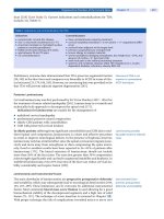
Fig. 1. Clinical features of phakomatosis pigmentovascularis. (A) The anterior abdomen and the
right thigh of this newborn show a combination of erythematous areas of nevus flammeus with
bluish areas of dermal melanocytosis. (B)The buttocks and the posterior thighs of the same baby
show a combination of similar features.
04/Sangüeza/19-26/F 01/14/2003, 10:52 AM20
Chapter 4 / Cutaneous Vascular Hamartomas 21
It is important to remember that lesions of nevus flammeus associated with
phakomatosis pigmentovascularis are indistinguishable from port wine stain not associ-
ated with melanocytic abnormalities. Ultrastructural studies indicate that nevus flammeus
associated with phakomatosis pigmentovascularis is surrounded by perivascular nerve
fibers, which are not present in isolated nevus flammeus (31).
T
REATMENT
The vascular component of phakomatosis pigmentovascularis may cause psychologi-
cal trauma to the patient, especially when it involves the face. In that case cosmetic
camouflage may be indicated. Laser therapy is also capable of producing good results in
treating nevus flammeus of phakomatosis pigmentovascularis (32).
There is no description of a melanoma originating in the melanocytic component of
phakomatosis pigmentovascularis.
References
1. Ota M, Kawanura T, Ito N. Phacomatosis pigmentovascularis (Ota). Jpn J Dermatol 1947;52:1–3.
2. Hasegawa Y, Yashura M. Phakomatosis pigmentovascularis type IVa. Arch Dermatol 1985;121:651–5.
3. Guillaume JC, Evenou P, Charpentier P, Avril MF. Phacomatose pigmento-vasculaire type IIa. Ann
Dermatol Venereol 1988;115:1113–5.
4. Mahroughan M, Mehregan AH, Mehregan DA. Phakomatosis pigmentovascularis. Report of a case.
Pediatr Dermatol 1996;13:36–8.
5. Stadhouders-Keet SA, Glastra A, Van Vloten WA. Phakomatosis pigmentovascularis (type IIIa). Ned
Tijdschr Geneeskd 1999;143:1337.
6. Cincinnati P, Carucci T, Rutiloni C. La facomatosi pigmento-vascolare. Minerva Pediatr 1996;48:225–8.
7. Murdoch SR, Keefe M. Phakomatosis pigmentovascularis type IIA in a Caucasian child. Pediatr
Dermatol 2000;17:157.
8. Toda K. A new type of phacomatosis pigmentovascularis (Ota). Jpn J Dermatol 1966;76:47–51.
9. Hasegawa Y, Yasuhara M. A variant of phakomatosis pigmentovascularis. Sin Res (Osaka)
1979;21:178–86.
10. Hasegawa Y, Yasuhara M. Phakomatosis pigmentovascularis type IVa. Arch Dermatol 1985;121:651–5.
11. Noriega Sanchez A, Markand ON, Herndon JH. Oculocutaneous melanosis associated with the Sturge-
Weber syndrome. Neurology 1972;22:256–62.
12. Furukawa T, Igata A, Toyokura Y, et al. Sturge-Weber and Klippel-Trenaunay syndrome with nevus of
Ota and Ito. Arch Dermatol 1970;102:640–5.
Fig. 2. Histopathologic features of phakomatosis pigmentovascularis. (A) Low-power magnifica-
tion shows dilated vascular structures at different levels of the dermis. (B) Higher magnification
shows the dilated and congestive capillary blood vessels and scattered spindle, dendritic melano-
cytes with abundant melanin interstitially arranged between collagen bundles of the dermis.
04/Sangüeza/19-26/F 01/14/2003, 10:52 AM21
22 Sangüeza and Requena / Pathology of Vascular Skin Lesions
13. Sigg C, Pelloni F. Oligosymptomatic form of Klippel-Trenaunay-Weber syndrome associated with
giant nevus spilus. Arch Dermatol 1989;125:1284–5.
14. Peyron N, Dereure O, Bessis D, Guilhou JJ, Guillot B. La phacomatose pigmento-vasculaire. A propos
de 2 cas associés à une angiodysplasie. J Mal Vasc 1993;18:336–9.
15. Teekhasaenee C, Ritch R. Glaucoma in phakomatosis pigmentovascularis. Ophthalmology 1997;
104:150–7.
16. Hagiwara K, Uezato H, Nonaka S. Phacomatosis pigmentovascularis type IIb associated with Sturge-
Weber syndrome and pyogenic granuloma. J Dermatol 1998;25:721–9.
17. Uysal G, Guven A, Ozhan B, Ozturk MH, Mutluay AH, Tulunay O. Phakomatosis pigmentovascularis
with Sturge-Weber syndrome: a case report. J Dermatol 2000;27:467–70.
18. Guiglia MC, Prendiville JS. Multiple granular cell tumors associated with giant speckled lentiginous
nevus and nevus flammeus in a child. J Am Acad Dermatol 1991;24:359–63.
19. Kikuchi I, Okazaki M. Congenital temporal alopecia in phakomatosis pigmentovascularis. J Dermatol
1982;9:485–7.
20. Kim HJ, Park KB, Yang JM, Park SH, Lee ES. Congenital triangular alopecia in phakomatosis
pigmentovascularis: report of 3 cases. Acta Derm Venereol 2000;80:215–6.
21. Zahorcsek Z, Schmelas A, Schneider I. Progrediente zirkumskripte Lentiginose Phakomatosis
pigmentovascularis III/A. Hautarzt 1988;39:519–23.
22. Gilliam AC, Ragge NK, Perez MI, Bolognia JL. Phakomatosis pigmentovascularis type IIb with iris
mammillations. Arch Dermatol 1993;129:340–2.
23. Van Gysel D, Oranje AP, Stroink H, Simonsz HJ. Phakomatosis pigmentovascularis. Pediatr Dermatol
1996;13:33–5.
24. Di Landro A, Tadini GL, Marchesi L, Cainelli T. Phakomatosis pigmentovascularis: a new case with
renal angiomas and some considerations about the classification. Pediatr Dermatol 1999;16:25–30.
25. Tsuruta D, Fukai K, Seto M, et al. Phakomatosis pigmentovascularis type IIIb associated with moyamoya
disease. Pediatr Dermatol 1999;16:35–8.
26. Joshi A, Garg VK, Agrawal S, Agarwalla A, Thakur A. Port-wine stain (nevus flammeus), congenital
Becker’s nevus, café-au-lait-macule and lentiginides: phakomatosis pigmentovascularis type Ia—a new
combination. J Dermatol 1999;26:834–6.
27. Huang C, Lee P. Phakomatosis pigmentovascularis IIb with renal anomaly. Clin Exp Dermatol
2000;25:51–4.
28. Cho S, Choi JH, Sung KJ, Moon KC, Koh JK. Phakomatosis pigmentovascularis type IIB with neuro-
logic abnormalities. Pediatr Dermatol 2001;18:263.
29. Bielsa I, Paradelo C, Ribera M, Ferrandiz C. Generalized nevus spilus and nevus anemicus in a patient
with a primary lymphedema: a new type of phakomatosis pigmentovascularis? Pediatr Dermatol
1998;15:293–5.
30. Libow LF. Phakomatosis pigmentovascularis type IIIb. J Am Acad Dermatol 1993;29:305–7.
31. Smoller BR, Rosen S. Port wine stains: a disease of altered neural modulation of blood vessels? Arch
Dermatol 1986;122:177–9.
32. Ono I, Tateshita T. Phacomatosis pigmentovascularis type IIa successfully treated with two types of
laser therapy. Br J Dermatol 2000;142:358–61.
04/Sangüeza/19-26/F 01/14/2003, 10:52 AM22
Chapter 4 / Cutaneous Vascular Hamartomas 23
2. ECCRINE ANGIOMATOUS HAMARTOMA
Eccrine angiomatous hamartoma (EAH) refers to a cutaneous hamartoma that com-
bines a proliferation of both eccrine glands and thin-walled blood vessels, usually of a
capillary nature. So far, 45 examples of EAH have been reported in the literature (1–35),
although some of the reported cases may be variations of normal skin (36) or simply
vascular malformations located in volar skin, where eccrine glands are normally abundant.
EAH was initially reported in 1859 by Lotzbeck (1), who described a lesion of angi-
omatous appearance on the cheek of a child. Histopathologically, the lesion was com-
posed of numerous clusters of eccrine glands within a stroma containing prominent blood
vessels. In 1895 (2), Beier used the term sudoriparous angioma to describe a painful
sudoriparous skin lesion of an angiomatous nature. In 1968, Hyman et al. (3) coined the
term eccrine angiomatous hamartoma for this lesion and published a literature review on
the subject. They noted that similar lesions were previously described under several
designations, including secreting sudoriparous angiomatous hamartoma (4), functioning
sudoriparous angiomatous hamartoma (5), nevus of sweat glands with angioma (6), and
cavernous angiomatosis of the sweat ducts (7).
C
LINICAL FEATURES
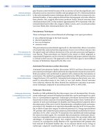
Clinically, most lesions of EAH present as solitary, erythematous nodules with an
angiomatous appearance, although multiple lesions have also been described (4,7–10,34).
They usually appear at birth or during early childhood, and several congenital examples
have been described (8,11,12). In an isolated case, the lesion developed after radio-
therapy (21). The most commonly affected areas are the acral zones, in particular the
palms and soles (Fig. 3), although lesions on the face, neck, and trunk have also been
reported. One patient with multiple lesions of EAH on the extensor surface of the wrists
also had lesions on the knuckle pads (34).
Biologically, the lesions are generally slow growing and asymptomatic, but pain and
hyperhidrosis may be an occasional feature of this lesion. The pain is probably owing to
nerve involvement (14,15) and the hyperhidrosis presumably results from stimulation of
the eccrine component owing to a local increase in temperature caused in turn by the
angiomatous component (8,10,14,16).
Fig. 3. Clinical features of eccrine angiomatous hamartoma. (A) Nodular lesion involving the
dorsum of the right toe in a newborn. (B) Nodular lesion involving the tip of the fifth finger.
04/Sangüeza/19-26/F 01/14/2003, 10:52 AM23
24 Sangüeza and Requena / Pathology of Vascular Skin Lesions
Although the lesions tend to grow slowly, as mentioned before, there are cases that
increase rapidly in size. In one reported case the increase in the size of the lesion was noted
during pregnancy, indicating a possible hormonal influence. In this particular case, par-
tial amputation of the involved finger was necessary because of severe pain (13).
H
ISTOPATHOLOGIC FEATURES
The characteristic histopathologic features of EAH include lobules of mature eccrine
glands and ducts closely associated with thin-walled blood vessels, usually of a capillary
nature (Fig. 4), although large venous channels have also been reported (12). In addition
to these two components that define the lesion, the presence of other structures including
fatty tissue (17,27,30), hair follicles (18,19), apocrine glands (10), neurovascular
glomic-like bodies (27), and occasional epidermal hyperplasia (32) has been described
and lends further support to the hamartomatous nature of the EAH. Immunohistochemi-
cal studies have demonstrated that antigens commonly found in eccrine glands, such as
carcinoembryonic antigen (CEA) and S-100 protein were qualitatively diminished in the
eccrine component of the EAH, whereas endothelial markers such as Ulex europaeus,
CD34, CD44, and factor VIII-related antigen were expressed by endothelial cells of the
vascular component (10,16). Immunoreactivity for gross cystic disease fluid protein-15
was detected in the eccrine gland component of one case (33).
T
REATMENT
Although it is benign and slow growing, EAH is often painful and may require surgical
excision. There have been reports, however, of troublesome lesions in which the pain
spontaneously resolved after some time (15).
Fig. 4. Histopathologic features of eccrine angiomatous hamartoma. (A) Scanning magnification
shows numerous dilated vascular structures involving both the upper and deeper dermis. (B)
Higher magnification demonstrates an abundant number of eccrine units intermingled with dilated
thin-walled vascular structures.
04/Sangüeza/19-26/F 01/14/2003, 10:52 AM24
Chapter 4 / Cutaneous Vascular Hamartomas 25
References
1. Lotzbeck C. Ein Fall von Schweissdrüsengeschwulst an der Wauge. Virchows Arch Pathol Anat
1859;16:160.
2. Beier E. Über einen Fall von Naevus Subcutaneous (Virchow) mit hochgradiqer Hyperplasie der
Knäuelodrüsen. Arch Dermat Syph 1895;31:337.
3. Hyman AB, Harris H, Brownstein MH. Eccrine angiomatous hamartoma. NY State J Med 1968;68:
2803–6.
4. Vilanova X, Piñol Aguade J, Castells A. Hamartoma angiomateux sudoripare sécretant. Dermatologica
1963;127:9–16.
5. Issa O. Hamartoma angiomatoso sudoriparo funcionante. Actas Dermosifiliogr 1964;55:361–5.
6. Söltz-Szötz K. Berich über Fall von Schweissdrüsen naevus Kombiniert mit einem Angiom. Z Hautkr
1958;24:189–92.
7. Archer BWC. Multiple cavernous angiomata of the sweat ducts associated with hemiplegia. Lancet
1927;2:595–6.
8. Domonkos AN, Suarez LS. Sudoriparous angioma. Arch Dermatol 1967;96:552–3.
9. Aloi F, Tomasini C, Pippione M. Eccrine angiomatous hamartoma: a multiple variant. Dermatology
1992;184:219–22.
10. Sulica RL, Kao GF, Sulica VJ, Penneys NS. Eccrine angiomatous hamartoma (nevus): immunohis-
tochemical findings and review of the literature. J Cutan Pathol 1994;21:71–5.
11. Kikuchi J, Kukari Y, Inoves S. Painful eccrine angiomatous nevus on the sole. J Dermatol 1982;9:329–32.
12. Sanmartin O, Botella R, Alegre V, Martinez A, Aliaga A. Congenital eccrine angiomatous hamartoma.
Am J Dermatopathol 1992;14:161–4.
13. Gabrielsen O, Elgjo K, Sommerschild H. Eccrine angiomatous hamartoma of the finger leading to
amputation. Clin Exp Dermatol 1991;16:44–5.
14. Challa VR, Jona J. Eccrine angiomatous hamartoma: a rare skin lesion with diverse histological features.
Dermatologica 1977;155:206–9.
15. Wolf R, Krakowski A, Dorfman B. Eccrine angiomatous hamartoma. A painful step. Arch Dematol
1989;125:1489–90.
16. Smith VC, Montesinos E, Revert A, Ramon D, Molina I, Jorda E. Eccrine angiomatous hamartoma:
report of three patients. Pediatr Dermatol 1996;13:139–42.
17. Donati P, Amantea A, Balus I. Eccrine angiomatous hamartoma. A lipomatous variant. J Cutan Pathol
1989:16:227–9.
18. Zeller DJ. Goldman RL. Eccrine-pilar angiomatous hamartoma. Dermatologica 1971;143:100–4.
19. Velasco, JA, Almeida V. Eccrine-pilar angiomatous nevus. Dermatologica 1988;177:317–22.
20. Aloi FG, Molinero A, Ronco A, Pippione M. Nevo eccrino-angiomatoso. G Ital Dermatol Venereol
1989;124:235–6.
21. Dallot A, Chemaly P, Kemeny JL, et al. Hamartome angio-eccrine chez un adulte après radiothérapie.
Ann Dermatol Venereol 1992;119:903–4.
22. Diaz-Landaeta L, Kerdel FA. Hyperhidrotic, painful lesion. Eccrine angiomatous hamartoma. Arch
Dermatol 1993;129:107.
23. Torres JE, Martin RF, Sanchez JL. Eccrine angiomatous hamartoma. PR Health Sci J 1994;13:159–60.
24. Nair LV, Kurien AM. Eccrine angiomatous hamartoma. Int J Dermatol 1994;33:650–1.
25. Nakayama H, Mihara M, Hattori K, Mishima E, Shimao S. Eccrine angiomatous hamartoma of the sacral
region. Acta Derm Venereol 1994;74:477.
26. Calderone DC, Glass LF, Seleznick M, Fenske NA. Eccrine angiomatous hamartoma. J Dermatol Surg
Oncol 1994;20:837–8.
27. Damiani S, Riccioni L. Palmar cutaneous hamartoma. Am J Dermatopathol 1998;20:65–8.
28. Michel JL, Secchi T, Balme B, Barrut D, Thomas L, Moulin G. Hamartome angio-eccrine congenital.
Ann Dermatol Venereol 1997;124:623–5.
29. Kwon OC, Oh ST, Kim SW, Park GS, Cho BK. Eccrine angiomatous hamartoma. Int J Dermatol
1998;37:787–9.
30. Cebreiro C, Sanchez Aguilar D, Gomez Centeno P, Fernandez Redondo V, Toribio J. Eccrine angioma-
tous hamartoma: report of seven cases. Clin Exp Dermatol 1998;23:267–70.
31. Nakatsui TC, Schloss E, Krol A, Lin AN. Eccrine angiomatous hamartoma: report of a case and literature
review. J Am Acad Dermatol 1999;41:109–11.
32. Tsuji T, Sawada H. Eccrine angiomatous hamartoma with verrucous features. Br J Dermatol
1999;141:167–9.
04/Sangüeza/19-26/F 01/14/2003, 10:52 AM25
26 Sangüeza and Requena / Pathology of Vascular Skin Lesions
33. Tanaka M, Shimizu S, Miyakawa S. Hypertrophic eccrine glands in eccrine angiomatous hamartoma
produce gross cystic disease fluid protein 15. Dermatology 2000;200:336–7.
34. Morell DS, Ghali FE, Stahr BJ, McCauliffe DP. Eccrine angiomatous hamartoma: a report of symmetric
and painful lesions of the wrists. Pediatr Dermatol 2001;18:117–9.
35. Lee SY, Chang SE, Choi JH, Sung KJ, Moon KC, Koh JK. Congenital eccrine angiomatous hamartoma:
report of two patients. J Dermatol 2001;28:338–40.
36. Laeng RH, Heilbrunner J, Itin PH. Late-onset eccrine angiomatous hamartoma: clinical, histological
and imaging findings. Dermatology 2001;203:70–4.
04/Sangüeza/19-26/F 01/14/2003, 10:52 AM26
Chapter 5 / Cutaneous Vascular Malformations 27
5
Cutaneous Vascular Malformations
CONTENTS
NEVUS ANEMICUS
CUTIS MARMORATA TELANGIECTATICA CONGENITA
NEVUS FLAMMEUS
HYPERKERATOTIC VASCULAR STAINS
VENOUS MALFORMATIONS
SUPERFICIAL CUTANEOUS LYMPHATIC MALFORMATIONS
CYSTIC LYMPHATIC MALFORMATIONS (CYSTIC HYGROMAS)
LYMPHANGIOMATOSIS
Malformation denotes an abnormal structure that results from an aberration in
embryologic development. Although the term malformation is conventionally used as a
synonym for hamartoma, the two are different, because hamartoma refers to a potpourri of
tissue elements normally present at a particular site. Vascular malformations can be either
functional or anatomic. In the case of functional abnormalities, the changes are related
mostly to physiologic changes, as is the case for nevus anemicus. In contrast, anatomic
vascular malformations exhibit evident morphologic abnormalities of the involved vessels.
Anatomic vascular malformations are subdivided into the following groups: capillary,
venous, arterial, lymphatic, and combined anomalies (Table 1). Clinically, it is important
to separate the vascular malformations into those of low flow and high flow. Low-flow
27
Table 1
Cutaneous Vascular Malformations
Functional
Nevus anemicus
Anatomical
Capillary
Cutis marmorata telangiectatica congenita
Nevus flammeus (port wine stain)
Hyperkeratotic vascular stains
Venous and arterial
Lymphatic
Superficial lymphatic
Deep (cystic) lymphatic
Lymphangiomatosis
Combined vascular malformations
05/Sangüeza/27-72/F 01/14/2003, 11:21 AM27
28 Sangüeza and Requena / Pathology of Vascular Skin Lesions
abnormalities include malformations of capillary, venous, lymphatic, or combination,
whereas high-flow abnormalities include arteriovenous malformations (1).
References
1. Mulliken JB. Classification of vascular birthmarks. In: Mulliken JB, Young AE, eds. Vascular Birth-
marks. Hemangiomas and Malformations. Philadelphia, WB Saunders, 1988;24–37.
05/Sangüeza/27-72/F 01/14/2003, 11:21 AM28
Chapter 5 / Cutaneous Vascular Malformations 29
1. NEVUS ANEMICUS
CLINICAL FEATURES
Nevus anemicus is an uncommon congenital vascular malformation observed more
frequently in women than in men. Clinically, the lesion consists of a localized circum-
scribed, pale macule with irregular margins occasionally surrounded by satellite macules
(1–14). Although the upper chest is the most commonly affected site (Fig. 1), it may occur
in any part of the body. Under diascopic pressure, the lesion becomes indistinguishable
from the blanched surrounding skin. Wood’s lamp examination does not accentuate the
lesion, and the application of friction, cold, or heat does not induce erythema in the
involved areas. All these maneuvers are helpful in distinguishing nevus anemicus from
vitiligo, hypochromic nevus, and other hypomelanosis. Nevus anemicus can be an addi-
tional feature in types II, III, and IV of phakomatosis pigmentovascularis (5,9,12,14).
The presence of persistent, localized areas of livid erythema, caused by an increase in
the vasoconstrictor tone of the thermoregulatory vessels of the involved skin and leading
to relative stasis in the superficial nutritional vasculature, is considered a clinical variant
Fig. 1. Clinical features of a nevus anemicus involving the anterior chest of an adult woman. (A)
Hypochromic macule on the right anterior chest. (B) Rubbing of the lesion results in peripheral
erythema secondary to vasodilation in adjacent noninvolved skin, whereas the lesion of nevus
anemicus remains a whitish color.
05/Sangüeza/27-72/F 01/14/2003, 11:21 AM29
30 Sangüeza and Requena / Pathology of Vascular Skin Lesions
of nevus anemicus (8). The term nevus oligemicus has been proposed for this clinical
variant of nevus anemicus. Another variant of nevus anemicus was described by Miura
et al. (15). This lesion is characterized by multiple anemic macules on the arms; however,
other authors (16) consider this lesion to be an expression of an exaggerated physiologic
speckled mottling of the limbs caused by transient changes in the vascular tone of the
cutaneous vessels, but not a true variant of nevus anemicus.
H
ISTOPATHOLOGIC FEATURES
Since nevus anemicus is a functional anomaly rather than an anatomic one, histopatho-
logic and ultrastructural examination of these lesions demonstrates that vascular struc-
tures are essentially normal (1). Intralesional injections of bradykinin, pilocarpine,
acetylcholine, 5-hydroxytryptamine, nicotine, or histamine fail to produce vasodilation
of the affected areas (1). However, erythema develops after an axillary sympathetic block
or the intralesional injection of an α-adrenergic blocking agent (2,3). A disturbance in the
regulation of vascular intramural adrenergic receptors may be demonstrated by means of
autoradiography, resulting in persistent vascular constriction (6,7). Furthermore, it has
been demonstrated that the blood vessels in nevus anemicus do not respond normally to
proinflammatory cytokines, at least at the level of E-selectin expression. Additionally, if
contact dermatitis affects the area of nevus anemicus, the keratinocytes overlying the area
do not express intercellular adhesion molecule-1 and HLA-DR, probably because of the
absence of infiltrating lymphocytes (11). All these findings support the conclusion that
nevus anemicus results from locally increased vascular reactivity to catecholamines.
Autograft transplant shows donor site dominance, lending further support to the idea that
the cause of nevus anemicus is either an increased sensitivity to stimulation by vasocon-
strictors or inhibition of vasodilator influences (2). The presence of vascular twin nevi,
i.e., telangiectatic nevus and nevus anemicus occurring together and adjacent to each
other, can be explained as twin spots resulting from allelism of somatic mutations (10).
T
REATMENT
Usually lesions of nevus anemicus do not require treatment. However, if the lesions
cause cosmetic disability then camouflage makeup is sufficient.
References
1. Greaves MW, Birkett D, Johson C. Nevus anemicus: a unique catecholamine-dependent nevus. Arch
Dermatol 1970;102:172–6.
2. Daniel RH, Hubler WR, Wolf JE, et al. Nevus anemicus: donor-dominant defect. Arch Dermatol
1977;113:53–6.
3. Mountcastle EA, Diestelmeier RM, Lupton GP. Nevus anemicus. J Am Acad Dermatol 1986;14:628–32.
4. Fleischer TL, Zeligman I. Nevus anemicus. Arch Dermatol 1969;100:750–5.
5. Ratz JL, Roenigk HH Jr. Multiple vascular anomalies: report of a case. J Dermatol Surg Oncol
1978;4:684–6.
6. Raff M. Die Bedeutung adrenerger Rezeptoren fur die Entstehung des naevus flammeus und des Naevus
anaemicus. Wien Klin Wochenschr 1981;129(suppl):1–14.
7. Dupre A, Bonafe JL, Jouas H. Naevus anemique generalisé acquis. Dermatologica 1981;163:276–81.
8. Davies MG, Greaves MW, Coutss A, Black AK. Nevus oligemicus. A variant of nevus anemicus. Arch
Dermatol 1981;117:111–3.
9. Hidano A, Arai Y. Hémihypertrophie congénitale associée à des anomalies cutanées pigmento-
vasculaires, cérébrales, viscèrales et squelettiques. Ann Dermatol Venereol 1987;114:665–9.
10. Happle R. Allelic somatic mutations may explain vascular twin nevi. Hum Genet 1991;86:321–3.
05/Sangüeza/27-72/F 01/14/2003, 11:21 AM30
Chapter 5 / Cutaneous Vascular Malformations 31
11. Mizutani H, Ohyanagi S, Umeda Y, Shimizu M, Kupper TS. Loss of cutaneous delayed hypersensitivity
reactions in nevus anemicus. Evidence for close concordance of cutaneous delayed hypersensitivity and
endothelial E-selectin expression. Arch Dermatol 1997;133:617–20.
12. Di Landro A, Tadini GL, Marchesi L, Cainelli T. Phakomatosis pigmentovascularis: a new case with
renal angiomas and some considerations about the classification. Pediatr Dermatol 1999;16:25–30.
13. Ahkami RN, Schwartz RA. Nevus anemicus. Dermatology 1999;198:327–9.
14. Hasegawa Y, Yasuhara M. Phakomatosis pigmentovascularis type IVa. Arch Dermatol 1985;121:651–5.
15. Miura Y, Tajima S, Ishibashi A, Hata Y. Multiple anemic macules on the arms: a variant form of nevus
anemicus? Dermatology 2000;201:180–3.
16. Plantin P, Schoenlaub P. Multiple anemic macules on the arms: not a variant form of nevus anemicus.
Dermatology 2001;202:271–2.
05/Sangüeza/27-72/F 01/14/2003, 11:21 AM31
32 Sangüeza and Requena / Pathology of Vascular Skin Lesions
2. CUTIS MARMORATA TELANGIECTATICA CONGENITA
Cutis marmorata telangiectatica congenita (CMTC) is usually present at birth and is
characterized by the presence of a reticulated cutaneous vascular network of a blue-violet
color. These lesions may be either localized or generalized. Van Lohuizen originally (1)
described CMTC in 1922 and coined the term. CMTC should be distinguished from cutis
marmorata, which is a physiologic response to cold and is prominent in neonates. To
emphasize the distinction, some authors have used the term reticulated vascular nevus for
CMTC (2). Although some familial cases have been reported (3–6), no recognizable
pattern of inheritance has been demonstrated by this disease. In about 50% of patients,
CMTC is associated with various other congenital anomalies (5), suggesting that CMTC
is the principal cutaneous manifestation of a complex syndrome (3).
C
LINICAL FEATURES
Clinically CMTC can be either localized (7) or generalized (8–10); when the lesions
are localized, the distribution is usually segmental, with a sharp midline demarcation.
However, the lesions occasionally involve the upper right limb and the lower left limb,
in a crossed limb dimelia distribution (11). The areas of involved skin show either a flat
or a reticulated erythema, producing a marbled appearance (Fig. 2). Telangiectasias are
generally visible within the reticulated lines, and the red-purple hue of the lesions may
become more prominent after crying, vigorous movements, or decrease in the ambient
temperature. The skin in the affected areas may be atrophic, and ulceration may be a
prominent feature (12). The lesions of CMTC show gradual spontaneous improvement
Fig. 2. Clinical features of cutis marmorata telangiectatica congenita. (A) Lesions involving the
lower left extremity. (B) Close-up view of the lesions involving the anterior aspect of the left leg.
05/Sangüeza/27-72/F 01/14/2003, 11:21 AM32
Chapter 5 / Cutaneous Vascular Malformations 33
with time and in some patients disappear entirely (8–10). In others, however, little or no
significant change occurs, and the lesions remain unmodified throughout life.
When the face is involved, there is either a diffuse erythema or nevus flammeus that
affects the upper lip and philtrum (12–16). Patients with facial lesions are at risk of
congenital glaucoma (17–22), which may be bilateral if the involvement is diffuse,
especially in those cases affected by nevus flammeus (12,13). However, congenital glau-
coma may also be present in patients with facial involvement of CMTC in the absence
of nevus flammeus (12,23). Other associated anomalies include hemiatrophy or hemihy-
pertrophy of the body (15,16), atrophy (2,5,6,14,15,24) or hypertrophy (5,14) of the
involved limb, macrocephaly (14,16,25–33), mental retardation (13,15,26,28), hydro-
cephalus (16,26,28,32), neuronal migrations defects (31), hearing loss (16), strabismus
(16), persistent ducts arteriosus (13) and other cardiovascular abnormalities (16), cardiac
arrhytmia (33), sudden infant death (33), internal arteriovenous malformations (28),
hypothyroidism (16), nevus anemicus (16), hemangiomas (13,16,29,32), congenital
melanocytic nevus (12), café-au-lait spots (16), asymmetric skull (13,34,35), microg-
nathia (13), triangular face (13), scaphoid scapulae (13), elevation of the hemidiaphragm
(16), lipomas (16), dystrophic teeth (13,31), anomalies of the growth of hair (31), high-
arched palate (13), cleft palate (14), syndactyly of the fingers or toes (13,15,16,28,32),
short fingers (13), polydactyly (32), acral cyanosis (13), simian lines on the palms (14),
anal atresia (16,36), rectovaginal and urethrovaginal fistulas (36), absent clitoris
(36), hypospadias (37), short stature (13), diffuse demineralization of bones (14),
lipoatrophy (34), weakness of the long extensor muscles of both thumbs (14), dislocated
hips (31), joint laxity (32), hypoplasia of a lumbar vertebra (35), stridor (31), Adams-
Oliver syndrome (aplasia cutis congenita with terminal transverse limb defects) (38–43),
spina bifida (44), neonatal ascites (45), congenital generalized fibromatosis (46),
porencephaly (46), soft tissue herniations on the lower legs (47), and bilateral retinal
detachment (48,49), tendinitis stenosans and bowing of the lower legs (50), congenital
hypothyroidism (51), double aortic arch (52), hypoplasia of the iliac and femoral veins
(53), moyamoya-like vascular abnormalities with factor V Leiden mutation resulting in
congenital hypercoagulable disorder (54), and elevated maternal serum human chorionic
gonadotrophin level with transitory isolated fetal ascites (55). Despite these numerous
anomalies associated with CMTC, most of the cases of this disease present as a solitary
abnormality.
H
ISTOPATHOLOGIC FEATURES
There is still some debate about the nature of the vascular lesions in CMTC. Although
most authors believe that there is true anatomic malformation, others believe that CMTC
is a functional malformation. Most of the skin biopsies exhibit dilated capillaries and
veins throughout the entire dermis and subcutaneous fat (1,3) whereas in a few cases (13),
no vascular abnormalities have been detected. Histopathologic examination of the lesions
in a patient with painful lesions of CMTC demonstrated an increased number of nerve
fibers (56). Ultrastructural studies have demonstrated an increased number of perithelial
cells, as well as atypical endothelium with a discontinuous basal lamina (5).
Colored echo-Doppler and phlebography studies have shown dilated deep venous
tracts with reflux to the superficial veins (57). Laser Doppler fluxmetry of the involved
skin has shown evidence of functional disturbance, which may be the expression of an
α-adrenergic innervation deficit of the cutaneous terminal blood vessels (58).
05/Sangüeza/27-72/F 01/14/2003, 11:21 AM33
34 Sangüeza and Requena / Pathology of Vascular Skin Lesions
The differential diagnosis of CMTC is with Bockenheimer’s syndrome or diffuse
genuine phlebectasia (12,59,60). Bockenheimer’s syndrome appears in childhood and
shows a progressive development of multiple large and painful venous ectasias involving
a single limb. Cutaneous lesions similar to those of CMTC may be also seen in newborns
with neonatal lupus erythematosus (61,62).
T
REATMENT
The cutaneous lesions of CMTC tend to fade with time, and there is no need to treat
these lesions during the first years of life. For persistent cutaneous lesions, treatment with
argon or dye laser may be helpful. The discovery of this malformation in the skin of an
infant, however, should prompt a diligent search for an associated congenital anomaly by
neuropediatric, ophthalmologic, and orthopedic explorations, in addition to dermato-
logic examination (63).
References
1. van Lohuizen CHJ. Über eine seltene angeborene Hautanomalie (cutis marmorata telangiectatica
congenita). Acta Derm Venereol 1922;3:202–11.
2. Brain RT. Naevus vascularis reticularis. Proc Soc Med 1954;47:172–3.
3. Andreev VC, Pramatarov K. Cutis mamorata telangiectatica congenita in two sisters. Br J Dermatol
1979;101:345–50.
4. Kurczinski TW. Hereditary cutis marmorata telangiectatica congenita. Pediatrics 1982;70:52–3.
5. Way BH, Herrmann, J, Gilbert EF, et al. Cutis marmorata telangiectatica congenita. J Cutan Pathol
1974;1:10–25.
6. Rogers M, Poyzer KG. Cutis marmorata telangiectatica congenita. Arch Dermatol 1982;118:895–9.
7. Suarez SM, Grossman ME. Localized cutis marmorata telangiectatica congenita. Pediatr Dermatol
1991;8:329–31.
8. Devillers AC, de Waard-van der Spek FB, Oranje AP. Cutis marmorata telangiectatica congenita:
clinical features in 35 cases. Arch Dermatol 1999;135:34–8
9. Amitai DB, Fichman S, Merlob P, Morad Y, Lapidoth M, Metzker A. Cutis marmorata telangiectatica
congenita: clinical findings in 85 patients. Pediatr Dermatol 2000;17:100–4.
10. Enjolras O. Cutis marmorata telangiectatica congenita. Ann Dermatol Venereol 2001;128:161–6
11. Sanchez P, Bosch RJ, Herrera E. Cutis marmorata telangiectatica congenita: forme dimelique croisée.
Ann Dermatol Venereol 1992;119:647–50.
12. Picascia DD, Esterly NB. Cutis marmorata telangiectatica congenita: report of 22 cases. J Am Acad
Dermatol 1989;20:1098–104.
13. Petrozzi WJ, Rahn EK, Mofenson H, et al. Cutis marmorata telangiectatica congenita. Arch Dermatol
1970;101:74–7.
14. Lee S, Lee JB, Kim JH, et al. Cutis marmorata congenita with multiple congenital abnormalities (van
Lohuizen’s syndrome). Dermatologica 1981;163:408–12.
15. Lopez-Herce Cid J, Roche Herrero MC, Pascual Castroviejo I. Cutis marmorata telangiectatica congenita.
Anomalías asociadas. An Esp Pediatr 1985;22:585–90.
16. Gerritsen MJ, Steijlen PM, Brunner HG, Rieu P. Cutis marmorata telangiectatica congenita: report of
18 cases. Br J Dermatol 2000;142:366–9.
17. Sato SE, Herschler J, Lynch PJ, et al. Congenital glaucoma associated with cutis marmorata congenita
telangiectatica: two case reports. J Pediatr Ophthalmol Strabismus 1988;25:13–7.
18. Vazquez F, Lopez B, Requena L, Garcia Perez A. Congenital glaucoma and cutis marmorata telangiecta-
sia: report of the second case. Dermatologica 1989;177:193–4.
19. Lynch PJ. Cutis marmorata telangiectatica congenita associated with congenital glaucoma. J Am Acad
Dermatol 1990;22:857.
20. Miranda I, Alonso MJ, Jimenez M, Tomas-Barberan S, Ferro M, Ruiz R. Cutis marmorata telangiectatica
congenita and glaucoma. Ophthalmic Paediatr Genet 1990;11:129–32.
21. Kremer I, Metzker A, Yassur Y. Intraoperative suprachoroidal hemorrhage in congenital glaucoma
associated with cutis marmorata telangiectatica congenita. Arch Ophthalmol 1991;109:1199–200.
22. Weilepp AE, Eichenfield LF. Association of glaucoma with cutis marmorata telangiectatica congenita:
a localized anatomic malformation. J Am Acad Dermatol 1996;35:276–8.
05/Sangüeza/27-72/F 01/14/2003, 11:21 AM34
Chapter 5 / Cutaneous Vascular Malformations 35
23. South DA, Jacobs AH. Cutis marmorata telangiectatica congenita (congenital generalized phlebectasia).
J Pediatr 1978;93:944–9.
24. Wong V. Cutis marmorata telangiectatica congenita: an unusual presentation with monoatrophy in two
Chinese children. J Paediatr Child Health 1997;33:71–3.
25. Stephan MJ, Hall BD, Smith DW, Cohen MM Jr. Macrocephaly in association with unusual cutaneous
angiomatosis. J Pediatr 1975;87:353–9.
26. Moore CA, Torriello HV, Abuelo DN, et al. Macrocephaly-cutis marmorata telangiectatica congenita: a
distinct disorder with developmental delay and connective tissue abnormalities. Am J Med Genet 1997;2:67–73.
27. Wroblewski I, Joannard A, Francois P, Baudain P, Beani JC, Beaudoing A. Cutis marmorata
telangiectatica congenita avec asymétrie corporalle. Pediatrie 1988;43:117–20
28. Clayton-Smith J, Kerr B, Brunner H, et al. Macrocephaly with cutis marmorata, haemangioma and
syndactyly—a distinctive overgrowth syndrome. Clin Dysmorphol 1997;6:291–302.
29. Carcao M, Blaser SI, Grant RM, Weksberg R, Siegel-Bartelt J. MRI findings in macrocephaly-cutis
marmorata telangiectatica congenita. Am J Med Genet 1998;76:165–7.
30. Vogels A, Devriendt K, Legius E, et al. The macrocephaly-cutis marmorata telangiectatica congenita
syndrome. Long-term follow-up data in 4 children adolescents. Genet Couns 1998;9:245–53.
31. Robertson SP, Gattas M, Rogers M, Ades LC. Macrocephaly-cutis marmorata telangiectatica congenita:
report of five patients and a review of the literature. Clin Dysmorphol 2000;9:1–9.
32. Franceschini P, Licata D, Di Cara G, Gaula A, Franceschini D, Genitori L. Macrocephaly-cutis
marmorata telangiectatica congenita without cutis marmorata? Am J Med Genet 2000;14:265–9.
33. Yano S, Watanabe Y. Association of arrhythmia and sudden death in macrocephaly-cutis marmorata
telangiectatica congenita syndrome. Am J Med Genet 2001;102:149–52.
34. Gelmetti C, Console V, Schianchi R, Missaglia R. Cutis marmorata telangiectatica congenita.
Descrizione di un nuovo caso. Pediatr Med Chir 1986;8:907–9.
35. Gelmetti C, Schianchi R, Ermacora E. Cutis marmorata telangiectatica congenita. Quatre nouveaux cas
et revue de la littérature. Ann Dermatol Venereol 1987;114:1517–28.
36. Del Giudice SM, Nydorf ED. Cutis marmorata telangiectatica congenita with multiple congenital anoma-
lies. Arch Dermatol 1986;122:1060–1.
37. Ben-Amitai D, Merlob P, Metzker A. Cutis marmorata telangiectatica congenita and hypospadias:
report of 4 cases. J Am Acad Dermatol 2001;45:131–2.
38. Powell ST, Su WPD. Cutis marmorata telangiectatica congenita: a report of 9 cases and review of the
literature. Cutis 1984;34:305–12.
39. Torrielo HV, Graff RG, Florentine MF, Lacina S, Moore WD. Scalp and limb defects with cutis
marmorata telangiectatica congenita: Adams-Oliver syndrome? Am J Med Genet 1988;29:269–76.
40. Bork K, Pfeifle J. Multifocal aplasia cutis congenita, distal limb hemimelia, and cutis marmorata
telangiectatica in a patient with Adams-Oliver syndrome. Br J Dermatol 1992;127:160–3.
41. Dyall-Smith D, Ramsden A, Laurie S. Adams-Oliver syndrome: aplasia cutis congenita, terminal trans-
verse limb defects and cutis marmorata telangiectatica congenita. Australas J Dermatol 1994;35:19–22.
42. Bjornsdottir US, Laxdal T, Bjornsson J. Cutis marmorata telangiectatica congenita with terminal trans-
verse limb defects. Acta Paediatr Scand 1988;77:780–2.
43. Mempel M, Abeck D, Lange I, et al. The wide spectrum of clinical expression in Adams-Oliver syn-
drome. A report of two cases. Br J Dermatol 1999;140:1157–60.
44. Schultz RB, Kocoshis S. Cutis marmorata telangiectatica congenita and neonatal ascites. J Pediatr
1979;95:157.
45. Spraker MK, Stack C, Esterly NB. Congenital generalized fibromatosis: a review of the literature and
report of a case associated with porencephaly, hemiatrophy, and cutis marmorata telangiectatica
congenita. J Am Acad Dermatol 1984;10:365–71.
46. Nicholls DSH, Harper JI. Cutis marmorata tetangiectatica congenita with soft-tissue herniations on the
lower legs. Clin Exp Dermatol 1989;14:369–70.
47. Freund E. Diffuse genuine phlebectasia. Arch Surg 1936;33:113–21.
48. Shields JA, Shields CL, Koller HP, Federman JL, Koblenzer P, Barbera LS. Cutis marmorata telan-
giectatica congenita associated with bilateral congenital retinal detachment. Retina 1990;10:135–9.
49. Pendergast SD, Trese MT, Shastry BS. Ocular findings in cutis marmorata telangiectatica congenita.
Bilateral exudative vitreoretinopathy. Retina 1997;17:306–9.
50. Kennedy C, Oranje AP, Keizer K, van den Heuvel MM, Catsman-Berrevoets CE. Cutis marmorata
telangiectatica congenita. Int J Dermatol 1992;31:249–52.
51. Pehr K, Moroz B. Cutis marmorata telangiectatica congenita: long-term follow-up, review of the literature
and report of a case in conjunction with congenital hypothyroidism. Pediatr Dermatol 1993;10:6–11.
05/Sangüeza/27-72/F 01/14/2003, 11:21 AM35
36 Sangüeza and Requena / Pathology of Vascular Skin Lesions
52 O’Toole EA, Deasy P, Watson R. Cutis marmorata telangiectatica congenita associated with a double
aortic arch. Pediatr Dermatol 1995;12:348–50.
53. Morgan JM, Naisby GP, Carmichael AJ. Cutis marmorata telangiectatica congenita with hypoplasia of
the right iliac and femoral veins. Br J Dermatol 1997;137:119–22.
54. Gruppo RA, DeGrauw TJ, Palasis S, Kalinyak KA, Bofinger MK. Strokes, cutis marmorata
telangiectatica congenita, and factor V Leiden. Pediatr Neurol 1998;18:342–5.
55. Chen CP, Chen HC, Liu FF, et al. Cutis marmorata telangiectatica congenita associated with an elevated
maternal serum human chorionic gonadotrophin level and transitory isolated fetal ascites. Br J Dermatol
1997;136:267–71.
56. Lentner A, Bohler U, Wittkopf-Baumann C, Younossi H, Grussendorf-Conen EI. Schmerzhafte Cutis
marmorata teleangiectatica congenita. Hautarzt 1992;43:657–60.
57. Lingier P, Munck D, Godart S. Cutis marmorata telangiectatica congenita. A propos de quatre nouveaux
cas. Phlebologie 1992;45:489–96.
58. Bormann G, Wohlrab J, Fisher M, Marsch WC. Cutis marmorata telangiectatica congenita: laser Dop-
pler fluxmetry evidence for a functional nervous defect. Pediatr Dermatol 2001;18:110–3.
59. Fitzsimmons JS, Starks M. Cutis marmorata telangiectatica congenita or congenital generalized
phlebectasia. Arch Dis Child 1970;45:724–6.
60. Atherton DJ. Naevi and other developmental defects. In: Champion RH, Burton JL, Ebling FJG eds.
Textbook of Dermatology, 5th ed., Oxford, Blackwell Scientific, 1992:445–526.
61. Greist MC, Probst E. Cutis marmorata telangiectatica congenita on neonatal lupus. Arch Dermatol
1980;116:1102–3.
62. Carrascosa JM, Ribera M, Bielsa I, Coroleu W, Ferrandiz C. Cutis marmorata telangiectatica congenita
or neonatal lupus? Pediatr Dermatol 1996;13:230–2.
63. Rupprecht R, Hundeiker M. Cutis marmorata teleangiectatica congenita. Wichtige Aspekte fur die
dermatologisches Praxis. Hautarzt 1997;48:21–5.
05/Sangüeza/27-72/F 01/14/2003, 11:21 AM36
Chapter 5 / Cutaneous Vascular Malformations 37
3. NEVUS FLAMMEUS
Nevus flammeus is a generic term used to describe congenital vascular malformations
that affect newborns. These commonly involve the forehead, face, and neck, although
lesions have been described in nearly all anatomic sites including mucous membranes (1).
Approximately 25–40% of newborns of all races are born with pink-red macular lesions that
involve the midline of the face, occiput, and nuchal regions (2–5). Most of these lesions are
small, and are located on the glabellar region of the forehead, where they are commonly
known as salmon patch. They tend to disappear during infancy or childhood. In other cases
the lesions are present on the nape of the neck; these lesions tend to be much more persistent
and in a significant percentage of patients they remain unchanged into adult life. The
combination of lesions on the glabellar region and the nape of the neck is known colloqui-
ally as stork bite. Inflammatory changes of dermatitis have been described in nuchal-
occipital nevus flammeus (6,7). Nuchal nevus flammeus is also considered a valuable skin
marker in patients with alopecia areata, indicating a more severe clinical course (8).
Involvement of the sacral skin by nevus flammeus is also frequent and, like the occipital
lesions, these lesions show a tendency to persist into adult life (9,10). Ocassionally, sacral
nevus flammeus is associated with occult spinal dysraphism (11). A less frequent variant
of nevus flammeus, is the port wine stain, consisting of large unilateral vascular lesions
affecting the face. These lesions also tend to persist during adulthood (2,4,5,12,13).
C
LINICAL FEATURES
Clinically, these small lesions are irregular, dull pinkish red macules that affect the
glabella, forehead, upper eyelids, upper lip (Fig. 3) and nuchal region. They usually affect
more than one site and have a midline distribution (Fig. 4). Most of the lesions affecting
the center of the face fade rapidly with age and disappear during the first year of life (3).
By contrast, nuchal and sacral lesions tend to persist into adulthood. Port wine stains
present as unilateral deep red or purple macules with a slow but progressive growth
throughout life. They also become increasingly darker to eventuate into raised and thick-
ened plaques. Sometimes small angiomatous nodules appear within an otherwise typical
port wine stain; in rare instances, a cobblestone pattern of red-purple nodules covers the
whole lesion (1,14) (Fig. 5). Although cases of congenital nevus flammeus are relatively
common, examples of acquired nevus flammeus are quite rare, and most of the reported
cases of the acquired type have been described in association with preceding trauma (15).
Fig. 3. Small nevus flammeus involving the skin of the upper lip.
05/Sangüeza/27-72/F 01/14/2003, 11:21 AM37
38 Sangüeza and Requena / Pathology of Vascular Skin Lesions
A variety of abnormalities have been associated with port wine stains. The association
of port wine stains and nevus anemicus is a fairly common event (16), and this combi-
nation has been termed naevus vascularis mixtus (17). Glaucoma is a frequent compli-
cation in facial port wine stains, not only in patients with the Sturge-Weber syndrome.
Approximately 10% of all patients with facial port wine stains have glaucoma without
Fig. 4. Extensive nevus flammeus involving the left chest.
Fig. 5. Nevus flammeus involving the face of an older woman. Long-standing lesions become
thicker, with red-purple nodules covering the whole lesion.
05/Sangüeza/27-72/F 01/14/2003, 11:21 AM38
Chapter 5 / Cutaneous Vascular Malformations 39
leptomeningeal involvement (18,19). Ipsilateral glaucoma is especially frequent when
the lesions of port wine stain involve the areas of inervation of both the ophthalmic and
maxillary divisions of the trigeminal nerve. It is less common when the areas of the face
affected are those that follow the distribution of the upper divisions of the fifth cranial
nerve or when the lesions are located solely below the eye (20). The presence of dilated
conjunctival vessels are common when the lids are involved, but this finding does not
correlate with either the presence or absence of glaucoma. Choroidal “angioma” is the
most characteristic ocular vascular malformation in patients with facial port wine stain
and glaucoma (21), and an ophthalmoscopic examination of the fundus of the eye is
imperative when a port wine stain is present in close proximity to the eye in a newborn.
The Sturge-Weber syndrome consists of a large facial nevus flammeus in the distri-
bution of the ophthalmic division of the trigeminal nerve (Fig. 6) accompanied by ipsi-
lateral leptomeningeal angiomatosis. Ocular involvement is not essential to establish the
diagnosis of this syndrome. Clinically, a unilateral port wine stain involving the forehead,
eye, and maxillary area characterizes the Sturge-Weber syndrome. Involvement of the
upper eyelid and the forehead is generally the rule 18 (Fig. 7). Usually the lesion is
unilateral, with a fairly sharp midline delimitation, but sometimes it extends beyond the
midline and bilateral facial port wine stains can be seen in approximately 40% of patients
(22). Homolateral leptomeningeal angiomatosis is the second component of the syn-
Fig. 6. Sturge-Weber syndrome. Large nevus flammeus involving the left side of the face.
Fig. 7. Sturge-Weber syndrome. Small nevus flammeus involving the left forehead, eyelid, and nose.
05/Sangüeza/27-72/F 01/14/2003, 11:21 AM39
40 Sangüeza and Requena / Pathology of Vascular Skin Lesions
drome, although there is no correlation between the extent of the cutaneous lesions and
the extent of leptomeningeal involvement. Some authors admit to the possibility of a
forme fruste of Sturge-Weber syndrome when unilateral leptomeningeal angiomatosis
occurs without facial port wine stain (23–25).
Among the neurologic manifestations, epilepsy is the most frequent (22), and there
is also some degree of mental retardation, which is more pronounced in patients with
bi-hemispheric involvement (26). X-rays of the skull, computed tomography (CT) scan-
ning, and magnetic resonance imaging (MRI) are useful in identifying early cortical
calcifications in these patients. Contralateral hemiplegia, hemisensory defects, and hom-
onymous hemianopsia may also occur. Ocular involvement is present in approximately
30–60% of all cases of Sturge-Weber syndrome (18,20). The manifestations are similar
to those seen in patients with facial port wine stain without leptomeningeal angiomatosis,
although they occur more frequently. Glaucoma, buphthalmos, and blindness are fre-
quent ophthalmologic complications in patients with Sturge-Weber syndrome.
Because of the increased cutaneous vascularity, sometimes an overgrowth of the
underlying soft tissue and bone, occurs, giving rise to a deformity very similar to that of
the Klippel-Trenaunay syndrome (27,28) (Fig. 8). As a matter of fact, Sturge-Weber
syndrome is often associated with Klippel-Trenaunay syndrome (29–31).
Klippel-Trenaunay syndrome is defined as a nevus flammeus (and sometimes other
vascular malformations) associated with soft tissue swelling involving a limb, with or
without bony hypertrophy of the affected extremity (31). If these lesions are associated
with an underlying arteriovenous anastomosis, then the syndrome is called Parkes-Weber
(32). The most common cutaneous lesions in these syndromes consist of one or several
port wine stains on the affected limb (33–36).
Fig. 8. Klippel-Trenaunay syndrome. Extensive nevus flammeus involving the posterior aspect of
a hypertrophic lower extremity.
05/Sangüeza/27-72/F 01/14/2003, 11:21 AM40
Chapter 5 / Cutaneous Vascular Malformations 41
Port wine stains have also been described in a large series of rare congenital disorders
including the following:
1. Phakomatosis pigmentovascularis (37–45).
2. Cobb’s syndrome, which is characterized by a nevus flammeus or other vascular malfor-
mations in a dermatomal distribution on the trunk or limb associated with vascular
malformations of the spinal canal at the same segmental level as that of the cutaneous
lesions (46) (Fig. 9).
3. Wyburn-Mason syndrome, which consists of unilateral retinal arteriovenous malforma-
tion associated with ipsilateral aneurysmal arteriovenous malformation and ipsilateral
port wine stain in the region of the affected eye (47). This syndrome has also been
described in association with Sturge-Weber syndrome (48).
4. Proteus syndrome—the association of hemihypertrophy, macrodactyly, verrucous epi-
dermal nevus, vascular malformations, soft subcutaneous masses, and cerebriform over-
growth of the palmar or plantar surfaces of the hypertrophied limb (49–51).
Fig. 9. Cobb syndrome. (A) Erythematous patch involving the left anterior chest. (B) The lesion
showed a zosteriform arrangement along the left flank. MRI studies demonstrated a vascular
malformation involving the same level of the spinal cord.
05/Sangüeza/27-72/F 01/14/2003, 11:21 AM41
42 Sangüeza and Requena / Pathology of Vascular Skin Lesions
5. Roberts’ syndrome, which consists of a mild facial port wine stain associated with
hypomelia, hypotrichosis, growth retardation, and cleft lip (52).
6. TAR syndrome—congenital thrombocytopenia, bilateral absence or hypoplasia of the
radius, and port wine stain (53).
7. von Hippel-Lindau syndrome or bilateral retinal angiomatosis in combination with cer-
ebellar or medullar hemangioblastoma and, in some instances, facial port wine stain (54).
8. Beckwith-Wiedemann syndrome, which combines exophthalmos, macroglossia, and
gigantism with facial nevus flammeus (55).
9. Rubinstein-Taybi syndrome, defined by mental deficiency, retarded somatic growth,
broad thumbs and big toes, antimongoloid palpebral fissures, high palate, and crowded
teeth (56).
10. Coats’ disease, which comprises retinal telangiectasia and ipsilateral facial port wine
stain (57).
HISTOPATHOLOGIC FEATURES
Histopathologically, the findings of isolated nevus flammeus and nevus flammeus
associated with other anomalies are identical. The main difference is an increased number
of dilated thin-walled capillaries and venules, most of which are situated in the upper part
of the reticular dermis (Fig. 10), although occasionally superficial areas of subcutaneous
fat are also involved. An increased number of mast cells has been described in the dermis
of the lesions of nevus flammeus (58). When angiomatous nodules develop in a patch of
Fig. 10. Histopathologic features of nevus flammeus. (A) Low power shows dilated blood vessels
in the papillary and reticular dermis. (B) Higher magnification demonstrates that these vessels
have thin walls and are lined by a single layer of endothelial cells.
05/Sangüeza/27-72/F 01/14/2003, 11:21 AM42
Chapter 5 / Cutaneous Vascular Malformations 43
nevus flammeus, they are made up either of an aggregation of numerous thin-walled
vessels whose lumina are of different calibers (59) or, in other instances, they are authen-
tic pyogenic granulomas developed within port wine stains (60–63). Other lesions that
may arise in a preexisting nevus flammeus include tufted angioma (63), angiolymphoid
hyperplasia with eosinophilia (64), and basal-cell carcinoma (65–68). In some of the
patients in whom basal cell carcinoma developed in a preexisting nevus flammeus, there
was antecedent of radiotherapy in this area (67).
P
ATHOGENESIS
The pathogenesis of port wine stains remains controversial, and the subject has been
recently revised (69). Three-dimensional reconstructions, thymidine uptake studies, and
ultrastructural observations support the view that port wine stains represent a vascular
ectasia rather than a proliferative process (70,71). Furthermore, a detailed study of port
wine stains in which vessel size, number, and dermal position were measured indicated
that these lesions are not proliferative but result from progressive vascular dilation of
preexisting blood vessels. There are two hypotheses that attempt to explain this dilation.
One suggests either a defect in the vascular wall or abnormalities of the supporting
structure of the dermis surrounding the dilated vessels. Immunofluorescence studies of
the different components of the vessel wall, including type IV collagen, fibronectin, and
factor VIII, disclosed no abnormalities in lesions of port wine stains (72). Comparative
studies of normal blood vessels and vessels of port wine stains utilizing immunohis-
tochemical analysis with four monoclonal antibodies specific for endothelial cells
(PAL-E, anti-factor VIII-related antigen, anti-intercellular adhesion molecule-1, and
anti-endothelial leukocyte adhesion molecule-1) demonstrated no substantial differences,
either in the intensity of staining or the distribution pattern of these antibodies (73).
Therefore, a functional alteration seems to be the most reasonable explanation for the
etiology of port wine stains. Immunoperoxidase studies with S-100 protein have demon-
strated a marked decrease in the nerve fibers associated with ectatic blood vessels of port
wine stains (45). Most likely, this is a primary factor in their etiology. A reduction of the
sympathetic innervation of the blood vessels leads to a failure to regulate vasoconstric-
tion, which in turn produces progressive vascular ectasia that characterize lesions of port
wine stain. In short, port-wine stain results from a neural deficiency of the sympathetic
innervation of the blood vessels (45).
T
REATMENT
Small, centrally located salmon patches tend to disappear by the end of the first year
of life. Port wine stains, however, persist into childhood and adult life, and newer forms
of laser therapy have been highly successful at producing good cosmetic results in facial
lesions (74–79). However, there are some anatomic differences. The most successful
responses of nevus flammeus treated by laser therapy are seen in young patients (less than
1 year old) with small lesions (under 20 cm
2
), and in lesions located over bony areas and
the central forehead (80). On the other hand, centrofacial lesions and lesions involving
maxillary areas in adults and children respond less favorably than lesions located else-
where on the head and neck (81–83). Since laser treatment has superficial penetration, it
cannot reach deeper vessels, so nevus flammeus lesions with a deep component have a
poor response to this type of therapy (82,83). Adverse cutaneous reactions after laser
therapy for nevus flammeus include hyperpigmentation (84) and atrophic and hyper-
trophic scars (84,85). Partial recurrences of the nevus flammeus after initially successful
laser treatment are common (86,87).
05/Sangüeza/27-72/F 01/14/2003, 11:21 AM43
44 Sangüeza and Requena / Pathology of Vascular Skin Lesions
References
1. Barsky SH, Rosen S, Geer DE, et al. The nature and evolution of port wine stains: a computer assisted
study. J Invest Dermatol 1980;74:154–7.
2. Hidano A, Purwoko R, Jitsukawa K. Statistical survey of skin changes in Japanese neonates. Pediatr
Dermatol 1986;3:140–4.
3. Leung AKC, Telmesani AMA. Salmon patches in Caucasian children. Pediatr Dermatol 1989;6:185–7.
4. Nanda A, Kaur S, Bhakoo ON, et al. Survey of cutaneous lesions in Indian newborns. Pediatr Dermatol
1989;6:39–42.
5. Rivers JK, Fredericksen PC, Dibdin C. A prevalence survey of dermatoses in the Australian neonate.
J Am Acad Dermatol 1990;23:77–81.
6. Tay YK, Morelli J, Weston WL. Inflammatory nuchal-occipital port-wine stains. J Am Acad Dermatol
1996;35:811–3.
7. Bonifazi E, Mazzota F. Inflammatory nuchal-occipital port-wine stains. J Am Acad Dermatol
1998;38:130.
8. Hatzis J, Kostakis P, Tosca A, et al. Nuchal nevus flammeus as a skin marker of prognosis in alopecia
areata. Dermatologica 1988;177:149–51.
9. Metzker A, Shamir R. Butterflay-shaped mark: a variant form of nevus flammeus simplex. Pediatrics
1990;85:1069–71.
10. Patrizi A, Neri I, Orlandi C, Marini R. Sacral medial telangiectatic vascular nevus: a study of 43 children.
Dermatology 1996;192:301–6.
11. Ben-Amital D, Davidson S, Schwartz M, et al. Sacral nevus flammeus simplex: the role of imaging.
Pediatr Dermatol 2000;17:469–71.
12. Alper JG, Holmes, LB. The incidence and significance of birthmarks in a cohort of 4641 newborns.
Pediatr Dermatol 1983;1:58–66.
13. Jacobs AH, Walton RG. The incidence of birthmarks in the neonate. Pediatrics 1976;58:218–22.
14. Finley JL, Noe JM, Arndt KA, et al. Port-wine stains: morphological variations and developmental
lesions. Arch Dermatol 1984;120:1453–5.
15. Adams BB, Lucky AW. Acquired port-wine stains and antecedent trauma: case report and review of the
literature. Arch Dermatol 2000;136:897–9.
16. Mills CM, Lanigan SW, Hughes J, Anstey AV. Demographic study of port wine stain patients attending
a laser clinic: family history, prevalence of naevus anemicus and results of prior treatment. Clin Exp
Dermatol 1997;22:166–8.
17. Hamm H, Happle R. Naevus vascularis mixtus. Hautarzt 1986;37:388–92.
18. Enjolras O, Riche MC, Merland JJ. Facial port-wine stains and Sturge-Weber syndrome. Pediatrics
1985;76:48–51.
19. Stevenson RF, Morin JD. Ocular findings in nevus flammeus. Can J Ophthalmol 1975;10:136–9.
20. Stevenson RF, Thompson HG, Morin JD. Unrecognized ocular problems associated with port-wine
stains of the face in children. Can Med Assoc J 1974;111:953–4.
21. Witschel H, Font RL. Hemangioma of the choroid: a clinicopathological study of 71 cases and a review
of the literature. Surv Ophthalmol 1976;20:415–31.
22. Uram M, Zubillaga C. The cutaneous manifestations of Sturge-Weber syndrome. J Clin Neuroophthalmol
1982;2:145–8.
23. Jacobs AH. Sturge-Weber syndrome without port-wine nevus. Pediatrics 1977; 60:785–6.
24. Andriola M, Stolfi J. Sturge-Weber syndrome: report of an atypical case. Am J Dis Child 1972;123:507–10.
25. Crosley CJ, Binet EF. Sturge-Weber syndrome: presentation as a focal seizure without nevus flammeus.
Clin Pediatr 1978;17:606–9.
26. Bebin EM, Gomer MR. The intelligence and social achievement of patients with unilateral and
bihemispheric Sturge-Weber syndrome. J Child Neurol 1988;3:181–90.
27. Royle HE, Lapp R, Ferrara ED. The Sturge-Weber syndrome. Oral Surg Oral Med Oral Pathol
1966;22:490–7.
28. Cosman B. Clinical experience in the laser therapy of port-wine stains. Lasers Surg Med 1980;1:133–52.
29. Harper PS. Sturge-Weber syndrome with Klippel-Trenaunay syndrome. Birth Defects 1971;7:314.
30. Schofield D, Zaatari GS, Gay BB. Klippel-Trenaunay and Sturge-Weber syndromes with renal heman-
gioma and double inferior vena cava. J Urol 1986; 136:442–5.
31. Klippel M, Trenaunay P. Du noevus variqueux osteohypertrophique. Arch Gen Med 1900;3:641–72.
32. Parkes Weber F. Angioma formation in connection with hypertrophy of limbs and hemi-hypertrophy.
Br J Dermatol 1907;19:231–5.
05/Sangüeza/27-72/F 01/14/2003, 11:21 AM44