The principles of toxicology environmental and industrial applications 2nd edition phần 5 ppsx
Bạn đang xem bản rút gọn của tài liệu. Xem và tải ngay bản đầy đủ của tài liệu tại đây (805.45 KB, 61 trang )
Two important areas for additional investigation are: 1) developing better tools for investigating
human cognitive function and abilities and 2) characterizing the relationship between the dose-
response characteristics of experimental animals and that of humans for lead. These questions are
important in terms of both public health and economics. In general, the scientific and regulatory
communities have regarded the clear and dramatic drop in children’s blood lead levels since the 1970s
as a real public health improvement realized through the control of lead from gasoline and paints. If
neurocognitive development turns out to be as sensitive as some suggest to the effects of lead, much
tougher questions about whether and how to address exposures, down to the range associated with
naturally occurring lead, will be up for consideration. Without obvious and readily replaceable major
exposure sources, like gasoline or paint, the costs associated with additional incremental reductions
in lead exposure for the population as a whole may be dramatic.
11.5 SUMMARY
This chapter has outlined the toxic responses of the male and female reproductive systems and the
developing fetus. Some of the mechanisms of toxicity, generally described using experimental
toxicants, have been presented to illustrate the types of responses and effects that should be considered.
In most cases, however, the experimental toxicants have limited direct application to human health
effects. Especially for occupational exposures, the gap between toxic potential and demonstrated
effects is large. Examples of actual human reproductive and developmental toxicants have been pointed
out so that those chemicals, which are currently known to represent a risk to humans, can be identified.
Some of the key points in the chapter included:
•
The differential sensitivity of various tissues and cell types in the male and female repro-
ductive organs to certain types of toxicants.
•
The functional and toxicological implications of the different patterns of cellular division
and germ cell maturation used by males and females.
•
The multiple interactions between the reproductive and endocrine systems and the balance
of endocrine regulation that may be vulnerable during certain toxic responses.
•
The relationship of the sequential course of developmental processes to toxic responses.
•
The major difference in toxic responses between the embryonic and fetal periods of
development.
REFERENCES AND SUGGESTED READING
Alvarez, J. G., and B. T. Storey, “ Evidence for increased lipid peroxidative damage and loss of superoxide dismutase
as a mode of sublethal damage to human sperm during cryopreservation.”
Jo. of Androl.
13:
232–241 (1992).
Arnold, S. F., D. M. Klotz, B. M. Collins, P. M. Vonier, L. J. Jr., Guillette, and J. A. McLachlan, Synergistic activation
of estrogen receptor with combinations of environmental chemicals [see comments] [retracted by McLachlan
JA. In: Science 1997 Jul 25;
277
(5325):462–463],
Science
, 1996;
272
: 1489–1492.
Ashby, J., J. Odum, H. Tinwell, and P. A. Lefevre, “Assessing the risks of adverse endocrine-mediated effects:
where to from here?” Regulatory Toxicology and Pharmacology
26
: 80–93 (1997).
Ashby, J., H. Tinwell, P. A. Lefevre, J. Odum, D. Paton, S. W. Millward, S. Tittensor, and A. N. Brooks, “ Normal
sexual development of rats exposed to butyl benzyl phthalate from conception to weaning.” Regulatory
Toxicology and Pharmacology
26
(1 Pt 1):102–118 (1997).
Auger, J., J. M. Kunstmann, F. Czyglik, and P. Jouannet, “ Decline in semen quality among fertile men in Paris
during the past 50 years.”
New England Journal of Medicine
332
: 281–285 (1995).
Bromwich, P., J. Cohen, I. Stewart, and A. Walker, “ Decline in sperm counts: and artefact or changed reference
range of ‘normal’?”
British Medical Journal
309
: 19–22 (1994).
236
REPRODUCTIVE TOXICOLOGY
Cagen, S. Z., J. M. Jr., Waechter, S. S. Dimond, W. J. Breslin, J. H. Butala, F. W. Jekat, R. L. Joiner, R. N. Shiotsuka,
G. E. Veenstra, and L. R. Harris, “ Normal reproductive organ development in CF-1 mice following prenatal
exposure to bisphenol A.” Toxicological Sciences 50(1): 36–44 (1999).
Carney, E. W., A. M. Hoberman, D. R. Farmer, R. W. Jr., Kapp, A. I. Nikiforov, M. Bernstein, M. E. Hurtt, W. J.
Breslin, S. Z. Cagen, and G. P. Daston. “ Estrogen modulation: tiered testing for human hazard evaluation.”
American Industrial Health Council, Reproductive and Developmental Effects Subcommittee. Reproductive
Toxicology 11(6): 879–892 (1997).
Colborn, T., F. S. Vom Saal, and A. M. Soto, “ Developmental effects of endocrine-disrupting chemicals in wildlife
and humans.” Environmental Health Perspectives 101: 378–384 (1993).
Crisp, T. M., E. M. Clegg, R. L. Cooper, W. P. Wood, D. G. Anderson, K. P. Baetcke, J. L. Hoffman, M. S. Morrow,
D. J. Rodier, J. E. Schaeffer, L. W. Touart, M. G. Zeeman, and Y. M. Patel, “Environmental endocrine disruption:
An effects assessment and analysis.” Environmental Health Perspectives 106 (Supplement 1): 11–56 (1998).
Endocrine Disruptor Screening and Testing Advisory Committee (EDSTAC), “ Endocrine Disruptor Screening and
Testing Advisory Commitee (EDSTAC) Final Report.” Washington, D.C. USEPA, editor, (1998).
Faber, K. A., and C. L., Jr., Hughes, ” Clinical Aspects of Reproductive Toxicology” in Witorsch, R. J., ed.,
Reproductive Toxicology. 2nd edition. New York: Raven Press, Ltd, (1995).
Gorospe, W. C., and M. Reinhard, “ Toxic Effects on the Ovary of the Nonpregnant Female.” in Witorsch, R. J.,
ed., Reproductive Toxicology. 2nd edition. New York: Raven Press, Ltd, (1995).
Koop, C. E., “The Latest Phoney Chemical Scare.” The Wall Street Journal, June 22, 1999.
Manson, J. M., and L. D. Wise, “Teratogens.” in Amdur, M. O., Doull, J., and Klaassen C. D., eds., Casarett and
Doull’s Toxicology: The Basic Science of Poisons. 4th edition. New York: Pergamon Press (1991).
Matt, D. W., and J. F. Borzelleca, “ Toxic Effects on the Female Reproductive System During Pregnancy, Parturition,
and Lactation.” in Witorsch, R. J., ed., Reproductive Toxicology. 2nd edition. New York: Raven Press, Ltd,
(1995).
Mattison, D. R., D. R. Plowchalk, M. J. Meadows, A. Z. Al-Juburi, J. Gandy, and A. Malek, “ Reproductive Toxicity:
Male and Female Reproductive Systems as Targets for Chemical Injury.” Medical Clinics of North America
74: 391–411 (1990).
McLachlan, J. A., Retraction: Synergistic activation of estrogen receptor with combinations of environmental
chemicals, Science 277: 462–463 (1997).
NagDas, S. K. “Effect of chlorpromazine on bovine sperm respiration.” Archives of Andrology 28: 195–200 (1992).
Nair, R. S., F. W. Jekat, D. H. Waalkens-Berendsen, R. Eiben, R. A. Barter, and M. A. Martens, “ Lack of
Developmental/Reproductive Effects with Low Concentrations of Butyl Benzyl Phthalate in Drinking Water in
Rats.” The Toxicologist, 48(1-S): 218 (1999).
National Research Council, Committee on Hormonally Active Agents in the Environment, Board on Environmental
Studies and Toxicology, Commission on Life Sciences, 1999. “ Hormonally Active Agents in the Environment.”
National Academy Press, Washington.
Nimrod, A. C. and W. H. Benson, “ Environmental estrogenic effects of alkylphenol ethoxylates.” Critical Reviews
in Toxicology 26: 335–364 (1996).
Olsen, G. W., K. M. Bodner, J. M. Ramlow, C. E. Ross, and L. I. Lipshultz, “ Have sperm counts been reduced 50
percent in 50 years? A statistical model revisited.” Fertility and Sterility 63: 887–893 (1995).
Peltola, V., E. Mantyla, I. Huhtaniemi, and M. Ahotupa, “ Lipid peroxidation and antioxidant enzyme activities in
the rat testis after cigarette smoke inhalation or administration of polychlorinated biphenyls or polychlorinated
naphthalenes.” Jo. of Androl. 15: 353–361 (1994).
Safe, S. H., “ Do environmental estrogens play a role in development of breast cancer in women and male
reproductive problems?” Human and Ecological Risk Assessment 1: 17–23 (1995).
Schardein, J. L. Chemically Induced Birth Defects. New York: Marcel Dekker, Inc (1985).
Schilling, K., C. Gembardt, and J. Hellwig, “ Reproduction toxicity of di-2-ethylhexyl phthalate (DEHP)” The
Toxicologist, 48; (1-S): 692 (1985, 1999).
Sharpe, R. M., J. S. Fisher, M. M. Millar, S. Jobling, and J. P. Sumpter, “ Gestational and lactational exposure of
rats to xenoestrogens results in reduced testicular size and sperm production.” Environmental Health Perspec-
tives 103(12): 1136–1143 (1995).
Shepard, T. H., Catalog of Teratogenic Agents. 6th edition. Baltimore: Johns Hopkins University Press (1989).
REFERENCES AND SUGGESTED READING
237
Sundaram, K., and R. J. Witorsch, “Toxic Effects on the Testes.” in Witorsch, R. J., ed., Reproductive Toxicology.
2nd edition. New York: Raven Press, Ltd, (1995).
Thomas, J. A. “ Toxic Responses of the Reproductive System.” In Amdur, M. O., Doull, J., and Klaassen, C. D.,
eds., Casarett and Doull’s Toxicology: The Basic Science of Poisons. 4th edition. New York: Pergamon Press
(1991).
vom Saal, F. S., B. G. Timms, M. M. Montano, P. Palanza, K. A. Thayer, S. C. Nagel, M. D. Dhar, V. K. Ganjam,
S. Parmigiani, and W. V. Welshons, “ Prostate enlargement in mice due to fetal exposure to low doses of estradiol
or diethylstilbestrol and opposite effects at high doses.” Proceedings of the National Academy of Sciences 94(5):
2056–2061 (1997).
238
REPRODUCTIVE TOXICOLOGY
12
Mutagenesis and Genetic Toxicology
MUTAGENESIS AND GENETIC TOXICOLOGY
CHRISTOPHER M. TEAF and PAUL J. MIDDENDORF
Genetic toxicology combines the study of physically or chemically induced changes in the hereditary
material (deoxyribonucleic acid or DNA) with the prediction and the prevention of potential adverse
effects. Modification of the human genetic material by chemical agents or physical agents (e.g.,
radiation) represents one of the most serious potential consequences of exposure to toxicants in the
environment or the workplace. Nevertheless, despite increasing research interest in this area, the
number of agents or processes that are known to cause such changes is quite limited. This chapter
presents information regarding the following areas:
•
Types and characteristics of genetic alteration
•
Common research methods for the assessment of genetic change
•
Practical significance of test results from animal and human studies in the identification of
potential mutagens
•
Theoretical relationships between mutagenesis and carcinogenesis
12.1 INDUCTION AND POTENTIAL CONSEQUENCES OF GENETIC CHANGE
Historical Perspective
The term
mutation
is defined as a transmissible change in the genetic material of an organism. This
actual heritable change in the genetic constitution of a cell or an individual is referred to as a genotypic
change because the genetic material has been altered. While all mutational changes result in alteration
of the genetic material in the parent cells, not all are immediately expressed in cell progeny as
functional, or phenotypic, changes. Thus, it is possible to have genetic change that is not associated
with a transmissible change. These distinctions are discussed in greater detail in subsequent sections.
Potential environmental and occupational mutagens may be classified as physical, biological, or
chemical agents. Ames and many subsequent researchers have identified representative chemical
mutagens in at least 10 classes of compounds, including the following: cyclic aromatics, ethers,
halogenated aliphatics, nitrosamines, selected pesticides, phthalate esters, selected phenols, selected
polychlorinated biphenyls, and selected polycyclic aromatics (PAHs). Despite nearly 50 years of
research concerning chemical-induced genetic change, ionizing radiation still represents the best
described example of a dose-dependent mutagen and was first demonstrated in the 1920s. Chemical
mutagenesis was first demonstrated in the 1940s, and many of the characteristics of radiation-induced
mutation are believed to be common to chemically induced mutation. This is particularly true for
molecules known as free radicals, which are formed in radiation events and some chemical toxic events.
Radicals contain unpaired electrons, are strongly electrophilic, and extraordinarily reactive, features
that are well correlated with both mutagenic and carcinogenic potency. Such reactive molecules
Principles of Toxicology: Environmental and Industrial Applications, Second Edition
, Edited by Phillip L. Williams,
Robert C. James, and Stephen M. Roberts.
ISBN 0-471-29321-0 © 2000 John Wiley & Sons, Inc.
239
probably are responsible for at least some of the alterations of nucleic acid sequences that are observed
in genotoxic processes.
Over 3500 functional disorders or disease states have been linked to heritable changes in humans,
and the ambient incidence of genetic disease may be as great as 10 percent in newborns. In the case
of some cancers, a change in the genotype of a cell results in a change in phenotype that is grossly
defined by rapid cellular division and a reversion of the cell to a less specialized type (dedifferentiation).
The subsequent generations eventually may form a growing tumor mass within the affected tissue.
This simplified sequence has been termed the
somatic cell mutation theory of cancer
. While not all
chemically-induced cancers can be explained by this hypothesis, general applicability of the somatic
cell mutation theory is supported by the following points:
•
Most demonstrated chemical mutagens are demonstrably carcinogenic in animal studies
•
Carcinogen-DNA complexes (adducts) often can be isolated from carcinogen-treated cells
•
Heritable defects in DNA-repair capability, such as in the sunlight-induced disease
xeroderma pigmentosum, predispose affected individuals to cancer
•
Tumor cells can be “initiated” by carcinogens but may remain in a dormant state for many
cell generations, an observation consistent with permanent DNA structural changes
•
Cancer cells generally display chromosomal abnormalities
•
Cancers display altered gene expression (i.e., a phenotypic change)
The issue of correlation between genotoxicity or mutagenicity assays and cancer is discussed in greater
detail in subsequent sections of this chapter.
Although genetic changes in somatic cells are of concern because consequences such as cancer
may be debilitating or lethal, mutational changes in germ cells (sperm or ovum) may have even more
serious consequences because of the potential for effects on subsequent human generations. If a lethal
and dominant mutation occurs in a germinal cell, the result is a nonviable offspring, and the change is
not transmissible. On the other hand, a dominant but viable mutation can be transmitted to the next
generation, and it need only be present in single form (heterozygous) to be expressed in the phenotype
of the individual. If the phenotypic change confers evolutionary disadvantage to the individual (e.g.,
renders it less fit), it is unlikely to become established in the gene pool. In contrast, individuals that
are heterozygous for recessive genes represent unaffected carriers that are essentially impossible to
detect in a population. Thus, recessive mutations are of the greatest potential concern. These mutations
may cause effects ranging from minor to lethal whenever two heterozygous carriers produce an
offspring that is homozygous for the recessive trait (i.e., the genes are present in both copies). Figure
12.1 describes the potential consequences of mutagenic events.
Occupational Mutagens, Spontaneous Mutations, and Naturally Occurring Mutagens
In considering the potential adverse effects of chemicals, it is important to recognize that both physical
and chemical mutagens occur naturally in the environment. Radiation is an ubiquitous feature of our
lives, sunlight representing the most obvious example.
Incomplete combustion produces mutagens such as benzo[
a
]pyrene, and some mutagens occur
naturally in the diet, or may be formed during normal cooking or food processing (e.g., nitrosamines).
In addition, drinking water and swimming-pool water have been shown to contain potential mutagens
that are formed during chlorination procedures. Thus, the genetic events that influence the human
evolutionary process appropriately may be viewed as a combination of normal background incidence
of spontaneous mutations that may be occurring during cellular division, coupled with the exposure
to naturally occurring chemical or physical mutagens.
Mutagenic chemicals in the workplace, or those that are introduced into the environment via
industrial operations, represent a potential contribution to the genetic burden, though the practical
significance of this contribution is not known with precision. It is estimated that over 70,000 synthetic
240
MUTAGENESIS AND GENETIC TOXICOLOGY
organic compounds currently are in use, a number which increases annually. Only a very small fraction
of these have been confirmed as human carcinogens (see Chapter 13), and no compound has been
shown unequivocally to be mutagenic in humans. However, animal and bacterial tests have demon-
strated a mutagenic potential for some occupational and environmental compounds at high exposure
levels, and it is reasonable to consider human exposure to these compounds, particularly in occupa-
tional situations where contact may be frequent and/or intense. This is not to suggest that very small
exposures common to environmental circumstances are likely to be associated with adverse effects.
12.2 GENETIC FUNDAMENTALS AND EVALUATION OF GENETIC CHANGE
Transcription and Translation
DNA (deoxyribonucleic acid) is the structural and biochemical unit on which heredity and genetics
are based for all species. It is the only cellular macromolecule that is self-replicating, alterable, and
transmissible. Subunits of the DNA molecule are grouped into genes that contain the information,
which is necessary to produce a cellular product. An example of such a cellular product is a polypeptide
or protein, which may have a structural, enzymatic, or regulatory function in the organism. Figure 12.2
illustrates how the sequence of messages on the DNA molecule is transcribed into the RNA (ribonucleic
acid) molecule and ultimately is translated into the polypeptide or protein. The sequence of base pairs
in the DNA molecule specifies the appropriate complementary (“mirror image”) sequence that governs
the formation of the messenger RNA (mRNA). Transfer RNAs (tRNA), each of which is specific for
a single amino acid, are matched to the appropriate segment of the mRNA. When the amino acids are
released from the tRNAs and are linked in a continuous string, the polypeptide (or protein) chain is
formed.
Recognition of the mRNA regions by the tRNA-amino acid complex is accomplished by a system
of triplet, or three-base, codons (in the mRNA) and complementary anticodons (in the tRNA). The
critical features of this coding system are that it is simultaneously
unambiguous
and
degenerate
. In
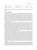
Figure 12.1 Possible consequences of mutagenic event in somatic and germinal cells.
12.2 GENETIC FUNDAMENTALS AND EVALUATION OF GENETIC CHANGE
241
other words, no triplet codon may call for more than a single specific tRNA-amino acid complex
(unambiguous), but several triplets may call for the same tRNA-amino acid (degenerate). This results
from the fact that four nucleotides, which form DNA (DNA is composed of adenine, cytosine, guanine,
and thymine), and the nucleotides forming RNA (RNA is made up of A, C, G, and uracil) may be
combined in triplet form in 64 different ways (4 × 4 × 4 or 4
3
). The 20 amino acids and three terminal
codes account for less than half of the available codons, leaving well over 30 codons of the possible
64. The biological significance of this degeneracy is that such a characteristic minimizes the influence
of minor mutations (e.g., single basepair deletions or additions) because codons differing only in minor
aspects may still code for the same amino acids. The significance of having an unambiguous code is
clear; the formation of proteins must be perfectly reproducible and exact. Table 12.1 depicts the amino
acids that are coded for by the various triplet codons of DNA, as well as the initiation and termination
signal triplets.
The process of mutagenesis results from an alteration in the DNA sequence. If the alteration is not
too radical, the rearrangement may be transmitted faithfully through the mRNA to protein synthesis,
Figure 12.2 Schematic representation of transcription and translation.
242
MUTAGENESIS AND GENETIC TOXICOLOGY
TABLE 12-1. Correspondence of the Genetic Code with the Appropriate Amino Acids (Note Unambiguity and Degeneracy)
First position in triplet
Second position in triplet
Third position in triplet
U C A G
A Isoleucine Threonine Asparagine Serine U
Isoleucine Threonine Asparagine Serine C
Isoleucine Threonine Lysine Arginine A
*Methionine Threonine Lysine Arginine G
C Leucine Proline Histidine Arginine U
Leucine Proline Histidine Arginine C
Leucine Proline Glutamate Arginine A
Leucine Proline Glutamate Arginine G
G Valine Alanine Aspartate Glycine U
Valine Alanine Aspartate Glycine C
Valine Alanine Glutamate Glycine A
Valine Alanine Glutamate Glycine G
U Phenylalanine Serine Tyrosine Cysteine U
Phenylalanine Serine Tyrosine Cysteine C
Leucine Serine STOP STOP A
Leucine Serine STOP Tryptophan G
*The sequence AUG, in addition to coding for methionine, is part of the initiator sequence that starts the translation process by which mRNA is formed from the DNA template.
243
which results in a gene product that is partially or completely unable to perform its normal function.
Such changes may be correlated with carcinogenesis, fetal death, fetal malformation, or biochemical
dysfunction, depending on the cell type that has been affected. However, cause and effect relationships
for such correlations typically are lacking.
Initiation and termination of DNA transcription are controlled by a separate set of regulatory genes.
Most regulatory genes respond to chemical cues, so that only those genes that are needed at a given
time are expressed or available. The remaining genes are in an inactive state. The processes of gene
activation and inactivation are believed to be critical to cellular differentiation, and interruption of these
processes may result in the expression of abnormal conditions such as tumors. This represents an
example of a case in which a non-genetic event may result in tumorigenesis. Oncogenes represent an
example of a situation where activation of a genetic phenomenon may act to initiate carcinogenicity.
In contrast, loss of “ tumor suppressor” genes may, by omission, result in initiation of the carcinogenic
process.
Chromosome Structure and Function
The DNA of mammalian species, including humans, is packaged in combination with specialized
proteins (predominantly histones) into units termed
chromosomes
, which are found in the nucleus of
the cell. The proteins are thought to “ cover” certain segments of the DNA and may act as inhibitors
of expression for some regions. Each normal human cell (except germ cells) contains 46 chromosomes
(23 pairs). Chromosomes may be present singly (haploid), as in germ cells (sperm or ovum), or in
pairs (diploid), as in somatic cells or in fertilized ova. In haploid cells, all functional genes present in
the cell can be expressed. In diploid cells, one allele may be dominant over the other, and in this case,
only the dominant gene of each functional pair is expressed. The unexpressed allele is termed
recessive
,
and recessive genes are expressed only when both copies of the recessive type are present. Some cell
types in mammals are found in forms other than diploid. Functionally normal liver cells, for example,
are occasionally found to be tetraploid (two chromosome pairs instead of one pair).
Some features and terminology that are important to cytogenetics, or the study of chromosomes,
include:
•
Karyotype—the array of chromosomes, typically taken at the point in the cell cycle known
as
metaphase
, which is unique to a species and forms the basis for cellular taxonomy; may
be used to detect physical or chemical damage
•
Centromere
—the primary constriction, which represents the site of attachment of the spindle
fiber during cell division; useful in identifying specific chromosomes, as its location is
relatively consistent
•
Nucleolar organizing region
—the secondary constriction, which represents the site of
synthesis of RNA, subsequently used in ribosomes for protein synthesis
•
Satellite
—the segment terminal to the nucleolar organizing region; useful in specific
chromosome identification
•
Heterochromatin
—tight-coiling region; relatively inactive
•
Euchromatin
—loose-coiling region; primary transcription site
Mitosis, Meiosis, and Fertilization
The process by which a somatic cell divides into two diploid daughter cells is called
mitosis
. The first
stage of mitosis is called
prophase
, during which the spindle is formed and the chromatin material
(DNA and protein) of the nucleus becomes shortened into well-defined chromosomes. During
metaphase
, the centriole pairs are pulled tightly by the attached microtubules to the very center of the
cell, lining up in the equatorial plane of the mitotic spindle. With still further growth of the spindle,
the chromatids in each pair of chromosomes are broken apart, a stage called
anaphase
. All 46 pairs
244
MUTAGENESIS AND GENETIC TOXICOLOGY
(in humans) of chromatids are thus separated, forming pairs of daughter chromosomes that are pulled
toward one mitotic spindle or the other. In
telophase
, the mitotic spindle grows longer, completing the
separation of daughter chromosomes. A new nuclear membrane is formed, and shortly thereafter the
cell constricts at the midpoint between the two nuclei, forming two new cells.
Meiosis
is the term for the process by which immature germ cells produce gametes (sperm or ova)
that are haploid. During meiosis, DNA is replicated, producing 46 chromosomes with sister chroma-
tids. The 46 chromosomes arrange into 23 pairs at the center of the nucleus, and in the first division
the pairs separate. In a second division, the sister chromatids separate, with one chromosome of each
pair being incorporated into four gametes. At the time of fertilization, or zygote formation, the fusion
of gametes once again forms a cell with a full complement of 46 chromosomes.
Genetic Alteration
Tests for genotoxicity in higher organisms may be placed into one of three basic categories: gene
mutation tests, chromosomal aberration tests, and DNA damage tests. These tests are conducted
individually or in combination to identify various types of mutagenic events (Figure 12.3) or other
genotoxic effects. For the purpose of this discussion, the principles of each test category will be
reviewed and specific tests will be discussed by broad phylogenetic classifications. Over 200 individual
test methods have been developed to assess the extent and magnitude of genetic alteration; however,
less than 20 have been validated or are in common use. Numerous mutagenic agents have the
demonstrated capacity to cause genetic change in one or more of these test systems, but no well-docu-
mented cases of human mutation are available. This latter conclusion may change as a result of
improvements in the ability to detect human genetic change. Nevertheless, as discussed in this section,
use of a reasonable battery of tests is capable of identifying almost all of the known human carcinogens,
consistent with the hypothesis that somatic cell mutations are, at least in part, responsible for a large
proportion of human cancers.
A transmissible change in the linear sequence of DNA can result from any one of three basic events:
•
Infidelity in DNA replication
•
Point mutation
•
Chromosomal aberration
Figure 12.3 Types of mutagenic changes (Adapted from Brusick, 1980).
12.2 GENETIC FUNDAMENTALS AND EVALUATION OF GENETIC CHANGE
245
One possibility, infidelity or inexact copying of a DNA strand during normal cellular replication, may
result from inaccurate initiation of replication, failure of the transcription enzymes to accurately “read”
the DNA, or interruption of the transcription process by agents that interpose (intercalate) themselves
within the DNA molecule or between the DNA and an enzyme.
Point mutations, as the second possibility, may be subdivided into basepair changes and frameshift
mutations (Figure 12.4). The former result from transition or transversion of DNA base pairs so that
Figure 12.4 Schematic representation of point mutations (frameshift and basepair changes).
246
MUTAGENESIS AND GENETIC TOXICOLOGY
the number of bases is unchanged but the sequence is altered. Because the genetic code is “ degenerate,”
this may or may not result in an altered product after transcription and translation. A frameshift
mutation, however, results from insertion or deletion of one or more bases from the linear sequence
of the DNA. This causes the transcription process to be displaced by the corresponding number of
bases and virtually assures an altered genetic product. Proflavine, which has been used as a bacte-
riostatic agent, is an example of a compound that intercalates within the DNA molecule. It is a flat,
planar molecule and inserts itself neatly between the bases. When it intercalates, it forces the DNA
strand out of its normal configuration, so that when the replication enzymes or transcription enzymes
try to read the bases, the bases are not spatially arranged the normal way, and the enzymes cannot read
the base sequence properly. The enzymes may skip over one or several bases, or may put an additional
base into the DNA or RNA strand at random. Proflavine does not chemically bind with the bases in
DNA. In contrast, many of the environmentally prevalent polynuclear aromatic hydrocarbons (PAHs)
may intercalate into the DNA, leading to frameshift, and also may chemically react directly with it, an
event that can lead to basepair substitution. An example of this is benzo(
a
)pyrene (BaP), which is found
at low concentrations throughout the environment as a product of combustion of fossil fuels, in grilled
steaks, tobacco smoke, and many other places. BaP by itself is seldom considered to be mutagenic.
However, after metabolism, many highly reactive epoxide intermediate metabolites are formed, one
of which (BPDE I) is highly mutagenic. BPDE I combines with guanine to form what is called a
DNA
adduct
. These adducts have been found in extremely small quantities by highly specialized and
sensitive techniques such as enzyme-linked immunosorbent assay (ELISA) and fluorescence. A
scheme of activation and adduct formation for BaP is given in Figure 12.5.
Basepair changes, described earlier, are of two kinds: transitions or transversions. In transitions,
one base is replaced by the base of the same chemical class. That is, a purine is replaced by the other
purine (e.g., adenine is replaced by guanine); in the case of pyrimidine bases, cytosine would be
replaced with thymine or vice versa. An example of a chemical that causes transitions is nitrous acid
(see Figure 12.6). Nitrous acid is formed from organic precursors such as nitrosamines, nitrite, and
nitrate salts. It reacts with amino (NH
2
) groups in nucleotides and converts them to keto (C
?
O) groups.
In transversions, a base pair is replaced in the DNA strand by a base of the other type: a purine is
replaced by a pyrimidine or vice versa.
Another group of chemicals that can cause mutations are alkylating agents. Some well-known
alkylating agents are the mustard gases, originally developed for chemical warfare. Chemicals in this
group add short carbon–hydrogen chains at specific locations on bases. The experimental agent ethyl
methanesulfonate (EMS) can alkylate guanine to form 7-ethylguanine (see Figure 12.7), which can
cause the bond between the base and deoxyribose in the backbone of the DNA strand to become
unstable and break. This leads to a gap in the DNA strand which, if unrepaired at the time of DNA
replication, is filled with any of the four available bases.
Not all point mutations are caused by radiation or chemicals; some may occur because of the nature
of the bases themselves. The bases have their preferred arrangement of hydrogen atoms, but on rare
occasions undergo rearrangements of the hydrogen atoms, called
tautomeric shifts
. The nitrogen atoms
attached to the purine and pyrimidine rings are usually in the amino (NH
2
) form and only rarely assume
the imino (NH) form. Similarly, the oxygen atoms attached to the carbon atoms of guanine and thymine
are normally arranged in the keto (C
?
O) form, but rarely rearrange to the enol (COH) form.
The changes in configuration lead to different hydrogen bonding patterns, and, if a base is in the
alternate form during replication, a wrong base can be put into the new growing strand leading to a
mutation. A group of chemicals, base analogs, that resemble the normal bases of DNA may lead to
mutations by being incorporated into DNA inadvertently during repair or replication. These chemicals
go through tautomeric shifts more often and result in inappropriate base pairing during replication so
that changes in the base sequence occur. An example of a base analogue is 5-bromouracil, which can
replace thymine.
Gene mutation tests measure those alterations of genetic material limited to the gene unit, that are
transmissible to progeny unless repaired. Brusick (1980) refers to gene mutations as “ microlesions”
because the actual genetic lesion is not microscopically visible. Microlesions are classified as either
12.2 GENETIC FUNDAMENTALS AND EVALUATION OF GENETIC CHANGE
247
Figure 12.5
Example of DNA adduct formation with benzo-a-pyrene.
248
basepair substitution mutations or frameshift mutations. These two categories of gene mutations are
induced by different mechanisms and, often, by distinctly different classes of chemical mutagens. Yet
both types of gene mutation are virtually always monitored by measuring some phenotypic change in
the test organism. Microlesions occur at a much lower frequency (10
–5
to 10
–6
) in comparison to
chromosome aberrations or “ macrolesions,” which may be as frequent as 10
–2
to 10
–3
.
As described earlier, the basepair changes induced by point mutations (Figure 12.4) will also alter
RNA codon sequences, which, in turn, change the amino acid sequence of the peptide chain being
Figure 12.6 Cytosine modified to uracil by nitrous acid.
Figure 12.7 Alkylating agent (EMS) effects on DNA.
12.2 GENETIC FUNDAMENTALS AND EVALUATION OF GENETIC CHANGE
249
formed, which may result in an alteration of some measurable cellular function. The phenotypic
changes that can be monitored by this type of test include auxotrophic changes (i.e., acquired
dependence on a formerly endogenously synthesized substance), altered proteins, color differences,
and lethality. It is extremely difficult to detect those alterations in mammalian DNA caused by insertions
or deletions of one or a few bases, except in rare instances where the specific protein product is known
and its formation can be monitored. It is somewhat easier in bacterial or prokaryotic systems, and this
has led to the use of bacterial or
in vitro
screening assays to detect potential mutagens. These issues
are discussed in greater detail in Brusick (1980, 1994).
Chromosomal aberrations, the third type of genetic change, may be present as chromatid gaps or
breaks, symmetrical exchange (exchange of corresponding segments between arms of a chromosome),
or asymmetric interchange between chromosomes. Point mutations can result in altered products of
gene expression, but chromosomal aberration or alteration in chromosome numbers passed on through
germ cells can have disastrous consequences, including embryonic death, teratogenesis, retarded
development, behavioral disorders, and infertility. Some naturally occurring abnormalities of human
chromosomal structure or number are shown in Table 12.2. The frequency of these events may be
increased by mutagenic agents. Because these genetic lesions may be visualized by microscopy, they
are referred to as
macrolesions
. One type of macrolesion is caused by an incomplete separation of
replicated chromosomes during cell division. This is characterized by the abnormal chromosome
numbers that result in the daughter cells and may be recognized as a change in the number of haploid
chromosome sets (
ploidy
changes) or in the gain or loss of single chromosomes (
aneuploidy
). A second
type of macrolesion caused by damage to chromosome structure (clastogenic effects) is categorized
by the abnormal chromosome morphology that results.
Two theories are currently available to explain the mechanism of chromosome aberration. One is
the classic “ breakage-first” hypothesis. This theory assumes that the initial lesion is a break in the
chromosomal backbone that is indicative of a broken DNA strand. Several possibilities exist following
such an event: (1) the ends may repair normally and rejoin to form a normal chromosome; (2) the ends
TABLE 12.2 Examples of Human Genetic Disorders
Chromosome Abnormalities
Cri-du-chat syndrome (partial deletion of chromosome 5)
Down’s syndrome (triplication of chromosome 21)
Klinefelter’s syndrome (XXY sex chromosome constitution; 47 chromosomes)
Turner’s syndrome (X0 sex chromosome constitution; 45 chromosomes)
Dominant Mutations
Chondrodystrophy
Hepatic porphyria
Huntington’s chorea
Retinoblastoma
Recessive Mutations
Albinism
Cystic fibrosis
Diabetes mellitus
Fanconi’s syndrome
Hemophilia
Xeroderma pigmentosum
Complex Inherited Traits
Anencephaly
Club foot
Spina bifida
Other congenital defects
250
MUTAGENESIS AND GENETIC TOXICOLOGY
may not be repaired, resulting in a permanent break; or (3) they may be misrepaired or join with another
chromosome to cause a translocation of genetic material. A second theory is the “chromatid exchange”
hypothesis. If the exchange occurs with a chromatid from another chromosome, an “ exchange figure”
results. This theory assumes that the initial lesion is not a break and that the lesion can either be repaired
directly or may interact with another lesion by a process called
exchange initiation
. Most chromosomal
abnormalities result in cell lethality and, if induced in germ cells, generally produce dominant lethal
effects that cannot be transmitted to the next generation. The traditional method for determining
chromosomal aberrations is the direct visual analysis of chromosomes in cells frozen at the metaphase
of their division cycle. Thus, metaphase-spread analysis evaluates both structural and numerical
chromosome anomalies directly.
Chemicals inducing changes in chromosomal number or structure also may be identified by the
micronucleus test, an assay that assesses genotoxicity by observing micronucleated cells. It is a
relatively simple assay because the number of cells with micronuclei are easily identified microscopi-
cally. At anaphase, in dividing cells that possess chromatid breaks or exchanges, chromatid and
chromosome fragments may lag behind when the chromosome elements move toward the spindle
poles. After telophase, the undamaged chromosomes give rise to regular daughter nuclei. The lagging
elements are also included in the daughter cells, but a considerable proportion are included in secondary
nuclei, which are typically much smaller than the principal nucleus and are therefore called
micronu-
clei
. Increased numbers of micronuclei represent increased chromosome breakage. Similar events can
occur if interference with the spindle apparatus occurs, but the appearance of micronuclei produced
in this manner is different, and they are usually larger than typical micronuclei. Historically, lympho-
cytes and epithelial cells have been the most commonly used cell populations.
Many point mutations are detected by the cell and are deleted by various repair mechanisms. Some,
however, remain undetected and are passed to daughter cells. The significance of the mutations varies
with the type of cell, and the location within the DNA. If the cell is of somatic lineage, altered gene
products can result from gene expression. If the cell is a gonadal cell (or germ cell), the change can be
passed on to offspring and may cause problems in future generations. Much of the DNA in organisms
is never expressed. If the mutation occurs in that portion of the DNA that is not expressed, no problem
occurs. However, if the mutation occurs in the active portion of the DNA, the altered gene products
can be expressed. An example of a problematic point mutation is in the gene that causes sickle cell
anemia. A change of one basepair (a transversion from thymine to adenine) results in the amino acid
glutamate being replaced by another amino acid, valine, in one of the molecules that makes up
hemoglobin, the oxygen-carrying molecule in red blood cells. When the blood becomes deoxygenated,
such as under heavy exercise conditions, the valine allows the red blood cells to assume a sickle shape
instead of the normal circular shape. This leads to clumping of blood cells in capillaries, which in turn
may limit blood flow to the tissues. This behavior of the blood cells exacerbates other effects of sickle
cell anemia, which result in oxygen deprivation because the hemoglobin content of the blood in persons
with sickle cell anemia is about half that of other persons.
12.3 NONMAMMALIAN MUTAGENICITY TESTS
Because results from bacterial or prokaryotic assays often establish priorities for other testing
approaches, it is of interest to briefly describe the assays currently used to screen for mutagenic
capacity, particularly those done in industrial settings.
Rapid cell division and the relative ease with which large quantities of data can be generated
(approximately 10
8
bacteria per test plate) have made bacterial tests the most widely utilized routine
means of testing for mutagenicity. These systems are the quickest and most inexpensive procedures.
However, bacteria are evolutionarily far removed from the human model. They lack true nuclei as well
as the enzymatic pathways by which most promutagens are activated to form mutagenic compounds.
Bacterial DNA has a different protein coat than seen in eukaryotes. Nevertheless, bacterial systems
have great utility as a preliminary screen for potential mutagens.
12.3 NONMAMMALIAN MUTAGENICITY TESTS
251
In addition to bacteria, fungi have been used in genotoxicity assays. The
Saccharomyces
and
Schizosaccharomyces
yeasts, as well as the molds
Neurospora
and
Aspergillus
, have been utilized in
forward mutation tests, which are similar in design to the salmonella histidine revertant assays that
will be described in the next section.
Typical Bacterial Test Systems
The most widely utilized bacterial test system for monitoring gene mutations and the most widely
utilized short-term mutagenicity test of any type is the
Salmonella typhimurium
microsome test
developed by Dr. Bruce Ames and co-workers and commonly called the Ames assay. The phenotypic
marker utilized for the detection of gene mutations in all the Ames
Salmonella
strains is the ability of
the bacteria to synthesize histidine, an amino acid essential for bacterial division. The tester strains of
bacteria have mutations rendering them unable to synthesize histidine; thus, they must depend on
histidine included in the culture medium in order to be able to multiply. Bacteria are taken directly
from a prepared culture and incorporated with a trace of histidine into soft agar overlay on a dish
containing minimal growth factors. The bacteria undergo several divisions, which are necessary for
the expression of mutagenicity and, after the available histidine has been used up, a fine bacterial lawn
is formed. Bacteria that have back-mutated in their histidine operon sites (and thus have reverted to
the ability to synthesize histidine) will keep on dividing to form discrete colonies, while the nonmutated
bacteria will die. A chemical that is a positive mutagen will demonstrate a statistically significant
dose-related increase in “ revertants” (colonies formed) when compared to the spontaneous revertants
in control plates.
Five Ames
S. typhimurium
tester strains are recommended for routine mutagenicity testing:
TA1535, TA1537, TA1538, TA98, and TA100. The TA1535 tester strain detects basepair substitution
mutations. The TA1538 tester strain detects frameshift mutagens that cause basepair deletions. The
TA1537 tester strain detects frameshift mutagens that cause basepair additions. The TA100 (basepair
substitution) and TA98 (frameshift) strains are sensitive to effects caused by certain compounds, such
as nitrofurans, which were not detectable with the previous three strains.
The lack of oxidative metabolism to transform promutagens (those mutagens requiring bioactiva-
tion to the active form) is overcome in these bacterial assays by two means. First, a suspension of rat
liver homogenate containing appropriate enzymes may be added to the bacterial incubation. The liver
preparation is centrifuged at 9000
g
for 20 min at 4°C, and the resultant supernatant (S9) is added to
the culture medium. In a slightly more complex procedure, called the
host-mediated assay
, the bacterial
tester strains are injected into the body cavity of a test animal such as the mouse. This host is treated
with the suspected mutagen and, after a selected period, the bacteria are harvested and assayed for
mutation (revertants) as described earlier. Other bacterial species used in mutagenicity screens include
Escherichia coli
and
Bacillus subtilis
.
Assays that measure DNA repair in bacterial systems have also been developed. These tests are
based on the premise that a strain deficient in DNA repair enzymes will be more susceptible to
mutagenic activity than will a similar strain that possesses repair enzymes that can correct the
mutagenic damage. A “ spot” test consists of placing the chemical to be tested in a well or on a paper
disk on top of the agar in a petri dish. The test chemical will diffuse from the central source, causing
a declining concentration gradient as the distance from the source increases. A strain deficient in repair
enzymes will exhibit a greater diameter of bacterial kill than the repair-sufficient strain tested with a
mutagen. In a “ suspension” test, a given number of bacteria are preincubated with and without the
test compound. The bacteria are then plated and the colonies counted. The repair-deficient strain will
demonstrate a greater percentage kill than will the sister DNA-repair-sufficient strain. A liver S9
activation system can also be incorporated in bacterial DNA repair tests. The most widely used bacterial
DNA repair test utilizes the polA
+
and polA
–
strains of
E. coli
. The polA
–
strain is deficient in DNA
polymerase I, whereas the polA
+
strain is sufficient in this enzyme.
252
MUTAGENESIS AND GENETIC TOXICOLOGY
Drosophila Test Systems
The fruitfly (
Drosophila melanogaster
) has received wide use in the sex-linked recessive lethal test.
The endpoint phenotypic change monitored in this test is the lethality of males in the F
2
generation.
Brusick has gone to the extent of labeling
Drosophila
an “ honorary mammalian model” by virtue of
its widespread application and correlation with positive mutagens in mammalian testing.
Drosophila
melanogaster
has also been utilized to monitor two types of chromosomal aberration endpoints through
phenotypic markers: loss and nondisjunction of X or Y sex chromosomes and heritable translocations.
The monitoring of translocations has the advantage of a very low background rate, facilitating
comparisons between controls and treated groups. Dominant lethal assays are also performed with
insects and can theoretically be applied in any organism where early embryonic death can be monitored.
The male is treated with the test agent, then mated with one or more females. If early fetal deaths occur,
these are demonstrative of a dominant lethal mutation in the germ cells of the treated male.
Plant Assays
A number of assay types are available in plant systems as well, including specific locus tests in corn
(
Zea maize
) and multilocus assays in
Arabidopsis
. Cytogenetic tests have been developed for
Trades-
cantia
(micronucleus test), as have chromosomal aberration assays in the root tips of onions (
Allium
sepa
) and beans (
Vicia faba
). Finally, DNA adducts analysis is applicable to somatic and germinal
plant cell systems. It is anticipated that one or more plant species may prove to be useful indicators of
the potential for genetic damage that may be related to emissions of environmental pollutants.
12.4 MAMMALIAN MUTAGENICITY TESTS
Testing chemicals for mutagenicity
in vivo
in mammalian systems is the most relevant method for
learning about mutagenicity in humans. Mammals such as the rat or mouse offer insights into human
physiology, metabolism, and reproduction that cannot be duplicated in other tests. Furthermore, the
route of administration of a chemical to a test animal can be selected to parallel normal human
environmental or occupational conditions of oral, dermal, or inhalation exposures.
Human epidemiologic findings may also be compared with the results of tests done in animals.
While the monitoring of human exposures and their effects does not constitute planned, controlled
mutagenicity testing, human epidemiology offers the opportunity to monitor and test for correlations
suggested by other mutagenicity tests. Thus, these studies are the only opportunity for direct human
modeling of a chemical’s mutagenic potential. It is worth noting that despite extensive investigation,
to date no chemical substances have been positively identified as human mutagens. The advantages
and limitations of a wide variety of genetic test systems are presented in Brusick (1994).
One perceived disadvantage of
in vivo
mammalian test systems is the time they require and their
cost. A larger commitment of physical resources and personnel is required than is required with
in
vitro
testing. Human epidemiology studies are further complicated by the fact that not all of the
environmental variables can be controlled. Frequently, the duration and extent of exposure to single
or multiple compounds can only be estimated. Nevertheless, progress is being made to lessen the cost
and decrease the time required for
in vivo
mammalian testing. Also, new data handling, statistical
techniques, and increased cooperation from industry have increased the reliability of human epidemiol-
ogy studies. More regular sampling of workplace exposures has helped to improve the quality and
accessibility of human data.
Mutagenic potential can vary greatly across a class of analytes, as shown for the metals (Costa,
1996). Mutagenicity data for metals can be quite difficult to interpret due to the breadth of mechanisms
at work, as illustrated by differences between Cr, Ni, As, and Cd.
12.4 MAMMALIAN MUTAGENICITY TESTS
253
Germ Cell Assays
A basic test used to detect specific gene mutations induced in germ cells of mammals is the
mouse-specific locus assay. This test involves treatment of wild-type mice, either male or female, with
a test compound before mating them to a strain homozygous for a number of recessive genes that are
expressed visibly in phenotype. If no mutations occur, then all offspring will be of the wild type. If a
mutation has occurred at one of the test loci in the treated mice, then the recessive phenotype will be
visibly detectable in the offspring. The mouse-specific locus test is of special significance in human
modeling because it is the only standardized assay that directly measures heritable germ cell gene
mutations in the mammal. A major drawback of the mouse-specific locus test is that extensive physical
plant facilities are required to execute this assay, and it has been estimated that one scientist and three
technicians could execute 10 single-dose mouse-specific locus tests in one year, provided there are
facilities for 5000 cages.
New and promising test procedures have been described for detecting germ cell mutations by using
alterations in selected enzyme activity as the phenotypic endpoint. A large group of somatic cell
enzymes can be monitored for changes in activity and kinetics in the F
1
generation. These changes
indicate changes in the parental genome.
A similar biochemical approach has been proposed for identifying germ cell mutations in humans
through the monitoring of placental cord blood samples. The activity of several erythrocyte enzymes,
such as glutathione reductase, can be monitored because the enzyme proteins are the products of a
single locus and because heterozygosity of a mutant allele for the chosen enzymes will result in
abnormal levels of enzyme activity. Likewise, it has been proposed that gene mutations be directly
monitored in mammalian germ cells by searching for phenotypic variants with biochemical markers
such as lactic acid dehydrogenase-X (LDH-X), an isozyme of lactic acid dehydrogenase found only
in testes and sperm. The test is based on the fact that a monospecific antibody for rabbit LDH-X reacts
with rat but not mouse LDH-X in sperm. The rat sperm fluoresce as a result of the reaction but the
mouse sperm do not fluoresce unless a phenotypic variant is present. If adapted to humans, this test
has potential use as a noninvasive screening test of germ cell mutations in males.
It has been proposed that the induction of behavioral effects in the offspring of male rats exposed
to a mutagenic agent may represent a genotoxic endpoint. For example, studies have demonstrated that
the mutagen cyclophosphamide can induce genotoxic behavioral effects in the progeny of male rats
and that these effects correspond to observed genetic damage caused in the spermatozoa following
meiosis. A similar effect has been attributed to vinyl chloride in at least one instance of occupational
exposures.
Mammalian germ cells can be monitored for chromosomal aberrations, and normally the testes are
used as the cell source. Mammalian male germ cells are protected by a biological barrier comparable
in function to the barrier which retards the penetration of chemicals to the brain. The blood–testes
barrier is a complex system composed of membranes surrounding the seminiferous tubules and the
several layers of spermatogenic cells organized within the tubules. This barrier restricts the permeabil-
ity of high-molecular-weight compounds to the developing male germ cell. An advantage of
in vivo
mammalian germ cell mutagenicity testing is that the protective contribution of this barrier is
automatically taken into account. Conventional procedures for harvesting mammalian male germ cell
tissue for metaphase-spread analysis involve mincing or teasing the seminiferous tubules to liberate
meiotic germ cells in suspension. This homogenate is centrifuged, the centrifuged pellet is discarded,
and the suspended cells are collected and analyzed. However, it was found that the tissue fragments
discarded during this conventional procedure contained more spermatogonial cells and meiotic
metaphases than did the suspension. Thus, the method has been refined by using tissue fragments and
adding collagenase to dissociate them. After collagenase treatment, the tissue fragments are gently
homogenized and centrifuged, and the pellet containing meiotic cells is resuspended and prepared for
microscopic analysis.
254
MUTAGENESIS AND GENETIC TOXICOLOGY
Dominant Lethal Assays
Dominant lethal assays can be performed in any organism where early embryonic death can be
monitored. As described earlier, mammals are commonly used in dominant lethal assays, although it
is possible to do so with insects as well. The male animals (typically mice or rats) are treated with the
suspected mutagen before being mated with one or more females. Each week these females are removed
and a new group of females is introduced to the treated male. This process is repeated for a period of
6–10 weeks. The females are sacrificed before parturition, and early fetal deaths are counted in the
uterine horns. This test has become standardized, and a large number of compounds have been screened
in mouse studies. As with most
in vivo
mammalian assays, costs and commitment of resources can be
extensive. However, the applicability of the data is typically quite good. The dominant lethal test in
rodents is of significance for human modeling because it gives an indication of heritable chromosomal
damage in a mammal. Even though the endpoint of early fetal death may seem of minor significance
when considering only its effects on the human gene pool, it does provide a signal that viable heritable
chromosomal damage and gene mutations may also be produced.
Heritable Translocation Assay
Results of dominant-lethal assays frequently correlate well with another test used for determining
clastogenic effects in mammalian germ cells: the heritable translocation assay (HTA). Translocation
represents complete transfer of material between two chromosomes. In HTA procedures, male mice
are mated to untreated females after treatment with the test compound, and the pregnant females are
allowed to deliver. Male offspring are subsequently mated to groups of females. If translocations are
produced through genotoxic action, then the affected first-generation male progeny will be partially
or completely sterile; this can be noticed in the litter size produced from those females to which they
were mated.
Micronucleus Tests
Application of the micronucleus test to mammalian germ cells recently has been reported. This test
procedure is analogous to the bone marrow micronucleus test (somatic cells), but it involves the
sampling of early spermatids from the seminiferous tubules of male rats. The number of micronuclei
are quantified by using fluorescent stain and counting micronucleated spermatids. To date, the
technique has not been widely used in occupational evaluations.
Spermhead Morphology Assay
Some relatively new tests have been developed that evaluate the ability of a test chemical to induce
abnormal sperm morphology when compared to controls. It has been proposed that an increase in
abnormal sperm morphology is evidence of genotoxicity because there seems to be an association
between abnormal sperm morphology and chromosome aberrations. However, recent investigations
have reported that induction of morphologically aberrant sperm can be caused by nongenotoxic actions,
such as dietary restriction. In addition, some known mutagens, including 1,2-dibromo-3-chloro-
propane (a pesticide with mutagenic, carcinogenic, and gonadotoxic properties), were reportedly
unable to induce production of spermhead abnormalities in mice, when tested. It should be noted that
sperm abnormalities are fairly common in humans and may occur at rates of 40–45 percent. Thus,
more verification is needed before strong conclusions can be drawn about the mammalian spermhead
morphology assay.
12.4 MAMMALIAN MUTAGENICITY TESTS
255
Tests for Primary DNA Damage
A historical test thought to monitor primary DNA damage in mammalian germ cells
in vivo
involves
the monitoring of sister chromatid exchange. The observation of sister chromatid exchanges through
differential staining involves exposing the cells to bromodeoxyuridine for two rounds of replication,
so that the chromosomes consist of one chromatid substituted on both arms with 5-bromodeoxyuridine
and the other substituted only on a single arm. Differential staining between sister chromatids is due
to the differences in bromodeoxyuridine incorporation in the sister chromatids.
Unscheduled DNA repair has been induced by chemical mutagens in mammalian male germ cells
from the spermatogonial to midspermatid stages of development. The test is based on the fact that cells
not undergoing replication (scheduled DNA synthesis) should not exhibit significant DNA synthesis.
Thus, incorporation of radiolabeled tracer molecules into the DNA of these cells should be minimal.
However, if a chemical mutagen damages the DNA, the DNA repair system may be activated, causing
unscheduled DNA synthesis (UDS). If such is the case, radiolabeled tracers will be incorporated into
the DNA; these can be monitored by autoradiography or by direct measurement of radioactivity in the
repaired DNA. Male germ cells lose DNA repair capability when they have advanced to the late
spermatid and mature spermatozoa stages; unscheduled DNA synthesis cannot then be induced by
chemical mutagens. The genotoxic agents methyl methanesulfonate, ethyl methanesulfonate, cyclo-
phosphamide, and Mitomen have been shown to induce unscheduled DNA repair
in vivo
in male mouse
germ cells. Similar procedures are available to evaluate UDS in some types of somatic cells as well.
Transgenic Mouse Assays
In the late 1980s and early 1990s the development of a new genotoxicity assay system was reported
by Gossen et al. (1989), Kohler et al. (1991), and colleagues. Briefly, the test system involves mutagen
dosing to a specific mouse strain (C57BL6) that has been infected with a viral “shuttle vector,”
isolation of the mouse DNA, recovery of the phage segment (
lac
I or
Lac
Z), and infection of an
E. coli
strain with the recovered phage. The phage will form plaques on a lawn of
E. coli
. The plaques are
colorless if no mutation has occurred or blue if a mutation has occurred. The assay may be performed
to gather information on mutations in somatic cells or in germ cells. The
lacZ
assay also is known as
the “ Muta-Mouse” assay, while the
lac
I assay also is known as the “ Big Blue” assay.
Advantages of the assay include its
in vivo
treatment regime, the fact that it can be conducted in a
few days from the isolation of DNA through plaque formation to mutation scoring. However, it may
be difficult to use extremely high dosages (e.g., approaching lethal doses), since the mice must survive
for 1–2 weeks in order to fix the mutation in the affected tissues. The performance of the transgenic
mouse assays that have been conducted on 26 substances was evaluated by Morrison and Ashby (1994),
including a review of the results of the tests that had been performed in the
lac
Z case (14 reports) and
in the lacI case (16 reports). These authors concluded that the variability of data reporting formats and
the rapid developments and modifications in the assay protocols make it difficult to perform direct
comparisons among tests or between this assay type and the results of other historically available
methods. Nevertheless, the initial results are generally promising, and there are no examples of internal
disagreement between responses for the same chemical in the same tissue.
In Vitro
Testing
Test systems have been developed that use mammalian cells in culture (
in vitro
) to detect chemical
mutagens. Disadvantages in comparison with
in vivo
mammalian tests are that
in vitro
tests lack
organ–system interaction, require a route for administration of the agent that cannot be varied, and
lack the normal distributional and metabolic factors present in the whole animal. The obvious
advantages are that costs are decreased and that experiments are more easily replicated, which
facilitates verification of results. Cases where human cells have been cultured successfully (e.g.,
lymphocytes) provide the only viable
in vitro
experiments on the human organism. Several endpoints
256
MUTAGENESIS AND GENETIC TOXICOLOGY
can be used during testing of potential mutagens
in vitro
. One of the most common involves the
monitoring of mutations in specific well-characterized gene loci, such as those coding for hypoxan-
thineguanine phosphoribosyl transferase (HGPRT), thymidine kinase (TK), or ouabain resistance
(OVA
r
). Mutagenic modification in the segments of the DNA coding for these proteins (enzymes)
results in an increased sensitivity of the cell, which can often be evaluated by the cell’s heightened
susceptibility to other agents (e.g., bromodeoxyuridine or 8-azaguanine).
As described in the section on
in vivo
mammalian testing, evaluations of sister chromatid exchange,
DNA repair activity, and chromosomal aberration through interpretation of metaphase spreads may be
applied to
in vitro
testing of mutagens. An additional procedure that has been correlated with chemical
mutagenicity is examination for cell culture transformation; following treatment with mutagens, some
cells in culture lose their normal, characteristic arrangement of monolayered attachment and begin to
pile up in a disorganized fashion. Two major drawbacks in looking for this feature are that considerable
expertise is necessary to interpret the results accurately and that the criteria for evaluation are more
subjective than for other mutagenicity assays.
A comparison of the sensitivity and specificity of selected short-term tests by two recognized
systems (National Toxicology Program (NTP) and Gene-Tox) is shown in Table 12.3.
12.5 OCCUPATIONAL SIGNIFICANCE OF MUTAGENS
Areas of Concern: Gene Pool and Oncogenesis
The potential significance of occupationally acquired mutations can be divided into two areas. The
first is concern for the protection of the human gene pool. This factor may represent the most significant
reason for genetic testing, but it is often underemphasized by nongeneticists involved with safety
evaluation because of the inability to demonstrate induced mutation in humans to date. The second
area is that of oncogenesis. The intimate relationship between the tumorigenic and genotoxic properties
of many chemicals (Figure 12.8) makes genetic testing a potentially powerful screening technique for
establishing priorities for future testing of chemicals of unknown cancer-causing potential. This factor
has been one of the primary driving forces behind the rapid expansion of genetic toxicology as a
discipline. Once again, however, the paucity of proved human carcinogens compared with the number
of demonstrated animal carcinogens suggests weaknesses in the process of extrapolating from animal
studies to human exposure in the workplace.
At the heart of the present legal and regulatory approach toward environmental and occupational
exposure to mutagens is the possibility that they may cause human genetic damage. Two important
assumptions underlie this central concept:
•
Environmental or occupational mutagens may cause aneuploidy, chromosome breaks, point
mutations, or other genetic damage in humans
•
Environmental or occupational mutagens that can be controlled by regulatory efforts
represent a significant component of total human exposure
Much of the interest in potential environmental and occupational mutagens is related to the prevalent
opinion that many cancers are initiated by a mutagenic event. This premise is supported by the strong
correlation between some specific occupational chemical exposures and cancer incidence in humans.
One good example is the relationship between liver cancer (angiosarcoma) and exposure to vinyl
chloride in some manufacturing operations. Another example is the respiratory tract cancers that may
be caused by exposure to bis(chloromethyl)ether.
12.5 OCCUPATIONAL SIGNIFICANCE OF MUTAGENS
257
TABLE 12-3 NTP and Gene-Tox Evaluation of Short-Term Test Sensitivities and Specificities
Sensitivity
a
Specificity
b
Gene-Tox
c
NTP
d
Gene-Tox
NTP
Assay (+)/Total %(+) (+)/Total %(+) (–)/Total %(–) (–)/Total %(–)
Ames/Salmonella 175/223 78 20/44 45 29/47 62 25/29 86
Mouse/lymphoma 45/54 87 31/44 70 0/5 0 13/29 45
CHO/HGPRT 40/41 98 — — 1/1 100 — —
V79 84/104 81 — — 3/3 100 — —
Drosophila SLRL 77/106 73 4/18 22 9/16 60 9/9 100
In vitro cytogenetics 40/54 74 24/44 55 2/6 33 20/29 69
In vivo cytogenetics 8/9 89 9/15 60 0/0 — 11/12 92
In vitro SCE 100/101 99 31/44 70 0/10 0 13/29 45
In vivo SCE 21/21 100 10/15 67 0/0 — 5/12 42
UDS in hepatocytes 19/22 86 6/30 20 0/0 — 13/14 93
Source:
Kier (1988).
a
Sensitivity is the proportion of positive results for carcinogens.
b
Specificity is the proportion of negative results for noncarcinogens.
c
Gene-Tox data include combined results for sufficient and limited-evidence carcinogens and noncarcinogens.
d
NTP specificity assumes that equivocal evidence compounds are noncarcinogens.
258
A Multidisciplinary Approach
Although each of the mutagenicity tests described in this chapter has individual strengths, likewise
each is weak in some facets of detection capacity. Clearly, therefore, the accurate and efficient testing
of chemicals and the protection from potential occupational mutagens require a multidisciplinary
approach that integrates toxicology, clinical chemistry, microbiology, pathology, epidemiology,
industrial hygiene, and occupational medicine. Testing intact animals has the advantage of increasing
the reliability of any extrapolations that must be made from the data; however, cost considerations
often limit the application of
in vivo
mammalian assays except when it is expected that they will
verify lower tier assays.
There are three areas where the results of mutagenicity testing of a given substance may be applied:
Figure 12.8 Proposed relationships between mutagenicity and carcinogenicity. (Adapted from Fishbein, 1979).
12.5 OCCUPATIONAL SIGNIFICANCE OF MUTAGENS
259
•
Extrapolation of test results in order to make a quantitative evaluation of the hazard of
exposures for humans
•
Prioritization of human hazards caused by specific compounds
•
Institution of remedial procedures that should be undertaken to minimize the human hazard
One of the most difficult areas of analysis is the correct application of mutagenicity tests to arrive
at a quantifiable human hazard from exposure to a given substance. There is frequently good
correlation between the mutagenicity and carcinogenicity of a substance in animal tests (Table
12.4). However, this may be misleading because models for carcinogenicity determination are
often characterized by chronic procedures utilizing very high doses in nonprimate species. These
may bear little resemblance to aspects of exposure in the human model, such as magnitude and
route of exposure, metabolic patterns, and environment (which are qualitative factors), and
exposure dose (which is quantitative).
As noted previously, the time and expense that are involved with lifetime carcinogenicity assays
have strongly influenced the use of test batteries as predictive measures of carcinogenic potential.
Among many others, Ashby and Tennant (1994), Anderson et al. (1994), Benigni and Giuliani (1987),
and Blake et al. (1990), have addressed the question of applicability of multiple test systems to the
classification of a substance as genotoxic or not, and carcinogenic or not. It is important in these efforts
to distinguish among “ sensitivity” (ability to identify a known carcinogen), “ specificity” (ability to
identify a noncarcinogen), and “ accuracy” (correct results of either type). Parodi et al. (1990) reported
on qualitative correlations associated with studies of up to 300 substances conducted during the period
1976 through 1988. Initial measures of sensitivity, specificity, and accuracy were approximately 90
percent, if the decision is based solely on
Salmonella
assays. As more substances have been tested,
this estimate has ranged from 45 to 91 percent. Best results typically are reported for sensitivity, where
accuracy generally is on the order of 65 to 75 percent. Consideration of the quantitative correlation
between short-term genotoxicity tests and carcinogenic potency has yielded extremely variable
estimates, ranging from approximately 30 to over 90 percent. The overwhelming conclusion was that
a battery of test systems that addresses differing endpoints is required if the goal is to develop a
confident conclusion regarding predictivity.
As is the case with many areas of toxicology, one may choose between
in vivo
and
in vitro
test
systems, each with their attendant advantages and disadvantages. The testing of chemicals in experi-
mental animals has all the advantages of any intact
in vivo
system; that is, it has all of the biochemical
and physiological requirements to make anthropomorphization more reliable. However,
in vivo
mutagenicity testing may require an investment of many thousands of dollars and a long period of
time. These disadvantages often force the tester to use a less expensive, well-established short-term
TABLE 12.4 Comparative Mutagenicity of Various Compounds
Compound
Established
human
carcinogen Bacteria Yeast
Drosophila
Mammalian
cells
Human
cells
Epichlorohydrin N N O Y Y Y
Ethyleneimine N O Y Y Y Y
Trimethyl phosphate O Y O Y Y Y
Tris NYOYYY
Ethylene dibromide N Y Y Y Y N
Vinyl chloride Y Y Y Y Y Y
Chloroprene Y Y O O O Y
Urethane N Y Y Y Y O
N = no; Y = yes; O = not tested.
260
MUTAGENESIS AND GENETIC TOXICOLOGY


