The Gale Encyclopedia of Neurological Disorders vol 1 - part 2 doc
Bạn đang xem bản rút gọn của tài liệu. Xem và tải ngay bản đầy đủ của tài liệu tại đây (1.35 MB, 52 trang )
Alzheimer disease
(NSAIDs) are currently being investigated for their use in
treating patients with Alzheimer disease.
Coping with the disorder
There are strategies to cope with this disorder and
these should be considered in the beginning stages of the
disease. Coping mechanisms depend on whether there are
family members available for support. If an individual is
without family members, relying on community support
through neighbors or volunteers of Alzheimer disease organizations will be necessary.
Many precautions can be made early on to avoid
difficult or life-threatening situations later, while maintaining everyday activities in the home environment. Dealing with a person with Alzheimer disease with patience is
important. Daily tasks should be performed when the person with Alzheimer disease feels best. Informing neighbors of the person’s condition is an important first step.
Arranging for assistance, depending on the stage of the disorder, will become necessary. As the ability to drive may
be compromised fairly early in the disorder, transportation
may need to be arranged. There are local chapters of the
Alzheimer’s Association that offer help with transportation
requirements.
In the early period of the disease when memory loss
is minimal, it is helpful for family and friends to interact
with the affected person, reminding him or her to take
medication, eat, keep appointments, and so forth. Family
and friends can help sustain the Alzheimer patient’s daily
living activities. Keeping records is also helpful, particularly if several people are overseeing the patient’s care.
Additionally, organizing the household so that it is easy to
find important items is recommended.
Other helpful coping mechanisms include posting
signs to remind patients of important phone numbers, to
turn off appliances, and to lock doors. It is important that
all electrical cords and appliances are arranged to minimize distraction, and to prevent danger of falling or misuse. Assistance in handling finances is usually necessary.
Providing an extra house key for neighbors and setting up
a schedule to check on persons with Alzheimer disease is
very helpful for both the patient and the family. By utilizing these and other family, neighborhood, and community
resources, many people with early Alzheimer disease are
able to maintain a successful lifestyle in their home environment for months or years.
Recovery and rehabilitation
For a person with Alzheimer disease, emphasis is
placed on maintaining cognitive and physical function for
as long as possible. Currently, there is no cure for
38
Alzheimer and, once the symptoms develop, patients do
not recover. Instead, they progressively worsen, usually
over a period of years. This has many psychosocial and financial ramifications for the patient and the patient’s caretakers. Social service workers can help families plan for
long-term care, as persons with Alzheimer disease most
often eventually require 24-hour assistance with feeding,
toileting, bathing, personal safety, and social interaction.
Taking care of patients in the later stages can be financially
and psychologically draining. Various support systems are
available through community mental health centers and
national support organizations.
Clinical trials
There are currently many clinical trials for the treatment or prevention of Alzheimer disease sponsored by the
National Institutes of Health (NIH). Large multi-center
clinical trials such as a Phase III clinical trail are aimed at
determining whether anti-inflammatory drugs delay agerelated cognitive decline. (Contact information: UCLA
Neuropsychiatric Institute, Los Angeles, California,
90024. Recruiter: Andrea Kaplan, (310) 825-0545 or her
email: ) A Phase III clinical
trial is also organized to test the drug Risperidone for the
treatment of agitated behavior in Alzheimer’s patients.
(Contact information: Palo Alto Veterans Administration
Health Care System, Menlo Park, California, 94025. Recruiter: Erin L. Cassidy, PhD, (650) 493-5000, ext.27013
or her email: )
Other trials include:
• A study on Valproate to prevent cognitive and behavioral
symptoms in patients. Contact information: Laura Jakimovich, RN, MS, (585) 760-6578 or her email:
• The drug Simvastatin, a cholesterol-lowering medication, is being studied to learn if it slows the progression
of Alzheimer disease. Contact information: Stanford
University, Palo Alto, California, 94304. Recruiter: Lisa
M. Kinoshita, PhD, (650) 493-0571 or her email:
• A study of the efficacy and dose of the drug NS 2330 to
improve cognition. Contact information: Peter Glassman, MD, PhD, (800) 344-4095, ext. 4776 or his email:
• A study of investigational medications for the treatment
of Alzheimer patients. Contact information: Eli Lilly and
Company, (877) 285-4559.
There are also many other studies that are investigating various other pharmacological agents such as vitamin
E and other currently available drugs.
GALE ENCYCLOPEDIA OF NEUROLOGICAL DISORDERS
There is considerable variability in the rate of
Alzheimer disease progression. The Alzheimer Disease Association claims that the time from the onset of clinical
symptoms to death can range from three to 20 years, with
an average duration of eight years. There are probably
many environmental and genetic factors that play a role in
the progression of the disease. The accumulation of damage and loss of brain cells eventually results in the failure
of many different organ systems in the body. According to
the National Institute of Neurological Disorders and
Stroke, the most common cause of death is due to infection.
Special concerns
Alzheimer disease should be distinguished from other
forms of dementia. In some cases, depression can result in
dementia-like symptoms. Other examples include chronic
drug use, chronic infections of the central nervous system, thyroid disease, and vitamin deficiencies. These
causes of dementia can often be treated. It is, therefore,
important to obtain an accurate diagnosis to avoid complications associated with the inappropriate treatment and
long-term care of these patients. There are also several genetically based syndromes in which dementia plays a role.
Genetic counseling
Genetic counseling is important for family members
biologically related to patients with Alzheimer disease because each first-degree relative has as much as a 20% lifetime risk of also being affected. The risk to immediate
relatives increases as more family members develop the
disease. In the early-onset form of the disease, the inheritance pattern is thought to be autosomal dominant. This
means that a carrier (who will eventually be affected) has
a 50% chance of passing on the mutated gene to his or her
offspring.
The general consensus in the scientific and medical
community is to not test children or adolescents in the absence of symptoms for adult-onset disorders. There are
many problems associated with predictive testing of
asymptomatic individuals who are not yet adults. Children
who undergo predictive testing lose the choice later in life
(when they are capable of understanding the full ramifications of the disease) to know or not to know this information. It is, therefore, an important consideration that
involves ethical and psychological implications.
Resources
BOOKS
Franci, E. Daunwald, and K. J. Isrelbacher. New York:
McGraw Hill, 2001.
Castleman, Michael, et al. There’s Still a Person in There: The
Complete Guide to Treating and Coping with Alzheimer’s.
New York: Perigee Books, 2000.
Mace, Nancy L., and Peter V. Rabins. The 36-Hour Day:
A Family Guide to Caring for Persons with
Alzheimer Disease, Related Dementing Illnesses,
and Memory Loss in Later Life. New York: Warner
Books, 2001.
PERIODICALS
Campion, D., et al. “Early-onset Autosomal Dominant
Alzheimer Disease: Prevalence, Genetic Heterogeneity,
and Mutation Spectrum.” Am J Hum Genet 65 (1999):
664–70.
Green, R.C. “Risk Assessment for Alzheimer’s Disease with
Genetic Susceptibility Testing: Has the Moment
Arrived?” Alzheimer’s Care Quarterly (2002):
3,208–14.
Rogan, S., and C. F. Lippa. “Alzheimer’s Disease and Other
Dementias: A Review.” Am J Alzheimers Dis Other
Demen (2002) 17: 11–7.
Romas, S. N., et al. “Familial Alzheimer Disease among
Caribbean Hispanics: A Reexamination of Its Association
with APOE.” Arch Neurol (2002) 59: 87–91.
Rosenberg, R. N. “The Molecular and Genetic Basis of AD:
The End of the Beginning: The 2000 Wartenberg
Lecture.” Neurology 54 (2000): 2045–54.
OTHER
ADEAR Alzheimer Disease Education and Referral Center.
National Institute on Aging about Alzheimer’s Disease—
General Information. February 10, 2004 (March 30,
2004). < />National Institutes of Health. Alzheimer’s Disease. February
10, 2004 (March 30, 2004). < />result.asp?disease_id=28>.
National Library of Medicine. Alzheimer’s Disease. MEDLINE plus Health Information. February 10, 2004 (March
30, 2004). < />alzheimersdisease.html>.
ORGANIZATIONS
Alzheimer’s Association. 919 North Michigan Avenue, Suite
1000, Chicago, IL 60611-1676. (312) 335-8700 or (800)
272-3900; Fax: (312) 335-1110.
<>.
Alzheimer’s Education and Referral Center. PO Box
8250, Silver Springs, MD 20907-8250. (800)
438-4380.
National Institute on Aging. Building 31, Room 5C27, 31
Center Drive, MSC 2292, Bethesda, MD 20892. (301)
496-1752. <>.
Bird, T. D. “Memory Loss and Dementia.” In Harrison’s
Principles of Internal Medicine, 15th ed. Edited by A. S.
GALE ENCYCLOPEDIA OF NEUROLOGICAL DISORDERS
Bryan Richard Cobb, PhD
39
Alzheimer disease
Prognosis
Amantadine
S Amantadine
Key Terms
Definition
Amantadine is a synthetic antiviral agent that also has
strong antiparkinsonian properties. It is sold in the United
States under the brand name Symmetrel, and is also available under its generic name.
Purpose
Amantadine is used to treat a group of side effects,
called parkinsonian side effects, that include tremors, difficulty walking, and slack muscle tone. These side effects
may occur in patients who are taking antipsychotic medications used to treat mental disorders such as schizophrenia. An unrelated use of amantadine is in the
treatment of viral infections of some strains of influenza A.
Description
Some medicines, called antipsychotic drugs, that are
used to treat schizophrenia and other mental disorders can
cause side effects similar to the symptoms of Parkinson’s
disease. The patient does not have Parkinson’s disease,
but may experience shaking in muscles while at rest, difficulty with voluntary movements, and poor muscle tone.
These symptoms are similar to the symptoms of Parkinson’s disease.
One way to eliminate these undesirable side effects is
to stop taking the antipsychotic medicine. Unfortunately,
the symptoms of the original mental disorder usually come
back; in most cases, simply stopping the antipsychotic
medication is not a reasonable option. Some drugs such as
amantadine that control the symptoms of Parkinson’s disease also control the parkinsonian side effects of antipsychotic medicines.
Amantadine works by restoring the chemical balance
between dopamine and acetylcholine, two neurotransmitter chemicals in the brain. Taking amantadine along with
the antipsychotic medicine helps to control symptoms of
the mental disorder, while reducing parkinsonian side effects. Amantadine is in the same family of drugs commonly known as anticholinergic drugs, including
biperiden and trihexyphenidyl.
Recommended dosage
Amantadine is available in 100 mg tablets and capsules, as well as a syrup containing 50 mg of amantadine
in each teaspoonful. For the treatment of drug-induced
parkinsonian side effects, amantadine is usually given in
a dose of 100 mg orally twice a day. Some patients may
need a total daily dose as high as 300 mg. Patients who are
40
Acetylcholine A naturally occurring chemical in
the body that transmits nerve impulses from cell to
cell. It causes blood vessels to dilate, lowers blood
pressure, and slows the heartbeat.
Anticholinergic Related to the ability of a drug to
block the nervous system chemical acetylcholine.
Dopamine A chemical in brain tissue that serves
to transmit nerve impulses (a neurotransmitter) and
helps to regulate movement and emotions.
Neurotransmitter A chemical in the brain that
transmits messages between neurons, or nerve
cells.
Parkinsonian Related to symptoms associated
with Parkinson’s disease, a nervous system disorder
characterized by abnormal muscle movement of
the tongue, face, and neck; inability to walk or
move quickly; walking in a shuffling manner; restlessness; and/or tremors.
taking other antiparkinsonian drugs at the same time may
require lower daily doses of amantadine (e.g., 100 mg
daily).
People with kidney disease or who are on hemodialysis must have their doses lowered. In these patients, doses
may range from 100 mg daily to as little as 200 mg every
seven days.
Precautions
Amantadine increases the amount of the dopamine (a
central nervous system stimulant) in the brain. Because
of this, patients with a history of epilepsy or other seizure
disorders should be carefully monitored while taking this
drug. This is especially true in the elderly and in patients
with kidney disease. Amantadine may cause visual disturbances and affect mental alertness and coordination.
People should not operate dangerous machinery or motor
vehicles while taking this drug.
Side effects
Five to 10% of patients taking amantadine may experience nervous system side effects, including:
• dizziness or lightheadedness
• insomnia
GALE ENCYCLOPEDIA OF NEUROLOGICAL DISORDERS
• impaired concentration
DeVane, C. Lindsay, PharmD. “Drug Therapy for Psychoses.”
In Fundamentals of Monitoring Psychoactive Drug
Therapy. Baltimore: Williams and Wilkins, 1990.
One to 5% of patients taking amantadine may experience other nervous system side effects, including:
• irritability or agitation
• depression
Jack Raber, PharmD
Ambenonium see Cholinergic stimulants
• confusion
• sleepiness or nightmares
S Amnestic disorders
• fatigue
Definition
• lack of coordination
• headache
In addition, up to 1% of patients may experience hallucinations, euphoria (excitement), extreme forgetfulness,
aggressive behavior, personality changes, or seizures.
Seizures are the most serious of all the side effects associated with amantadine.
Gastrointestinal side effects may also occur in patients taking amantadine. Five to 10% of people taking this
drug experience nausea and up to 5% have dry mouth, loss
of appetite, constipation, and vomiting. In most situations,
amantadine may be continued and these side effects
treated symptomatically.
One to 5% of patients taking amantadine have also reported a bluish coloring of their skin (usually on the legs)
that is associated with enlargement of the blood vessels
(livedo reticularis). This side effect usually appears
within one month to one year of starting the drug and subsides within weeks to months after the drug is discontinued. People who think they may be experiencing this or
other side effects from any medication should tell their
physician.
Interactions
Taking amantadine along with other drugs used to
treat parkinsonian side effects may cause increased confusion or even hallucinations. The combination of amantadine and central nervous system stimulants (e.g.,
amphetamines or decongestants) may cause increased central nervous stimulation or increase the likelihood of
seizures.
Resources
BOOKS
American Society of Health-System Pharmacists. AHFS Drug
Information 2002. Bethesda: American Society of HealthSystem Pharmacists, 2002.
Amnestic disorders are conditions that cause memory
loss.
Description
Memory is the ability to retain and recall new information. Memory can be subdivided into short-term memory, which involves holding onto information for a minute
or less, and long-term memory, which involves holding
onto information for over a minute. Long-term memory
can be further subdivided into recent memory, which involves new learning, and remote memory, which involves
old information. In general, amnestic disorders more frequently involve deficits in new learning or recent memory.
There are a number of terms that are crucial to the understanding of amnestic disorders. In order to retain information, an individual must be able to pay close enough
attention to the information that is presented; this is referred to as registration. The process whereby memories
are established is referred to as encoding or storage. Retaining information in the long-term memory requires passage of time during which memory is consolidated. When
an individual’s memory is tested, retrieval is the process
whereby the individual recalls the information from memory. Working memory is the ability to manipulate information from short-term memory in order to perform some
function. Amnestic disorders may affect any or all of these
necessary steps.
The time period affecting memory is also described.
Anterograde amnesia is more common. Anterograde amnesia begins at a certain point in time and continues to interfere with the establishment of memory from that point
forward in time. Retrograde amnesia refers to a loss of
memory for information that was learned prior to the onset
of amnesia. Retrograde amnesia often occurs in conjunction with head injury, and may result in erasure of memory of events or information from some time period
(ranging from seconds to months) prior to the head injury.
Over the course of recovery and rehabilitation from a head
GALE ENCYCLOPEDIA OF NEUROLOGICAL DISORDERS
41
Amnestic disorders
• nervousness or anxiety
Amnestic disorders
Key Terms
Acetylcholine A brain chemical or neurotransmitter that carries information throughout the nervous
system.
Anterograde Memory loss for information/events
occurring after the onset of the amnestic disorder.
Delirium A condition characterized by waxingand-waning episodes of confusion and agitation.
Dementia A chronic condition in which thinking
and memory are progressively impaired. Other
symptoms may also occur, including personality
changes and depression.
Retrograde Memory loss for information/events
prior to the onset of the amnestic disorder.
Transient ischemic attack (TIA) A stroke-like
phenomenon in which a brief blockage of a brain
blood vessel causes short-term neurological deficits
that are completely resolved within 24 hours of
their onset.
injury, memory may be restored or the period of amnesia
may eventually shorten.
Demographics
About 7% of all individuals over the age of 65 have
some form of dementia that involves some degree of amnesia, as do about 50% of all individuals over the age of 85.
Causes and symptoms
A number of brain disorders can result in amnestic
disorders, including various types of dementia (such as
Alzheimer’s disease), traumatic brain injury (such as
concussion), stroke, accidents that involve oxygen deprivation to the brain or interruption of blood flow to the
brain (such as ruptured aneurysms), encephalitis, tumors
in the thalamus and/or hypothalamus, Wernicke-Korsakoff
syndrome (a sequelae of thiamine deficiency usually due
to severe alcoholism), and seizures. Psychological disorders can also cause a type of amnesia called “psychogenic
amnesia.”
A curious condition called transient global amnesia
causes delirium (a period of waxing and waning confusion and agitation), anterograde amnesia, and retrograde
amnesia for events and information from the several hours
prior to the onset of the attack. Transient global amnesia
usually only lasts for several hours. Ultimately, the individual recovers completely, with no lasting memory
42
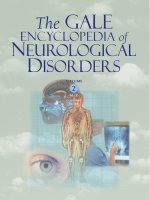
Hippocampus
Amygdala
Memory loss may result from bilateral damage to the limbic
system of the brain responsible for memory storage, processing, and recall. (Illustration by Electronic Illustrators Group.)
deficit. The cause of transient global amnesia is poorly understood; researchers are suspicious that it may be due to
either seizure activity in the brain or a brief blockage in a
brain blood vessel, which causes a brief stroke-like event
that completely resolves without permanent sequelae
(similar to a transient ischemic attack).
Symptoms of amnestic disorders may include difficulty recalling remote events or information, and/or difficulty learning and then recalling new information. In some
cases, the patient is fully aware of the memory impairment, and frustrated by it; in other cases, the patient may
seem completely oblivious to the memory impairment or
may even attempt to fill in the deficit in memory with confabulation. Depending on the underlying condition responsible for the amnesia, a number of other symptoms
may be present as well.
Diagnosis
Diagnosis of amnestic disorders begins by establishing an individual’s level of orientation to person, place,
and time. Does he or she know who he or she is? Where
he or she is? The day/date/time? An individual’s ability to
recall common current events (who is the president?) may
reveal information about the memory deficit. A family
member or close friend may be an invaluable part of the
examination, in order to provide some background information on the onset and progression of the memory loss,
GALE ENCYCLOPEDIA OF NEUROLOGICAL DISORDERS
A variety of memory tests can be utilized to assess an
individual’s ability to attend to information, utilize shortterm memory, and store and retrieve information from
long-term memory. Both verbal and visual memory should
be tested. Verbal memory can be tested by working with an
individual to memorize word lists, then testing recall after
a certain amount of time has elapsed. Similarly, visual
memory can be tested by asking an individual to locate
several objects that were hidden in a room in the individual’s presence.
Depending on what types of conditions are being considered, other tests may include blood tests, neuroimaging
(CT, MRI, or PET scans of the brain), cerebrospinal fluid
testing, and EEG testing.
Treatment team
A neurologist and/or psychiatrist may be involved in
diagnosing and treating amnestic disorders. Depending on
the underlying condition responsible for the memory
deficit, other specialists may be involved as well. Occupational and speech and language therapists may be involved in rehabilitation programs for individuals who have
amnestic disorders as part of their clinical picture.
remove the tumor. Individuals with transient global amnesia can be expected to fully recover from their memory impairment within hours or days of its onset. In the case of
some traumatic brain injuries, the amnesia may improve
with time (as brain swelling decreases, for example), but
there may always remain some degree of amnesia for the
events just prior to the moment of the injury.
Resources
BOOKS
Cummings, Jeffrey L. “Disorders of Cognition.” In Cecil
Textbook of Internal Medicine, edited by Lee Goldman, et
al. Philadelphia: W. B. Saunders Company, 2000.
Gabrieli, John D., et al. “Memory.” In Textbook of Clinical
Neurology, edited by Christopher G. Goetz. Philadelphia:
W. B. Saunders Company, 2003.
Mesulam, M.-Marsel. “Aphasias and Other Focal Cerebral
Disorders.” In Harrison’s Principles of Internal Medicine,
edited by Eugene Braunwald, et al. New York: McGrawHill Professional, 2001.
Rosalyn Carson-DeWitt, MD
Amphetamine see Central nervous system
stimulants
Treatment
In some cases, treatment of the underlying disorder
may help improve the accompanying amnesia. In mild
cases of amnesia, rehabilitation may involve teaching
memory techniques and encouraging the use of memory
tools, such as association techniques, lists, notes, calendars, timers, etc. Memory exercises may be helpful. Recent treatments for Alzheimer’s disease and other
dementias have involved medications that interfere with
the metabolism of the brain chemical (neurotransmitter)
called acetylcholine, thus increasing the available quantity
of acetylcholine. These drugs, such as donepezil and
tacrine, seem to improve memory in patients with
Alzheimer’s disease. Research studies are attempting to
explore whether these drugs may also help amnestic disorders that stem from other underlying conditions.
Prognosis
The prognosis is very dependent on the underlying
condition that has caused the memory deficit, and on
whether that condition has a tendency to progress or stabilize. Alzheimer’s disease, for example, is relentlessly progressive, and therefore the memory deficits that accompany
this condition can be expected to worsen considerably over
time. Individuals who have memory deficits due to a brain
tumor may have their symptoms improve after surgery to
S Amyotrophic lateral sclerosis
Definition
Amyotrophic lateral sclerosis (ALS) is a disease that
breaks down tissues in the nervous system (a neurodegenerative disease) of unknown cause that affects the
nerves responsible for movement. It is also known as
motor neuron disease and Lou Gehrig’s disease, after the
baseball player whose career it ended.
Description
ALS is a disease of the motor neurons, those nerve
cells reaching from the brain to the spinal cord (upper
motor neurons) and the spinal cord to the peripheral nerves
(lower motor neurons) that control muscle movement. In
ALS, for unknown reasons, these neurons die, leading to
a progressive loss of the ability to move virtually any of
the muscles in the body. ALS affects “voluntary” muscles,
those controlled by conscious thought, such as the arm,
leg, and trunk muscles. ALS, in and of itself, does not affect sensation, thought processes, the heart muscle, or the
“smooth” muscle of the digestive system, bladder, and
other internal organs. Most people with ALS retain function of their eye muscles as well. However, various forms
GALE ENCYCLOPEDIA OF NEUROLOGICAL DISORDERS
43
Amyotrophic lateral sclerosis
as well as information regarding the individual’s original
level of functioning.
Amyotrophic lateral sclerosis
Key Terms
Aspiration Inhalation of food or liquids into the
lungs.
Bulbar muscles Muscles of the mouth and throat
responsible for speech and swallowing.
Fasciculations Involuntary twitching of muscles.
Motor neuron A nerve cell that controls a muscle.
Riluzole (Rilutek) The first drug approved in the
United States for the treatment of ALS.
Voluntary muscle A muscle under conscious
control; contrasted with smooth muscle and heart
muscle, which are not under voluntary control.
of ALS may be associated with a loss of intellectual function (dementia) or sensory symptoms.
“Amyotrophic” refers to the loss of muscle bulk, a
cardinal sign of ALS. “Lateral” indicates one of the regions of the spinal cord affected, and “sclerosis” describes
the hardened tissue that develops in place of healthy
nerves. ALS affects approximately 30,000 people in the
United States, with about 5,000 new cases each year. It
usually begins between the ages of 40 and 70, although
younger onset is possible. Men are slightly more likely to
develop ALS than women.
ALS progresses rapidly in most cases. It is fatal
within three years for 50% of all people affected, and
within five years for 80%. Ten percent of people with ALS
live beyond eight years.
Causes and symptoms
Causes
The symptoms of ALS are caused by the death of
motor neurons in the spinal cord and brain. Normally, these
neurons convey electrical messages from the brain to the
muscles to stimulate movement in the arms, legs, trunk,
neck, and head. As motor neurons die, the muscles they enervate cannot be moved as effectively, and weakness results. In addition, lack of stimulation leads to muscle
wasting, or loss of bulk. Involvement of the upper motor
neurons causes spasms and increased tone in the limbs, and
abnormal reflexes. Involvement of the lower motor neurons causes muscle wasting and twitching (fasciculations).
Although many causes of motor neuron degeneration
have been suggested for ALS, none has yet been proven responsible. Results of recent research have implicated toxic
44
molecular fragments known as free radicals. Some evidence suggests that a cascade of events leads to excess free
radical production inside motor neurons, leading to their
death. Why free radicals should be produced in excess
amounts is unclear, as is whether this excess is the cause
or the effect of other degenerative processes. Additional
agents within this toxic cascade may include excessive levels of a neurotransmitter known as glutamate, which may
over-stimulate motor neurons, thereby increasing free-radical production, and a faulty detoxification enzyme
known as SOD-1, for superoxide dismutase type 1. The
actual pathway of destruction is not known, however, nor
is the trigger for the rapid degeneration that marks ALS.
Further research may show that other pathways are involved, perhaps ones even more important than this one.
Autoimmune factors or premature aging may play some
role, as could viral agents or environmental toxins.
Two major forms of ALS are known: familial and
sporadic. Familial ALS accounts for about 10% of all ALS
cases. As the name suggests, familial ALS is believed to be
caused by the inheritance of one or more faulty genes.
About 15% of families with this type of ALS have mutations in the gene for SOD-1. SOD-1 gene defects are dominant, meaning only one gene copy is needed to develop
the disease. Therefore, a parent with the faulty gene has a
50% chance of passing the gene along to a child.
Sporadic ALS has no known cause. While many environmental toxins have been suggested as causes, to date
no research has confirmed any of the candidates investigated, including aluminum and mercury and lead from
dental fillings. As research progresses, it is likely that
many cases of sporadic ALS will be shown to have a genetic basis as well.
A third type, called Western Pacific ALS, occurs in
Guam and other Pacific islands. This form combines
symptoms of both ALS and Parkinson’s disease.
Symptoms
The earliest sign of ALS is most often weakness in the
arms or legs, usually more pronounced on one side than
the other at first. Loss of function is usually more rapid in
the legs among people with familial ALS and in the arms
among those with sporadic ALS. Leg weakness may first
become apparent by an increased frequency of stumbling
on uneven pavement, or an unexplained difficulty climbing stairs. Arm weakness may lead to difficulty grasping
and holding a cup, for instance, or loss of dexterity in the
fingers.
Less often, the earliest sign of ALS is weakness in the
bulbar muscles, those muscles in the mouth and throat that
control chewing, swallowing, and speaking. A person with
bulbar weakness may become hoarse or tired after speaking at length, or speech may become slurred.
GALE ENCYCLOPEDIA OF NEUROLOGICAL DISORDERS
Normal nerve fiber
Amyotrophic lateral sclerosis
NORMAL SPINAL NEURON
DISEASED SPINAL NEURON
Affected nerve fiber
Normal skeletal muscle
Wasted skeletal muscle
Amyotrophic lateral sclerosis (ALS) is caused by the degeneration and death of motor neurons in the spinal cord and brain.
These neurons convey electrical messages from the brain to the muscles to stimulate movement in the arms, legs, trunk,
neck, and head. As motor neurons degenerate, the muscles are weakened and cannot move as effectively, leading to muscle wasting. (Illustration by Electronic Illustrators Group.)
In addition to weakness, the other cardinal signs of
ALS are muscle wasting and persistent twitching (fasciculation). These are usually seen after weakness becomes
obvious. Fasciculation is quite common in people without
the disease, and is virtually never the first sign of ALS.
While initial weakness may be limited to one region,
ALS almost always progresses rapidly to involve virtually
all the voluntary muscle groups in the body. Later symptoms include loss of the ability to walk, to use the arms and
hands, to speak clearly or at all, to swallow, and to hold the
head up. Weakness of the respiratory muscles makes
breathing and coughing difficult, and poor swallowing
control increases the likelihood of inhaling food or saliva
(aspiration). Aspiration increases the likelihood of lung infection, which is often the cause of death. With a ventilator and scrupulous bronchial hygiene, a person with ALS
may live much longer than the average, although weakness
and wasting will continue to erode any remaining functional abilities. Most people with ALS continue to retain
function of the extraocular muscles that move their eyes,
allowing some communication to take place with simple
blinks or through use of a computer-assisted device.
Diagnosis
The diagnosis of ALS begins with a complete medical
history and physical exam, plus a neurological examination to determine the distribution and extent of weakness.
An electrical test of muscle function, called an electromyogram, or EMG, is an important part of the diagnostic process. Various other tests, including blood and
urine tests, x rays, and CT scans, may be done to rule out
other possible causes of the symptoms, such as tumors of
GALE ENCYCLOPEDIA OF NEUROLOGICAL DISORDERS
45
Amyotrophic lateral sclerosis
the skull base or high cervical spinal cord, thyroid disease,
spinal arthritis, lead poisoning, or severe vitamin deficiency. ALS is rarely misdiagnosed following a careful review of all these factors.
Treatment
There is no cure for ALS, and no treatment that can
significantly alter its course. There are many things which
can be done, however, to help maintain quality of life and
to retain functional ability even in the face of progressive
weakness.
As of early 1998, only one drug had been approved
for treatment of ALS. Riluzole (Rilutek) appears to provide on average a three-month increase in life expectancy
when taken regularly early in the disease, and shows a significant slowing of the loss of muscle strength. Riluzole
acts by decreasing glutamate release from nerve terminals.
Experimental trials of nerve growth factor have not
demonstrated any benefit. No other drug or vitamin currently available has been shown to have any effect on the
course of ALS.
A physical therapist works with an affected person
and family to implement exercise and stretching programs
to maintain strength and range of motion, and to promote
general health. Swimming may be a good choice for people with ALS, as it provides a low-impact workout to most
muscle groups. One result of chronic inactivity is contracture, or muscle shortening. Contractures limit a person’s range of motion, and are often painful. Regular
stretching can prevent contracture. Several drugs are available to reduce cramping, a common complaint in ALS.
An occupational therapist can help design solutions to
movement and coordination problems, and provide advice
on adaptive devices and home modifications.
Speech and swallowing difficulties can be minimized
or delayed through training provided by a speech-language pathologist. This specialist can also provide advice
on communication aids, including computer-assisted devices and simpler word boards.
Nutritional advice can be provided by a nutritionist. A
person with ALS often needs softer foods to prevent jaw
exhaustion or choking. Later in the disease, nutrition may
be provided by a gastrostomy tube inserted into the stomach.
Mechanical ventilation may be used when breathing
becomes too difficult. Modern mechanical ventilators are
small and portable, allowing a person with ALS to maintain the maximum level of function and mobility. Ventilation may be administered through a mouth or nose piece,
or through a tracheostomy tube. This tube is inserted
through a small hole made in the windpipe. In addition to
46
providing direct access to the airway, the tube also decreases the risk aspiration. While many people with rapidly progressing ALS choose not to use ventilators for
lengthy periods, they are increasingly being used to prolong life for a short time.
The progressive nature of ALS means that most persons will eventually require full-time nursing care. This
care is often provided by a spouse or other family member.
While the skills involved are not difficult to learn, the
physical and emotional burden of care can be overwhelming. Caregivers need to recognize and provide for
their own needs as well as those of people with ALS, to
prevent depression, burnout, and bitterness.
Throughout the disease, a support group can provide
important psychological aid to affected persons and their
caregivers as they come to terms with the losses ALS inflicts. Support groups are sponsored by both the ALS Society and the Muscular Dystrophy Association.
Alternative treatment
Given the grave prognosis and absence of traditional
medical treatments, it is not surprising that a large number
of alternative treatments have been tried for ALS. Two
studies published in 1988 suggested that amino-acid therapies may provide some improvement for some people
with ALS. While individual reports claim benefits for
megavitamin therapy, herbal medicine, and removal of
dental fillings, for instance, no evidence suggests that
these offer any more than a brief psychological boost,
often followed by a more severe letdown when it becomes
apparent the disease has continued unabated. However,
once the causes of ALS are better understood, alternative
therapies may be more intensively studied. For example,
if damage by free radicals turns out to be the root of most
of the symptoms, antioxidant vitamins and supplements
may be used more routinely to slow the progression of
ALS. Or, if environmental toxins are implicated, alternative therapies with the goal of detoxifying the body may be
of some use.
Prognosis
ALS usually progresses rapidly, and leads to death
from respiratory infection within three to five years in
most cases. The slowest disease progression is seen in
those who are young and have their first symptoms in the
limbs. About 10% of people with ALS live longer than
eight years.
Prevention
There is no known way to prevent ALS or to alter its
course.
GALE ENCYCLOPEDIA OF NEUROLOGICAL DISORDERS
BOOKS
Adams, Raymond D., Maurice Victor, and Allan H. Ropper.
Adams’ & Victor’s Principles of Neurology, 6th ed. New
York: McGraw Hill, 1997.
Brown, Robert H. “The motor neuron diseases.” In Harrison’s
Principles of Internal Medicine, 14th ed., edited by
Anthony S. Fauci, et al., pp. 2368-2372. New York:
McGraw-Hill, 1998.
Feldman, Eva L. “Motor neuron diseases.” In Cecil Textbook of
Medicine, 21st ed., edited by Lee Goldman and J. Claude
Bennett, pp. 2089-2092. Philadelphia: W. B. Saunders,
2000.
Kimura, Jun, and Ryuji Kaji. Physiology of ALS and Related
Diseases. Amsterdam: Elsevier Science, 1997.
Mitsumoto, Hiroshi, David A. Chad, Erik Pioro, and Sid
Gilman. Amyotrophic Lateral Sclerosis. New York:
Oxford University Press, 1997.
Muscular Dystrophy Association. 3300 East Sunrise Drive,
Tucson, AZ 85718-3208. (520) 529-2000 or (800) 5721717; Fax: (520) 529-5300. <www.mdausa.org>.
WEBSITES
ALS Society of Canada. < />ALS Survival Guide. < />American Academy of Family Physicians.
National Organization for Rare Diseases.
rdb_sum&id=57.htm>.
National Institute of Neurological Disorders and Stroke.
< />disorders/amyotrophiclateralsclerosis_doc.htm>.
National Library of Medicine. < />medlineplus/amyotrophiclateralsclerosis.html>.
World Federation of Neurology. < />
PERIODICALS
Ansevin, C. F. “Treatment of ALS with pleconaril.” Neurology
56, no. 5 (2001): 691-692.
Eisen, A., and M. Weber. “The motor cortex and amyotrophic
lateral sclerosis.” Muscle and Nerve 24, no. 4 (2001):
564-573.
Gelanis, D. F. “Respiratory Failure or Impairment in
Amyotrophic Lateral Sclerosis.” Current treatment
options in neurology 3, no. 2 (2001): 133-138.
Ludolph, A. C. “Treatment of amyotrophic lateral sclerosis—
what is the next step?” Journal of Neurology 246, Suppl 6
(2000): 13-18.
Pasetti, C., and G. Zanini. “The physician-patient relationship
in amyotrophic lateral sclerosis.” Neurological Science
21, no. 5 (2000): 318-323.
Robberecht, W. “Genetics of amyotrophic lateral sclerosis.”
Journal of Neurology 246, Suppl 6 (2000): 2-6.
Robbins, R. A., Z. Simmons, B. A. Bremer, S. M. Walsh, and S.
Fischer. “Quality of life in ALS is maintained as physical
function declines.” Neurology 56, no. 4 (2001): 442-444.
ORGANIZATIONS
ALS Association of America. 27001 Agoura Road, Suite 150,
Calabasas Hills, CA 91301-5104. (800) 782-4747
(Information and Referral Service) or (818) 880-9007;
Fax: (818) 880-9006. < />American Academy of Family Physicians. 11400 Tomahawk
Creek Parkway, Leawood, KS 66211-2672. (913) 9066000. < />American Academy of Neurology. 1080 Montreal Avenue, St.
Paul, Minnesota 55116. (651) 695-1940; Fax: (651) 6952791. < />American Medical Association, 515 N. State Street, Chicago,
IL 60610. (312) 464-5000. < />Centers for Disease Control and Prevention. 1600 Clifton
Road, Atlanta, GA 30333. (404) 639-3534 or (800) 3113435. < />< />
L. Fleming Fallon, Jr., MD, DrPH
S Anatomical nomenclature
Over the centuries, anatomists developed a standard
nomenclature, or method of naming anatomical structures.
Terms such as “up” or “down” obviously have no meaning unless the orientation of the body is clear. When a
body is lying on its back, the thorax and abdomen are at
the same level. The upright sense of up and down is lost.
Further, because anatomical studies and particularly embryological studies were often carried out in animals, the
development of the nomenclature relative to comparative
anatomy had an enormous impact on the development of
human anatomical nomenclature. There were obvious difficulties in relating terms from quadrupeds (animals that
walk on four legs) who have abdominal and thoracic regions at the same level as opposed to human bipeds in
whom an upward and downward orientation might seem
more obvious.
In order to standardize nomenclature, anatomical
terms relate to the standard anatomical position. When the
human body is in the standard anatomical position it is upright, erect on two legs, facing frontward, with the arms at
the sides each rotated so that the palms of the hands turn
forward.
In the standard anatomical position, superior means
toward the head or the cranial end of the body.
The term inferior means toward the feet or the caudal
end of the body.
GALE ENCYCLOPEDIA OF NEUROLOGICAL DISORDERS
47
Anatomical nomenclature
Resources
Anatomical nomenclature
The frontal surface of the body is the anterior or ventral surface of the body. Accordingly, the terms “anteriorly” and “ventrally” specify a position closer to—or
toward—the frontal surface of the body. The back surface
of the body is the posterior or dorsal surface and the terms
“posteriorly” and “dorsally” specify a position closer to—
or toward—the posterior surface of the body.
The terms superficial and deep relate to the distance
from the exterior surface of the body. Cavities such as the
thoracic cavity have internal and external regions that correspond to deep and superficial relationships in the midsagittal plane.
The bones of the skull are fused by sutures that form
important anatomical landmarks. Sutures are joints that
run jaggedly along the interface between the bones. At
birth, the sutures are soft, broad, and cartilaginous. The sutures eventually fuse and become rigid and ossified near
the end of puberty or early in adulthood.
The sagittal suture unties the parietal bones of the
skull along the midline of the body. The suture is used as
an anatomical landmark in anatomical nomenclature to establish what are termed sagittal planes of the body. The
primary sagittal plane is the sagittal plane that runs
through the length of the sagittal suture. Planes that are
parallel to the sagittal plane, but that are offset from the
midsagittal plane are termed parasagittal planes. Sagittal
planes run anteriorly and posteriorly, are always at right
angles to the coronal planes. The medial plane or midsagittal plane divides the body vertically into superficially
symmetrical right and left halves.
The medial plane also establishes a centerline axis for
the body. The terms medial and lateral relate positions relative to the medial axis. If a structure is medial to another
structure, the medial structure is closer to the medial or
center axis. If a structure is lateral to another structure, the
lateral structure is farther way from the medial axis. For
example, the lungs are lateral to the heart.
The coronal suture unites the frontal bone with the
parietal bones. In anatomical nomenclature, the primary
coronal plane designates the plane that runs through the
length of the coronal suture. The primary coronal plane is
also termed the frontal plane because it divides the body
into frontal and back halves.
Planes that divide the body into superior and inferior
portions, and that are at right angles to both the sagittal and
coronal planes are termed transverse planes. Anatomical
planes that are not parallel to sagittal, coronal, or transverse planes are termed oblique planes.
The body is also divided into several regional areas.
The most superior area is the cephalic region that includes
the head. The thoracic region is commonly known as the
chest region. Although the celiac region more specifically
48
refers to the center of the abdominal region, celiac is
sometimes used to designate a wider area of abdominal
structures. At the inferior end of the abdominal region lies
the pelvic region or pelvis. The posterior or dorsal side of
the body has its own special regions, named for the underlying vertebrae. From superior to inferior along the
midline of the dorsal surface lie the cervical, thoracic,
lumbar, and sacral regions. The buttocks are the most
prominent feature of the gluteal region.
The term upper limbs or upper extremities refers to
the arms. The term lower limbs or lower extremities refers
to the legs.
The proximal end of an extremity is at the junction of
the extremity (i.e., arm or leg) with the trunk of the body.
The distal end of an extremity is the point on the extremity farthest away from the trunk (e.g., fingers and toes).
Accordingly, if a structure is proximate to another structure it is closer to the trunk (e.g., the elbow is proximate
to the wrist). If a structure is distal to another, it is farther
from the trunk (e.g., the fingers are distal to the wrist).
Structures may also be described as being medial or
lateral to the midline axis of each extremity. Within the
upper limbs, the terms radial and ulnar may be used synonymous with lateral and medial. In the lower extremities,
the terms fibular and tibial may be used as synonyms for
lateral and medial.
Rotations of the extremities may de described as medial rotations (toward the midline) or lateral rotations
(away from the midline).
Many structural relationships are described by combined anatomical terms (e.g., the eyes are anterio-medial
to the ears).
There are also terms of movement that are standardized by anatomical nomenclature. Starting from the
anatomical position, abduction indicates the movement of
an arm or leg away from the midline or midsagittal plane.
Adduction indicates movement of an extremity toward the
midline.
The opening of the hands into the anatomical position
is supination of the hands. Rotation so the dorsal side of
the hands face forward is termed pronation.
The term flexion means movement toward the flexor
or anterior surface. In contrast, extension may be generally
regarded as movement toward the extensor or posterior
surface. Flexion occurs when the arm brings the hand from
the anatomical position toward the shoulder (a curl) or
when the arm is raised over the head from the anatomical
position. Extension returns the upper arm and or lower to
the anatomical position. Because of the embryological rotation of the lower limbs that rotates the primitive dorsal
GALE ENCYCLOPEDIA OF NEUROLOGICAL DISORDERS
The term palmar surface (palm side) is applied to the
flexion side of the hand. The term plantar surface is applied to the bottom sole of the foot. From the anatomical
position, extension occurs when the toes are curled back
and the foot arches upward and flexion occurs as the foot
is returned to anatomical position.
Rolling motions of the foot are described as inversion
(rolling with the big toe initially lifting upward) and eversion (rolling with the big toe initially moving downward).
K. Lee Lerner
S Anencephaly
Key Terms
Alpha-fetoprotein (AFP) A chemical substance
produced by the fetus and found in the fetal
circulation.
Causes and symptoms
As an isolated defect, anencephaly appears to be
caused by a combination of genetic factors and environmental influences that predispose to faulty formation of
the nervous system. The specific genes and environmental insults that contribute to this multifactorial causation
are not completely understood. It is known that nutritional
insufficiency, specifically folic acid insufficiency, is one
predisposing environmental factor, and that mutations of
genes involved in folic acid metabolism are genetic risk
factors. The recurrence risk after the birth of an infant with
anencephaly is 3–5%. The recurrence may be anencephaly
or another neural tube defect such as spina bifida.
Anencephaly is readily apparent at birth because of
exposure of all or part of the brain. Not only is the brain
malformed, but it is also damaged because of the absence
of the overlying protective encasement.
Definition
Anencephaly is a lethal birth defect characterized by
the absence of all or part of the skull and scalp and malformation of the brain.
Description
Anencephaly is one of a group of malformations of
the central nervous system collectively called neural
tube defects. Anencephaly is readily apparent at birth because of the absence of the skull and scalp and exposure
of the underlying brain. The condition is also called acrania (absence of the skull) and acephaly (absence of the
head). In its most severe form, the entire skull and scalp
are missing. In some cases, termed “meroacrania” or
“meroanencephaly,” a portion of the skull may be present.
In most instances, anencephaly occurs as an isolated birth
defect with the other organs and tissues of the body forming correctly. In approximately 10% of cases, other malformations coexist with anencephaly.
Demographics
Anencephaly occurs in all races and ethnic groups.
The prevalence rates range from less than one in 10,000
births (European countries) to more than 10 per 10,000
births (Mexico, China).
Diagnosis
Anencephaly is diagnosed by observation. Prenatal
diagnosis may be made by ultrasound examination after
12–14 weeks’ gestation. Prenatal diagnosis of anencephaly
can also be detected through maternal serum alpha-fetoprotein screening. The level of alpha-fetoprotein in the
maternal blood is elevated because of the leakage of this
fetal protein into the amniotic fluid.
There are no treatments for anencephaly. A pregnant
woman or couple expecting an anencephalic baby will
need a sensitive and supportive health care team, and perhaps some additional psychological support as they face
the inevitable death of their infant, usually before or
shortly after birth.
Treatment and management
No treatment is indicated for anencephaly. Affected
infants are stillborn or die within the first few days of life.
The risk for occurrence or recurrence of anencephaly may
be reduced by half or more by the intake of folic acid during the months immediately before and after conception.
Natural folic acid, a B vitamin, may be found in many
foods (green leafy vegetables, legumes, orange juice,
liver). Synthetic folic acid may be obtained in vitamin
preparations and in certain fortified breakfast cereals. In
GALE ENCYCLOPEDIA OF NEUROLOGICAL DISORDERS
49
Anencephaly
side to the adult form ventral side, flexion occurs as the
thigh is raised anteriorly and superiorly toward the anterior
portion of the pelvis. Extension occurs when the thigh is
returned to anatomical position. Specifically, due to the
embryological rotation, flexion of the lower leg occurs as
the foot is raised toward the back of the thigh and extension of the lower leg occurs with the kicking motion that
returns the lower leg to anatomical position.
Anencephaly
Diagram of Anencephaly
NORMAL INFANT
ANENCEPHALIC INFANT
Brain
Brain Stem
Brain Stem
Infants born with anencephaly have either a severely underdeveloped brain or total brain absence. A portion of the brainstem usually protrudes through the skull, which also fails to develop properly. (Gale Group.)
the United States, all enriched cereal grain flours have
been fortified with folic acid.
National Birth Defects Prevention Network. Atlanta, GA. (770)
488-3550. <>.
Roger E. Stevenson, MD
Rosalyn Carson-DeWitt, MD
Clinical Trials
Research is primarily directed at understanding the
underlying factors that affect early neurological development in the fetus.
Prognosis
Anencephaly is uniformly fatal at birth or soon thereafter.
Resources
PERIODICALS
Czeizel, A. E., and I. Dudas. “Prevention of the First
Occurrence of Neural Tube Defects by Preconceptional
Vitamin Supplementation.” New England Journal of
Medicine 327 (1992): 1832–1835.
Medical Research Council Vitamin Study Research Group.
“Prevention of Neural Tube Defects: Results of the
Medical Research Council Vitamin Study.” Lancet 338
(1991): 131–137.
Sells, C. J., and J. G. Hall. “Neural Tube Defects.” Mental
Retardation and Developmental Disabilities Research
Reviews 4, no. 4, 1998.
Definition
Cerebral aneurysm is the enlargement, distention, dilation, bulging, or ballooning of the wall of a cerebral artery or vein. Aneurysms affect arteries throughout the
body, including blood vessels in the brain (intracerebral
aneurysm). Ruptures of intracerebral aneurysm result in
stroke (loss of blood supply to tissue) and bleeding into
the subarachnoid space). The most common aneurysm is
an abdominal aneurysm.
Description
ORGANIZATIONS
March of Dimes Birth Defects Foundation. 1275 Mamaroneck
Ave., White Plains, NY 10605. (888) 663-4637.
<>.
50
S Aneurysms
Dilations, or ballooning, of blood vessels to form an
aneurysm are particularly dangerous because they increase
the chance of arterial rupture and subsequent bleeding into
brain tissues (a hemorrhagic stroke). Rupture of an
aneurysm can lead to the leakage of blood into the tissues
and spaces surrounding the brain. This leaked blood then
clots to form an intracranial hematoma. Aneurysms that
rupture can result in severe disability or death.
GALE ENCYCLOPEDIA OF NEUROLOGICAL DISORDERS
Aneurysms
Key Terms
Aneurysm A bulging, weakened area in a blood
vessel.
Common complications of cerebral aneurysms that
leak include hydrocephalus (the excessive accumulation
of cerebrospinal fluid) and persistent spasms of blood vessels that adversely affect the maintenance of arterial blood
pressure.
Once they rupture or bleed, aneurysms have a tendency toward recurrent bleeding episodes. This tendency
to rebleed is particularly high in the first few days following the initial bleed. Intracerebral bleeds are often accompanied by increases in cerebrospinal fluid and an increased
intracranial pressure (hydrocephalus).
Once they occur, aneurysms are dynamic and can increase in size over time. The increase in size is not always
linear and can advance sporadically until they expand to a
critical size. As they grow, aneurysms begin to put pressure on surrounding tissues. In addition, as they grow,
aneurysms usually result in progressively more difficult
problems.
The larger the size of an aneurysm, regardless of location, the greater the chance it will ultimately bleed.
Cerebral aneurysm ruptures usually lead to subarachnoid
hemorrhage (SAH).
Demographics
Although more common in adults than children, cerebral aneurysms occur in all age groups. Cerebral
aneurysms are more common—and the risk of aneurysm
generally increases—with age.
Aneurysm sufferers are rarely young; the incidence of
aneurysm is low in those under 20 years of age. In contrast, aneurysms are relatively common in people over 65
years of age. Risk indicators for some groups such as Caucasian males begin to increase at age 55. Some studies indicate that up to 5% of the population over 65 suffer some
form of aneurysm.
Incidence of specific aneurysms varies, but in general
within the United States they are occur less frequently in
Caucasian women, and are relatively uncommon in
African Americans.
Of those affected with an aneurysm anywhere in the
body, the National Institute of Health (NIH) estimates that
approximately 30,000 people in the United States will suffer an aneurysm rupture.
Arteriograph of the head from behind, showing an
aneurysm, the balloon-like smooth swelling just below and
to the right of center. (CNRI/National Audubon Society
Collection/Photo Researchers, Inc. Reproduced by permission.)
Cigarette smoking and excess alcohol use substantially increase the risk of aneurysm rupture.
Causes and symptoms
An aneurysm may be a congenital defect in the structure of the muscular wall of affected blood vessels (e.g.,
the intima of an artery), or arise secondary to trauma, atherosclerosis, or high blood pressure. The defect results in
an abnormal thinning of the arterial or venous wall that
makes the wall subsequently susceptible to aneurysm.
Research data appears to show that some individuals
have a basic genetic susceptibility or predisposition to
aneurysms. The genetic inheritance patterns resemble
characteristics linked to an autosomal dominant gene.
Within some families, rates of aneurysms can run as high
as five to 10 times those found in the general population.
Direct causes of intracerebral aneurysms include infection, trauma, or neoplastic disease. If infection is the
cause, the infection may be from a remote site. For example, an aneurysm in the brain may result from the loosed
embolus such as plaque, fatty deposit, clot, or clump of
cells, originating at an infection in another part of the
body. The embolus is transported to the site of the future
cerebral aneurysm by the bloodstream and cerebral circulation. An aneurysm formed in this manner is termed a
mycotic aneurysm.
GALE ENCYCLOPEDIA OF NEUROLOGICAL DISORDERS
51
Aneurysms
Prior to rupture, the symptoms associated with an
aneurysm depend upon its location, size, and rate of expansion. A static aneurysm that does not leak (bleed) or
adversely affect cerebral circulation or neighboring tissue
may be asymptomatic (without symptoms). In contrast,
larger aneurysms or aneurysms with a rapid growth rate
may produce pronounced symptoms such as swelling, loss
of sensation, blurred vision, etc.
Just prior to an aneurysm rupture, patients typically
experience some symptoms commonly associated with
stroke. Depending on the size and location of the
aneurysm about to rupture, a patient may suffer a severe
headache, deterioration or disturbances of hearing, and
disturbances of vision such as double vision, severe nausea and vomiting, and syncopal episodes (periodic fainting or loss of consciousness).
A severe headache that is unresponsive to standard
analgesics is the most common sign of a leaking or bleeding aneurysm. Many patients experience a series of sentinel (warning) headaches if the aneurysm begins to leak
prior to rupture. A fully ruptured aneurysm presents with
a severe headache that is frequently accompanied by fainting or temporary (transient) loss of consciousness, often
with severe nausea, vomiting, and rapidly developing stiff
neck (nuchal rigidity).
Aneurysms normally rupture while the patient is active and awake.
Diagnosis
The severe headache that accompanies a cerebral
aneurysm is often the principle complaint upon which the
diagnosis of aneurysm begins to build.
Angiography provides the most definitive diagnosis
of an intracerebral aneurysm by determining the specific
site of the aneurysm. A computed tomography (CT) scan
can also diagnose a bleeding cerebral aneurysm. Arteriography is an x ray of the carotid artery taken when a special dye is injected into the artery.
The presence of blood in the cerebrospinal fluid withdrawn during a lumbar puncture is also diagnostic evidence for blood leaking into the subarachnoid space.
Magnetic resonance imaging (MRI) studies can
also be useful in accessing the extent of damage to surrounding tissues and are often used to study aneurysms
prior to leakage or rupture. MRI uses magnetic fields to
detect subtle changes in brain tissue content. The benefit
of MRI over CT imaging is that MRI is better able to localize the exact anatomical position of an aneurysm. Other
types of MRI scans are magnetic resonance angiography
(MRA) and functional magnetic resonance imaging
(fMRI). Neurosurgeons use MRA to detect stenosis
(blockage) of the brain arteries inside the skull by mapping
52
flowing blood. Functional MRI uses a magnet to pick up
signals from oxygenated blood and can show brain activity through increases in local blood flow.
Duplex Doppler ultrasound and arteriography are two
additional diagnostic imaging techniques used to decide if
an individual would benefit from a surgical procedure
called carotid endarterectomy. This surgery is used to
remove fatty deposits from the carotid arteries and can
help prevent stroke. Doppler ultrasound is a painless, noninvasive test in which sound waves bounce off the moving
blood and the tissue in the artery and can be formed into
an image.
Treatment team
Management and treatment of aneurysms require a
multi-disciplinary team. Physicians are responsible for
caring for general health and providing guidance aimed at
preventing a stroke. Neurologists and neurosurgeons usually lead acute-care teams and direct patient care during
hospitalization and recovery from surgery. Neuroradiologists help pinpoint the location and extent of aneurysms.
Treatment
Treatment for ruptures of cerebral aneurysms includes
measures to stabilize the emergency by assuring cardiopulmonary functions (adequate heart rate and respiration) while simultaneously moving to decrease intracranial
pressure and surgically clip (repair and seal) the ruptured
cerebral aneurysm.
Surgery is often performed as soon as the patient is stabilized; ideally within 72 hours of the onset of rupture. The
goal of surgery is to prevent rebleeding. Surgery is performed to expose the aneurysm and allow the placement of
a clip across a strong portion of the vessel to obstruct the
flow of blood through the weakened aneurysm. Repeat surgical procedures to seal an aneurysm are not uncommon.
Treatment of unruptured aneurysms is certainly less
dramatic, but presents a more deliberate and complex path.
Microcoil thrombosis or balloon embolization (the insertion via the arterial catheter of a balloon or other obstruction that blocks blood flow through the region of
aneurysm) are alternatives to full surgical intervention.
Other nonsurgical interventions include rest, medications, and hypertensive-hypervolemic therapy to drive
blood around obstructed vessels.
Treatment decisions are made between the treatment
team and family members with regard to the best course of
treatment and the probable outcomes for patients suffering
a severe aneurysm rupture with extensive damage to surrounding brain tissue.
Asymptomatic aneurysms allow the treatment team to
more fully evaluate surgical and nonsurgical options.
GALE ENCYCLOPEDIA OF NEUROLOGICAL DISORDERS
The recovery and rehabilitation of patients suffering
a cerebral aneurysm depend on the location and size of the
aneurysm. The course of recovery and rehabilitation is
also heavily influenced by whether the aneurysm ruptures.
Key to recovery is the prevention of aneurysm rebleeding, the management of swelling in the ventricular
system (hydrocephalus), seizures, cardiac arrhythmias,
and vasospasm. The onset of vasospasm within the first
two weeks of the initial bleeding incident is the major
cause of death in those who survive the initial rupture of
the aneurysm.
Ventricular drains are used to control the buildup of
cerebrospinal fluid in the ventricular system.
Clinical trials
As of May 2004, current studies sponsored by the National Institute of Neurological Disorders and Stroke
(NINDS) include a study on the effect of the drug ProliNO
on brain artery spasms after aneurysm rupture and a study
of the role of genetics on the development of intracranial
aneurysms (Familial Intracranial Aneurysm Study). Further information is available at <>.
Prognosis
The overall prognosis for a patient with a cerebral
aneurysm depends on several factors including the size, location, and stability of the aneurysm. Facets of the patient’s
general health, neurological health, age, and familial history must also be evaluated in forming a prognosis.
Although each patient is different, and each aneurysm
must be individually evaluated, in general, the prognosis
for patients who have suffered a bleed is guarded at best,
with mortality rates up 60% within a year of the initial
bleeding incident. Approximately half of the survivors suffer some long-lasting disability. Patients with cerebral
aneurysm can, however, fully recover with no long-lasting
disorder.
Data regarding the prognosis for unruptured
aneurysms is more tentative and not specific for cerebral
aneurysms. Some long-term studies give evidence that
only 10% of patients might suffer leakage or bleeding
from their aneurysm over a period of 10 years and only
about a quarter of patients would experience bleeding
from the aneurysm over a period of 25 years.
aneurysms also occur in persons with normal blood pressure.
Other physiological stresses such as pregnancy have
not been demonstrated to have a correlation to the rupture
of cerebral aneurysm.
Resources
BOOKS
Bear, M., et al. Neuroscience: Exploring the Brain. Baltimore:
Williams & Wilkins, 1996.
Goetz, C. G., et al. Textbook of Clinical Neurology.
Philadelphia: W.B. Saunders Co., 1999.
Goldman, Cecil. Textbook of Medicine, 21st ed. New York:
W.B. Saunders Co., 2000.
Guyton & Hall. Textbook of Medical Physiology, 10th ed. New
York: W.B.Saunders Co., 2000.
Wiebers, David. Stroke-Free for Life: The Complete Guide to
Stroke Prevention and Treatment. New York: Harper,
2002.
OTHER
“Stroke Risk Factors.” American Stroke Association. April 20,
2004 (May 22, 2004).
< />ORGANIZATIONS
American Stroke Association: A Division of American Heart
Association. 7272 Greenville Avenue, Dallas, TX
75231-4596. (214) 706-5231 or (888) 4STROKE
(478-7653).
< />Brain Aneurysm Foundation. 12 Clarendon Street, Boston,
MA 02116. (617) 723-3870; Fax: (617) 723-8672.
<>.
National Stroke Association. 9707 East Easter Lane,
Englewood, CO 80112-3747. (303) 649-9299 or (800)
STROKES (787-6537); Fax: (303) 649-1328.
<http//www.stroke.org/>.
Paul Arthur
S Angelman syndrome
Definition
Angelman syndrome (AS) is a genetic condition that
causes severe mental retardation, severe speech impairment, and a characteristic happy and excitable demeanor.
Description
Special concerns
Intracerebral aneurysms are sometimes associated
with other diseases such as fibromuscular hyperplasia or
other disorders such as high blood pressure (although
Individuals with AS show evidence of delayed development by 6–12 months of age. Eventually, this delay is
recognized as severe mental retardation. Unlike some genetic conditions causing severe mental retardation, AS is
GALE ENCYCLOPEDIA OF NEUROLOGICAL DISORDERS
53
Angelman syndrome
Recovery and rehabilitation
Angelman syndrome
Angelman Syndrome
1. Etiology: Deletion, Uniparental Disomy, or Unknown
88y
d.71y
61y
36y
d.83y
Colon
cancer
54y
23y
49y
18y
2. Etiology: UBE3A mutation, Imprinting mutation, or Unknown
78y
46y
d.71y d.88y
Liver
cirrhosis
63y
39y
60y
d.62y
Stroke
48y
25y 21y 17y 14y
12y
75y
39y
12y d.2mos
Congenital
heart defect
2
2y
See Symbol Guide for Pedigree Charts. (Gale Group.)
not associated with developmental regression (loss of previously attained developmental milestones).
Severe speech impairment is a striking feature of AS.
Speech is almost always limited to a few words. However,
receptive language skills (listening to and understanding
the speech of others) and non-verbal communication are
not as severely affected.
Individuals with AS have a balance disorder, causing
unstable and jerky movements. This typically includes gait
ataxia (a slow, unbalanced way of walking) and tremulous
movements of the limbs.
AS is also associated with a unique “happy” behavior,
which may be the best-known feature of the condition.
This may include frequent laughter or smiling, often with
no apparent stimulus. Children with AS often appear
happy, excited, and active. They may also sometimes flap
their hands repeatedly. Generally, they have a short attention span. These characteristic behaviors led to the original name of this condition, the “Happy Puppet” syndrome.
However, this name is no longer used as it is considered
insensitive to AS individuals and their families.
Demographics
AS has been reported in individuals of diverse ethnic
backgrounds. The incidence of the condition is estimated
at 1/10,000 to 1/30,000.
54
Causes and symptoms
Most cases of AS have been traced to specific genetic
defects on chromosomes received from the mother. In
about 8% of individuals with AS, no genetic cause can be
identified. This may reflect misdiagnosis, or the presence
of additional, unrecognized mechanisms leading to AS.
The first abnormalities noted in an infant with AS are
often delays in motor milestones (those related to physical
skills, such as sitting up or walking), muscular hypotonia
(poor muscle tone), and speech impairment. Some infants
seem unaccountably happy and may exhibit fits of laughter. By age 12 months, 50% of infants with AS have microcephaly (a small head size). Tremulous movements
are often noted during the first year of life.
Seizures occur in 80% of children with AS, usually
by three years of age. No major brain lesions are typically
seen on cranial imaging studies.
The achievement of walking is delayed, usually occurring between two-and-a-half and six years of age. The
child with AS typically exhibits a jerky, stiff gait, often
with uplifted and bent arms. About 10% of individuals
with AS do not walk. Additionally, children may have
drooling, protrusion of the tongue, hyperactivity, and a
short attention span.
Many children have a decreased need for sleep and
abnormal sleep/wake cycles. This problem may emerge in
GALE ENCYCLOPEDIA OF NEUROLOGICAL DISORDERS
Diagnosis
The clinical diagnosis of AS is made on the basis of
physical examination and medical and developmental history. Confirmation requires specialized laboratory testing.
There is no single laboratory test that can identify all
cases of AS. Several different tests may be performed to
look for the various genetic causes of AS. When positive,
these tests are considered diagnostic for AS. These include
DNA methylation studies, UBE3A mutation analysis, and
fluorescent in situ hybridization (FISH).
Individuals with AS may be more likely to develop
particular medical problems which are treated accordingly.
Newborn babies may have difficulty feeding and special
bottle nipples or other interventions may be necessary.
Gastroesophageal reflux (heartburn) may lead to vomiting
or poor weight gain and may be treated with drugs or surgery. Constipation is a frequent problem and is treated
with laxative medications. Many individuals with AS have
strabismus (crossed eyes), which may require surgical correction. Orthopedic problems, such as tightening of tendons or scoliosis, are common. These problems may be
treated with physical therapy, bracing, or surgery.
Prognosis
Individuals with AS have significant mental retardation and speech impairment that are considered to occur in
all cases. However, they do have capacity to learn and
should receive appropriate educational training.
Young people with AS typically have good physical
health aside from seizures. Although life span data are not
available, the life span of people with AS is expected to be
normal.
Special concerns
Educational concerns
Treatment team
Children with Angelman syndrome will need help
from a variety of professionals, including a general pediatrician and pediatric neurologist. A child psychiatrist
and/or psychologist may be helpful as well, particularly to
help design and implement various behavioral plans.
Physical, occupational, and speech and language therapists may help support specific deficits. A learning specialist may be consulted for help with an individualized
educational plan.
Treatment
There is no specific treatment for AS. A variety of
symptomatic management strategies may be offered for
hyperactivity, seizures, mental retardation, speech impairment, and other medical problems.
The typical hyperactivity in AS may not respond to
traditional behavior modification strategies. Children
with AS may have a decreased need for sleep and a tendency to awaken during the night. Drug therapy may be
prescribed to counteract hyperactivity or aid sleep. Most
families make special accommodations for their child by
providing a safe yet confining environment.
Seizures in AS are usually controllable with one or
more anti-seizure medications. In some individuals with
severe seizures, dietary manipulations may be tried in
combination with medication.
Children with AS appear to benefit from targeted educational training. Physical and occupational therapy may
improve the disordered, unbalanced movements typical of
AS. Children with a severe balance disorder may require
special supportive chairs. Speech therapy is often directed
towards the development of nonverbal communication
strategies, such as picture cards, communication boards, or
basic signing gestures.
Legal issues
The most pressing long-term concern for patients
with AS is working out a life plan for ongoing care, since
many are likely to outlive their parents. The parents of a
child diagnosed with AS should consult an estate planner,
an attorney, and a certified public accountant (CPA) in
order to draft a life plan and letter of intent. A letter of intent is not a legally binding document, but it gives the patient’s siblings and other relatives or caregivers necessary
information on providing for her in the future. The attorney can help the parents decide about such matters as
guardianship as well as guide them through the legal
process of appointing a guardian, which varies from state
to state.
Resources
PERIODICALS
“Angelman syndrome.” The Exceptional Parent 30, no. 3
(March 2000): S2.
GALE ENCYCLOPEDIA OF NEUROLOGICAL DISORDERS
55
Angelman syndrome
infancy and persist throughout childhood. Upon awakening at night, children may become very active and destructive to bedroom surroundings.
The language impairment associated with AS is severe. Most children with AS fail to learn appropriate and
consistent use of more than a few words. Receptive language skills are less severely affected. Older children and
adults are able to communicate by using gestures or communication boards (special devices bearing visual symbols
corresponding to commonly used expressions or words).
Some individuals with AS may have a lighter skin
complexion than would be expected given their family
background.
Angiography
Lombroso, Paul J. “Genetics of Childhood Disorders: XVI.
Angelman Syndrome: A Failure to Process.” Journal of
the American Academy of Child and Adolescent
Psychiatry 39, no. 7 (July 2000): 931.
ORGANIZATION
Angelman Syndrome Foundation, Inc. 414 Plaza Drive, Suite
209, Westmont, IL 60559. (800) IF-ANGEL or (630) 7349267. Fax: (630) 655-0391.
<>.
WEBSITES
Williams, Charles A., M.D., Amy C. Lossie, Ph.D., and Daniel
J. Driscoll, Ph.D. “Angelman Syndrome.” (November 21,
2000). GeneClinics. University of Washington, Seattle.
< />
Jennifer Ann Roggenbuck, MS, CGC
Rosalyn Carson-DeWitt, MD
S Angiography
Definition
Angiography is the x-ray (radiographic) study of the
blood vessels. An angiogram uses a radiopaque substance,
or contrast medium, to make the blood vessels visible
under x ray. The key ingredient in most radiographic contrast media is iodine.
Purpose
Angiography is used to detect abnormalities, including narrowing (stenosis) or blockages in the blood vessels
(called occlusions) throughout the circulatory system and
in some organs. The procedure is commonly used to identify atherosclerosis; to diagnose heart disease; to evaluate
kidney function and detect kidney cysts or tumors; to map
renal anatomy in transplant donors; to detect an aneurysm
(an abnormal bulge of an artery that can rupture leading to
hemorrhage), tumor, blood clot, or arteriovenous malformations (abnormal tangles of arteries and veins) in the
brain; and to diagnose problems with the retina of the eye.
It is also used to provide surgeons with an accurate vascular map of the heart prior to open-heart surgery, or of the
brain prior to neurosurgery. Angiography may be used
after penetrating trauma, like a gunshot or knife wound, to
detect blood vessel injury. It may also be used to check the
position of shunts and stents placed by physicians into
blood vessels.
Precautions
Patients with kidney disease or injury may have further kidney damage from the contrast media used for angiography. Patients who have blood-clotting problems,
have a known allergy to contrast media, or are allergic to
56
iodine may not be suitable candidates for an angiography
procedure. Newer types of contrast media classified as
non-ionic are less toxic and cause fewer side effects than
traditional ionic agents. Because x rays carry risks of ionizing radiation exposure to the fetus, pregnant women are
also advised to avoid this procedure.
Description
Angiography requires the injection of a contrast
medium that makes the blood vessels visible to x ray. The
contrast medium is injected through a procedure known as
arterial puncture. The puncture is usually made in the
groin area, armpit, inside of the elbow, or neck.
Patients undergoing an angiogram are advised to stop
eating and drinking eight hours prior to the procedure.
They must remove all jewelry before the procedure and
change into a hospital gown. If the arterial puncture is to
be made in the armpit or groin area, shaving may be required. A sedative may be administered to relax the patient
for the procedure. An intravenous (IV) line is also inserted
into a vein in the patient’s arm before the procedure begins, in case medication or blood products are required
during the angiogram, or if complications arise.
Prior to the angiographic procedure, patients are
briefed on the details of the test, the benefits and risks, and
the possible complications involved, and asked to sign an
informed consent form.
The site is cleaned with an antiseptic agent and injected with a local anesthetic. Then, a small incision is
made in the skin to help the needle pass. A needle containing a solid inner core called a stylet is inserted through
the incision and into the artery. When the radiologist has
punctured the artery with the needle, the stylet is removed
and replaced with another long wire called a guide wire.
It is normal for blood to spurt out of the needle before the
guide wire is inserted.
The guide wire is fed through the outer needle into the
artery to the area that requires angiographic study. A fluoroscope displays a view of the patient’s vascular system
and is used to direct the guide wire to the correct location.
Once it is in position, the needle is then removed, and a
catheter is threaded over the length of the guide wire until
it reaches the area of study. The guide wire is then removed, and the catheter is left in place in preparation for
the injection of the contrast medium.
Depending on the type of angiographic procedure
being performed, the contrast medium is either injected by
hand with a syringe or is mechanically injected with an automatic injector, sometimes called a power injector, connected to the catheter. An automatic injector is used
frequently because it is able to deliver a large volume of
contrast medium very quickly to the angiographic site.
Usually a small test injection is made by hand to confirm
GALE ENCYCLOPEDIA OF NEUROLOGICAL DISORDERS
Angiography
A female patient undergoing a cerebral angiography. The arteries of her brain are seen in the angiograms (arterial x rays)
on the monitors at the upper left; a radio-opaque dye has been injected into her arterial system. (© Laurent. Photo
Researchers. Reproduced by permission.)
that the catheter is in the correct position. The patient is
told that the injection will start, and is instructed to remain
very still. The injection causes some mild to moderate discomfort. Possible side effects or reactions include
headache, dizziness, irregular heartbeat, nausea, warmth,
burning sensation, and chest pain, but they usually last
only momentarily. To view the area of study from different angles or perspectives, the patient may be asked to
change positions several times, and subsequent contrast
medium injections may be administered. During any injection, the patient or the imaging equipment may move.
Throughout the injection procedure, radiographs (xray pictures) or fluoroscopic images are obtained. Because
of the high pressure of arterial blood flow, the contrast
medium dissipates through the patient’s system quickly
and becomes diluted, so images must be obtained in rapid
succession. One or more automatic film changers may be
used to capture the required radiographic images. In many
imaging departments, angiographic images are captured
digitally, negating the need for film changers. The ability
to capture digital images also makes it possible to manipulate the information electronically, allowing for a procedure known as digital subtraction angiography (DSA).
Because every image captured is comprised of tiny picture
elements called pixels, computers can be used to manipulate the information in ways that enhance diagnostic information. One common approach is to electronically
remove or (subtract) bony structures that otherwise would
be superimposed over the vessels being studied, hence the
name digital subtraction angiography.
Once the x rays are complete, the catheter is slowly
and carefully removed from the patient. Manual pressure
is applied to the site with a sandbag or other weight for
10–20 minutes to allow for clotting to take place and the
arterial puncture to reseal itself. A pressure bandage is then
applied, usually for 24 hours.
GALE ENCYCLOPEDIA OF NEUROLOGICAL DISORDERS
57
Angiography
Key Terms
Arteriosclerosis A chronic condition characterized by thickening and hardening of the arteries and
the build-up of plaque on the arterial walls. Arteriosclerosis can slow or impair blood circulation.
Fluoroscope An imaging device that displays x rays
of the body. Fluoroscopy allows the radiologist to visualize the guide wire and catheter moving through
the patient’s artery.
Carotid artery An artery located in the neck that
supplies blood to the brain.
Guide wire A wire that is inserted into an artery to
guide a catheter to a certain location in the body.
Catheter A long, thin, flexible tube used in angiography to inject contrast material into the arteries.
Cirrhosis A condition characterized by the destruction of healthy liver tissue. A cirrhotic liver is
scarred and cannot function properly (i.e., breaks
down the proteins in the bloodstream). Cirrhosis is
associated with portal hypertension.
Embolism A blood clot, air bubble, or clot of foreign
material that travels and blocks the flow of blood in
an artery. When blood supply blocks a tissue or organ
with an embolism, infarction (death of the tissue the
artery feeds) occurs. Without immediate and appropriate treatment, an embolism can be fatal.
Femoral artery An artery located in the groin area
that is the most frequently accessed site for arterial
puncture in angiography.
Fluorescein dye An orange dye used to illuminate
the blood vessels of the retina in fluorescein angiography.
Most angiograms follow the general procedures outlined above, but vary slightly depending on the area of the
vascular system being studied. There is a variety of common angiographic procedures.
Cerebral angiography
Cerebral angiography is used to detect aneurysms,
stenosis, blood clots, and other vascular irregularities in
the brain. The catheter is inserted into the femoral or
carotid artery and the injected contrast medium travels
through the blood vessels in the brain. Patients frequently
experience headache, warmth, or a burning sensation in
the head or neck during the injection portion of the procedure. A cerebral angiogram takes two to four hours to
complete.
Coronary angiography
Coronary angiography is administered by a cardiologist with training in radiology or, occasionally, by a radiologist. The arterial puncture is typically made in the
femoral artery, and the cardiologist uses a guide wire and
catheter to perform a contrast injection and x-ray series on
58
Ischemia A lack of normal blood supply to a organ
or body part because of blockages or constriction of
the blood vessels.
Necrosis Cellular or tissue death; skin necrosis may
be caused by multiple, consecutive doses of radiation from fluoroscopic or x-ray procedures.
Plaque Fatty material that is deposited on the inside
of the arterial wall.
Portal hypertension A condition caused by cirrhosis of the liver, characterized by impaired or reversed
blood flow from the portal vein to the liver. The resulting pressure can cause an enlarged spleen and dilated, bleeding veins in the esophagus and stomach.
Portal vein thrombosis The development of a
blood clot in the vein that brings blood into the liver.
Untreated portal vein thrombosis causes portal
hypertension.
the coronary arteries. The catheter may also be placed in
the left ventricle to examine the mitral and aortic valves of
the heart. If the cardiologist requires a view of the right
ventricle of the heart or of the tricuspid or pulmonic
valves, the catheter is inserted through a large vein and
guided into the right ventricle. The catheter also serves the
purpose of monitoring blood pressures in these different
locations inside the heart. The angiographic procedure
takes several hours, depending on the complexity of the
procedure.
Pulmonary (lung) angiography
Pulmonary, or lung, angiography is performed to
evaluate blood circulation to the lungs. It is also considered the most accurate diagnostic test for detecting a pulmonary embolism. The procedure differs from cerebral
and coronary angiography in that the guide wire and
catheter are inserted into a vein instead of an artery, and
are guided up through the chambers of the heart and into
the pulmonary artery. Throughout the procedure, the patient’s vital signs are monitored to ensure that the catheter
doesn’t cause arrhythmias, or irregular heartbeats. The
GALE ENCYCLOPEDIA OF NEUROLOGICAL DISORDERS
thrombosis and to assess the patency and location of the
vascular system prior to liver transplantation.
Kidney (renal) angiography
Patients with chronic renal disease or injury can suffer further damage to their kidneys from the contrast
medium used in a renal angiogram, yet they often require
the test to evaluate kidney function. These patients should
be well hydrated with an intravenous saline drip before the
procedure, and may benefit from available medications
(e.g., dopamine) that help to protect the kidney from further injury associated with contrast agents. During a renal
angiogram, the guide wire and catheter are inserted into
the femoral artery in the groin area and advanced through
the abdominal aorta, the main artery in the abdomen, and
into the renal arteries. The procedure takes approximately
one hour.
Computerized tomographic angiography (CTA), a
new technique, is used in the evaluation of patients with
intracranial aneurysms. CTA is particularly useful in delineating the relationship of vascular lesions with bony
anatomy close to the skull base. While such lesions can be
demonstrated with standard angiography, it often requires
studying several projections of the two-dimensional films
rendered with standard angiography. CTA is ideal for more
anatomically complex skull-base lesions because it clearly
demonstrates the exact relationship of the bony anatomy
with the vascular pathology. This is not possible using
standard angiographic techniques. Once the information
has been captured a workstation is used to process and reconstruct images. The approach yields shaded surface displays of the actual vascular anatomy that are three
dimensional and clearly show the relationship of the bony
anatomy with the vascular pathology.
Fluorescein angiography
Fluorescein angiography is used to diagnose retinal
problems and circulatory disorders. It is typically conducted as an outpatient procedure. The patient’s pupils are
dilated with eye drops and he or she rests the chin and
forehead against a bracing apparatus to keep it still.
Sodium fluorescein dye is then injected with a syringe into
a vein in the patient’s arm. The dye travels through the patient’s body and into the blood vessels of the eye. The procedure does not require x rays. Instead, a rapid series of
close-up photographs of the patient’s eyes are taken, one
set immediately after the dye is injected, and a second set
approximately 20 minutes later once the dye has moved
through the patient’s vascular system. The entire procedure takes up to one hour.
Celiac and mesenteric angiography
Celiac and mesenteric angiography involves radiographic exploration of the celiac and mesenteric arteries,
arterial branches of the abdominal aorta that supply blood
to the abdomen and digestive system. The test is commonly
used to detect aneurysm, thrombosis, and signs of ischemia
in the celiac and mesenteric arteries, and to locate the
source of gastrointestinal bleeding. It is also used in the diagnosis of a number of conditions, including portal hypertension, and cirrhosis. The procedure can take up to three
hours, depending on the number of blood vessels studied.
Splenoportography
A splenoportograph is a variation of an angiogram
that involves the injection of contrast medium directly into
the spleen to view the splenic and portal veins. It is used
to diagnose blockages in the splenic vein and portal-vein
Most angiographic procedures are typically paid for by
major medical insurance. Patients should check with their
individual insurance plans to determine their coverage.
Angiography can also be performed using magnetic
resonance imaging (MRI) scanners. The technique is
called MRA (magnetic resonance angiography). A contrast medium is not usually used, but may be used in some
body applications. The active ingredient in the contrast
medium used for MRA is one of the rare earth elements,
gadolinium. The contrast agent is injected into an arm
vein, and images are acquired with careful attention being
paid to the timing of the injection and selection of MRI
specific imaging parameters. Once the information has
been captured, a workstation is used to process and reconstruct the images. The post-processing capabilities associated with CTA and MRA yield three-dimensional
representations of the vascular pathology being studied
and can also be used to either enhance or subtract adjacent
anatomical structures.
Aftercare
Because life-threatening internal bleeding is a possible
complication of an arterial puncture, an overnight stay in
the hospital is sometimes recommended following an angiographic procedure, particularly with cerebral and coronary angiography. If the procedure is performed on an
outpatient basis, the patient is typically kept under close observation for a period of six to 12 hours before being released. If the arterial puncture was performed in the femoral
artery, the patient is instructed to keep his or her leg straight
and relatively immobile during the observation period. The
patient’s blood pressure and vital signs are monitored, and
the puncture site observed closely. Pain medication may be
prescribed if the patient is experiencing discomfort from the
GALE ENCYCLOPEDIA OF NEUROLOGICAL DISORDERS
59
Angiography
contrast medium is then injected into the pulmonary artery
where it circulates through the lungs’ capillaries. The test
typically takes up to 90 minutes and carries more risk than
other angiography procedures.
Angiography
puncture, and a cold pack is often applied to the site to reduce swelling. It is normal for the puncture site to be sore
and bruised for several weeks. The patient may also develop
a hematoma at the puncture site, a hard mass created by the
blood vessels broken during the procedure. Hematomas
should be watched carefully, as they may indicate continued bleeding of the arterial puncture site.
Results
The results of an angiogram or arteriogram depend on
the artery or organ system being examined. Generally, test
results should display a normal and unimpeded flow of
blood through the vascular system. Fluorescein angiography should result in no leakage of fluorescein dye through
the retinal blood vessels.
Angiography patients are also advised to have two to
three days of rest after the procedure in order to avoid
placing any undue stress on the arterial puncture site. Patients who experience continued bleeding or abnormal
swelling of the puncture site, sudden dizziness, or chest
pain in the days following an angiographic procedure
should seek medical attention immediately.
Abnormal results of an angiogram may display a narrowed blood vessel with decreased arterial blood flow (ischemia) or an irregular arrangement or location of blood
vessels. The results of an angiogram vary widely by the
type of procedure performed, and should be interpreted by
and explained to the patient by a trained radiologist.
Patients undergoing a fluorescein angiography should
not drive or expose their eyes to direct sunlight for 12
hours following the procedure.
Resources
Risks
Because angiography involves puncturing an artery,
internal bleeding or hemorrhage are possible complications
of the test. As with any invasive procedure, infection of the
puncture site or bloodstream is also a risk, but this is rare.
A stroke or heart attack may be triggered by an angiogram if blood clots or plaque on the inside of the arterial wall are dislodged by the catheter and form a blockage
in the blood vessels or artery, or if the vessel undergoes
temporary narrowing or spasm from irritation by the
catheter. The heart may also become irritated by the movement of the catheter through its chambers during pulmonary and coronary angiographic procedures, and
arrhythmias may develop.
Patients who develop an allergic reaction to the contrast medium used in angiography may experience a variety of symptoms, including swelling, difficulty breathing,
heart failure, or a sudden drop in blood pressure. If the patient is aware of the allergy before the test is administered,
certain medications (e.g., steroids) can be administered at
that time to counteract the reaction.
Angiography involves minor exposure to radiation
through the x rays and fluoroscopic guidance used in the
procedure. Unless the patient is pregnant, or multiple radiological or fluoroscopic studies are required, the dose of
radiation incurred during a single procedure poses little
risk. However, multiple studies requiring fluoroscopic exposure that are conducted in a short time period have been
known to cause skin necrosis in some individuals. This risk
can be minimized by careful monitoring and documentation of cumulative radiation doses administered to these
patients, particularly in those who have therapeutic procedures performed along with the diagnostic angiography.
60
BOOKS
Baum, Stanley, and Michael J. Pentecost, eds. Abrams’
Angiography, 4th ed. Philadelphia: Lippincott-Raven,
1996.
LaBergem, Jeanne, ed. Interventional Radiology Essentials, 1st
ed. Philadelphia: Lippincott Williams & Wilkins, 2000.
Ziessman, Harvey, ed. The Radiologic Clinics of North
America, Update on Nuclear Medicine. Philadelphia: W.
B. Saunders Company, 2001.
OTHER
Food and Drug Administration. Public Health Advisory:
Avoidance of Serious X-Ray-Induced Skin Injuries to
Patients during Fluoroscopically Guided Procedures.
September 30, 1994. Rockville, MD: Center for Devices
and Radiological Health, FDA, 1994.
Radiological Society of North America CMEJ. Renal MR
Angiography. April 1, 1999 (February 18, 2004).
< />
Stephen John Hage, AAAS, RT(R), FAHRA
Lee Alan Shratter, MD
Angiomatosis see von Hippel-Lindau
disease
S Anosmia
Definition
The term anosmia means lack of the sense of smell. It
may also refer to a decreased sense of smell. Ageusia, a
companion word, refers to a lack of taste sensation. Patients who actually have anosmia may complain wrongly
of ageusia, although they retain the ability to distinguish
salt, sweet, sour, and bitter—humans’ only taste sensations.
GALE ENCYCLOPEDIA OF NEUROLOGICAL DISORDERS
Of the five senses, smell ranks fourth in importance
for humans, although it is much more pronounced in other
animals. Bloodhounds, for example, can smell an odor that
is a thousand times weaker than one perceptible by humans. Taste, considered the fifth sense, is mostly the smell
of food in the mouth. The sense of smell originates from
the first cranial nerves (the olfactory nerves), which sit at
the base of the brain’s frontal lobes, right behind the eyes
and above the nose. Inhaled airborne chemicals stimulate
these nerves.
There are other aberrations of smell beside a decrease.
Smells can be distorted, intensified, or hallucinated. These
changes usually indicate a malfunction of the brain.
Causes and symptoms
The most common cause of anosmia is nasal occlusion caused by rhinitis (inflammation of the nasal membranes). If no air gets to the olfactory nerves, smell will not
happen. In turn, rhinitis and nasal polyps (growths on
nasal membranes) are caused by irritants such as allergens,
infections, cigarette smoke, and other air pollutants. Tumors such as nasal polyps can also block the nasal passages and the olfactory nerves and cause anosmia. Head
injury or, rarely, certain viral infections can damage or destroy the olfactory nerves.
Diagnosis
It is difficult to measure a loss of smell, and no one
complains of loss of smell in just one nostril. So a physician usually begins by testing each nostril separately with
a common, non-irritating odor such as perfume, lemon,
vanilla, or coffee. Polyps and rhinitis are obvious causal
agents a physician looks for. Imaging studies of the head
may be necessary in order to detect brain injury, sinus infection, or tumor.
Treatment
Cessation of smoking is one step. Many smokers who
quit discover new tastes so enthusiastically that they immediately gain weight. Attention to reducing exposure to
other nasal irritants and treatment of respiratory allergies
or chronic upper respiratory infections will be beneficial.
Corticosteroids are particularly helpful.
Key Terms
Allergen Any substance that irritates only those
who are sensitive (allergic) to it.
Corticosteroids Cortisone, prednisone, and related drugs that reduce inflammation.
Rhinitis Inflammation and swelling of the nasal
membranes.
Nasal polyps Drop-shaped overgrowths of the
nasal membranes.
the body heals. If chronic rhinitis is present, this is often
related to an environmental irritant or to food allergies. Removal of the causative factors is the first step to healing.
Nasal steams with essential oils offer relief of the blockage and tonification of the membranes. Blockages can
sometimes be resolved through naso-specific therapy—a
way of realigning the nasal cavities. Polyp blockage can be
addressed through botanical medicine treatment as well as
hydrotherapy. Olfactory nerve damage may not be regenerable. Some olfactory aberrations, like intensified sense
of smell, can be resolved using homeopathic medicine.
Prognosis
If nasal inflammation is the cause of anosmia, the
chances of recovery are excellent. However, if nerve damage is the cause of the problem, the recovery of smell is
much more difficult.
Resources
BOOKS
Bennett, J. Claude, and Fred Plum, eds. Cecil Textbook of
Medicine. Philadelphia: W. B. Saunders Co., 1996.
Harrison’s Principles of Internal Medicine. Ed. Anthony S.
Fauci, et al. New York: McGraw-Hill, 1997.
“Olfactory Dysfunction.” In Current Medical Diagnosis and
Treatment, 1996. 35th ed. Ed. Stephen McPhee, et al.
Stamford: Appleton & Lange, 1995.
PERIODICALS
Davidson, T. M., C. Murphy, and A. A. Jalowayski. “Smell
Impairment. Can It Be Reversed?” Postgraduate Medicine
98 (July 1995): 107-109, 112.
Alternative treatment
Finding and treating the cause of the loss of smell is
the first approach in naturopathic medicine. If rhinitis is
the cause, treating acute rhinitis with herbal mast cell stabilizers and herbal decongestants can offer some relief as
J. Ricker Polsdorfer, MD
Anoxia see Hypoxia
GALE ENCYCLOPEDIA OF NEUROLOGICAL DISORDERS
61
Anosmia
Description
Anticholinergics
S Anticholinergics
Key Terms
Definition
Anticholinergics are a class of medications that inhibit parasympathetic nerve impulses by selectively
blocking the binding of the neurotransmitter acetylcholine
to its receptor in nerve cells. The nerve fibers of the
parasympathetic system are responsible for the involuntary movements of smooth muscles present in the
gastrointestinal tract, urinary tract, lungs, etc. Anticholinergics are divided into three categories in accordance with
their specific targets in the central and/or peripheral nervous system: antimuscarinic agents, ganglionic blockers,
and neuromuscular blockers.
Purpose
Anticholinergic drugs are used to treat a variety of
disorders such as gastrointestinal cramps, urinary bladder
spasm, asthma, motion sickness, muscular spasms, poisoning with certain toxic compounds, and as an aid to
anesthesia.
Description
Antimuscarinic agents are so called because they
block muscarine, a poisonous substance found in the
Amanita muscaria, a nonedible mushroom species. Muscarine is a toxic compound that competes with acetylcholine for the same cholinoreceptors. Antimuscarinic
agents are atropine, scopolamine, and ipratropium bromide. Atropine and scopolamine are alkaloids naturally
occurring in Atropa belladonna and Datura stramonium
plants, whereas ipratropium bromide is a derivative of atropine used to treat asthma.
Under the form of atropine sulfate, atropine is used in
the treatment of gastrointestinal and bladder spasm, cardiac arrhythmias, and poisoning by cholinergic toxins
such as organophosphates or muscarine. Atropine is used
in ophthalmology as well when the measurement of eye
refractive errors (i.e., cyclopegia) is required, due to its
papillary dilation properties. Scopolamine shows an effect
in the peripheral nervous system similar to those of atropine. However, scopolamine is a central nervous system (CNS) depressant and constitutes a highly effective
treatment to prevent motion sickness, although at high
doses it causes CNS excitement with side effects similar
to those caused by high doses of atropine. Its use in ophthalmology is identical in purpose to that of atropine. The
main use of ipratropium is for asthma treatment. Ipratropium is also administered to patients with chronic obstructive pulmonary disease.
Benapryzine, benzhexol, orphenadrine, and bornaprine are other examples of anticholinergic drugs used
62
Acetylcholine The neurotransmitter, or chemical
that works in the brain to transmit nerve signals, involved in regulating muscles, memory, mood, and
sleep.
Neuromuscular junction The junction between a
nerve fiber and the muscle it supplies.
Neurotransmitter Chemicals that allow the movement of information from one neuron across the
gap between the adjacent neuron.
Parasympathetic nervous system A branch of the
autonomic nervous system that tends to induce secretion, increase the tone and contraction of
smooth muscle, and cause dilation of blood vessels.
alone or in combination with other medications in Parkinson’s disease to improve motor function. Disturbances in
dopaminergic transmissions are associated with the symptoms observed in Parkinson’s disease. The beneficial effects of anticholinergics in this disease are due to the
resulting imbalance between dopamine and acetylcholine
ratio in neurons (e.g., levels of acetylcholine lower than
dopamine levels). These anticholinergic agents may interfere with mood and also decrease gastrointestinal movements, causing constipation; and the positive effects on
motor functions vary among patients. Other classes of
drugs available today that act on the pathways of dopamine
and its receptors to treat Parkinson’s disease, such as levodopa, tolcapone, and pramipexol, effectively increase the
levels of dopamine at dopaminergic receptors in neurons.
Ganglionic blockers are anticholinergic agents that
target nicotinic receptors in nerve cells of either sympathetic or parasympathetic systems. The most used ganglionic blockers are trimethaphan and mecamylamine.
Trimethaphan is administered by intravenous infusion for
the emergency short-term control of extreme high blood
pressure caused by pulmonary edema, or in surgeries that
require a controlled lower blood pressure, such as the repair of an aortic aneurysm. Mecamylamine is used to treat
moderately severe and severe hypertension (high blood
pressure), as the drug is easily absorbed when taken orally.
Neuromuscular anticholinergic agents act on motornerve cholinoreceptors. They prevent the transmission of
signals from motor nerves to neuromuscular structures of
the skeletal muscle. Neuromuscular blockers are very useful as muscle relaxants in several surgical procedures, either as an adjuvant to anesthesia or as a pre-anesthetic.
Their main therapeutic use is in surgical procedures. Examples of the first group are mivacurium, tubocurarine,
GALE ENCYCLOPEDIA OF NEUROLOGICAL DISORDERS