The Gale Encyclopedia of Neurological Disorders vol 2 - part 2 ppsx
Bạn đang xem bản rút gọn của tài liệu. Xem và tải ngay bản đầy đủ của tài liệu tại đây (999.37 KB, 51 trang )
GALE ENCYCLOPEDIA OF NEUROLOGICAL DISORDERS
547
Movement disorders
Key Terms
Botulinum toxin Any of a group of potent bacterial
toxins or poisons produced by different strains of the
bacterium Clostridium botulinum. The toxins cause
muscle paralysis, and thus force the relaxation of a
muscle in spasm.
Cerebral palsy A movement disorder caused by a
permanent brain defect or injury present at birth or
shortly after. It is frequently associated with prema-
ture birth. Cerebral palsy is not progressive.
Computed tomography (CT) An imaging technique
in which cross-sectional x rays of the body are com-
piled to create a three-dimensional image of the
body’s internal structures.
Encephalopathy An abnormality in the structure or
function of tissues of the brain.
Essential tremor An uncontrollable (involuntary)
shaking of the hands, head, and face. Also called fa-
milial tremor because it is sometimes inherited, it
can begin in the teens or in middle age. The exact
cause is not known.
Fetal tissue transplantation A method of treating
Parkinson’s and other neurological diseases by graft-
ing brain cells from human fetuses onto the basal
ganglia. Human adults cannot grow new brain cells
but developing fetuses can. Grafting fetal tissue stim-
ulates the growth of new brain cells in affected adult
brains.
Hereditary ataxia One of a group of hereditary de-
generative diseases of the spinal cord or cerebellum.
These diseases cause tremor, spasm, and wasting of
muscle.
Huntington’s disease A rare hereditary condition
that causes progressive chorea (jerky muscle move-
ments) and mental deterioration that ends in de-
mentia. Huntington’s symptoms usually appear in
patients in their 40s. There is no effective treatment.
Levodopa (L-dopa) A substance used in the treat-
ment of Parkinson’s disease. Levodopa can cross the
blood-brain barrier that protects the brain. Once in
the brain, it is converted to dopamine and thus can
replace the dopamine lost in Parkinson’s disease.
Magnetic resonance imaging (MRI) An imaging
technique that uses a large circular magnet and radio
waves to generate signals from atoms in the body.
These signals are used to construct images of internal
structures.
Parkinson’s disease A slowly progressive disease
that destroys nerve cells in the basal ganglia and thus
causes loss of dopamine, a chemical that aids in
transmission of nerve signals (neurotransmitter).
Parkinson’s is characterized by shaking in resting
muscles, a stooping posture, slurred speech, muscu-
lar stiffness, and weakness.
Positron emission tomography (PET) A diagnostic
technique in which computer-assisted x rays are used
to track a radioactive substance inside a patient’s
body. PET can be used to study the biochemical ac-
tivity of the brain.
Progressive supranuclear palsy A rare disease that
gradually destroys nerve cells in the parts of the brain
that control eye movements, breathing, and muscle
coordination. The loss of nerve cells causes palsy, or
paralysis, that slowly gets worse as the disease pro-
gresses. The palsy affects ability to move the eyes,
relax the muscles, and control balance.
Restless legs syndrome A condition that causes an
annoying feeling of tiredness, uneasiness, and itching
deep within the muscle of the leg. It is accompanied
by twitching and sometimes pain. The only relief is in
walking or moving the legs.
Tourette syndrome An abnormal condition that
causes uncontrollable facial grimaces and tics and
arm and shoulder movements. Tourette syndrome is
perhaps best known for uncontrollable vocal tics that
include grunts, shouts, and use of obscene language
(coprolalia).
Wilson’s disease An inborn defect of copper me-
tabolism in which free copper may be deposited in a
variety of areas of the body. Deposits in the brain can
cause tremor and other symptoms of Parkinson’s
disease.
progress, “sculpting” the barrage of voluntary commands
into a tightly controlled, constantly evolving pattern. Cere-
bellar disorders cause inability to control the force, fine
positioning, and speed of movements (ataxia). Disorders
of the cerebellum may also impair the ability to judge dis-
tance so that a person under- or overreaches the target
(dysmetria). Tremor during voluntary movements can also
result from cerebellar damage.
LetterM.qxd 10/1/04 11:07 AM Page 547
548
GALE ENCYCLOPEDIA OF NEUROLOGICAL DISORDERS
Movement disorders
THE BASAL GANGLIA Both the cerebellum and the
motor cortex send information to a set of structures deep
within the brain that help control involuntary components
of movement (basal ganglia). The basal ganglia send out-
put messages to the motor cortex, helping to initiate move-
ments, regulate repetitive or patterned movements, and
control muscle tone.
Circuits within the basal ganglia are complex. Within
this structure, some groups of cells begin the action of
other basal ganglia components and some groups of cells
block the action. These complicated feedback circuits are
not entirely understood. Disruptions of these circuits are
known to cause several distinct movement disorders. A
portion of the basal ganglia called the substantia nigra
sends electrical signals that block output from another
structure called the subthalamic nucleus. The subthalamic
nucleus sends signals to the globus pallidus, which in turn
blocks the thalamic nuclei. Finally, the thalamic nuclei
send signals to the motor cortex. The substantia nigra,
then, begins movement and the globus pallidus blocks it.
This complicated circuit can be disrupted at several
points. For instance, loss of substantia nigra cells, as in
Parkinson’s disease, increases blocking of the thalamic nu-
clei, preventing them from sending signals to the motor
cortex. The result is a loss of movement (motor activity),
a characteristic of Parkinson’s.
In contrast, cell loss in early Huntington’s disease de-
creases blocking of signals from the thalamic nuclei, caus-
ing more cortex stimulation and stronger but uncontrolled
movements.
Disruptions in other portions of the basal ganglia are
thought to cause tics, tremors,dystonia, and a variety of
other movement disorders, although the exact mechanisms
are not well understood.
Some movement disorders, including Huntington’s
disease and inherited ataxias, are caused by inherited ge-
netic defects. Some diseases that cause sustained muscle
contraction limited to a particular muscle group (focal dys-
tonia) are inherited, but others are caused by trauma. The
cause of most cases of Parkinson’s disease is unknown, al-
though genes have been found for some familial forms.
Symptoms
Abnormal movements are broadly classified as either
hyperkinetic—too much movement—and hypokinetic—
too little movement. Hyperkinetic movements include:
• Dystonia: sustained muscle contractions, often causing
twisting or repetitive movements and abnormal postures.
Dystonia may be limited to one area (focal) or may affect
the whole body (general). Focal dystonias may affect the
neck (cervical dystonia or torticollis), the face (one-sided
or hemifacial spasm, contraction of the eyelid or ble-
pharospasm, contraction of the mouth and jaw or oro-
mandibular dystonia, simultaneous spasm of the chin and
eyelid or Meige syndrome), the vocal cords (laryngeal
dystonia), or the arms and legs (writer’s cramp, occupa-
tional cramps). Dystonia may be painful as well as inca-
pacitating.
•Tremor: uncontrollable (involuntary) shaking of a body
part. Tremor may occur only when muscles are relaxed
or it may occur only during an action or holding an ac-
tive posture.
•Tics: involuntary, rapid, nonrhythmic movement or
sound. Tics can be controlled briefly.
• Myoclonus:a sudden, shock-like muscle contraction.
Myoclonic jerks may occur singly or repetitively. Unlike
tics, myoclonus cannot be controlled even briefly.
• Chorea: rapid, nonrhythmic, usually jerky movements,
most often in the arms and legs.
• Ballism: like chorea, but the movements are much larger,
more explosive and involve more of the arm or leg. This
condition, also called ballismus, can occur on both sides
of the body or on one side only (hemiballismus).
• Akathisia: restlessness and a desire to move to relieve
uncomfortable sensations. Sensations may include a feel-
ing of crawling, itching, stretching, or creeping, usually
in the legs.
• Athetosis. slow, writhing, continuous, uncontrollable
movement of the arms and legs.
Hypokinetic movements include:
•Bradykinesia: slowness of movement.
•Freezing: inability to begin a movement or involuntary
stopping of a movement before it is completed.
• Rigidity: an increase in muscle tension when an arm or
leg is moved by an outside force.
• Postural instability: loss of ability to maintain upright
posture caused by slow or absent righting reflexes.
Diagnosis
Diagnosis of movement disorders requires a careful
medical history and a thorough physical and neurological
examination. Brain imaging studies are usually performed.
Imaging techniques include computed tomography scan
(CT scan), positron emission tomography (PET),or
magnetic resonance imaging (MRI) scans. Routine
blood and urine analyses are performed. A lumbar punc-
ture (spinal tap) may be necessary. Video recording of the
abnormal movement is often used to analyze movement
patterns and to track progress of the disorder and its treat-
ment. Genetic testing is available for some forms of move-
ment disorders.
LetterM.qxd 10/1/04 11:07 AM Page 548
GALE ENCYCLOPEDIA OF NEUROLOGICAL DISORDERS
549
Moyamoya disease
Treatment
Treatment of a movement disorder begins with deter-
mining its cause. Physical and occupational therapy may
help make up for lost control and strength. Drug therapy
can help compensate for some imbalances of the basal
ganglionic circuit. For instance, levodopa (L-dopa) or re-
lated compounds can substitute for lost dopamine-pro-
ducing cells in Parkinson’s disease. Conversely, blocking
normal dopamine action is a possible treatment in some
hyperkinetic disorders, including tics. Oral medications
can also help reduce overall muscle tone. Local injections
of botulinum toxin can selectively weaken overactive
muscles in dystonia and spasticity. Destruction of periph-
eral nerves through injection of phenol can reduce spas-
ticity. All of these treatments may have some side effects.
Surgical destruction or inactivation of basal gan-
glionic circuits has proven effective for Parkinson’s dis-
ease and is being tested for other movement disorders.
Transplantation of fetal cells into the basal ganglia has
produced mixed results in Parkinson’s disease.
There are several alternative therapies that can be use-
ful when treating movement disorders. The progress made
will depend on the individual and his/her condition.
Among the therapies that may be helpful are acupunc-
ture, homeopathy, touch therapies, postural alignment
therapies, and biofeedback.
Prognosis
The prognosis for a patient with a movement disorder
depends on the specific disorder.
Resources
BOOKS
Martini, Frederic. Fundamentals of Anatomy and Physiology.
Englewood Cliffs, NJ: Prentice Hall, 1989.
Watts, Ray L., and William C. Koller, eds. Movement
Disorders: Neurologic Principles and Practice. New
York: McGraw-Hill, 1997.
ORGANIZATIONS
Worldwide Education and Awareness for Movement Disorders.
One Gustave L. Levy Place, Box 1052, New York, NY
10029. (800) 437-6683. <>.
Richard Robinson
❙
Moyamoya disease
Definition
Moyamoya disease is a rare disorder of blood vessels
in the brain known as internal carotid arteries (ICA). The
condition is characterized by stenosis (narrowing) or oc-
clusion (blockage) of one or both ICA with subsequent
formation of an abnormal network of blood vessels adja-
cent to the ICA.
Description
Moyamoya disease was first described in Japan in
1955. The term moyamoya,a Japanese word that means
“puff of smoke,” describes the appearance of the abnormal
vessels that form adjacent to the internal carotid arteries.
Alternate names for the disorder include spontaneous oc-
clusion of the circle of Willis, and basal occlusive disease
with telangiectasia.
Moyamoya disease can occur in children (juvenile
type) or in adults (adult type). Children tend to be less than
age 10 and adults are usually between ages 30 and 49. Af-
fected individuals typically present with signs of stroke or
other types of cerebral ischemia (decreased blood flow to
an area of the brain due to obstruction in an artery), cere-
bral hemorrhage (bleeding), or seizures (mainly in chil-
dren). Symptoms in an affected child or adult may include
disturbed consciousness, speech deficits, sensory and cog-
nitive impairment, involuntary movements, or vision prob-
lems. Options for treatment for people with moyamoya
disease consist of medications and brain surgery. Without
treatment, repeated strokes, transient ischemic attacks,
brain hemorrhages, or seizures can lead to serious cogni-
tive impairment, physical disability, or death.
Demographics
Moyamoya disease occurs worldwide and is most
prevalent in Asia, and especially in Japan. According to a
report in 1998, more than 6000 cases had been described.
The disease occurs in about one in a million people per
year. Estimates of disease incidence in Japan are as much
as ten times greater. Slightly more females than males are
affected. The male-to-female ratio has been reported to be
around 2:3. Approximately 10% of cases of moyamoya
disease are familial.
Causes and symptoms
The cause of moyamoya disease is unknown. Possible
explanations for the disorder include injuries to the brain,
infection, multifactorial inheritance, genetic factors, or
other causes. For example, moyamoya disease has been
associated with meningitis, radiation therapy to the skull
in children, and genetic conditions such as Down syn-
drome, neurofibromatosis, and sickle cell anemia. Also,
there have been reports linking a region on chromosome 3
(named MYM1) and a region on chromosome 17 (named
MYM2) to moyamoya disease in some families.
The initial symptoms of moyamoya disease are some-
what different in children and adults. In children, there is
ischemia due to stenosis and occlusion of the circle of
LetterM.qxd 10/1/04 11:07 AM Page 549
550
GALE ENCYCLOPEDIA OF NEUROLOGICAL DISORDERS
Moyamoya disease
Key Terms
Stroke Interruption of blood flow to a part of the
brain with consequent brain damage. A stroke may
be caused by a blood clot or by hemorrhage due to
a burst blood vessel. Also known as a cerebrovas-
cular accident.
Transient ischemic attacks A brief interruption of
the blood supply to part of the brain that causes a
temporary impairment of vision, speech, or move-
ment. Usually, the episode lasts for just a few mo-
ments, but it may be a warning sign for a full-scale
stroke.
Willis, a ring of arteries at the base of the brain. In chil-
dren, the disease tends to cause repeated “mini-strokes”
known as transient ischemic attacks (TIAs) or, less often,
seizures. The TIAs usually manifest as weakness of one
side of the body (hemiparesis), speech disturbances, and
sensory deficits. TIAs may be made worse by hyperventi-
lation, such as with intense crying. Involuntary movements
may occur. Mental retardation may be present.
Adults with moyamoya disease typically present with
bleeding in the brain (cerebral hemorrhage) or strokes.
Cerebral hemorrhage occurs as a result of breakdown of
the coexisting blood vessels that formed earlier in life due
to stenosis or occlusion of the ICA. The cerebral hemor-
rhages are commonly located in the thalamus, basal gan-
glia, or deep white matter of the brain. Symptoms can
include disturbance of consciousness and/or hemiparesis.
Adult patients with moyamoya disease may go on to have
further hemorrhages and strokes which can result in sig-
nificant and irreversible brain damage.
Diagnosis
A diagnosis of moyamoya disease is based on find-
ings from neuroradiologic studies and on clinical signs
consistent with this diagnosis. Neuroradiologic studies
used to establish the diagnosis of moyamoya disease in-
clude cerebral angiography, magnetic resonance im-
aging (MRI),magnetic resonance angiography (MRA),
and computed tomography (CT) scan. Cerebral angiogra-
phy is the most common means of confirming a diagnosis
of moyamoya disease. There are reports indicating that
MRI and MRA, which are less invasive procedures, may
be used instead of cerebral angiography. CT scan findings
tend to be non-specific and not as useful as CA, MRI, and
MRA in making the diagnosis.
Characteristic brain findings in moyamoya disease
include narrowing or occlusion of the end portion of one
or both internal carotid arteries, an abnormal network
or blood vessels at the base of the brain, and presence of
these findings on both sides of the brain. In about 10%
of cases, cerebral aneurysms may also be found. Nuclear
medicine studies such as Xenon-enhanced CT, posi-
tron emission tomography (PET),orsingle photon
emission computed tomography (SPECT) may be per-
formed in order to evaluate cerebral blood flow (CBF)
patterns. The information obtained from CBF studies
helps the neurologist and/or neurosurgeon to devise a
treatment plan.
Treatment
There is no cure for moyamoya disease. Early treat-
ment is important to avoid mental and physical impair-
ment. Treatment options include medications and surgical
revascularization.
Medications. Individuals having TIAs and stroke may
be given antiplatelet drugs, vasodilators, or anticoagulants
to help prevent future attacks. Steroid therapy may be
prescribed for a person who has involuntary movements.
For a patient with a cerebral hemorrhage, treatment may
include management of hypertension, if present.
Surgery. The purpose of revascularization surgery in
moyamoya disease is to augment or redirect blood flow in
the brain. Surgical revascularization has been reported to
improve cerebral blood flow, to reduce ischemic attacks,
and, in children, to increase IQ. The optimal method of
surgery depends on the patient’s history and clinical status.
There are various direct and indirect methods of restoring
blood supply in the brain. Examples of direct bypass sur-
gery include techniques known as superficial temporal
artery to middle cerebral artery bypass, and extracranial-
intracranial bypass to anterior or posterior cerebral artery.
Examples of indirect bypass surgery include techniques
known as encephaloduroarteriosynangiosis, encephalo-
myosynangiosis, and encephaloarteriosynangiosis.
Treatment team
Management of moyamoya disease requires a multi-
disciplinary approach. In addition to the patient’s primary
health care professionals, medical professionals involved
in the care of patients with moyamoya disease generally
include specialists in neurology, neurosurgery, neuroradi-
ology, and anesthesiology. Specialists in orthopedic sur-
gery, ophthalmology, rehabilitation, physical therapy,
occupational therapy, speech therapy, and mental health
may also be involved in the care of affected individuals.
Psychological counseling and contact with other affected
LetterM.qxd 10/1/04 11:07 AM Page 550
GALE ENCYCLOPEDIA OF NEUROLOGICAL DISORDERS
551
Mucopolysaccharidoses
patients may assist families in coping with this condition,
especially given it’s rarity.
Recovery and rehabilitation
The potential for rehabilitation in moyamoya disease
depends in part on the degree of impairment caused by
complications such as strokes, cerebral hemorrhages, and
seizures. Interventions such as physical, occupational, and
speech therapy may be recommended for management of
problems such as hemiparesis, speech problems, and sen-
sory deficits. Some patients may require assistance with
daily living. In cases in which there is significant disabil-
ity, consideration may be given to in-home nursing care or
placement in a residential care facility that can provide 24-
hour care and support services.
Clinical trials
As of 2004, there were no clinical trials specifically
for patients with moyamoya disease. As more is learned
about the causes of moyamoya disease, it is hoped that
novel therapies may be developed in the future. As of 2004,
one laboratory listed on the GeneTests web site (www.
genetests.org) was conducting genetic research on moya-
moya disease. Interested patients may discuss the feasibil-
ity of participating in this research with their physician.
Prognosis
As of 2004, the prognosis for moyamoya disease was
not well defined. The prognosis depends in part on the ex-
tent of brain injury present at the time of diagnosis and the
success of treatment. For example, a person who had a
major stroke or cerebral hemorrhage may already be per-
manently impaired, both physically and mentally. Reports
of clinical outcome after treatment are mixed. Some indi-
viduals experience improvement of symptoms while oth-
ers continue to show progressive decline. Moyamoya
disease tends to be more progressive in children than in
adults. In those patients who don’t stabilize clinically, sig-
nificant disability or death may occur.
Special concerns
Children with moyamoya disease may have learning
disabilities or mental retardation. They may also experi-
ence physical disabilities that impact academic perform-
ance. Such children may be eligible to have an Individual
Education Plan (IEP). An IEP provides a framework from
which administrators, teachers, and parents can meet the
educational needs of a child with special learning needs.
Depending upon severity of symptoms and the degree of
learning difficulties, some children with moyamoya dis-
ease may be best served by special education classes or a
private educational setting.
Resources
BOOKS
Ikezaki, Kiyonobu and Christopher M. Loftus, eds. Moyamoya
Disease. Rolling Meadows, IL: American Association of
Neurological Surgeons, 2001.
Parker, James N., and Philip M. Parker, eds. The Official
Parent’s Sourcebook on Moyamoya Disease: A Revised
and Updated Directory for the Internet Age. San Diego,
CA: ICON Health Publications, 2002.
PERIODICALS
Ikezaki, K. “Rational approach to treatment of moyamoya dis-
ease in childhood.” Journal of Child Neurology 15
(November 2000): 350–6.
Kobayashi, E., N. Saeki, H. Oishi, S. Hirai, and A. Yamaura.
“Long-term natural history of hemorrhagic moyamoya
disease in 42 patients.” Journal of Neurosurgery 93
(December 2000): 976–80.
Lamphere, K. “Moyamoya disease. An uncommon cause of
stroke in the young.” Adv Nurse Pract 11 (2003): 63–6.
Shetty-Alva, N., and S. Alva. “Familial moyamoya disease in
Caucasians.” Pediatric Neurology 23 (November 2000):
445–7.
Yonekawa, Y., and N. Kahn. “Moyamoya disese.” Advances in
Neurology 92 (2003): 113–118.
WEBSITES
The National Institute of Neurological Disorders and Stroke
(NINDS). Moyamoya Disease Information Page.
< />disorders/moyamoya.htm>.
Online Mendelian Inheritance In Man (OMIM). Moyamoya
Disease 1. <:80/entrez/
dispomim.cgi?id=252350htm>.
ORGANIZATIONS
Children’s Hemiplegia and Stroke Association (CHASA). 4101
West Green Oaks Blvd., PMB #149, Arlington, TX
76016. (817) 492-4325.
<>.
Families with Moyamoya Support Network. 4900 McGowan
Street SE, Cedar Rapids, IA 52403.
National Stroke Association. 9707 East Easter Lane,
Englewood, CO 80112-3747. (303) 649-9299 or 800-
STROKES (787-6537); Fax: (303) 649-1328.
<>.
Dawn J. Cardeiro, MS, CGC
❙
Mucopolysaccharidoses
Definition
The mucopolysaccharidoses (MPS) are a number of
metabolic disorders that follow a chronic and progressive
course and involve many body systems.
LetterM.qxd 10/1/04 11:07 AM Page 551
552
GALE ENCYCLOPEDIA OF NEUROLOGICAL DISORDERS
Mucopolysaccharidoses
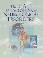
14
15
16
13
12
11
11
12
13
2
1
HD: Huntington disease
MPS: Mucopolysaccharidoses
Achondroplasia
RIEG: Rieger syndrome
p
q
25
27
31
33
34
35
32
28
24
26
23
22
21
3
1
LQT4: Long QT syndrome 4
alpha-synuclein: Parkinson’s disease
Chromosome 4
EVC: Ellis-van Creveld
Mucopolysaccharidoses, on chromosome 4. (Gale Group.)
Description
Though the symptoms and severity vary for each
MPS disorder, common features include enlarged organs
(organomegaly), dysostosis multiplex (abnormal bone for-
mation), and a characteristic facial appearance. Hearing,
vision, breathing, heart function, joint mobility, and men-
tal capacity may also be affected. As of 2003, seven types
of MPS have been classified. The MPS disorders are
caused by absent or insufficient production of proteins
known as lysosomal enzymes The specific enzyme that is
deficient or absent distinguishes one type of MPS from an-
other. However, before these enzymes were identified, the
signs and symptoms expressed by an affected individual
led to the diagnosis. The discovery of these enzymes re-
sulted in a reclassification of some of the MPS disorders.
These conditions are often referred to as MPS I, MPS II,
MPS III, MPS IV, MPS VI, MPS VII, and MPS IX and
may also referred to by their original names, which are
Hurler (MPS I H), Hurler-Scheie (MPS I H/S), Scheie
(MPS I S), Hunter (MPS II), Sanfilippo (MPS III),
Morquio (MPS IV), Maroteaux-Lamy (MPS VI), Sly
(MPS VII), and Hyaluronidase deficiency (MPS IX).
Demographics
The MPS syndromes are considered to be rare. San-
filippo syndrome appears to be the most common MPS
with a reported incidence of one in 70,000. The incidence
of Hyaluronidase deficiency is not yet known. The inci-
dence of the remaining six classes of MPS are estimated
to be: one in 100,000 for Hurler syndrome; one in 500,000
for Scheie syndrome; one in 115,000 for Hurler/Scheie
disease; one in 100,000 (male live births) for Hunter syn-
drome (mild and severe combined); one in 100,000 to one
in 300,000 for Morquio syndrome (types A and B in-
cluded); one in 215,000 for Maroteaux-Lamy syndrome;
and less than one in 250,000 for Sly syndrome. These fig-
ures are general; more exact figures have been reported for
individual MPS disorders in certain countries.
Causes and symptoms
All of the MPS are genetic conditions. MPS I, MPS
III, MPS IV, MPS VI, MPS VII, and MPS IX are inherited
in an autosomal recessive manner which means that af-
fected individuals have two altered or non-functioning
genes, one from each parent, for a specific enzyme that is
needed to break down mucopolysaccharides. MPS II
(Hunter syndrome) is inherited in an X-linked manner
which means that the gene for MPS II is located on the X
chromosome, one of the two sex chromosomes. Hunter
syndrome primarily affects males because they have only
one X chromosome and therefore lack a second, normal
copy of the gene responsible for the condition. Carriers for
the autosomal recessive forms of MPS have one normal
copy and one non-working copy of the MPS gene in ques-
tion. Female carriers of the X-linked MPS (MPS II) have
one X chromosome with a normal gene for the condition
(the IDS gene) and one X chromosome with a non-work-
ing IDS gene.
The enzymes that are deficient in the MPS disorders
normally break down a type of mucopolysaccharide (a
long chain of sugar molecules) in the body known as gly-
cosaminoglycans (GAGs). Glycosaminoglycans are es-
sential for building the bones, cartilage, skin, tendons, and
other tissues in the body. Normally, the human body con-
tinuously breaks down and builds GAGs. There are several
enzymes involved in breaking down each GAG and a de-
ficiency or absence of any of the essential enzymes can
cause one or more GAGs to accumulate in the tissues and
organs in the body. When too much GAG is stored, organs
and tissues can be damaged or not function properly. The
LetterM.qxd 10/1/04 11:07 AM Page 552
GALE ENCYCLOPEDIA OF NEUROLOGICAL DISORDERS
553
Mucopolysaccharidoses
accumulating material is stored in cellular structures called
lysosomes, and these disorders are also known as lysoso-
mal storage diseases.
MPS I
Mutations in the alpha-L-iduronidase (IDUA) gene
located on chromosome 4 cause the MPS I disorders
(Hurler, Hurler-Scheie, and Scheie syndromes). Initially,
these three disorders were believed to be separate because
each was associated with different physical symptoms and
prognoses. However, once the underlying cause of these
conditions was identified, it was recognized that all three
were variants of the same disorder.
MPS I H (HURLER SYNDROME) Individuals with
Hurler syndrome tend to have the most severe form of
MPS I. Hurler syndrome may also be referred to as severe
MPS I. Infants with Hurler syndrome appear normal at
birth and typically begin to develop normally. Symptoms
of Hurler syndrome are often evident within the first year
or two after birth. Many of these infants may initially grow
faster than expected, but their growth slows and typically
stops by age three. Facial features also begin to appear
coarse; affected children develop a short nose, flatter face,
thicker skin, and a protruding tongue. Additionally, their
heads become larger and they develop more hair on their
bodies with the hair becoming coarser. Affected children
with Hurler syndrome lose previously attained skills
(milestones) and eventually suffer from profound mental
retardation. Progressive abnormal development of all
bones of the body (dysostosis multiplex) occurs in all chil-
dren with Hurler syndrome. Children usually develop joint
contractures (stiff joints), kyphosis (a “hunchback” curve
of the spine), and broad hands with short fingers. Many of
these children experience breathing difficulties, and res-
piratory infections are common. Other common problems
include heart valve dysfunction, cardiomyopathy (weak-
ness of the heart muscle), hepatosplenomegaly (enlarged
spleen and liver), clouding of the cornea, hearing loss, and
carpal tunnel syndrome. Children with Hurler syndrome
typically die within the first ten years of life.
MPS I H/S (HURLER-SCHEIE SYNDROME) Hurler-
Scheie syndrome is felt to be the intermediate form of MPS
I, meaning that the symptoms are not as severe as those in
individuals who have Hurler syndrome but not as mild as
those with Scheie syndrome. Hurler-Scheie syndrome may
also be referred to as intermediate MPS I. Individuals with
Hurler-Scheie syndrome tend to be shorter than expected
and may develop some of the physical features seen in
Hurler syndrome, but usually they are not as severe. Intel-
lectual ability varies; individuals have normal or near nor-
mal intelligence. The prognosis for children with
Hurler-Scheie syndrome is variable with some individuals
dying during childhood and others living to adulthood.
MPS I S (SCHEIE SYNDROME) Scheie syndrome is con-
sidered the mild form of MPS I. Individuals with Scheie
syndrome usually have normal intelligence, but there have
been some reports of affected individuals developing psy-
chiatric problems. Common physical problems include
corneal clouding, heart abnormalities, and orthopedic dif-
ficulties involving the hands and back. Individuals with
Scheie syndrome do not develop the facial features seen
with severe MPS I. Usually life span is normal.
MPS II (Hunter syndrome)
Mutations in the iduronate-2-sulphatase (IDS) gene
cause both forms of MPS II (mild and severe). Nearly all
individuals with Hunter syndrome are male, because the
gene that causes the condition is located on the X chro-
mosome. The severe form is associated with progressive
mental retardation and physical disability, with most indi-
viduals dying before age 15. Males with the mild form of
Hunter syndrome usually have have normal or near normal
intelligence. They tend to develop physical differences
similar to males with the severe form, but not as quickly.
Most males with Hunter syndrome develop joint stiffness,
chronic diarrhea, enlarged liver and spleen, heart valve
problems, hearing loss, kyphosis, and tend to be shorter
than expected. Men with mild Hunter syndrome can have
a normal life span and some have had children.
MPS III (Sanfilippo syndrome)
MPS III is a variable condition with symptoms be-
ginning to appear between ages two and six years of age.
The condition is characterized by developmental delay, be-
havioral problems, and mild physical problems (as com-
pared to other types of MPS). Specific problems include:
seizures, sleeplessness, thick skin, joint contractures, en-
larged tongues, cardiomyopathy, hyperactivity, and men-
tal retardation. The life expectancy in MPS III is also
variable. On average, individuals with MPS III live until
adolescence. Initially, the diagnosis of MPS III, like the
other MPS conditions, was clinical; the diagnosis was
made by observation of certain physical characteristics. It
was later discovered that a deficiency in one of four en-
zymes could lead to the developmental delay and physical
symptoms associated with MPS III. Each type of MPS III
is now subdivided into four groups, labeled A-D, accord-
ing to the specific enzyme deficiency. All four of these en-
zymes help to break down the same GAG, heparan sulfate.
MPS IIIA (SANFILIPPO SYNDROME TYPE A) MPS IIIA
is caused by a deficiency of the enzyme heparan sulfate
sulfamidase, due to mutations in the SGSH gene on chro-
mosome 17. Type IIIA is felt to be the most severe of the
four types, in which symptoms appear and death occurs at
an earlier age.
LetterM.qxd 10/1/04 11:07 AM Page 553
554
GALE ENCYCLOPEDIA OF NEUROLOGICAL DISORDERS
Mucopolysaccharidoses
Key Terms
Carpal tunnel syndrome A condition caused by
compression of the median nerve in the carpal tun-
nel of the hand, characterized by pain.
Cornea The clear, dome-shaped outer covering of
the eye that lies in front of the iris and pupil. The
cornea lets light into the eye.
Gene A building block of inheritance, which
contains the instructions for the production of a
particular protein, and is made up of a molecular
sequence found on a section of DNA. Each gene is
found on a precise location on a chromosome.
Hydrops fetalis A condition in which a fetus or
newborn baby accumulates fluids, causing swollen
arms and legs and impaired breathing.
Metabolic Refers to the chemical reactions in liv-
ing organisms.
Mucopolysaccharide A complex molecule made
of smaller sugar molecules strung together to form
a chain. It is found in mucous secretions and inter-
cellular spaces.
Mutation A permanent change in the genetic ma-
terial that may alter a trait or characteristic of an in-
dividual, or manifest as disease. This change can be
transmitted to offspring.
MPS IIIB (SANFILIPPO SYNDROME TYPE B) MPS IIIB
is due to a deficiency in N-acetyl-alpha-D-glu-
cosaminidase due to mutations in the NAGLU gene, also
located on chromosome 17. This type of MPS III is not felt
to be as severe as Type IIIA and the characteristics vary.
Type IIIB is the most common of the four types of MPS III
in southeastern Europe.
MPS IIIC (SANFILIPPO SYNDROME TYPE C) A defi-
ciency in the enzyme acetyl-CoA-alpha-glucosaminide
acetyltransferase causes MPS IIIC. This is considered a
rare form of MPS III. The gene involved in MPS IIIC is
believed to be located on chromosome 14.
MPS IIID (SANFILIPPO SYNDROME TYPE D) MPS IIID
is caused by a deficiency in the enzyme N-acetylglu-
cosamine-6-sulfatase, due to mutations in the GNS gene lo-
cated on chromosome 12. This form of MPS III is also rare.
MPS IV (Morquio syndrome)
Morquio syndrome is characterized by severe skele-
tal deformities and their secondary effects on the nervous
system. Intelligence is usually normal. One of the earliest
symptoms seen in this condition is a difference in the way
the child walks. Skeletal abnormalities can be extreme and
include dwarfism, kyphosis (outward-curved spine),
prominent breastbone, flat feet, and genu-valgum (knock-
knees). A bone deformity known as odontoid hypoplasia
(improper formation of the bones that stabilize the head
and neck) can result in compression of the spinal cord, a
potentially serious and life-threatening complication. As
with several of the MPS disorders, Morquio syndrome was
originally diagnosed by the presence of particular signs
and symptoms. However, it is now known that the defi-
ciency of two different enzymes can result in MPS IV.
These two types of MPS IV are called MPS IV A and MPS
IV B. MPS IV is variable in its severity. MPS IV A is the
classic (typical) or the severe form of the condition and is
caused by a deficiency in the enzyme galactosamine-6-sul-
phatase. The gene involved with MPS IV A (GALNS) is
located on chromosome 16. MPS IV B is considered the
milder form of the condition. The enzyme, beta-galac-
tosidase, is deficient in MPS IV B. The gene involved with
MPS IV B (GLB1) is located on chromosome 3.
MPS VI (Maroteaux-Lamy syndrome)
MPS VI is caused by deficiency of the enzyme N-
acetylglucosamine-4-sulphatase (arylsulfatase B), due to
mutations in the ARSD gene located on chromosome 5.
Affected individuals may have a mild or severe form of the
condition. Typically, the nervous system and intelligence
are not affected. Individuals with a more severe form of
MPS VI can develop airway obstruction, hydrocephalus
(extra fluid accumulating in the brain), and abnormal
growth and formation of the bones. Additionally, individ-
uals with a severe form of MPS VI are more likely to die
while in their teens. With a milder form of the condition,
individuals tend to be shorter than expected for their age,
develop corneal clouding, and live longer.
MPS VII (Sly syndrome)
MPS VII, an extremely rare form of MPS, results
from a deficiency of the enzyme beta-glucuronidase due to
mutations in the GUSB gene on chromosome 7. MPS VII
is also highly variable, but symptoms are generally simi-
lar to those seen in individuals with Hurler syndrome. In
severe cases, infants may be born with hydrops fetalis.
MPS IX (Hyaluronidase deficiency)
MPS IX is a condition that was first described in 1996
and has been grouped with the other MPS conditions by
some researchers. MPS IX is caused by the deficiency of
the enzyme hyaluronidase due to mutations in the HYAL1
gene on chromosome 3. In the few individuals described
with this condition, the symptoms are variable, but some
develop soft-tissue masses (growths under the skin). Also,
these individuals are shorter than expected for their age.
LetterM.qxd 10/1/04 11:07 AM Page 554
GALE ENCYCLOPEDIA OF NEUROLOGICAL DISORDERS
555
Mucopolysaccharidoses
Diagnosis
Identification of symptoms is usually the first step in
making an MPS diagnosis. Doctors will then use labora-
tory tests to establish an accurate diagnosis. They may first
use a screening test that looks for glycosaminoglycans in
the urine. The definitive diagnosis of an MPS is made
using a biochemical test that measures the specific enzyme
(known to be reduced or absent) in the individual’s tissues
or bodily fluids. Genetic testing may also be used to con-
firm a suspected diagnosis and, in some cases, to provide
limited information about potential disease severity. Ge-
netic testing is accomplished by looking for specific
changes known as mutations in the gene responsible for
the MPS disorder. Genetic testing is available for all of the
MPS disorders except MPS IIIC, MPS IVB, and MPS IX.
If the gene mutation(s) have been found in an affected in-
dividual, the same genetic test may be used for carrier
screening in unaffected family members, such as adult sib-
lings, and for prenatal diagnosis. If the DNA mutations are
not found or if genetic testing is not available, carrier
screening and prenatal diagnosis may be accomplished
using biochemical methods. Preimplantation genetic di-
agnosis (PGD) is available on a research basis for MPS I
and MPS II. More information on PGD for these types of
MPS can be found by contacting the Reproductive Genet-
ics Institute at (773) 472-4900 or at
Treatment team
Treatment of MPS disorders requires a multidiscipli-
nary approach. In addition to the patient’s primary health
care professionals, medical professionals involved in the
care of patients with an MPS usually includes specialists in
neurology, neurosurgery, ophthalmology (eyes), otolaryn-
gology (ear-nose-throat), audiology (hearing), cardiology,
pulmonology (lungs), anesthesiology, gastroenterology,
nutrition, orthopedic surgery, rehabilitation (physical, oc-
cupational, and speech therapy) and genetics. Some pa-
tients with MPS may receive comprehensive services
through a specialty clinic such as metabolic or neuroge-
netics clinic. A genetic specialist, such as a clinical geneti-
cist or a genetic counselor, may be helpful to the patient
and family, especially at the time of diagnosis or prior to
genetic testing. Psychological counseling and MPS sup-
port groups may also assist families in coping with this
condition.
Treatment
Treatment of the MPS disorders primarily consists of
supportive care and management of complications. Bone
marrow transplant (BMT) and enzyme replacement are
two promising therapies that offer the possibility of alter-
ing the course of these conditions. Due to the progressive
nature of the MPS disorders, regular evaluations by pri-
mary care providers and specialists is required to detect
problems early. Treatment for the most common problems
found in the MPS disorders is listed below.
Symmtomatic care and treatment
HYDROCEPHALUS Hydrocephalus (increased fluid in
the ventricles of the brain) commonly occurs in MPS I,
MPS II, MPS VI, and MPS VII due to a blocked circulation
of cerebral spinal fluid in the brain. If the hydrocephalus is
detected early, a surgical procedure known as ventricu-
loperitoneal shunting or a VP shunt may afford the affected
individual with a better outcome. Periodic CT or MRI
scans may be recommended to monitor for hydrocephalus
in a child with MPS. In MPS III, enlarged ventricles
(spaces in the brain) may occur but here the enlargement is
thought to be due to cortical atrophy (loss of brain cells).
It has been reported that shunting may decrease behavior
problems associated with this form of MPS.
SEIZURES Seizures are a problem found in severe
forms of MPS and especially in MPS III (Sanfilippo syn-
drome). Patients with seizures are given a type of pre-
scription medication known as an anticonvulsant.
VISION AND HEARING Regular evaluation by an oph-
thalmologist is recommended to look for common eye
problems including changes in the retina, glaucoma, and
corneal clouding. Retinal degeneration, an eye problem
that leads to night blindness and loss of peripheral vision,
is common in MPS I, MPS II, and MPS III. Adding a night
light to a hall or bedroom may help with this. Glaucoma
is especially common in MPS I and is usually treated with
medications. Corneal clouding is found in MPS I, MPS IV,
MPS VI and MPS VII. People with corneal clouding have
photophobia (the inability to tolerate bright light). Caps
with a visor or sunglasses may be recommended to help
reduce this problem. Corneal transplantation is possible
for people with significantly reduced vision yet transplants
may not always result in improved vision in the long term.
Hearing problems are common in the MPS disorders.
Regular hearing evaluations are important so that children
with hearing loss can be treated early. Hearing aids may
provide some degree of improvement. Recurrent otitis
media (middle ear infections) significantly contribute to
hearing loss in individuals with MPS. Prescription med-
ications are used to treat otitis media. Ventilating tubes in
the ears may be used to minimize the long term effects of
these infections.
CARDIOVASCULAR Many individuals with MPS show
some signs of heart disease. Common problems include
abnormal heart valves, narrowing of the blood vessels in
the heart, and weak heart muscles (cardiomyopathy). Pa-
tients with MPS I H and the severe form of MPS II usually
LetterM.qxd 10/1/04 11:07 AM Page 555
556
GALE ENCYCLOPEDIA OF NEUROLOGICAL DISORDERS
Mucopolysaccharidoses
have damage to the mitral valve. In MPS I H/S, MPS IS,
MPS IV, and MPS VI, aortic valvular disease is more com-
mon. Medications may be prescribed for congestive heart
failure and hypertension associated with underlying heart
disease. Valve replacement surgery is possible and has
been reported in the MPS disorders.
AIRWAY DISEASE Obstruction of the airway is a com-
mon and significant problem for individuals with MPS.
This problem can be due to a narrowed trachea (wind
pipe), thickened vocal cords, large adenoids or tonsils, de-
creased rib movement with breathing, and a large tongue.
A condition known as obstructive sleep apnea (temporary
cessation of breathing while asleep) is the most common
airway problem in MPS. Treatment for sleep apnea may
include: removal of adenoids and tonsils, CPAP or BiPAP
treatment, or a tracheostomy. CPAP (continuous positive
airway pressure) and BiPAP (bilevel positive airway pres-
sure) are treatments that help to keep the airway open at
nighttime. A tracheostomy, an permanent opening through
the neck into the trachea, may be needed in severe cases of
sleep apnea.
FEEDING PROBLEMS For many individuals with MPS,
neurological problems eventually lead to significant prob-
lems with chewing and swallowing. Surgical placement of
gastrostomy tube (G-tube) or a jejunostomy tube (J-tube)
may be recommended when feeding problems cause
weight loss, choking, gagging, or episodes of pneumonia.
SKELETAL DEFORMITIES Bony problems, especially of
the neck, spine, and hips may require orthopedic inter-
vention. Problems of the cervical spine due to odontoid
hypoplasia (improper formation bones that stabilize the
head and neck) can be quite serious. Odontoid hypoplasia
can lead to slippage of the bones in the neck and com-
pression of the spine in the cervical (neck) region. In se-
vere cases, this spinal cord compression may result in
nerve damage, paralysis or death. Odontoid hypoplasia is
common in MPS IV (Morquio syndrome). Treatment in-
cludes regular monitoring with MRI or X-rays and cervi-
cal fusion surgery for severe cases. Other bony problems
seen in the MPS disorders include progressive scoliosis or
kyphosis (curvatures of the spine ) and hip dysplasia (ab-
normal hip joint). Bracing and sometimes surgery may be
used to treat spine curvature. A surgical procedure known
as spinal fusion may be considered in patients with sig-
nificant curvature. Patients with hip dysplasia may be
given non-steroidal anti-inflammatory medications.
CARPAL TUNNEL SYNDROME Carpal tunnel syn-
drome is a common problem in MPS. Although many in-
dividuals with MPS may not have typical symptoms
(numbness, tingling, pain), the carpal tunnel syndrome
can and may be severe. Treatment options include splint-
ing, anti-inflammatory medications and surgery.
Bone marrow transplantation (BMT)
Bone marrow transplants have been used to treat chil-
dren with MPS I, MPS II, MPS III, and MPS VI. Some
success has been achieved with BMT in MPS I and in
MPS VI; however, this treatment is not a cure and is con-
sidered experimental due to the associated risks, including
death. Some children who have undergone BMT have
shown reduced progression of some disease symptoms. It
remains uncertain whether BMT can prevent brain dam-
age. BMT is not recommended as a treatment for MPS II
or MPS III.
Enzyme replacement therapy
Enzyme replacement therapy is available for MPS I.
A pharmaceutical form of alpha-L-iduronidase known as
laronidase is available in the United States. More infor-
mation may be obtained at<>.
Enzyme therapy may be an option in the future for indi-
viduals with MPS IV.
Recovery and rehabilitation
Rehabilitation for the MPS disorders consists of phys-
ical, occupational, and possibly speech therapy. For ex-
ample, physical therapy may help preserve joint function
for individuals with joint stiffness. Joint stiffness is pres-
ent in all of the MPS disorders except MPS IV and MPS
IX. In physical therapy, patients may undergo range-of-
motion exercises (passive bending and stretching of the
arms and legs). Also, physical therapy after neck, spine or
knee surgery can help patients (who could walk prior to
surgery) to walk again. Occupational therapy can teach pa-
tients to use adaptive techniques and devices that may help
compensate for loss of mobility and/or for loss of speech.
Speech therapy may be indicated for individuals with
MPS; however, this intervention may not be useful in cases
in which the mental condition is rapidly deteriorating.
Hyperactivity can be a severe problem in individuals
with MPS, especially in MPS III and MPS II. Medications
may or may not be successful in treating this problem. Be-
havior modification programs may be helpful for some hy-
peractive MPS children. It may also be necessary to adapt
the house and yard to the child.
Clinical trials
As of December 2003, there were four clinical trials
related to the MPS disorders that were recruiting patients.
A phase II/II trial to determine whether the administration
of iduronate-2-sulfatase enzyme is safe and efficacious in
patients with MPS II will be conducted in the United
States, Brazil, Germany and England. Information on this
trial can be found at <> or by
contacting Transkaryotic Therapies at 617-613-4499. A
LetterM.qxd 10/1/04 11:07 AM Page 556
GALE ENCYCLOPEDIA OF NEUROLOGICAL DISORDERS
557
Mucopolysaccharidoses
phase III trial to evaluate the ability of recombinant human
arylsulfatase B enzyme to enhance endurance in patients
with Mucopolysaccharidosis VI (MPS VI) will be con-
ducted in the United States. Information on this trial can
be found at <> or by con-
tacting BioMarin Pharmaceuticals at 415-884-6700. A
phase II study of allogeneic bone marrow or umbilical
cord blood transplantation in patients with mucopoly-
saccharidosis I will be conducted in the United States.
Information on this trial can be found at <http://www.
clinicaltrials.gov> or by contacting the Study Chair at the
Fairview University Medical Center in Minneapolis, Min-
nesota, at 612-624-5407. A phase II study of bone marrow
or umbilical cord blood transplantation in patients with
lysosomal or peroxisomal inborn errors of metabolism. In-
formation on this trial can be found at <ni-
caltrials.gov> or by contacting the Study Chair at the
Fairview University Medical Center in Minneapolis, Min-
nesota at 612-624-5407.
Prognosis
Life expectancy for individuals with an MPS is ex-
tremely varied. In severe forms of MPS, affected individ-
uals may die in infancy such as in the severe cases of Sly
syndrome, or they may die in in childhood or adolescence
such as in Hurler syndrome and severe Hunter syndrome.
In milder forms of MPS such as Scheie syndrome, mild
Hunter syndrome individuals can live well into adulthood.
Life spans for individuals with Sanfillipo syndrome,
Maroteaux-Lamy syndrome, Morquio syndrome and mild
Sly syndrome are quite variable. As more MPS I patients
utilize enzyme replacement therapy, new information
about prognosis and life span for this disorder will be
learned.
Special concerns
Many individuals with an MPS condition have prob-
lems with airway constriction. This constriction may be so
serious as to create significant difficulties in administering
general anesthesia. Therefore, it is recommended that sur-
gical procedures be performed under local anesthesia
whenever possible. If general anesthesia is needed, it
should be administered by an anesthesiologist experienced
in the MPS disorders.
Children and families affected by an MPS may bene-
fitfrom social services. A social worker may be able to
help families obtain Social Security, Medicaid, or other as-
sistance available from agencies that specialize in the care
of persons with disabilities. A child with MPS may bene-
fitfrom an Individual Education Plan (IEP). An IEP
provides a framework from which administrators, teach-
ers, and parents can meet the educational needs of a child
with MPS.
Resources
BOOKS
Neufeld, Elizabeth F. and Joseph Muenzer.“The
Mucopolysaccharidoses.” Chapter 136. In The Metabolic
and Molecular Bases of Inherited Disease, 8th ed., Vol. 3,
edited by Charles R. Scriver, Arthur L. Beaudet, William
S. Sly, and David Valle. New York: McGraw-Hill Medical
Publishing Division, 2001.
Parker, James N., and Philip M. Parker, eds. The Official
Parent’s Sourcebook on Mucopolysachharidoses: A
Revised and Updated Directory for the Internet Age. San
Diego, CA: ICON Health Publications, 2002.
PERIODICALS
Froissart, R., I. Moreira da Silva, N. Guffon, D. Bozon, and I.
Maire. “Mucopolysaccharidosis type II-genotype/pheno-
type aspects.” Acta Paediatrica Supplement 91 (2002):
82–87.
Gulati, M. S., and M. A. Agin. “Morquio syndrome: a rehabili-
tation perspective.” Journal of Spinal Cord Medicine 19
(January 1996): 12–16.
Kakkis, E. D. “Enzyme replacement therapy for the
mucopolysaccharide storage disorders.” Expert Opinion
on Investigational Drugs 11 (May 2002): 675–685.
Robertson, S. P., G. L. Klug, and J. G. Rogers. “Cerebrospinal
fluid shunts in the management of behavioral problems in
Sanfilippo syndrome.” European Journal of Pediatrics
157 (August 1998): 653–655.
Vougioukas, V. I., A. Berlis, M. V. Kopp, R. Korinthenberg, J.
Spreer, and V. van Velthoven. “Neurosurgical interven-
tions in children with Maroteaux-Lamy syndrome. Case
report and review of the literature.” Pediatric
Neurosurgery 35 (July 2001): 35–38.
WEBSITES
Online Mendelian Inheritance in Man (OMIM). National
Center for Biotechnology Information. <http://
www.ncbi.nlm.nih.gov/Omim/>.
The National Institute of Neurological Disorders and Stroke
(NINDS). Mucopolysaccharidoses Information Page.
< />disorders/mucopolysaccharidoses.htm>.
OTHER
The National MPS Society. MPS Disorder booklets. 45
Packard Drive, Bangor, ME: The National MPS Society,
2001-2003. < />ORGANIZATIONS
Canadian Society for Mucopolysaccharide and Related
Diseases. PO Box 64714, Unionville, Ontario L3R-OM9,
CA. (904) 479-8701 or (800) 667-1846. rldillio@
interlog.com. <>.
National MPS Society, Inc. 45 Packard Drive, Bangor, ME
04401. (207) 947-1445; Fax: (207) 990-3074.
<>.
LetterM.qxd 10/1/04 11:07 AM Page 557
558
GALE ENCYCLOPEDIA OF NEUROLOGICAL DISORDERS
Multi-infarct dementia
Society for Mucopolysaccharide Diseases. 46 Woodside Road,
Amersham, Buckinghamshire HP6-6AJ, UK. (149) 443-
4252; Fax: (149) 443-4252.
<>.
Dawn J. Cardeiro, MS, CGC
❙
Multi-infarct dementia
Definition
Multi-infarct dementia is one form of dementia that
occurs when small blood vessels in the brain are blocked
by blood clots or fatty deposits. The blockage interrupts the
flow of blood to regions of the brain (a stroke), which, if
sustained, causes the death of cells in numerous areas of the
brain. Another form of multi-infarct dementia is inherited.
Description
Blockage or narrowing of small blood vessels by
blood clots or by deposits of fat can impede the flow of
blood through the vessel. Deprivation of the essential blood
is catastrophic for the regions that are supplied by the ves-
sels. In the brain, such vessel blockage can cause the death
of brain cells. This event is also called a stroke. The stroke-
related cell death affects the functioning of the brain.
Multi-infarct dementia is the most common form of
dementia (the loss of cognitive brain due to disease or in-
jury) due to changes in blood vessels. Alzheimer’s dis-
ease is the most common of these so-called vascular
dementias. The term multi-infarct is used because there
are many areas in the brain where cell damage or death
occurs. Besides dementia, multi-infarct dementia can
cause stroke, headaches of migraine-like intensity, and
behavioral disturbances.
An inherited form of multi-infarct dementia is desig-
nated as CADASIL, which is an acronym for cerebral au-
tosomal dominant arteriopathy with subcortical infarcts
and leukoencephalopathy.
Demographics
Multi-infarct dementia usually begins between the
ages of 60–75 years. For as-yet-undetermined reasons, it
affects men more than women. Multi-infarct dementia is
the second most common cause of dementia in older peo-
ple after Alzheimer’s disease, accounting for up to 20% of
all progressively worsening dementias.
CADASIL occurs in young male and female adults. It
has been diagnosed in Americans, Africans, and Asians,
and may occur in other racial groups.
Causes and symptoms
The root cause of multi-infarct dementia is usually
small blood clots that lodge in blood vessels in the brain,
which results in the death of brain cells. Over time, the se-
ries of small strokes (also known as mini-strokes, transient
ischemic attacks, or TIAs) magnifies the brain cell dam-
age. Blood clots can result from an elevated blood pres-
sure. Indeed, it is uncommon for someone affected with
multi-infarct dementia not to have a history of high blood
pressure.
There are a variety of symptoms caused by the brain
cell loss. These include mental confusion, problems re-
taining information even for a short time, loss of recogni-
tion of surroundings that are familiar (which can lead to
getting lost in previously familiar territory), loss of control
of urination and defecation, moving with a rapid shuffling
motion, difficulty in following instructions, rapid swings
in emotion, and difficulty performing tasks that were pre-
viously routine. These symptoms appear in a stepwise
manner, from less to more severe. As well, the initial
symptoms can be so slight as to be unrecognized, disre-
garded, or rationalized as being due to other causes such
as a temporarily stressful period. These early problems in-
clude a mild weakness in an arm or a leg, slurred speech,
or dizziness that only lasts for a few days. As more blood
vessels become blocked with the occurrence of more
strokes, the more severe symptoms associated with men-
tal decline become apparent.
CADASIL is characterized by a series of strokes,
which is thought to be triggered by genetically determined
deficiencies of small cerebral arteries. The defects affect
blood flow to the brain in a similar fashion as occurs in
multi-infarct dementia. The symptoms associated with
CADASIL range from migraines to a slowly progressing
series of symptoms that is similar to the symptoms that de-
velop in multi-infarct dementia.
Diagnosis
Multi-infarct dementia is diagnosed based on the his-
tory of symptoms, especially of high blood pressure and
strokes. A physician will look for several features during
the examination, which include arm or leg weakness,
speech difficulties, or dizziness. Tests that can be per-
formed in the doctor’s office include taking a blood
pressure reading, recording the heartbeat (an electro-
encephalogram, or EEG), and obtaining blood for labora-
tory analysis. Ultrasound studies of the carotid artery may
also be performed.
Diagnosis most often involves the non-destructive im-
aging of the brain by means of computed tomography (CT)
or magnetic resonance imaging (MRI) to reveal blood
clots or the characteristic damaged regions of the brain.
LetterM.qxd 10/1/04 11:07 AM Page 558
GALE ENCYCLOPEDIA OF NEUROLOGICAL DISORDERS
559
Multi-infarct dementia
Key Terms
Dementia A chronic loss of mental capacity due
to an organic cause.
Infarct Tissue death due to lack of oxygen result-
ing from a blood clot, plaque, or inflammation that
blocks an artery.
Transient ischemic attack (TIA) A temporary,
stroke-like event that lasts for only a short time and
is caused by a blood vessel that is temporarily
blocked.
Diagnosis can also be aided by an examination by a
psychologist or a psychiatrist to test a person’s degree of
mental reasoning, ability to learn and retain new informa-
tion, and attention span. Symptoms can be similar to those
of Alzheimer’s disease, which can complicate and delay the
diagnosis of both disorders. Indeed, a person can have both
disorders at the same time, as their causes are different.
Treatment team
A person with multi-infarct dementia can benefit from
a support network that includes a family physician, neu-
rologist, pharmacist, nurses, and supportive family mem-
bers and other care givers. Community resources are also
important, such as assisted living facilities, adult day or
respite care centers, and local agencies on aging.
Treatment
There is no specific treatment for multi-infarct de-
mentia, as the damage to the brain cells cannot be re-
versed. Treatment typically involves trying to limit further
deterioration. This focuses on establishing and/or main-
taining a lower blood pressure, which lessens the tendency
of blood clot formation. Those people who are diabetic
will be treated for this condition, as diabetes can contribute
to stroke. Other factors that can be involved in lessening
blood pressure include maintaining a target cholesterol
level, exercise,avoiding smoking, and moderation in al-
cohol consumption.
Aspirin is known to reduce the tendency of the blood
to clot. Some physicians will prescribe aspirin or similarly
acting drugs for this purpose. As well, those with high cho-
lesterol may benefit from a diet change and/or the use of
cholesterol-lowering drugs such as statins. In some people,
surgery that removes blockages in the main blood vessel
to the brain (the carotid artery) can be done. Other surgi-
cal treatments that increase blood flow through vessels in-
clude angioplasty and stenting to increase arterial flow to
the brain.
Recovery and rehabilitation
As damage to the brain cannot be reversed, the focus
for a person with multi-infarct dementia is placed upon
prevention of further brain tissue injury, and maintaining
optimum independent functioning.
Clinical trials
As of May 2004, there were no clinical trials under-
way or in the process of recruiting patients for either
multi-infarct dementia or CADASIL. However, research is
being funded by agencies such as the National Institute of
Neurological Disorders and Stroke and is aimed at under-
standing the development of dementia. The hope is that the
diagnosis of dementias will be improved. Ultimately, the
goal is to reverse or prevent the disorder.
Prognosis
The outlook for people with multi-infarct dementia is
poor. While some improvement in mental faculty may
occur, this is typically of short-term duration. Over longer
time, mental decline is inevitable and marked.
Special concerns
A person with multi-infarct dementia is often reliant
on family and friends for daily care and support. Family
and caregivers can help by stimulating a person’s mental
activity and prompting the individual to recall past expe-
riences. Eventually, around-the-clock care may become
necessary to provide a safe and stimulating environment.
Resources
BOOKS
Bird, T. D. “Memory Loss and Dementia.” In Harrison’s
Principles of Internal Medicine, 15th Edition, edited by
Franci, A. S., E. Daunwald, and K. J. Isrelbacher. New
York: McGraw Hill, 2001.
Mace Nancy L. The 36-Hour Day: A Family Guide to Caring
for Persons with Alzheimer Disease, Related Dementing
Illnesses, and Memory Loss in Later Life. New York:
Warner Books, 2001.
OTHER
“Multi-Infarct Dementia.” National Mental Health
Association. May 14, 2004 (June 1, 2004).
< />“Multi-Infarct Dementia Fact Sheet.” Alzheimer’s Disease
Education & Referral Center (ADEAR). May 15, 2004
(June 1, 2004). < />pubs/mid.htm>.
“NINDS Multi-Infarct Dementia Information Page.” National
Institute of Neurological Disorders and Stroke. May 14,
2004 (June 1, 2004). < />health_and_medical/disorders/multi-infarctdementia.doc>.
LetterM.qxd 10/1/04 11:07 AM Page 559
560
GALE ENCYCLOPEDIA OF NEUROLOGICAL DISORDERS
Multifocal motor neuropathy
ORGANIZATIONS
National Institute for Neurological Diseases and Stroke
(NINDS). P.O. Box 5801, Bethesda, MD 20824. (301)
496-5751. (800) 352-9424. <ds/nih.gov>.
National Institute on Aging (NIA). 31 Center Drive,
Rm. 5C27 MSC 2292, Bethesda, MD 20892-2292.
(301) 496-1752 or (800) 222-2225.
<>.
National Institute of Mental Health (NIMH). 6001 Executive
Blvd. Rm. 8184, MSC 9663, Bethesda, MD 20892-9663.
(301) 443-4513 or (866) 615-6464; Fax: (301) 443-4279.
<>.
Brian Douglas Hoyle, PhD
❙
Multifocal motor neuropathy
Definition
Multifocal motor neuropathy is a rare condition in
which the muscles in the body become progressively
weaker over months to years.
Description
Multifocal motor neuropathy is often mistaken for the
more catastrophic, inevitably fatal condition called amy-
otrophic lateral sclerosis (ALS). Unlike ALS, however,
multifocal motor neuropathy can be treated; therefore, dis-
tinguishing between these two conditions is crucial.
Demographics
Multifocal motor neuropathy is a very rare condition,
affecting only about 1 per 100,000 people in the popula-
tion. Men are about three times as likely to be affected as
women. Most patients are between the ages of thirty and
fifty when symptoms are noted, with the average age of
onset being 40 years.
Causes and symptoms
Multifocal motor neuropathy is thought to result
from an autoimmune disorder; that is, the body’s immune
system accidentally misidentifies markers on the body’s
own nerve cells as foreign. The immune system then be-
gins to produce cells that attack and injure or destroy ei-
ther the nerve cells or the myelin sheath wrapped around
the nerve cells. Because the myelin sheath allows mes-
sages to be conducted down a nerve quickly, injury to the
sheath or to the nerve itself results in slowed or faulty
nerve conduction.
Symptoms of multifocal motor neuropathy usually
begin with gradually progressive weakness of the hands.
Leg and foot weakness may follow, as well as decreased
muscle volume (called muscle wasting), muscle cramps,
and involuntary twitching and cramping of muscles. The
weakness is asymmetric; that is, a muscle group on only
one side of the body may be affected. Over time, numb-
ness or tingling of affected areas may occur, although sen-
sation is not lost.
Diagnosis
Diagnosis of multifocal motor neuropathy usually re-
quires both a careful physical examination, as well as elec-
tromyographic (EMG) testing. Physical examination will
reveal weakness and decreased muscle size, abnormal re-
flexes, muscle twitches, and totally normal sensation. EMG
involves inserting a needle electrode into a muscle, and
measuring the electrical activity within the muscle at rest
and during use. A characteristic pattern of abnormal nerve
conduction and muscle contraction will be noted on EMG.
Blood tests will usually reveal the presence of anti-
bodies (immune cells) directed against ganglioside, a com-
ponent of nerve cells.
Treatment team
Patients with multifocal motor neuropathy are usually
cared for by neurologists.
Treatment
Treatment for multifocal motor neuropathy involves
using intravenous immunoglobulin (IVIg) to dampen
down the immune system’s overactivity. If IVIg is not suc-
cessful, then the immunosuppressant drug cyclophos-
phamide may be administered.
In very mild, early cases, treatment may not be nec-
essary. If the condition progresses or prompts serious dis-
ability, treatment may be necessary. Treatment may then be
required intermittently, if the condition progresses again.
Prognosis
Muscle strength usually begins to improve within
three to six weeks of the initiation of treatment. Early
treatment of multifocal motor neuropathy usually results
in sufficient symptom resolution to prevent any permanent
disability. Over many years, however, many patients will
note a continued, slow progression of muscle weakness.
Resources
BOOKS
Asbury, Arthur K., and Stephen L. Hauser. “Guillain-Barré
Syndrome and Other Immune-mediated Neuropathies.”
In Harrison’s Principles of Internal Medicine, edited by
Eugene Braunwald, et al. NY: McGraw-Hill
Professional, 2001.
LetterM.qxd 10/1/04 11:07 AM Page 560
GALE ENCYCLOPEDIA OF NEUROLOGICAL DISORDERS
561
Multiple sclerosis
Griffin, John W. “Immune Mediated Neuropathies.” In Cecil
Textbook of Internal Medicine, edited by Lee Goldman, et
al. Philadelphia: W. B. Saunders Company, 2000.
Shields, Robert W., and Asa J. Wilbourn. “Demyelinating dis-
orders of the peripheral nervous system.” In Textbook of
Clinical Neurology, edited by Christopher G. Goetz.
Philadelphia: W. B. Saunders Company, 2003.
WEBSITES
National Institute of Neurological Disorders and Stroke
(NINDS). NINDS Multifocal Motor Neuropathy
Information Page. November 1, 2003 (June 3, 2004).
< />multifocal_neuropathy.htm>.
Rosalyn Carson-DeWitt, MD
❙
Multiple sclerosis
Definition
Multiple sclerosis is an inflammatory demyelinating
disease of the central nervous system. The disease re-
sults in injury to the myelin sheath (the fatty matter that
covers the axons of the nerve cells), the oligodendrocytes
(the cells that produce myelin) and, to a lesser extent, the
axons and nerve cells themselves. The symptoms of mul-
tiple sclerosis vary, depending in part on the location of
plaques (areas of thick scar tissue) within the central nerv-
ous system. Common symptoms include weakness and fa-
tigue, sensory disturbances in the limbs, bladder or bowel
dysfunction, problems with sexual function, and ataxia
(loss of coordination). Although the disease may not be
cured or prevented at this time, treatments are available to
reduce severity and delay progression.
Description
Multiple, or disseminated, sclerosis (MS) is a slowly
progressive disease of the central nervous system (CNS),
that comprises the brain and spinal cord. In 1868, French
physician Jean-Martin Charcot (1825–1893) produced his
lectures on “Sclerose en plaques,” providing the first de-
tailed clinical description of the disease. The cause of mul-
tiple sclerosis is unknown, and it cannot be prevented or
cured. Great progress, however, is being made in treating
and identifying underlying mechanisms that trigger the
disease. The primary characteristic of MS is the destruc-
tion of myelin, a fatty insulation covering the nerve fibers.
The end results of this process, called demyelination, are
multiple patches of hard, scarred tissue called plaques.
Another important feature in the disease is destruction of
axons, the long filaments that carry electric impulses away
from a nerve cell, which is now considered to be a major
factor in the permanent disability that occurs with MS.
Multiple sclerosis is usually characterized by a re-
lapsing remitting course in the early stages, with full or
nearly full recovery initially. In the early stages, there may
be little damage to axons. Over time, the disease enters an
irreversible progressive phase of neurological deficit. Each
relapse causes further loss of nervous tissue and progres-
sive dysfunction. In some cases there may be chronic pro-
gression without remission or acute disease rapidly leading
to death.
MS is a diverse disease. No two affected persons are
the same and each will experience different combinations
of symptoms with differing severity. The most common
form is relapsing-remitting multiple sclerosis (RRMS),
which affects 80–85% of people with MS. These patients
develop disease relapses, often without a specific trigger,
but possibly associated with infections. Disease relapses
can last between 24 hours and several months, and the per-
son may, or may not, completely recover. The disease is
stable between relapses, although affected persons can
have residual symptoms and disability.
After several years, the majority (70%) of persons
with MS will develop secondary progressive multiple scle-
rosis (SPMS), whereby they experience a progressive neu-
rological deterioration. They may still suffer from
superimposed relapses. A subcategory of RRMS patients
(around 20%) has benign MS. These patients have rare
and mild relapses and a long course of disease with mini-
mal or no disability. If patients have a steady neurological
decline from the onset, without relapses, they are de-
scribed as having primary-progressive multiple sclerosis
(PPMS). This comprises approximately 15–20% of people
with the disease
Afourth, rare type of MS is progressive-relapsing
multiple sclerosis (PRMS), which is considered a variant of
PPMS with similar prognosis. In patients with PRMS,
there is a gradual neurological decline from the beginning.
It is similar to PPMS, but has superimposed, acute relapses.
Demographics
According to the National Multiple Sclerosis Society,
approximately 400,000 Americans acknowledge having
MS, and every week about 200 people are diagnosed.
Worldwide, MS may affect 2.5 million individuals. The
usual age of onset is within the third and fourth decades,
although the disease can begin in childhood and also
above the age of 60 years. Overall, MS occurs more fre-
quently in women than in men, and the female-to-male
ratio is approximately of 2:1. This female predominance is
less defined in patients with PPMS, which typically de-
velops at a later age.
There is a variation in the worldwide distribution of
MS, with the highest prevalence in the northern and cen-
tral Europe, northern North America and southeastern
LetterM.qxd 10/1/04 11:07 AM Page 561
562
GALE ENCYCLOPEDIA OF NEUROLOGICAL DISORDERS
Multiple sclerosis
Nerve cell
Myelin sheath
Absence of
myelin sheath
MS results in injury to the myelin sheath that covers the axons of the nerve cells, the cells that produce myelin (oligoden-
drocytes), and, to a lesser extent, the axons and nerve cells themselves. (Illustration by Electronic Illustrators Group.)
Australia. Clusters, or areas with more than the expected
amount, occur. There are also racial differences, with a low
prevalence in Asians and Africans or people of African de-
scent, and a higher frequency in Caucasians, especially of
northern European descendent. MS is rare between the
equator and latitudes 30°–35° north and south. The preva-
lence of MS increases proportionally with increased dis-
tance from the equator. There is no satisfactory explanation
of this phenomenon, although certain variables have been
researched. These include environmental factors, such as
climate, humidity, hours of daily sunshine, resistance to
certain viruses, and even consumption of cow’s milk.
Causes and symptoms
The causes of multiple sclerosis remain unknown, but
it is widely accepted that susceptibility to MS is deter-
mined by a complex interaction between susceptibility
genes and environment. The most popular current theory
is that the disease occurs in people with a genetic suscep-
tibility, who are exposed to some environmental assault (a
virus or a toxin) that disrupts the blood-brain barrier, a
protective membrane that controls the passage of sub-
stances from the blood into the central nervous system.
Most researchers consider MS to be an autoimmune dis-
ease-one in which the body, through its immune system,
launches a defensive attack against its own tissues. Im-
mune factors converge in the nerve cells and trigger in-
flammation and an autoimmune attack on myelin and
axons. Still, a number of disease patterns have been ob-
served in MS patients, and some experts believe that MS
may prove to be not a single disorder, but may represent
several diseases with different causes.
Components of myelin such as myelin basic protein
have been the focus of much research because, when in-
jected into laboratory animals, they can precipitate exper-
imental allergic encephalomyelitis (EAE), a chronic
LetterM.qxd 10/1/04 11:07 AM Page 562
GALE ENCYCLOPEDIA OF NEUROLOGICAL DISORDERS
563
Multiple sclerosis
relapsing brain and spinal cord disease that resembles MS.
The injected myelin probably stimulates the immune sys-
tem to produce anti-myelin T-cells that attack the animal’s
own myelin.
Increasing scientific evidence suggests that genetics
may play a role in determining a person’s susceptibility to
MS. No specific gene has been identified and it seems to
have a mode of inheritance that involves multiple genes.
Twin studies have shown an increased risk of 30% in iden-
tical twins, and around 5% in fraternal twins. First-degree
relatives of a person with MS have a two or three percent
increased risk, which, although small, is higher than in the
general population. Further indications that more than one
gene is involved in MS susceptibility comes from studies
of families in which more than one member has MS.
Several research teams found that people with MS in-
herit certain regions on individual genes more frequently
than people without MS. Of particular interest is the
human leukocyte antigen (HLA) or major histocompati-
bility complex region on chromosome 6. HLAs are ge-
netically determined proteins that influence the immune
system. Another interesting candidate is CD24, which has
shown to be essential for the induction of EAE in mice.
CD24 is a cell surface protein with expression in a variety
of cell types that can participate in the rise of MS, includ-
ing activated T-cells.
An infectious cause of MS has been indicated by
some studies as well as by similarities to infectious de-
myelinating diseases. However, infectious agents more
likely shape the immune response that may induce the dis-
ease under special circumstances. Evidence is mounting
that infection with the Epstein-Barr virus (EBV), which
can cause mononucleosis, may also increase the risk of de-
veloping multiple sclerosis later in life. Researchers have
shown that people with multiple sclerosis tend to carry
higher levels of antibodies to the Epstein-Barr virus and
that they seem to be at higher risk for the disease. Some of
the immune cells that become programmed to attack the
Epstein-Barr virus may begin to attack myelin as well.
Environmental factors, other than infectious agents,
for which there is some evidence of an association with
MS, include toxins, low sunlight exposure, diet factors,
and trauma.
Almost any neurological deficit can occur in MS, but
there are several signs and symptoms that are characteris-
tic and their presence should suggest MS as a possible di-
agnosis, particularly in a young adult.
Vision disorders such as optic neuritis can occur. Optic
neuritis (ON) is an inflammation of the optic nerve char-
acterized by acute or subacute loss of vision usually in one,
but occasionally in both eyes. The visual loss evolves over
a period of hours or days. Vision returns to normal within
two months, but may deteriorate in later years. Previous
history of optic neuritis in a person who develops a neuro-
logical illness will strongly support the diagnosis of MS.
Cognitive (thought) impairment is thought to affect
40–70% of MS patients and can be present even in the
early stages of MS. Approximately one-third of people
with MS have some degree of memory loss. Other areas of
cognitive function particularly affected in the MS patient
include sustained attention, verbal fluency, and spatial per-
ception. Dementia (loss of intellectual function) is often
common in the latter stages of MS.
Many MS patients are temperature sensitive. In hot-
ter weather or during a period of raised body temperature,
their MS symptoms worsen. Most frequently, vision is af-
fected and muscle weakness occurs.
About two-thirds of MS patients experience pain at
some point during the course of the disease and 40% are
never pain free. MS causes many pain syndromes; some
are acute, while others are chronic. Some worsen with age
and disease progression. Pain syndromes associated with
MS are trigeminal (facial) pain, powerful spasms and
cramps, optic neuritis pain, pressure pain, stiffened joints,
and a variety of sensations including feelings of itching,
burning, and shooting pain.
The Lhermitte’s sign can occur, which is actually
more of a symptom than a sign. A tingling or electric-like
sensation down the back and legs is felt upon flexing the
neck. The symptom is non-specific, but occurs more fre-
quently in MS than in any other condition and provides an
important clue to the correct diagnosis.
Urinary incontinence affects up to 90% of people with
multiple sclerosis and usually occurs before major physi-
cal disability is apparent. Bladder problems are due to
plaques in the spinal cord. If demyelination occurs in both
controlling pathways, the bladder will neither store urine
nor empty it properly. Constipation affects about 40% of
people with MS. Bowel incontinence and urgency of defe-
cation can also occur in about half of people with MS.
Fatigue is a common complaint in MS. Characteris-
tics of fatigue include muscle weakness, coordination
problems, ataxia, transient deafness, changes in taste or
smell and numbness of the extremities. Spasticity occurs
in up to 90% of MS patients and it can be painful and dis-
tressing. Spasticity is characterized by weakness, loss of
dexterity, and the inability to control specific movements.
It is usually more severe in the legs and torso.
Sexual dysfunction is common among people with
multiple sclerosis. If MS damages the nerve pathways
from the brain to the sexual organs via the spinal cord, sex-
ual response can be directly affected. Physicians and peo-
ple with MS often neglect to deal with this aspect of the
LetterM.qxd 10/1/04 11:07 AM Page 563
564
GALE ENCYCLOPEDIA OF NEUROLOGICAL DISORDERS
Multiple sclerosis
Key Terms
Autoimmune disease One of a group of diseases,
like rheumatoid arthritis and systemic lupus erythe-
matosus, in which the immune system is overactive
and has lost the ability to distinguish between self
and non-self. The body’s immune cells turn on the
body, attacking various tissues and organs.
Axon A long, threadlike projection that is part of
a neuron (nerve cell).
Myelin A fatty sheath surrounding nerves through-
out the body that helps them conduct impulses
more quickly.
disease, and both treatments and strategies for success are
available.
Depression is common in MS; some studies show
that over 50% of people with MS have depression at some
point in their lifetime. There is also an increased risk of
suicide. If depression is present, it should be treated prior
to initiating MS therapy. Depression in those with MS is
treated in the same way as the general population.
Diagnosis
MS diagnosis is based upon an individual’s history of
clinical symptoms and neurological examination. A qual-
ified physician, often a neurologist,must thoroughly re-
view all symptoms experienced by an individual to suspect
MS. Other conditions with similar symptoms must be
ruled out, often requiring various tests.
The diagnosis of MS is usually made in a young adult
with relapsing and remitting symptoms referable to dif-
ferent areas of CNS white matter. Diagnosis is more dif-
ficult in a patient with the recent onset of neurological
complaints or with a primary progressive clinical course.
Laboratory studies include blood work to exclude
collagen vascular disease, infections (ie, Lyme disease,
syphilis), endocrine abnormalities, vitamin B-12 defi-
ciency, sarcoidosis, and vasculitis. The examination of
cerebrospinal fluid (CSF) has been used to support the di-
agnosis of MS. The presence of myelin basic protein in
the CSF of an MS patient may be highly suggestive of ac-
tivity of the MS process, but its absence does not rule out
active disease.
A newer neuroimaging technique, magnetic reso-
nance spectroscopy (MRS), has been useful in following
NAA (N-acetyl-aspartate) levels in patients with multiple
sclerosis. NAA is an amino acid found in neurons and
axons of the mature brain. In patients with relapsing-re-
mitting MS, NAA levels are reduced, suggesting axonal
loss; however, in patients with secondary progressive MS
with more disability, the NAA levels are reduced more sig-
nificantly. In fact, patients with MS had lower levels of
NAA even in areas of the brain previously thought to be
unaffected, when compared with levels in normal persons.
Magnetic resonance imaging (MRI) remains the
imaging procedure of choice for diagnosing and monitor-
ing disease progression in the brain and spinal cord. This
test can show brain abnormalities in 90–95% of patients
and spinal cord lesions in up to 75% of cases, especially
in elderly patients. However, MRI alone cannot be used to
diagnose MS. Evoked potential tests that measure how
quickly and accurately a person’s nervous system re-
sponds to certain stimulation have been the most useful
neurophysiological studies for evaluation of MS.
At the onset, MS may be mistaken for other inflam-
matory diseases of the central nervous system, such as
Behçet disease, antiphospholipid syndrome or acute dis-
seminated encephalomyelitis (ADEM). Pseudotumoral
MS may be reminiscent of lymphoma, other tumors (glial
tumors), or infectious diseases (like Lyme disease, HTLV1
infection or abcess). Recurrent relapses of neurological
impairment may also be mistaken for cavernomatosis. In
most cases, MRI findings, cerebrospinal fluid analysis,
evoked potentials, the association with systemic signs and
the relapsing remitting nature of the disease allow physi-
cians to exclude other diseases, and to arrive at a diagno-
sis of MS.
Treatment team
The multidisciplinary team usually includes special-
ists in neurology, urology, ophthalmology, neuropsychol-
ogy, and social work.
Treatment
The three goals of drug therapy in the treatment of
MS are management of acute episodes, prevention of dis-
ease progression, and treatment of chronic symptoms.
Specific symptoms that may be treated include muscle
spasticity, lack of co-ordination, tremor, fatigue, pain,
bladder and bowel dysfunctions, sexual dysfunction and
depression.
Exacerbations (episodes of worsening symptoms) can
be defined as temporary flare-ups, sometimes referred to
as attacks or relapses. Most relapses show a degree of
spontaneous recovery, but treatment is offered for those re-
lapses that have a severe impact on function. Steroids are
the treatment of choice for relapses, usually methyl-pred-
nisolone given orally or by intravenous infusion. Before
starting steroids, infection should be excluded because
LetterM.qxd 10/1/04 11:07 AM Page 564
GALE ENCYCLOPEDIA OF NEUROLOGICAL DISORDERS
565
Multiple sclerosis
steroids have immunosuppressant action and can exacer-
bate the infection.
Disease modifying treatments are aimed at slowing
disease progression. The two current types of im-
munomodulatory agents used as a first line treatment are
interferon beta and glatiramer acetate. Interferon beta has
proved effective with RRMS and SPMS. There is currently
no evidence for improvement with PPMS. Discontinuation
of the treatment may be necessary because of intolerance
to side effects, when a pregnancy is planned, or when it is
no longer effective. Glatiramer is the appropriate treatment
to reduce relapse frequency in patients with RRMS and it
should not be used for both PPMS and SPMS. Stopping
criteria for glatiramer are the same of interferon beta.
A number of treatments are available for managing
MS chronic symptoms and complications, each one with
specific drugs. Indeed, symptomatic treatment, along with
supportive measures and rehabilitation, are a major part of
the MS treatment.
Recovery and rehabilitation
When recovering from a symptom flare-up or learn-
ing to cope with a change in mobility, rehabilitation
through physical therapy can be of great value training pa-
tients to improve mobility and to decrease spasticity and
strengthen muscles. Some of those who have a physically
demanding or highly stressful job may choose to make a
career change, in which case vocational training is helpful.
Occupational therapy helps in assessing the patient’s
functional abilities in completing activities of daily living,
assessing fine motor skills, and evaluating for adaptive
equipment and assistive technology needs. Speech thera-
pists assess the patient’s speech, language, and swallow-
ing and may work with the patient on compensatory
techniques to manage cognitive problems.
Clinical trials
The National Institute of Neurological Disorders and
Stroke (NINDS) is recruiting patients to evaluate the safety,
tolerability, and effect of the drug Rolipram on MS. The
NINDS is also recruiting patients with relapsing-
remitting or secondary progressive multiple sclerosis to
examine the safety and effectiveness of Zenapax (a labo-
ratory-manufactured antibody) in treatment of MS. More
information is available at the website: <http://www.
clinicaltrials.gov>, a clinical trial service sponsored by the
United States government.
Prognosis
It is generally very difficult to predict the course of
MS. The disorder varies greatly in each individual, but
most people with MS can expect to live 95% of the normal
life expectancy. Some studies have shown that people who
have few attacks in the first several years after diagnosis,
long intervals between attacks, complete recovery from at-
tacks, and attacks that are sensory in nature (i.e., numbness
or tingling) tend to fare better. People who have early
symptoms of tremor, difficulty in walking, or who have
frequent attacks with incomplete recoveries, or more le-
sions visible on MRI scans early on, tend to have a more
progressive disease course.
Special concerns
People with should avoid caffeine-containing bever-
age, which can actually be dehydrating. The diet should
also be rich in fiber, particularly from whole grains, fruits
and vegetables to increase digestive motility and reduce
constipation. Maintenance of weight in the normal range
is also desirable in order to diminishes stress on the joints
and skeletal muscles.
Gait difficulty (difficulty with walking) may worsen
during pregnancy, and assistive devices for walking or a
wheelchair are useful at this time. During pregnancy, blad-
der and bowel problems may also be aggravated in women
with MS who already have these dysfunctions.
Resources
BOOKS
O’Connor, Paul. Multiple Sclerosis: The Facts You Need.
Firefly Books, 1999.
Warren, Sharon, and Kenneth Warren. Multiple Sclerosis.
World Health Organization, 2001.
PERIODICALS
Myles, Mary L. “The ongoing battle against multiple sclero-
sis.” Canadian Journal of Diagnosis (June, 2003):
108–117.
OTHER
“About MS.” Multiple Sclerosis Association of America.
<> (February 12, 2004).
National Institute of Neurological Disorders and Stroke.
NINDS Multiple Sclerosis Information Page.
< />disorders/multiple_sclerosis.htm> (February 12, 2004).
National Multiple Sclerosis Society. Living with MS.
<> (February 1, 2004).
ORGANIZATIONS
The National Multiple Sclerosis Society. 733 Third Avenue,
6th floor, New York, NY 10017. (212) 986-3240 or (800)
344-4867; Fax: (212) 986-7981.
<>.
Marcos do Carmo Oyama
Iuri Drumond Louro
LetterM.qxd 10/1/04 11:07 AM Page 565
566
GALE ENCYCLOPEDIA OF NEUROLOGICAL DISORDERS
Multiple system atrophy
Key Terms
Atrophy The progressive wasting and loss of func-
tion of any part of the body.
Cerebellum The part of the brain involved in the
coordination of movement, walking, and balance.
Neurodegeneration The deterioration of nerve
tissues.
❙
Multiple system atrophy
Definition
Multiple system atrophy (MSA) is a neurodegenera-
tive disease characterized by parkinsonism, cerebellar dys-
function, and autonomic disturbances.
Description
MSA causes a wide range of symptoms, in keeping
with its name of “multiple system” atrophy. Parkinsonian
symptoms include tremor, rigidity and slowed movements;
cerebellar symptoms include incoordination and un-
steady gait; and autonomic symptoms include orthostatic
hypotension (drop in blood pressure upon standing) and
urinary incontinence. Because of this wide variety of
symptoms, it was originally thought of as three distinct
diseases: striatonigral degeneration (parkinsonian
symptoms), olivopontocerebellar atrophy (cerebellar
symptoms) and Shy-Drager syndrome (autonomic symp-
toms). Further study showed the overlap among these con-
ditions was best explained by considering them as a single
disease with symptoms clustered into three groups. His-
torically, confusion about the disease was made even
worse because olivopontocerebellar atrophy is also the
name of an unrelated genetically inherited disease. It is
hoped that widespread use of the name MSA will clear up
some of this confusion.
Demographics
Because MSA is often misdiagnosed, figures on its
prevalence are not known with certainty. It is estimated
there are between 25,000 and 100,000 people in the United
States with MSA. Onset is usually in the early fifties, and
men are slightly more likely to be affected than women.
Causes and symptoms
The cause or causes of MSA are unknown. No genes
have been found for MSA. Some evidence indicates that
toxins may be responsible, but no specific agents have
been identified. The brains of MSA patients reveal that
cells called glia undergo characteristic changes. Glia are
supportive cells for neurons, brain cells that conduct elec-
trical signals. In MSA, glia develop tangles of proteins
within them, called glial cytoplasmic inclusions. It is not
known whether these actually cause MSA, or are caused
by some other problem that is the real culprit.
The symptoms of MSA fall into three separate areas-
parkinsonism, cerebellar symptoms, and autonomic dis-
turbances. The distribution and severity of individual
symptoms varies among patients. MSA is a progressive
disease, and symptoms worsen over time.
Parkinsonism is the initial symptom in almost half of
all patients. The classic symptoms of Parkinson’s disease
(PD)—tremor, stiffness or rigidity, and slowed move-
ments—are seen in MSA, although tremor is not as com-
mon, and is jerkier than the tremor of PD.
Cerebellar symptoms are the initial feature in very
few MSA patients, but occur in about half of patients at
some point during the disease. The cerebellum is an im-
portant center for coordination, and degeneration of the
cerebellum in MSA leads to loss of balance, incoordina-
tion in the limbs, and loss of smooth eye movements. A
person with cerebellar dysfunction in MSA typically
walks with a wide stance to improve stability, and may
lose the hand-eye coordination that makes so many simple
activities possible.
Autonomic symptoms refer to those involving the au-
tonomic nervous system. The autonomic nervous system
controls a variety of “automatic” body functions, includ-
ing blood pressure, heart rate, sweating, and bladder func-
tion. Autonomic symptoms are the initial complaint in half
or more of all MSA patients. The most common initial
problem is urinary dysfunction in women, and erectile
dysfunction in men. Urinary dysfunction may be inconti-
nence, or inability to void the bladder. Other autonomic
symptoms include lack of sweating, constipation, and
fecal incontinence.
Orthostatic hypotension is a common autonomic
symptom. It refers to a significant drop in blood pressure
shortly after standing. It can cause dizziness, lighthead-
edness, fainting, weakness, fatigue,yawning, slurred
speech, headache, neck ache, cognitive impairment, and
blurred vision.
Other symptoms may also occur in MSA. These may
include:
•vocal cord paralysis, leading to hoarseness
•swallowing difficulty
• sleep apnea
• spasticity
LetterM.qxd 10/1/04 11:07 AM Page 566
GALE ENCYCLOPEDIA OF NEUROLOGICAL DISORDERS
567
Muscular dystrophy
•myoclonus
•Raynaud’s phenomenon (cold extremities)
Diagnosis
The diagnosis of MSA is difficult, because it is eas-
ily mistaken in its earlier stages for Parkinson’s disease,
which is much more common. Autonomic disturbance
also occurs in PD, but is much more pronounced in MSA.
MSA is the more likely diagnostic choice when disease
progression is rapid, and when the patient responds mildly
or poorly to levodopa, the mainstay of PD treatment. Some
centers use electromyography of the anal sphincter (the
muscles surrounding the anus) in order to confirm the di-
agnosis of MSA. Abnormal results indicate MSA rather
than PD, although this method is not universally recog-
nized as valid.
Neuroimaging may be used to rule out other causes of
similar symptoms, such as lesions in the brain or normal
pressure hydrocephalus.
Treatment team
The treatment team includes the neurologist, possi-
bly a movement disorders specialist, a urologist, and a
speech/language pathologist.
Treatment
There are no treatments that halt or slow the degen-
eration of brain cells that causes MSA. Treatment is aimed
at relieving symptoms.
Treatment of parkinsonian symptoms is attempted
with standard PD drugs, namely levodopa and the
dopamine agonists. Unfortunately, these are rarely as ef-
fective in MSA as they are in PD, although about one-third
of patients have at least a moderate response. In the best
case, treatment relieves stiffness, tremor and slowed move-
ments, allowing increased activities of daily living.
Orthostatic hypotension is treated with medications to
increase retention of fluids (fludrocortisone), compressive
stockings to keep blood from pooling in the legs, increas-
ing fluids, and increasing salt intake. Midodrine, a drug
that helps maintain blood pressure is often prescribed.
A urologist may be needed to define the type of uri-
nary dysfunction the patient has, and to manage treatment.
A bedside commode or condom catheter may be helpful
for urge incontinence, or inability to hold urine once the
urge to urinate occurs. If incomplete voiding is the prob-
lem, intermittent catheterization may be needed. Detrusor
hyperreflexia, in which the bladder muscle undergoes
spasms, may be treated with drugs to reduce these spasms.
Male erectile dysfunction may be treated with silde-
nafil or other medications.
Anhidrosis, or lack of sweating, can be dangerous in
an active patient, because of the risk of overheating.
Awareness of the problem and avoidance of prolonged ex-
ercise are helpful.
Gait ataxia may require a mobility aid, such as a
cane, walker, or eventually a wheelchair.
Speech and swallowing problems are dealt with by a
speech/language pathologist, who may work with the pa-
tient to develop swallowing strategies, and instruct in the
use of assistive communication devices. Sleep apnea may
be treated with continuous positive airway pressure venti-
lation.
Clinical trials
Clinical trials for MSA are usually directed toward
better diagnosis, or symptomatic treatment. Until re-
searchers develop a better understanding of the causes of
the disease, little progress can be expected in development
of treatments to slow its progression.
Prognosis
The average survival after diagnosis is 9-10 years.
Death usually occurs from pneumonia or suddenly from
insufficient respiration, due to degeneration of the respi-
ratory centers in the brain.
Special concerns
Resources
PERIODICALS
Wenning, G. K., et al. “Multiple System Atrophy.” Lancet
Neurology 3 (2004): 93-103.
WEBSITES
Shy-Drager Syndrome/Multiple System Atrophy Support
Group. <www.shy-drager-syndrome.org>.
WE MOVE. <www.wemove.org>.
Richard Robinson
Muscle-nerve biopsy see Biopsy
❙
Muscular dystrophy
Definition
Muscular dystrophies (MD) are inherited disorders
characterized by progressive weakness and degeneration
of the skeletal or voluntary muscles which control move-
ment, without a central or peripheral nerve abnormality.
The muscles of the heart and other involuntary muscles are
LetterM.qxd 10/1/04 11:07 AM Page 567
568
GALE ENCYCLOPEDIA OF NEUROLOGICAL DISORDERS
Muscular dystrophy
also affected in some forms of MD, and a few forms in-
volve other organs as well. The major forms of muscular
dystrophy include myotonic, Duchenne, Becker, limb-gir-
dle, facioscapulohumeral, congenital, oculopharyngeal,
distal, Emery-Dreifuss and Fukuyama muscular dystrophy.
Description
The commonest form of these inherited disorders is
the Duchenne muscular dystrophy (DMD). The disorder
was originally described in the mid-nineteenth century by
the English physician Edward Meryon. At a meeting of the
Royal Medical and Chirurgical Society in 1851, and later
published in the transactions of the society, he described
in detail the clinical presentation of Duchenne muscular
dystrophy, beginning in early childhood with progressive
muscle wasting and weakness and leading to death in late
adolescence. Furthermore, his detailed histological studies
led him to conclude that the muscle membrane or sar-
colemma was broken down and destroyed.
Duchenne muscular dystrophy will usually produce
symptoms between the ages of three and seven in young
boys. It begins with a weakness in the pelvic area first and
then progresses to the shoulder muscles. As the disorder
escalates, the muscles enlarge although the muscle tissue
is weak. The heart muscle will also enlarge, creating prob-
lems with the heartbeat that can be detected on an elec-
trocardiogram. In most cases, the affected child has a
waddling walk, often falls, has difficulty rising from a sit-
ting position, has a difficult time climbing stairs, is unable
to fully extend the arms and legs, and may develop scol-
iosis (an abnormally curved spine). In most cases, children
with DMD are confined to a wheel chair between the ages
of ten and twelve.
Most people with Becker muscular dystrophy (BMD)
first experience difficulties between the ages of five and
fifteen years, although onset in the third or fourth decade
or even later can occur. By definition, patients with BMD
are able to walk beyond age fifteen, while patients with
DMD are typically in a wheelchair by the age of twelve.
Patients with BMD have a reduced life expectancy, but
most survive into the fourth or fifth decade. Mental re-
tardation may occur in BMD, but it is not as common as
in DMD. Cardiac (heart muscle) involvement occurs in
BMD and may result in heart failure.
Myotonic muscular dystrophy (MMD) affects the
muscles in the hands and feet. Limb-girdle muscular dys-
trophy (LGMD) begins late in childhood affecting mainly
the muscles of the shoulders and hips. Facioscapulo-
humeral muscular dystrophy (FSHD) affects only the
muscles of the upper arms, face and shoulder girdle. Lan-
douzy-Dejerine muscular dystrophy (LDMD), which is
transmitted by an autosomal dominant gene, affects the
face, shoulder and lower leg muscles.
Other disorders related to muscular dystrophy include
Steinert’s disease, Thomsen’s disease, and Pompe’s dis-
ease. Steinert’s disease affects both males and females,
causing the muscles to be unable to relax after contracting,
while Thomsen’s disease causes a stiffness of the legs,
hands and eyelids. Pompe’s disease, which is a glycogen
storage disease, affects the liver, heart, nerves and muscles.
Demographics
United States
The incidence of muscular dystrophy varies, depend-
ing on the specific type. Duchenne muscular dystrophy is
the most common condition. It is inherited on the X chro-
mosome, primarily affects boys, and is the most severe
type of the disease. Although women with the defective
gene are carriers, they usually show no symptoms. DMD
has an inheritance pattern of 1 case per 3,500 live male
births, and one-third of cases are due to spontaneous new
mutations.
Becker muscular dystrophy is the second most com-
mon form, with an incidence of 1 case per 30,000 live
male births. Like DMD, BMD is linked to the X chromo-
some. Other types of MD are rare. Limb-girdle muscular
dystrophy includes several different illnesses, which can
be inherited by both males and females, as can fa-
cioscapulohumeral muscular dystrophy.
International
The incidence of muscular dystrophies internationally
is similar to that of the United States, however some types
are especially frequent in certain populations and are rare
elsewhere. For example, autosomal dominant distal mus-
cular dystrophy occurs more often in Scandinavia than
elsewhere, Fukuyama muscular dystrophy in Japan, ocu-
lopharyngeal muscular dystrophy in French Canada, and
several autosomal recessive LGMD in communities in
Brazil, North America, and the Middle East.
Causes and symptoms
All types of muscular dystrophy are inherited. They
are caused by a defect in one or more of the genes that
control muscle structure and function. Some types are in-
herited as a dominant gene abnormality, while others are
inherited as a recessive gene abnormality or an X-linked
recessive gene abnormality. In an X-linked recessive gene
abnormality, the gene is on the X chromosome, one of the
pair of chromosomes that determine a person’s sex.
Both DMD and BMD are inherited X-linked recessive
diseases affecting primarily skeletal muscle and the my-
ocardium (heart muscle). Dystrophin, a large protein that
stabilizes the plasma membrane during muscle contrac-
tions, is absent in DMD and reduced in BMD. This results
LetterM.qxd 10/1/04 11:07 AM Page 568
GALE ENCYCLOPEDIA OF NEUROLOGICAL DISORDERS
569
Muscular dystrophy
Jerry Lewis, talking with Sarah Schwegel, MDA National Goodwill Ambassador, during the Muscular Dystrophy Association
Labor Day Telethon. (Reproduced by permission of the Muscular Dystrophy Association.)
in an unstable muscle cell membrane and impaired func-
tion in the cell. Muscle fibers continually deteriorate and
regenerate until the capacity for repair is no longer suffi-
cient. Muscle fiber tissue is eventually replaced by fat and
connective tissue. The abnormal gene for DMD and BMD
is on the short arm of the X chromosome at position Xp21.
Two types of MMD are well recognized: noncongen-
ital (NC-MMD, not present at birth) and congenital (C-
MMD, present at birth). In MMD, a DNA sequence within
the gene on chromosome 19q 13.3, is repeated many
times, leading to an enlarged, unstable area of the chro-
mosome. Called a triplet repeat mutation, the flawed gene
grows by sudden leaps when transmitted from generation
to generation, causing the disease to occur at a younger
age and in a more severe form (a phenomenon called an-
ticipation). C-MMD patients have been shown to have
substantially more repeats than those found in NC-MMD
patients.
In FSHD, the abnormal gene is known to be near the
end of chromosome 4. Exact DNA testing for diagnostic
purposes is not yet available except in some cases, a de-
tailed genetic analysis of a particular family can be ac-
complished.
Genetic studies with LGMD have identified one form
linked to chromosome 15q, another form to chromosome
2p, and two more severe forms to 13ql2 and 17ql2-q21.
Symptoms can first appear during early childhood or
late in adult life, depending on the type of muscular dys-
trophy.
• Duchenne muscular dystrophy—Symptoms usually
begin between ages two and four. Because of a progres-
sive weakening of leg muscles, the child falls frequently
and has difficulty getting up from the ground. The child
also has trouble walking or running normally. By age 12,
most patients are unable to walk and are limited to a
LetterM.qxd 10/1/04 11:07 AM Page 569
570
GALE ENCYCLOPEDIA OF NEUROLOGICAL DISORDERS
Muscular dystrophy
wheelchair. As the illness progresses, there also is an ab-
normal curvature of the spine.
• Becker muscular dystrophy—Symptoms are similar to
those of DMD, but they are milder and begin later, usu-
ally between ages five and fifteen.
•Myotonic muscular dystrophy—Muscle myotonia may
develop soon after birth or begin as late as early adult-
hood, and especially affects the hands, wrists and tongue.
There also is wasting and weakening of facial muscles,
neck muscles, and muscles of the wrists, fingers and an-
kles. Involvement of the tongue and throat muscles
causes speech problems and difficulty swallowing. If the
diaphragm and chest muscle also are involved, there may
be breathing problems.
•Limb-girdle muscular dystrophy—Symptoms begin in
late childhood or early adulthood. They include pro-
gressive muscle weakness in the shoulders and hips, to-
gether with breathing problems (if the diaphragm is
involved). If illness also affects the heart muscle, there
may be heart failure or abnormal heart rhythms.
•Facioscapulohumeral muscular dystrophy—Symptoms
may begin during infancy, late childhood, or early adult-
hood. Usually, the first sign is facial weakness with dif-
ficulty smiling, whistling and closing the eyes. Later,
there is difficulty raising the arms or flexing the wrists
and/or ankles.
Diagnosis
The diagnosis of muscular dystrophy is made with a
physical examination and diagnostic testing by the child’s
physician. During the examination, the child’s physician
obtains a complete prenatal and birth history and asks if
other family members are known to have MD. In addition
to a clinical history and a physical exam, others exams
may be suggested:
• Serum creatine kinase—Measurement of serum (a blood
component) concentration of creatine kinase is a simple
and inexpensive diagnostic test for severe forms of dys-
trophy known to be associated with high concentrations
of creatine in the blood. In DMD, serum creatine kinase
values are raised from birth, and testing in newborns for
early diagnosis could reduce the possibility of further af-
fected boys in a family and improve medical assistance
before the onset of symptoms.
• Electromyography—This test is important in the estab-
lishment of the myopathic (muscle disease not caused by
nerve dysfunction) nature of the disease and for the ex-
clusion of neurogenic (from the nerves) causes of weak-
ness, including peripheral nerve disorders. Because
electromyography is an invasive technique involving a
needle stick, it is becoming less favored in the investi-
gation of children, but it still has an important role in the
diagnosis of adult disease.
• Muscle histology—The one unifying feature of the dys-
trophies is their similar muscle histological (in the tis-
sues) findings, such as variation in muscle fiber size,
muscle fiber death, invasion by macrophages (a versatile
immune cell), and ultimately, replacement by fat and
connective tissue. This picture is aggravated in the more
severe forms of dystrophy, such as Duchenne type. How-
ever, in FSHD and LGMD, inflammatory changes in tis-
sues are often the main features.
• Immunohistochemistry and mutation analysis—In some
muscular dystrophies, certain proteins are deficient in
muscle tissue. Immunohistochemistry involves methods
of detecting the presence of these specific proteins in
muscle cells or tissues. A diagnosis can be made when
these protein deficiencies are identified. Analysis of
genetic mutations associated with muscular dystrophies
is also important for genetic counseling and prenatal
diagnosis.
Treatment team
There are many professionals available to help the
child with muscular dystrophy, depending on the patient’s
needs. These include physicians, orthopedic surgeons
(bone specialists), physical therapists, orthotists (special-
ists on equipment to maintain posture and mobility), oc-
cupational therapists, dietitians, nurses, social workers,
psychologists, teachers, religious advisers, staff from the
Muscular Dystrophy Association, parents, and other per-
sons with MD.
Physical therapy involves a program of stretching ex-
ercises to maintain muscle length and the flexibility of
joints. Physical therapists also work with orthotists. Night
splints, calipers, swivel walkers, and braces are some of
the aids employed. Physical therapists are the main pro-
fessionals involved in teaching parents the appropriate ex-
ercises and in making sure that any mobility aids are
comfortable. Both physical therapy and hydrotherapy
(water therapy) contribute significantly to mobility and
respiratory function.
Treatment
Although there is no known cure for muscular dys-
trophy, exercise and physical therapy are recommended to
maintain mobility for as long as possible. Corticosteroid
drugs and gene therapies are being studied to help relieve
the symptoms.
Specific treatment for muscular dystrophy is deter-
mined by the child’s physician based on age, overall
LetterM.qxd 10/1/04 11:07 AM Page 570
GALE ENCYCLOPEDIA OF NEUROLOGICAL DISORDERS
571
Muscular dystrophy
Key Terms
Autosomal dominant disorder A genetic disorder
caused by a dominant mutant gene that can be in-
herited by either parent.
Autosomal recessive disorder A genetic disorder
that is inherited from parents that are both carriers,
but do not have the disorder. Parents with an af-
fected recessive gene have a 25% chance of pass-
ing on the disorder to their offspring with each
pregnancy.
Dystrophin A large protein that stabilizes the
plasma membrane of a muscle cell during muscle
contractions. Dystrophin is absent or reduced in the
most common forms of muscular dystrophy.
Electromyography A test used to detect nerve
function. It measures the electrical activity gener-
ated by muscles.
Immunohistochemistry A method of detecting
the presence of specific proteins in cells or tissues.
Macrophage A large, versatile immune cell that
acts as a scavenger, engulfing dead cells, foreign
substances, and other debris.
Mutation A permanent, heritable change in a
gene or chromosome structure.
Myopathy Refers to a disorder of the muscle, usu-
ally associated with weakness.
Myotonia Abnormally long muscular contractions.
health, medical history, extent of the condition, type of
condition, child’s tolerance for specific medications, pro-
cedures or therapies.
Drug therapies
In children with Duchenne muscular dystrophy, corti-
costeroids (such as prednisone) may be prescribed to tem-
porarily delay progression of their illness; however, some
patients cannot tolerate this medication because of side ef-
fects. Powerful medications that suppress the immune sys-
tem have been reported to help some patients, but their use
is controversial. In patients with MMD, myotonia (abnor-
mally long muscular contractions) may be treated with
medications such as carbamazepine or phenytoin.
Gene therapy
With advances in molecular biology techniques, an-
other method of treatment currently under intense investi-
gation is somatic gene therapy. The idea is to introduce
healthy immature cells into affected muscles, which would
fuse and stimulate production of enough dystrophin to re-
verse the degeneration already taking place. Although this
has been achieved successfully in mice, the benefit may not
translate into humans. The mice cannot demonstrate mus-
cle strength, and the laboratory-raised mice were not able
to mount a rejection response that may occur in humans.
Other therapies
The orthopedic problems in children with MD lead to
progressive weakness with walking difficulties, soft-tissue
contractures, and spinal deformities. The role of the or-
thopedic surgeon is to correct deformities and help main-
tain the child’s ambulatory status for as long as possible.
The modalities available to obtain these goals include:
functional testing; physical therapy; use of orthoses (spe-
cialized aids); fracture management; soft tissue, bone, and
spinal surgeries; use of a wheelchair when indicated; and
genetic and/or psychological testing.
Recovery and rehabilitation
To date, there is no known treatment, medicine, or
surgery that will cure MD, or stop the muscles from weak-
ening. The goal of treatment is to prevent deformity and
allow the child to function as independently as possible at
home and in the community.
Physical therapy
In general, patients are given supportive care, together
with leg braces and physical therapy to maximize their
ability to function in daily life. Stretching limbs to avoid
tightened tendons and muscles is particularly important.
When tightness of tendons develops (called contractures),
surgery can be performed. When chest muscles are in-
volved, respiratory therapy may be used to delay the onset
of breathing problems. In addition, people with MD are
given age-appropriate dietary therapy to help them avoid
obesity. Obesity is especially harmful to patients with MD
because it places additional strain on their already weak
muscles. Unfortunately, many MD patients are at a high
risk of obesity because their limited physical activity pre-
vents them from exercising.
Wheelchair prescription
If the person with MD becomes nonambulatory,
wheelchair mobility is essential. The wheelchair should
complement the patient’s lifestyle, providing comfort,
safety, and functionality. Special attention should be
given to the frame, seat, backrest, front rigging, rear
wheels, and casters because of the functional weakness
and contractures in the upper and lower extremities of pa-
tients with limb-girdle dystrophy. An accessible home
LetterM.qxd 10/1/04 11:07 AM Page 571