Atlas of Neuromuscular Diseases - part 6 docx
Bạn đang xem bản rút gọn của tài liệu. Xem và tải ngay bản đầy đủ của tài liệu tại đây (619.97 KB, 46 trang )
237
The sural nerve is formed from two branches: the medial cutaneous nerve of the
calf (tibial nerve) and the lateral cutaneous nerve of the calf (common peroneal
nerve). In general, the sural nerve contains only sensory fibers. It runs along the
middle of the calf region, lateral to the Achilles tendon and lateral malleolus.
The nerve innervates the lateral ankle and lateral aspect of the sole, to the base
of the 5th toe. The sural nerve gives rise to the lateral calcaneal nerves posterior
and proximal to the tip of the lateral malleolus. At the proximal fifth metatarsal
tuberosity the nerve divides into a lateral branch (the dorsolateral cutaneous
nerve of the fifth toe) and a medial branch, providing sensation to the dorsome-
dial fifth toe and dorsolateral fourth toe.
Numbness, pain, and paresthesias at the lateral side of the foot.
Symptoms after excision:
Dysesthesias occur in 40–50% of cases. Neuroma formation may also occur.
Postoperative scarring may result in dysesthesias. There is no difference in
outcome between whole nerve biopsy or fascicular biopsy.
Tinel’s sign may indicate the site of the lesion.
Baker’s Cyst
Arthroscopy, operation for varicose veins
Calf muscle biopsies
Elastic socks
Footwear
Tight lacing
Acute or chronic ankle sprain
Avulsion fracture of base of 5th metatarsal bone
Adhesion after soft tissue injury
Fractured sesamoid bone in peroneus longus tendon
Ganglion
Idiopathic neuroma
Osteochondroma
Sitting with crossed ankles
Shoes
Sural nerve
Symptoms
Signs
Pathogenesis
Popliteal fossa
Ankle
Anatomy
Genetic testing NCV/EMG Laboratory Imaging Biopsy
+ + used
Calf
238
Surgery:
Ankle fractures, talus, calcaneus, base of fifth metatarsal, Achilles tendon
rupture
Laboratory (include genetics), electrophysiology, imaging, biopsy, sensory NCV
Diagnosis of neuroma:
Tinel‘s sign, pain and paresthesias below distal fibula or along the lateral or
dorsolateral border of the foot.
Asymmetric neuropathy
Herpes zoster (rare)
S1 irritation
Padding of shoewear, steroids, excision and transposition of the nerve stump
Depends upon the etiology
Dawson DM, Hallet M, Wilbourn AJ (1999) Entrapment neuropathies of the foot and ankle.
In: Dawson DM, Hallet M, Wilbourn AJ (eds) Entrapment neuropathies. Lippincott Raven,
Philadelphia, pp 297–334
Gabriel CM, Howard R, Kinsella N, et al (2000) Prospective study of the usefulness of sural
nerve biopsy. J Neurol Neurosurg Psychiatry 69: 442–446
Killian JM, Foreman PJ (2001) Clinical utility of dorsal sural nerve conduction studies.
Muscle Nerve 24: 817–820
Pollock M, Nukada N, Taylor P, et al (1983) Comparison between fascicular and whole
nerve biopsy. Ann Neurol 13: 65–68
Staal A, van Gijn J, Spaans F (1999) The sural nerve. In: Staal A, van Gijn J, Spaans F (eds)
Mononeuropathies. Saunders, London, pp 143–144
Differential diagnosis
Therapy
Prognosis
References
Diagnosis
239
Terminal branch of tibial nerve at the head of III and IV metatarsal bone, and
toes.
Pain in the forefoot, localized to the second and third interdigital space.
Numbness and paresthesias of adjacent toes may be present. Aggravated by
shoes (e.g., high heels).
Worsened by standing and walking.
Sometimes sensory loss at opposing side of affected toes.
Pain may be provoked by compression of metatarsal 3,4 or 3,5.
Interdigital tenderness.
Pain might be elicited by adduction of metatarsals and metatarsal compression.
Pain and paresthesias of adjacent toes may be present.
Forefoot pain and numbness may also occur.
Mechanical irritation of the nerve may cause neuroma and neuritis.
Lateral pressure from adjacent metatarsal heads result in neuritis and neuroma
formation.
NCV (SNAP reduction) – difficult to assess.
Ultrasound
MRI
Local injection: lidocaine
Studies:
Electrophysiology, imaging
Freiberg’s infarction
Metatarsophalangeal pathology (instability, synovitis)
Metatarsal stress fracture
Plantar keratosis
Avoidance of high heeled shoes
Anti-inflammatory drugs and pain therapy
Steroid or local anesthetic agent injection
Surgery
Mononeuropathy: interdigital neuroma and neuritis
Symptoms
Clinical syndrome
Causes
Diagnosis
Differential diagnosis
Anatomy
Genetic testing NCV/EMG Laboratory Imaging Biopsy
++
Therapy
240
Dawson DM (1999) Interdigital (Morton’s) neuroma and neuritis. In: Dawson DM, Hallet
M, Wilbourn AJ (eds) Entrapment neuropathies. Little Brown and Company, Philadelphia,
pp 328–331
Kaminsky S, Griffin L, Milsap J, et al (1997) Is ultrasonography a reliable way to confirm the
diagnosis of Morton’s neuroma? Orthopedics 20: 37–39
Lassmann G, Lassmann H, Stockinger L (1976) Morton’s metatarsalgia: light and electron
microscopic observations and their relations to entrapment neuropathies. Virchows Arch
370: 307–321
Levitsky KA, Alman BA, Jessevar DS, et al (1993) Digital nerves of the foot: anatomic
variations and implications regarding the pathogenesis of interdigital neuroma. Foot Ankle
14: 208–214
Oh S, Kim HS, Ahmad BK (1984) Electrophysiological diagnosis of interdigital neuropathy
of the foot. Muscle Nerve 7: 218–225
Zanetti M, Lederman T, Zollinger H, et al (1997) Efficacy of MR imaging in patients
suspected of having Morton’s neuroma. Am J Neuroradiol 168: 529–532
References
241
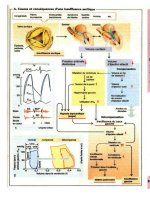
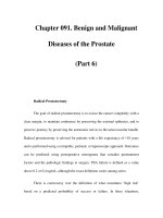
Nerves of the foot
May be involved in tarsal tunnel. Also, ganglion in tarsal tunnel may involve the
nerve.
The calcaneal nerve (pure sensory) originates at the point of the tarsal tunnel,
to innervate the medial part of the heel.
Both nerves pass through the tarsal tunnel, though the arch and sole of the foot.
Causes: trauma, tendon sheath cysts, Schwannomas, hypertrophy or fibrosis
of abductor hallucis muscle, sometimes from a discernible cause.
Fig. 49. Foot nerves.
1
Medial
plantar branch.
2
Lateral plan-
tar branch
Plantar nerves
(medial and lateral)
Calcaneal nerve
242
Isolated lateral plantar nerve lesion: occurs less frequently, from a foot
fracture or ankle sprain.
Entrapment of the first branch of the lateral plantar nerve has been described.
(Affects intrinsic foot muscles, and periosteum of calcaneus. Occurs in athletes
with heel pain).
Occurs at adjacent metatarsal bones before the division into two digital nerves.
Symptoms:
Radiating pain into one or two toes. Worse while standing and walking.
Sitting and removing shoes improves symptoms.
Often from fibrous nodules that are called “neuromas”.
Therapy:
Carbamazepine or other drugs used in neuropathic pain.
Electrocoagulation
Injections
Local anesthetic block
Pads
Shoes
Surgery
Diagnosis:
NCV, CT, MRI
This nerve crosses the first metatarsophalangeal joint on the medial side of the
big toe. Damage to the medial plantar proper digital nerve occurs where it
crosses the first metatarsophalangeal joint, or on the medial side of the big toe.
Symptoms:
Pain or paresthesias on the medial side of the big toe, especially when walking.
Often mild, but may also be disabling.
Sign:
Tinel’s at base of big toe.
Causes:
Acute blunt blows, lacerations,
Blunt trauma
Poor fitting shoes
Scars
Medial plantar proper digital nerve syndrome (Joplin’s neuroma)
Differential diagnosis: arthritis of big toe.
Marques WJ, Barreira AA (1996) Joplin’s neuroma. Muscle Nerve 19: 1361
Park TA (1996) Isolated inferior calcaneal neuropathy. Muscle Nerve 19: 106–108
Staal A, van Gijn J, Spaans F (2000) The tibial nerve. In: Staal A, van Gijn J, Spaans F (eds)
Mononeuropathies: examination, diagnosis and treatment. Saunders, London, pp 125–132
Interdigital nerves
(Morton’s
metatarsalgia)
Medial plantar proper
digital nerve
(Joplin’s neuroma)
References
243
Peripheral nerve tumors
Peripheral nerve tumors usually present with a slowly progressive mononeur-
opathy. Initially paresthesia, pain, followed by motor or sensory loss, or both
occur. The tumors may be seen, palpated or detected in imaging.
Mechanical factors (e.g. sitting , stretching the sciatic nerve, walking if tumor is
on the foot) can exacerbate pain or paresthesias. Patient’s often experience
anemia and weight loss.
Tumor can be palpated or a mass can be seen (e.g. supraclavicular fossa).
MRI can give a precise location. NCV and EMG can be used to assess the
functional impairment of the nerve lesion.
Metastasis of solid tumors into peripheral nerves are rare, but have been
described in lymphoma (particularly in neurolymphomatosis) and metastatic
cancer. Local involvement of peripheral nerves with either compression or
infiltration can be seen more frequently at the brachial plexus and sacral
plexus, also at a radicular level in association with metastatic vertebral column
disease.
Classification of peripheral nerve tumors: adapted from Birch 1993
Nerve sheath Schwannoma (neurolemmomas, Malignant
tumors neurinomas): (cellular, plexiform Schwannoma
and melanotic)
Neurofibroma: Solitary neuro- Neurofibrosarcoma
fibroma Plexiform neuro- (4–29% as a mani-
fibroma, fascicular spread festation of NF1)
through peripheral nerve tissue
Fibrolipoma Neuroepithelioma
Neuronal tumors Ganglioneuroma Ganglioneuroblastoma
Neuroblastoma
Schwannomas are the commonest benign nerve sheath tumors. They are
encapsulated and displace adjacent nerve fascicles. Schwannomas can present
as a painless, palpable mass on upper or lower extremities. A Tinel’s sign can
usually be elicited.
They can be divided into a) with association with Recklinghausen‘s disease and
b) without association with Recklingshausen disease.
a) Neurinomas and van Recklinghausen‘s disease: Neurofibromas occur in
cutaneous nerves and in larger nerves. The neurinomas in this patient group
have a 15% risk of malignant transformation.
Clinical development
Signs
Diagnosis
Metastasis
Schwannomas
Neurofibroma
244
b) Neurinomas occur on extremities. These are more likely to arise from the
motor portion of the nerve than from the sensory. They can occur as a
localized mass or involve longer nerve segments. Histologically they in-
volve the entire cross section of the nerve.
Other benign nonneuronal nerve sheath tumors are: desmoids, myoblastomas
and lymphangiomas, lipomas, lipohamartomas, hemangiomas, hemangioperi-
cytomas , arteriovenous fistulae, ganglions, end epidermoid cysts.
Localized hypertrophic mononeuropathy: is a slowly progressive mononeur-
opathy with little pain or numbness (may occur with NF1, or isolated). Any
nerve can be affected as well as nerve roots.
Malignant neural sheath tumors:
Consist of malignant Schwannomas, neurofibromas, usually termed as “sarco-
ma”. Malignant transformation of a benign nerve sheath cell tumor is more
likely in patients with von Recklinghausen’s disease. The tumors occur in long
nerves of the extremities and in the nerve plexus.
Other tumors of the neural crest:
Neuroblastoma
Ganglioneuroblastoma
Ganglioneuroma
Paraganglioma
Pheochromocytoma
Cranial nerves, nerve roots, the nerve plexus and single nerves can be affected
in cancer patients. The table gives an overview over the most frequently
affected nerves (Table 12).
Table 12. Involvement of peripheral nerves in cancer patients
Nerve Neoplastic Therapy-related Other causes
CN Base of skull meta
stasis
Toxicity of chemo- and
Leptomeningeal
carcinomatosis radiotherapy
Head and neck tumors
Axillary nerve Surgery, mastectomy,
neck dissection
Long thoracic nerve Mastectomy Inflammatory neuropathy
Radiotherapy
Phrenic nerve Lung cancer, lymphoma, Thoracic surgery Critical illness
thymoma thymectomy neuropathy in intesive care
patients and sepsis
Pectoral nerves Neck dissection
Musculocutaneus nerve Local metastasis Perioperative position
Peripheral nerve
involvement in cancer
patients
245
Table 12. Continued
Nerve Neoplastic Therapy-related Other causes
Cutaneous antebrachii Paravenous injection
medialis nerve
Median nerve Neurolymphomatosis
Amyloid deposition
Paraproteinemia
Ulnar nerve C8 lesion, Pancoast Radiotherapy
Tumor Malpostioning
Radial nerve Malpositioning, chemotherapy
(vincristine)
Truncal nerves Metastasis, local metastasis Operations Herpes Zoster
into vertebral column, Longterm steroid treatment
collapse of vertebral column with osteoporotic bone lesions
Iliohypogastric nerve Renal operations
Ilioinguinal nerve Abdominal surgery
Genitofemoral nerve Renal surgery
Cutaneus femoris Surgery radiotherapy
lateral nerve
Femoral nerve Local pelvic tumor, inguinal Surgery, anticoagulation,
tumor or lymph nodes radiotherapy
Obturator nerve
Metastasis, obturator
foramen Surgery pelvis
Gluteus medius Recurrence of local tumor
Sciatic nerve Metastasis, Foramen Intraarterial cytostatic Injections, malpositioning
piriforme
perfusion, radiotherapy
Tibial nerve Rarely affected: cauda
equina,
sacral plexus lesion
Peroneal nerve Local destruction of Malpositioning, cytostatic Paraneoplastic
vertebral column, meningeal drugs (vincristine) Cachexia
carcinomatosis Peroneal lesion may be
Compression of cauda equina part of sciatic nerve lesion
Osteolysis of capitulum fibulae
All local Intravenous
mononeuropathies Intraarterial perfusions
References
Basheer H, Rabia F, el-Hewl K (1997) Neurofibromas of digital nerves. J Hand Surg (Br) 22:
61–63
Birch B (1993) Peripheral nerve tumors. In: Dyck PJ, Thomas PK, Griffin JP, Low PA,
Poduslo JF (eds) Peripheral neuropathy. Saunders WB, Philadelphia, pp 1623–1640
Ferner RE, Lucas JD, O’Doherty MJO, et al (2000) Evaluation of 18 fluorodeoxyglucose
positron emission tomography (18 FDG PET) in the detection of malignant peripheral nerve
sheath tumours arising from within plexiform neurofibromas in neurofibromatosis 1. J
Neurol Neurosurg Psychiatry 68: 353–357
Foley KM, Woodruff M, Ellis FT (1980) Radiation induced malignant and atypical
peripheral nerve sheath tumors. Ann Neurol 7: 311–318
Gabet JY (1989) Amyloid pseudotumor of the sciatic nerve. Rev Neurol 145: 872–876
246
Gijtenbeek JMM, Gabreels-Festen AAWM, Lammens M, et al (2001) Mononeuropathy
multiplex as the initial manifestation of neurofibromatosis type 2. Neurology 56: 1766–
1768
Krücke W (1955) Erkrankungen der peripheren Nerven. In: Lubarsch O, Henke F, Rössle R
(Hrsg) Handbuch der speziellen pathologischen Anatomie und Histologie. Springer, Berlin,
S 1–248
Mitsumoto H (1992) Perineural cell hypertrophic mononeuropathy manifesting as CTS.
Muscle Nerve 15: 1364–1368
Roncaroli F, Poppi M, Riccioni L, et al (1997) Primary non Hodgkin’s lymphoma of the
sciatic nerve folowed by localization in the central nervous system. Neurosurgery 40: 618–
621
Tang JB, Ishii S, Usui M, et al (1990) Multifocal neurilemomas in different nerves of the
same upper extermity. J Hand Surg (Am) 15: 788–792
Thomas PK, King RHMT, Chiang TR, et al (1990) Neurofibromatous neuropathy. Muscle
Nerve 13: 93–101
Yassini PR, Sauter K, Schochet SS, et al (2000) Localized hypertrophic mononeuropathy
involving spinal roots and associated with sacral meningocele. Case report. J Neurosurg
79: 774–778
247
Polyneuropathies
249
Introduction
The peripheral nervous system (PNS) is defined as cell bodies or axons support-
ed by Schwann cells. The PNS includes the cranial nerves (except the second
cranial nerve), the dorsal root ganglia, the spinal nerve roots, the peripheral
nerve trunks, and peripheral nerves. The peripheral autonomic system also lies
within the PNS.
Peripheral neuropathy in its broadest definition encompasses any injury to
the PNS. More precise terminology describes the specific site of PNS injury.
Neuronopathies are direct injury to the cell bodies with a secondary axonal
loss. Axonopathies represent a primary insult to axons; axonopathies, particu-
larly when severe, can result in a secondary loss of cell bodies. A radiculopathy
Fig. 1. Common stocking and
glove distribution in polyneur-
opathies
250
is injury to spinal nerve roots while a plexopathy denotes injury in peripheral
nerves as they course through a plexus. Polyneuropathy, the main focus of this
chapter, refers to bilateral symmetrical injury to the peripheral nerves.
Polyneuropathy is commonly secondary to more generalized disease pro-
cesses including systemic, metabolic or rheumatological disorders, cancer,
vitamin deficiency states, exposure and/or ingestion of toxins and drugs, infec-
tions, immune reactions and inherited disorders of Schwann cell function.
Table 13 provides a more complete list of disorders that lead to polyneuropathy.
Multiple isolated peripheral nerve injuries, known as multiple mononeuropa-
thies or mononeuropathy multiplex, are also usually due to systemic disease. It
can be difficulty to distinguish near confluent mononeuropathy multiplex from
generalized polyneuropathy. In contrast, isolated peripheral nerve injury is
usually due to focal injury and is termed mononeuropathy. The mononeuropa-
thies are discussed in chapter mononeuropathy.
The most common polyneuropathy has a distal distribution with loss of
sensory function beginning in the toes. As the sensory loss progresses to mid
calf, the patient experiences sensation loss in the fingertips, resulting in the
classic stocking-glove distribution of
distal symmetric polyneuropathy
(Fig. 1)
.
Reflex changes parallel sensory disturbances with ankle reflexes being first
decreased then absent. Symptomatic distal motor nerve involvement is less
common and, when present, suggests specific underlying systemic disease
processes, particularly immune mediated and toxic neuropathies. Motor weak-
ness can occur in a proximal distribution, leading to a
proximal symmetric
polyneuropathy.
This pattern is also most commonly present in immune or
toxic neuropathies. A pure sensory
proximal symmetric polyneuropathy
is very
rare but can occur in acute intermittent porphyria. Another less common
distribution of symmetric polyneuropathies is with initial motor or sensory loss
in the arms. This can occur in immune mediated neuropathies, porphyria and
inherited disorders of the PNS.
Patients with polyneuropathy generally fall into two major classes: patients
with negative symptoms and patients with positive symptoms. This distinction
can be helpful to the clinician in both the diagnosis and care of the patient. As
the term suggests, patients with negative symptoms have painless loss of
sensory function or motor loss that does not perturb the patient’s functional
ability. Loss of sensation most commonly reflects loss of both large and small
nerve fibers. Patients with negative symptoms develop the insensate foot with
loss of vibratory perception and proprioception (large fiber) and light touch,
temperature and pain sensation (small fiber). Eighty five percent of patients with
diabetic polyneuropathy have no symptomatic complaints (i.e. negative senso-
ry symptoms). This group of patients however is at high risk for ulcer formation
because of their lack of pain sensation. In parallel negative motor symptoms,
particularly atrophy of distal foot musculature, can lead to foot deformities and
can also increase the risk of ulcers. Positive sensory symptoms can occur in
patients with polyneuropathy in the absence or presence of external stimuli. At
rest patients can experience painful parasthesias and/or frank pain. In response
to normal stimuli such as light touch, patients may develop symptoms of
hyperalgesia, dysesthesias or allodynia. Positive motor symptoms include
cramps, fasciculations and functional weakness.
In summary, this chapter discusses the main polyneuropathies encountered
by a physician in daily practice. It is not intended to be inclusive of all
Anatomical
distribution
Clinical syndrome
251
polyneuropathies but the disorders discussed should provide the clinician with
the knowledge required to diagnose and treat nearly all patients seen in an
outpatient clinic. The neuropathies will be discussed in the order outlined in
Table 13. Some key abbreviations used in this discussion include CMAP
(compound muscle action potential), SNAP (sensory nerve action potential),
and CSF (cerebrospinal fluid).
Table 13. Differential diagnosis of polyneuropathy
Metabolic Disease
Diabetic distal symmetric polyneuropathy
Diabetic autonomic neuropathy
Diabetic mononeuritis multiplex
Diabetic polyradiculopathy
Renal Disease
Systemic Disease
Systemic vasculitis
Non-systemic vasculitis
Paraproteinemia
Amyloidosis
Cancer
Neoplastic disease
Paraneoplastic disease
Motor neuron disease syndrome
Critical Illness
Infectious
Human Immunodeficiency Virus (HIV)
Hepatitis B
Lyme
Diphtheria
Leprosy
Syphylis
Parasites
Inflammatory
Acute motor axonal neuropathy
Acute motor and sensory axonal neuropathy
Acute inflammatory demyelinating polyradiculoneuropathy
Chronic inflammatory demyelinating polyradiculoneuropathy
Chronic demyelinating polyradiculoneuropathy with anti-MAG antibodies
Miller-Fisher Syndrome
Multifocal Motor Neuropathy
Nutritional
Cobalamin
Post-gastroplasty
Pyridoxine
Strachan’s syndrome
Thiamine
Tocopheral
Industrial Agents, Metals and Drugs
Industrial Agents
Acrylamide
Carbon Disulfide
Hexacarbons
Organophosphorous Agents
252
Table 13. Continued
Drugs
Alcohol
Amiodarone
Chloramphenicol
Colchicine
Dapsone
Disulfiram
Vinka alkaloids
Platinum
Taxol
Metals
Arsenic
Mercury
Thallium
Hereditary
Hereditary Autonomic and Sensory Neuropathy
Hereditary Motor Sensory Neuropathy (Charcot-Marie-Tooth Disease) Types 1, 2
Hereditary Neuropathy with Pressure Palsies
Porphyria
253
Diabetes is the most common cause of neuropathy in the Western World.
The 4 main peripheral nervous system complications of diabetes will be
discussed: distal symmetric polyneuropathy, autonomic neuropathy, mononeu-
ritis multiplex and the syndrome of plexopathy/polyradiculopathy that is fre-
quently termed amyotrophy.
Diabetic distal symmetric polyneuropathy
Genetic testing NCV/EMG Laboratory Imaging Biopsy
+++ +++ +
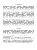
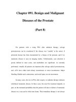
Fig. 2. Diabetic neuropathy. Pes
planus A, sensory loss may in-
duce osteous changes with col-
lapse of the small foot bones-
see X ray B
Metabolic diseases
254
Both large and small sensory and motor nerves are affected in diabetic distal
symmetric polyneuropathy (DPN). DPN is a length dependent neuropathy
affecting the feet first.
DPN is most commonly a slowly progressive disorder. A rapid onset can be
seen in newly diagnosed type 1 patients when rigorous glycemic control is
abruptly instituted. Equally common among men and women, 85% of patients
have an insensate foot with negative sensory and motor symptoms. Fifteen
percent of patients have positive symptoms with paresthesias, dysesthesias,
pain and muscle cramps. Patients with an insensate foot are at risk for foot
injury and ulceration.
DPN occurs in both type 1 and type 2 diabetic patients. The severity of DPN
correlates with the degree and duration of diabetes. After 25 years of diabetes,
at least 50% if not more of patients have DPN. Examination of the feet reveals
atrophic skin changes, callous and fissure formation (Fig. 2). Commonly all
sensory modalities are decreased in a stocking-glove pattern with loss of ankle
reflexes. Weakness is uncommon and present distally in only the most severe
cases. When sensation loss reaches the midcalf, early sensory loss is found in
the fingers.
The Diabetes Control and Complications Trials (DCCT) confirmed that hyper-
glycemia underlies the development of DPN. It is likely that the hyperglycemic
state disrupts both the normal metabolism and blood flow of peripheral nerves.
Laboratory:
HbA1C is frequently elevated. Serum proteins, vitamin levels, hepatic function
and serological markers of vasculitis should be normal. Frequently patients
have serologic evidence of mild renal dysfunction and measurable proteinuria.
Unless renal dysfunction is severe, the diabetic state itself, and not the second-
ary loss of renal function, is the primary cause of neuropathy.
Electrophysiology:
Early in neuropathy NCV reveal low normal or absent sural sensory responses
with mild decreases in peroneal motor conduction velocities. As the neuropa-
Anatomy/distribution
Symptoms
Clinical syndrome/
signs
Pathogenesis
Diagnosis
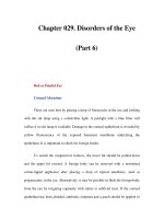
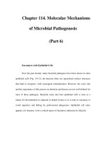
Fig. 3. Sural nerve biopsy from a
patient with diabetic neuropa-
thy and an asymptomatic con-
trol subject. A Normal sural
nerve showing an abundant and
normal distribution of myelinat-
ed fibers. B Sural nerve from a
patient with diabetes showing
severe loss of axons. C High
magnification view of B show-
ing loss of myelinated fibers,
splaying of myelin with early
onion bulb form formation
255
thy progresses, sensory amplitudes in the hand decline and there is evidence of
denervation by EMG in distal foot muscles.
Nerve Biopsy:
There is loss of large and small axons in the absence of inflammation with
thickening of blood vessel basement membrane (Fig. 3). Nerve biopsy is usually
not required for the diagnosis.
A systematic stepwise elimination of other common causes is required. See
Table 13.
DPN requires preventative and, in some cases, symptomatic therapy. Preventa-
tive therapy consists of optimal glycemic control coupled with daily foot
hygiene. The patient should inspect his feet each night and keep his feet clean
and dry. Painful DPN can be treated with gabapentin at doses up to 800 mg/
QID and amitryptiline or nortryptiline (25 to 150 mg/QHS). Please see the
review by Simmons (2002) for a complete approach to the treatment of painful
neurpathy.
Fifteen percent of patients with neuropathy develop an ulcer in their lifetime.
Prognosis is dependent on daily foot hygiene and care.
Feldman EL, Stevens MJ, Russell JW, et al (2001) Diabetic neuropathy. In: Becker KL (ed)
Principles and practice of endocrinology and metabolism, 3rd edn. Lippincott, Williams &
Wilkins, pp 1391–1399
Feldman EL, Stevens MJ, Russell JW, et al (2002) Somatosensory neuropathy. In: Porte D Jr,
Sherwin RS, Baron A (eds) Ellenberg and Rifkin’s diabetes mellitus, 6th edn. McGraw Hill,
pp 771–788
Simmons Z, Feldman EL (2002) Update on diabetic neuropathy. Curr Opin Neurol 15:
595–603
Windebank AJ, Feldman EL (2001) Diabetes and the nervous system. In: Aminoff MJ (ed)
Neurology and general medicine, 3rd edn. Churchill Livingstone, pp 341–364
Differential diagnosis
Therapy
Prognosis
References
256
Both sympathetic and parasympathetic fibers are affected in diabetic autonom-
ic neuropathy (DAN). Like DPN, DAN is a length dependent neuropathy with
loss of autonomic function that can vary from mild to severe.
Mild subclinical DAN is common and occurs in patients with DPN. Symptom-
atic DPN can vary from mild to severe. Cardiac symptoms include fixed
tachycardia, orthostatic/postprandial hypotension, arrhythmias, and in severe
cases, sudden cardiac death. Gastrointestinal symptoms include constipation,
nightime diarrhea and gastroparesis with early satiety, nausea and vomiting.
Genitourinary symptoms are common in men, with impotence present in
nearly all males after 25 years of diabetes. Urinary retention occurs in men and
women. Abnormal pupillary responses and abnormal sweating occurs, with
anhydrosis of the feet and hands, and gustatory sweating in more severe cases.
Abnormal neuroendocrine responses likely contribute to hypoglycemic un-
awareness in type 1 patients.
Symptomatic DAN is more common in type 1 patients, although subclinical
DAN (diagnosed by cardiovascular testing) is common in type 2 patients. The
signs in DAN parallel the symptoms. Patients have an abnormal heart rate, poor
cardiac beat to beat variation, orthostasis, weight loss from gastroparesis,
urinary tract infections from urinary retention, poor pupillary responses and
absent sweating.
Like DPN, it is generally held that hyperglycemia underlies the development of
DAN. It is likely that the hyperglycemic state disrupts both the normal metab-
olism and blood flow of autonomic ganglia and nerves.
Laboratory:
As with DPN.
Electrophysiology:
Standard measures of cardiac autonomic function are required for the diagnosis
and include measures of heart rate (R) variability conducted in the supine
position with the patient breathing at a fixed rate of 6 breaths per minute during
a 6 minute period. The maximum and minimum R-R intervals during each
breathing cycle are measured and converted to beats a minute. The 30: 15 ratio
Diabetic autonomic neuropathy
Anatomy/distribution
Genetic testing NCV/EMG Laboratory Imaging Biopsy
++ ++
Symptoms
Clinical syndrome/
signs
Pathogenesis
Diagnosis
257
is calculated for patients. The heart rate response is determined on changing
from the lying to standing position. The shortest R-R interval around the 15th
beat and the longest R-R interval around the 30th beat upon standing is
measured to calculate the ratio. Orthostatic hypotension is measured. Patients
can also undergo a bladder cystoscopy, gastroesophageal manometry, sweat
testing and an eye exam.
Imaging:
Positron emission tomography (PET) quantitates sympathetic cardiac innerva-
tion and is an excellent measure of left ventricular function.
Biospy:
None.
It is essential to exclude atherosclerotic heart disease, primary gastrointestinal
disease such as peptic ulcer disease or colitis, bladder or urinary tract anatom-
ical abnormalities leading to retention (in males, consider prostatism) and drug
induced changes in pupils and sweating.
Like DPN, therapy is preventive and symptomatic. Preventive therapy is based
on optimal glycemic control. Symptomatic treatment is targeted toward the
symptom i.e. hydration and support stockings for orthostasis with extreme cases
requiring midodrine 5 mg/TID. Therapy is discussed in detail in Vinik (2002).
Like DPN, DAN usually progresses slowly over years, with a patient becoming
more symptomatic. It is estimated that sudden cardiac death due to DAN
occurs in 1–2% of all type 1 diabetic patients.
Feldman EL, Stevens MJ, Russell JW (2002) Diabetic peripheral and autonomic neuropathy.
In: Sperling MA (ed) Contemporary endocrinology: type 1 diabetes: etiology and treatment.
Humana Press, pp 437–461
Vinik AI, Erbas T, Pfeifer MA, et al (2002) Diabetic autonomic neuropathy. In: Porte Jr D,
Sherwin RS, Baron A (eds) Ellenberg and Rifkin’s diabetes mellitus, 6th edition. McGraw
Hill, pp 789–804
Differential diagnosis
Therapy
Prognosis
References
258
Diabetic mononeuritis multiplex (DMM) and diabetic polyradiculopathy (DPR)
are due to the loss of motor and sensory axons in one or more named nerves or
nerve roots. The term mononeuritis multiplex refers to multiple mononeuro-
pathies in conjunction with polyneuropathy.
Patients experience proximal and distal weakness and sensory loss in specific
named peripheral nerves (including cranial or truncal nerves) or nerve roots.
The onset is sudden and usually extremely painful in the sensory distribution of
the nerve/nerve root. In DMM, the most commonly involved named nerves
include the median, radial and femoral nerve and cranial nerve III. In DPR,
thoracic and high lumbar nerve roots are frequently affected, initially unilater-
ally, but frequently with later bilateral involvement.
DMM and DPR are sudden in onset, often self-limited, and occur primarily in
older, poorly controlled type 2 patients. In DMM, patients experience sudden
pain, weakness and sensory loss in a named peripheral nerve. Patients with
DMM of cranial nerve III, present with unilateral pain, diplopia, and ptosis with
pupillary sparing. In DPR, involvement of thoracic nerve roots presents as
band-like abdominal pain that is often misdiagnosed as an acute intraabdomi-
nal emergency. L2-L4 DPR is often confused with a pure femoral neuropathy;
the former is common while the later is rare. Patients are weak in hip flexion
and knee extension with an absent knee reflex; frequently weakness will spread
to involve L5-S1 anterior myotomes.
Unlike DPN or DAN, DMM and DPR are due to discreet infarcts in nerves due
to vascular occlusions. Epineural vessels are inflamed with IgM and comple-
ment deposition.
Laboratory:
It is essential to exclude vasculitis by appropriate serological screening (see
p. 262).
Electrophysiology:
NCV reveals loss of sensory and in advanced cases motor amplitude and mildly
slowed conduction velocities in distinct nerves. EMG reveals denervation in
myotomes corresponding with the named nerves.
Diabetic mononeuritis multiplex and diabetic polyradiculopathy
(amyotrophy)
Anatomy/distribution
Genetic testing NCV/EMG Laboratory Imaging Biopsy
++ ++ ++
Symptoms
Clinical syndrome/
signs
Pathogenesis
Diagnosis
259
Imaging:
Cranial aneurysm should be excluded in cranial nerve III palsies by cranial
MRI. Abdominal and lumbosacral plexus CAT scans are routine to rule out
intraabdominal pathology in patients with diabetic thoracic radiculopathy and
a mass lesion in the lumbosacral plexus in patients with diabetic lumbar
polyradiculopathy.
Biospy:
None.
Patients usual require aggressive pain management. Glycemic control is essen-
tial to prevent reoccurrence. Physical therapy and supportive care help accel-
erate recovery. There are reports of using intravenous gammaglobulin (IVIG) in
DPR, but efficacy remains unproven.
DMM and DPR improve spontaneously in most cases, but may leave mild
residual deficits. It is essential to achieve improved glycemic control in affected
patients; if not, it is highly likely that the patient will experience recurrent
episodes.
Dyck JB, Norell JE, Dyck PJ (1999) Microvasculitis and ischemia in diabetic lumbosacral
radiculoplexus neuropathy. Neurology 53: 2113–2121
Feldman EL, Stevens MJ, Russell JW, Greene DA (2001) Diabetic neuropathy. In: Becker KL
(ed) Principles and practice of endocrinology and metabolism, 3rd edition. Lippincott,
Williams & Wilkins, pp 1391–1399
Simmons Z, Feldman EL (2002) Update on diabetic neuropathy. Curr Opin Neurol 15:
595–603
Windebank AJ, Feldman EL (2001) Diabetes and the nervous system. In: Aminoff MJ (ed)
Neurology and general medicine, 3rd edition. Churchill Livingstone, pp 341–364
Therapy
Prognosis
References
260
Both large and small sensory and motor nerves are affected in distal symmetric
polyneuropathy due to renal disease. Like DPN, this is a length dependent
neuropathy.
This is most commonly a slowly progressive disorder. Patients present with
pain, dyesthesias, sensory loss, muscle cramps, restless legs and, in more
advanced cases, leg weakness.
This neuropathy commonly occurs in patients with end-stage renal disease on
dialysis; 60% of patients on dialysis have some degree of neuropathy. Neuro-
pathy secondary to renal disease is 2 times more common in men. Examination
reveals a symmetric stocking-glove loss to all sensory modalities with distal
weakness, absent ankle and depressed knee reflexes.
While the definitive cause is unknown, the neuropathy may be due to accumu-
lation of metabolites or loss of unknown renal factors.
Laboratory:
Serum BUN and Cr and 24 hour urine collection all indicate renal failure.
Electrophysiology:
Early in neuropathy there are prolonged distal latencies, slowed motor conduc-
tion velocities and prolonged F waves. The relationship between conduction
slowing and renal failure is well established. Lowered sensory and motor
amplitudes are present, and in severe cases, are absent. There is evidence of
denervation by EMG in distal foot muscles.
Imaging:
None.
Nerve Biopsy:
There is evidence of axonal degeneration, with loss of large and small axons in
the absence of inflammation. Nerve biopsy is usually not required for the
diagnosis.
Diabetes and other drugs, such as colchicine, may mimic or exacerbate the
neuropathy.
Distal symmetric polyneuropathy of renal disease
Genetic testing NCV/EMG Laboratory Imaging Biopsy
++ ++
Anatomy/distribution
Symptoms
Clinical syndrome/
signs
Pathogenesis
Diagnosis
Differential diagnosis
261
Therapy consists of pain management and physical therapy. Optimizing renal
function may improve the neuropathy.
The neuropathy progresses over a period of months and is rarely fulminant.
Prognosis is improved following renal transplant, and sometimes with more
intensive dialysis.
Burns DJ, Bate D (1998) Neurology and the kidney. J Neurol Neurosurg Psychiatry 65:
810–821
Reference
Therapy
Prognosis
262
Systemic disease
Vasculitic neuropathy, systemic
Genetic testing NCV/EMG Laboratory Imaging Biopsy
++ ++ ++
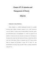
Fig. 4. Sural nerve biopsy from a
patient with isolated peripheral
nerve vasculitis. A Infiltration of
a perineurial vessel wall by mul-
tiple inflammatory cells includ-
ing lymphocytes and macroph-
ages (black arrows). There is
also evidence of pink fibrin de-
posits consistent with the pres-
ence of fibrinoid necrosis. B
Teased fiber preparations show-
ing multiple axon balls (white
arrows) and evidence of empty
strands consistent with axonal
degeneration
Fig. 5. Dorsal root ganglion bi-
opsy from a patient with severe
sensory ataxia due to dorsal root
ganglionitis. There are clusters
of inflammatory cells (white ar-
rows) surrounding the dorsal
root ganglion neurons (black ar-
rows). Many of the neurons
show evidence of degeneration