Atlas of Neuromuscular Diseases - part 10 docx
Bạn đang xem bản rút gọn của tài liệu. Xem và tải ngay bản đầy đủ của tài liệu tại đây (379.37 KB, 45 trang )
425
This is variable and depends on the specific systemic disorder, however proxi-
mal muscles are most usually affected.
This is variable depending on the specific cause of myopathy. Most of these
myopathies progress slowly, although rapid progression of symptoms may be
observed with thyrotoxicosis. If treated most endocrine related myopathies are
self limiting. Myopathies related to paraneoplastic disorders are usually not
treatable.
Any age although most are observed in adults. Paraneoplastic related myopa-
thies are more common in older patients.
This disorder may be associated with a painful myopathy that can simulate
polymyalgia or polymyositis. In severely hypothyroid children a syndrome
characterized by weakness, slow movements, and striking muscle hypertrophy
may be observed. Percussion myotonia and myoedema may be observed in
patients with hypothyroidism.
Myopathies associated with endocrine/metabolic disorders
and carcinoma
Distribution/anatomy
Onset/age
Clinical syndrome
Hypothyroidism
Time course
Genetic testing NCV/EMG Laboratory Imaging Biopsy
– ++ +++ + +++
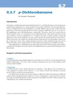
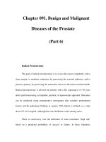
Fig. 32. Muscle from a patient
with diabetes mellitus showing
myolysis with degenerating fi-
bers (arrow heads)
426
Thyrotoxicosis is associated with muscle atrophy and weakness. It may also be
associated with a progressive extraocular muscle weakness, ptosis, periodic
paralysis, myasthenia gravis, spastic paraparesis and bulbar palsy. Subjects may
have brisk reflexes and fasciculations similar to amyotrophic lateral sclerosis.
Affected patients may have tetany, muscle spasm, and occasionally weakness.
Patients may have proximal weakness, muscle atrophy, hyperreflexia, and
fasciculations.
Occasionally muscle atrophy and weakness may be observed under conditions
of hypercortisolemia.
The muscles may appear enlarged, however this disorder is usually associated
with mild proximal upper or lower extremity muscle weakness.
Diabetes is not associated with a generalized myopathy, however muscle
necrosis or inflammation may occur in diabetic amyotrophy. In Flier’s syn-
drome, there is muscle pain, cramps, fatigue, acanthosis nigricans and pro-
gressing enlargement of the hands and feet, and impaired glucose tolerance.
Hypoglycemia may be associated with muscle atrophy as part of a motor
neuron type syndrome. It does not produce primary myopathy.
In chronic renal failure patients may have proximal weakness and in addition
myoglobinuria may occur.
This may be seen as part of an inflammatory myopathy
,
may also be observed
in carcinoid syndrome, or may occur due to a metabolic disturbance. Direct
invasion of muscle is rare although it may be observed with leukemias and
lymphomas.
The pathogenesis depends on the specific muscle disorders indicated above.
Laboratory:
A variety of electrolyte and endocrine changes support the diagnosis as indicat-
ed under the specific disease. The CK may be normal or significantly elevated
e.g. in diabetic muscle infarction or with hypothyroidism.
Electrophysiology:
The EMG is dependent on the specific disorder, but in general there is evidence
of myopathic changes in affected muscles.
Imaging:
Muscle imaging may be of value.
Muscle biopsy:
In both hypo and hyperthyroidism the muscle biopsy is often normal, although
there may be evidence of mild fiber atrophy. In hyperparathyroidism and
acromegaly there may be mild type 2 fiber atrophy. Evidence of inflammation
and muscle infarction may be observed in affected muscle in diabetic amyotro-
phy. Muscle destruction following rhabdomyolysis may also be seen in this
condition (Fig. 32). Inflammatory changes may be observed in carcinomatous
myopathy, or as part of a paraneoplastic syndrome.
Acromegaly
Hyperthyroidism
Hypoparathyroidism
Hyperparathyroidism
Cushing syndrome and
corticosteroid atrophy
Diabetes
Uremia and myopathy
Carcinomatous myopathy
Pathogenesis
Diagnosis
427
This is wide and includes the different causes of metabolic and systemic disease
associated with myopathy. In addition the inflammatory myopathies e.g. PM,
DERM, and IBM may resemble these disorders. Lambert-Eaton myasthenic
syndrome (LEMS) may mimic a paraneoplastic myopathy. Type 2 fiber atrophy
due to any cause may mimic a metabolic myopathy.
The therapy of the underlying systemic disease often leads to improvement of
the myopathy.
This is dependent on the specific disorder, but if appropriate therapy is institut-
ed the prognosis is usually good for the endocrine disorders such as hypothy-
roidism, hyperthyroidism, hyperparathyroidism, acromegaly, and diabetes.
Dyck PJ, Windebank AJ (2002) Diabetic and nondiabetic lumbosacral radiculoplexus
neuropathies: new insights into pathophysiology and treatment. Muscle Nerve 25: 477–
491
Horak HA, Pourmand R (2000) Endocrine myopathies. Neurol Clin 18: 203–213
Madariaga MG (2002) Polymyositis-like syndrome in hypothyroidism: review of cases
reported over the past twenty-five years. Thyroid 12: 331–336
Differential diagnosis
Therapy
Prognosis
References
428
Myotonia congenita
Genetic testing NCV/EMG Laboratory Imaging Biopsy
++ +++ – – +
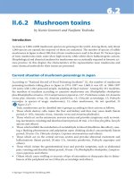
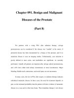
Fig. 33. Myotonia congenita. A
Muscle myotonia in the hypoth-
enar muscles. B Myotonic dis-
charges in the EMG from affect-
ed muscle
Fig. 34. Thomson’s myotonia
congenita. A Increased muscle
bulk in the arms and chest in a
patient with Thomson’s disease.
B Hypertrophy of the extensor
digitorum brevis muscle
429
Variable, may affect both limb and facial muscles.
Progresses very slowly over a lifetime. Usually strength is spared.
– Myotonia congenita (Thomsen): onset in infancy.
– Myotonia congenita (Becker): onset is usually in early childhood.
Myotonia is usually mild, approximately 50% may have percussion myotonia.
The myotonia (Fig. 33) is associated with fluctuations, and may be worsened by
cold, hunger, fatigue and emotional upset. Muscle hypertrophy is seen in many
patients (Fig. 34), and occasionally patients may complain of myalgias. Patients
may report a “warm-up” phenomenon, in which the myotonia decreases after
repeated activity. Muscle strength is usually normal.
Patients may also have a “warm-up” phenomenon. The disease is more severe
than Thomsen’s, and although strength is usually normal in childhood, there is
often mild distal weakness in older individuals. Strength often deteriorates after
short periods of exercise. Hypertrophy may also be observed in the leg muscles,
although it is less common than in Thomsen’s disease.
Mild myotonia occurring late in life, with less muscle hypertrophy.
Thomsen’s disease is due to a defect of the muscle chloride channel (CLCN1).
Thomsen’s disease is an autosomal dominant disorder, with the gene abnormal-
ity localized on chromosome 7q35. The mutation interferes with the normal
tetramer formation on the chloride channel. Chloride conductance through the
channel is eliminated or reduced. Normal chloride conduction is necessary to
stabilize the membrane potential. Without chloride conductance there is in-
creased cation conductance after depolarization, and spontaneous triggering of
action potentials. In missense mutations of the chloride channel there is a
partial defect in normal conductance of chloride. In contrast, with frame shift
mutations there is complete loss of chloride conductance. In Becker’s disease
there is likewise a defect of the muscle chloride channel (CLCN1), with a
recessive mode of inheritance linked to chromosome 7q35. A variety of genetic
defects have been described including more than 20 missense mutations, and
deletions. Depending on the type of mutation there may be low or reduced
opening of chloride channels, or there may be chloride efflux but not influx. A
final type of congenital myotonia, myotonia levior, is autosomal dominant and
again is related to a mutation of the CLCN1 channel.
Laboratory:
Laboratory tests are generally of limited value. CK is usually normal.
Electrophysiology:
90% of subjects with congenital myotonia will have electrophysiological evi-
dence of myotonia (Fig. 33B). The myotonia is present even in early childhood,
and is greater in distal than in proximal muscles. MUAPs are usually normal,
and there is no evidence of myopathic discharges on EMG. With repetitive
stimulation a decrement may be observed, especially at high stimulation
Distribution/anatomy
Clinical syndrome
Myotonia congenita
(Thomsen)
Myotonia congenita
(Becker)
Myotonia levior
Time course
Onset/age
Diagnosis
Pathogenesis
430
frequencies in excess of 25 Hz. Cooling does not affect the nerve response. In
Becker’s disease there may be a “warm-up” effect with less myotonia after
maximal contraction, and unlike Thomsen’s there may be occasional small,
short duration MUAPs.
Genetic testing:
Testing for mutations of the CLCN1 gene may be diagnostically useful.
Muscle biopsy:
Muscle biopsy findings are variable, and are not specific for the diagnosis.
Myopathic changes are more likely with Becker’s, which is a more severe form
of myotonia than Thomsen’s disease. In more severe cases there may be
increased fiber diameter variation, internalization of nuclei, and vacuolation.
– Paramyotonia
– Hyperkalemic periodic paralysis
– Hypokalemic periodic paralysis
– Mild DM1 or DM2
The following medications may help with symptoms, and control of myotonia:
quinine (200 to 1200 mg/d), mexiletine (150 to 1000 mg/d), dilantin (300 to
400 mg/d), procainamide (125 to 1000 mg/d), tocainide, carbamazepine, ace-
tazolamide (125 to 1000 mg/d). Procainamide is rarely used because of con-
cerns with bone marrow suppression. Several medications should be avoided
in these patients including depolarizing muscle relaxants, and β2 agonists.
The prognosis for Thomson’s disease is good, with mild progression over many
years. Patients with Becker’s myotonic dystrophy may develop more significant
weakness later in life.
George AL Jr, Crackower MA, Abdalla JA, et al (1993) Molecular basis of Thomsen’s disease
(autosomal dominant myotonia congenita). Nat Genet 3: 305–310
Jentsch TJ, Stein V, Weinreich F, et al (2002) Molecular structure and physiological function
of chloride channels. Physiol Rev 82: 503–568
Ptacek LJ, Tawil R, Griggs RC, et al (1993) Sodium channel mutations in acetazolamide-
responsive myotonia congenita, paramyotonia congenita, and hyperkalemic periodic
paralysis. Neurology 44: 1500–1503
Wu FF, Ryan A, Devaney J, et al (2002) Novel CLCN1 mutations with unique clinical and
electrophysiological consequences. Brain 125: 2392–2407
Differential diagnosis
Therapy
Prognosis
References
431
Many patients who have myotonia have only minimal or no symptoms. In more
severely affected subjects myotonia may affect both proximal and distal mus-
cles.
Many subjects are asymptomatic. In those who develop symptoms the condi-
tion either remains stable or only slowly progresses.
The disorder may present at any age, most commonly in late adolescence.
Weakness develops in late adolescence, although myotonia may present in
infancy.
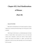
Patients may develop weakness or stiffness, which may be coupled with
myotonia. Myotonia is often worse with cold and exercise and may affect the
face, neck and upper extremities (Fig. 35). Episodic weakness may occur after
exercise, cold exposure, or may occur spontaneously. The weakness usually
lasts for a few minutes but may extend to several days. In some patients
weakness may be worse after potassium load, or may be exacerbated by
hyperthyroidism. Myotonia is usually paradoxical in that it worsens with exer-
cise, in comparison to that observed in myotonia congenita.
Paramyotonia congenita is an autosomal dominant disorder associated with a
gain of function mutation of the SCN4A gene on chromosome 17q23. At least
eleven missense mutations have been described.
Genetic testing NCV/EMG Laboratory Imaging Biopsy
++ +++ – – +
Paramyotonia congenita
Distribution/anatomy
Time course
Onset/age
Clinical syndrome
Pathogenesis
Fig. 35. Myotonia of the hand in
a patient with cold induced my-
otonia (Von Eulenburg’s dis-
ease). The patient is trying to
open his hand
432
Laboratory:
Laboratory studies are usually normal.
Electrophysiology:
With cooling of the muscle there is a decrease in the CMAP amplitude and with
prolonged cooling it may disappear entirely. The amplitude usually recovers
with warming. With cooling, the myotonia on EMG may initially worsen, but
with prolonged cooling there is usually depolarization and paralysis, and the
mytonia disappears.
Genetic testing:
Testing for mutations of the SCN4A gene.
Muscle biopsy:
Muscle biopsy may be unremarkable with occasional central nuclei with
hypertrophic, split, rare atrophic, or regenerating fibers. In some areas there
may be focal myofibril degeneration, with lipid deposits, myelin bodies, and
subsarcolemmal vacuoles.
– Myotonia congenita
– Myotonia fluctuans
– Myotonia permanens
– Acetazolamide responsive myotonia
– Hyperkalemic periodic paralysis
Several medications may be helpful in decreasing the symptoms in paramyoto-
nia. These include mexiletine 150–1000 mg/d, acetazolamide 125–1000 mg/d,
dichlorphenamide 50–150 mg/d. Tocainide may help some patients, however
there is a concern about myelosuppression.
Prognosis in paramyotonia congenita is usually good.
Bendahhou S, Cummins TR, Kwiecinski H, et al (1999) Characterization of a new sodium
channel mutation at arginine 1448 associated with moderate Paramyotonia congenita in
humans. J Physiol 518: 337–344
Chahine M, George AL Jr, Zhou M, et al (1994) Sodium channel mutations in paramyotonia
congenita uncouple inactivation from activation. Neuron 12: 281–294
Ptacek LJ, Tawil R, Griggs RC, et al (1994) Sodium channel mutations in acetazolamide-
responsive myotonia congenita, paramyotonia congenita, and hyperkalemic periodic
paralysis. Neurology 44: 1500–1503
Diagnosis
Differential diagnosis
Therapy
Prognosis
References
433
This from of periodic paralysis usually affects proximal muscles and is symmet-
ric. Occasionally distal muscles may be affected, or the disease may occur
asymmetrically in excessively exercised muscles.
Usually progresses slowly over several decades.
Onset is usually in the first decade.
Hyperkalemic periodic paralysis is characterized by flaccid, episodic weakness.
The disorder frequently occurs in the early morning before eating, and may also
be associated with rest after exercise. Episodes last up to 60 minutes on average,
however occasionally the flaccid episodic weakness may last for hours or even
days. The weakness is provoked by exercise, potassium loading, pregnancy,
ingestion of glucocorticoids, stress, fasting, and ethanol use. The episodes of
weakness may be relieved by carbohydrate intake or by mild exercise.
Hyperkalemic periodic paralysis is an autosomal dominant disorder of the
sodium channel subunit SCN4A localized to chromosome 17q35. In hyper-
kalemic periodic paralysis there is a gain-of-function of the sodium channel,
resulting from one or more of seven missense mutations. There is also uncon-
trolled repetive firing of action potentials due to a non-inactivating Na+ inward
current.
Laboratory:
Patients often have an elevated serum K
+
greater than 4.5 mEq/l and a high
urinary potassium. The serum CK is usually normal or mildly elevated.
Electrophysiology:
The CMAP amplitude increases immediately after 5 minutes of sustained
exercise, and reduces by 40% or greater during rest following the exercise. In
the form with myotonia, the EMG shows trains of positive sharp waves,
fibrillation potentials, and myotonic discharges between attacks. The motor
unit potentials are usually normal.
Muscle biopsy:
Tubular aggregates may be observed in muscle fibers, along with dilatations of
the sarcoplasmic reticulum. Vacuolation may be observed, and usually vacu-
oles contain amorphous material surrounded by glycogen granules.
Hyperkalemic periodic paralysis
Genetic testing NCV/EMG Laboratory Imaging Biopsy
+++ ++ +++ – ++
Distribution/anatomy
Time course
Onset/age
Clinical syndrome
Pathogenesis
Diagnosis
434
Provocative test:
An oral potassium load administered in a fasting patient in the morning after
exercise may induce weakness. The study should only be done if renal and
cardiac function, and the serum potassium are normal. The patient is given
0.05g/kg KCl in a sugar free liquid over 3 minutes. The patient’s electrolytes,
EKG and strength are monitored every 20 minutes. Weakness typically occurs
in 1 to 2 hours. If the test is negative, a higher dose of KCl up to 0.15
g/kg may be required. An exercise test may also induce hyperkalemic paralysis.
The subject works out for 30 minutes, increasing their pulse rate beyond 120
beats per minute. They are then rested and the serum potassium is measured.
Normally potassium will rise during exercise and then fall to near pre-exercise
levels. In hyperkalemic periodic paralysis there is a second hyperkalemic
period with associated paralysis that occurs approximately 15 to 20 minutes
after exercise.
– Paramyotonia
– Hypokalemic periodic paralysis
– Acetazolamide responsive myotonia congenita
– Myotonia permanens
– Myotonia fluctuans
– Normokalemic periodic paralysis
– Andersen’s syndrome
In Andersen’s syndrome there is a potassium sensitive periodic paralysis with
cardiac dysrhythmias and dysmorphic features. Acetazolamide-responsive my-
otonia congenita is an autosomal dominant sodium channel defect in which
there is muscle hypertrophy, and “paradoxical” myotonia. The disorder is
associated with muscle pain and stiffness, is aggravated by potassium, and
improved by acetazolamide. It is not associated with weakness. Myotonia
permanens is a sodium channel defect associated with severe continuous
myotonia that may interfere with breathing. There is usually marked muscle
hypertrophy in this disorder. Myotonia fluctuans is an autosomal dominant
defect of the SCN4A subunit of the muscle sodium channel. In this disorder
there is mild myotonia that varies in severity. Stiffness develops during rest
approximately 30 minutes after exercise and may last for up to 60 minutes.
Stiffness is worsened by potassium, or depolarizing agents. The stiffness may
interfere with respiration if there is no weakness or cold sensitivity.
In hyperkalemic periodic paralysis, many of the attacks are short lived and do
not require treatment. During an acute attack, carbohydrate ingestion may
improve the weakness. Use of acetazolamide or thiazide diuretics may help
prevent further attacks. Mexiletine is of no benefit in hyperkalemic periodic
paralysis.
This is variable, with most patients having a fairly good prognosis. One muta-
tion (T704M) is associated with severe myopathy and permanent weakness.
Fontaine B, Khurana TS, Hoffman EP,
et al
(1990) Hyperkalemic periodic paralysis and the adult
muscle sodium channel alpha subunit gene. Science 250: 1000–1002
Differential diagnosis
Therapy
Prognosis
References
435
Ptacek LJ, George AL Jr, Griggs RC, et al (1991) Identification of a mutation in the gene
causing hyperkalemic periodic paralysis. Cell 67: 1021–1027
Rojas CV, Neely A, Velasco-Loyden G, et al (1999) Hyperkalemic periodic paralysis
M1592V mutation modifies activation in human skeletal muscle Na+ channel. Am J
Physiol 276: C259–266
Wagner S, Lerche H, Mitrovic N, et al (1997) A novel sodium channel mutation causing a
hyperkalemic paralytic and paramyotonic syndrome with variable clinical expressivity.
Neurology 49: 1018–1025
436
Hypokalemic periodic paralysis may affect both proximal and distal muscles,
although proximal muscles are often more severely affected.
The disorder gradually worsens over many years.
Onset usually as a teenager.
Hypokalemic periodic paralysis is associated with acute episodes of flaccid
weakness. In contrast to hyperkalemic periodic paralysis, the hypokalemic
variant is associated with less frequent attacks, although the attacks are often
longer and more severe than in the hyperkalemic variant. Hypokalemic period-
ic paralysis also is associated with a higher rate of degenerative myopathy and
disabling weakness in the limbs. It is not associated with myotonia. The
disorder is evoked by glucose ingestion, and improved by potassium intake.
Hypokalemic periodic paralysis is inherited as an autosomal dominant disor-
der. The disease may be associated with a defect in several genes. These
include a loss of function mutation of the calcium channel α-1 subunit on
chromosome 1q42 (CACNA1S), a loss of function mutation of the sodium
channel α subunit on chromosome 17q23 (SCN4A), and a loss of function
mutation of the KCNE3 gene coding for the potassium channel b subunit
(MiRP2) on chromosome 11q13-14. The defects in CACNA1S, SCN4A, and
KCNE3 are associated with a variety of missense mutations. The mutations of
the CACNA1S gene are the most frequent.
Laboratory:
Calcium levels are usually low to low normal. CK levels are usually normal, but
may be increased during attacks.
Electrophysiology:
CMAP amplitudes are decreased during attacks, and increased immediately
after sustained (5 min) maximal contraction between attacks. In most affected
subjects, there is then a progressive reduction in the CMAP amplitude during
rest 20 to 40 min after the initial increment. An infusion of glucose and insulin
may provoke the symptoms, but needs to be used with EKG monitoring. During
an attack there is an increase in insertional activity, and an increase in short
duration, polyphasic motor unit potentials that disappear as the muscle be-
comes paralyzed. In most subjects the needle EMG is normal between attacks.
Hypokalemic periodic paralysis
Genetic testing NCV/EMG Laboratory Imaging Biopsy
+++ ++ +++ – ++
Distribution/anatomy
Time course
Onset/age
Clinical syndrome
Pathogenesis
Diagnosis
437
Differential diagnosis
Therapy
Prognosis
References
Genetic testing:
Testing for SCN4A, CACNA1S, l KCNE3 mutations may be useful in individual
cases.
Muscle biopsy:
Clear central vacuoles are observed, along with tubular aggregates. In addition,
there may be myopathic changes including variation in muscle size, split fibers,
and internalized nuclei. There is vacuolar dilation of the sarcoplasmic reticu-
lum during attacks.
– Thyrotoxic periodic paralysis
– Hyperkalemic periodic paralysis
– Myotonia fluctuans
Potassium supplementation of 40 to 80 mEq 2–3 times per day will often
decrease the severity of the attacks. Acetazolamide sustained release tablets
(500–2000 mg/d) or dichlorphenamide (50–150 mg/d) may reduce the frequen-
cy of the attacks. Use of potassium sparing diuretics (triamterene or spironolac-
tone) in combination with acetazolamide or dichlorphenamide may also re-
duce the frequency of periodic paralysis.
With appropriate treatment the prognosis is usually good.
Cannon SC (2002) An expanding view for the molecular basis of familial periodic paralysis.
Neuromuscul Disord 12: 533–543
Davies NP, Eunson LH, Samuel M, et al (2001) Sodium channel gene mutations in
hypokalemic periodic paralysis: an uncommon cause in the UK. Neurology 57: 1323–
1325
Dias da Silva MR, Cerutti JM, Tengan CH, et al (2002) Mutations linked to familial
hypokalaemic periodic paralysis in the calcium channel alpha1 subunit gene (Cav1.1) are
not associated with thyrotoxic hypokalaemic periodic paralysis. Clin Endocrinol (Oxf) 56:
367–375
Lehmann-Horn F, Jurkat-Rott K, Rudel R (2002) Periodic paralysis: understanding channel-
opathies. Curr Neurol Neurosci Rep 2: 61–69
Moxley III RT (2000) Channelopathies. Curr Treat Options Neurol 2: 31–47
439
Motor neuron disease
441
Amyotrophic lateral sclerosis (ALS) causes the loss of both upper and lower
motor neurons. On autopsy, there is loss of the pyramidal cells of the motor
cortex, with atrophy of the brainstem and spinal cord. The corticospinal tracts
are degenerated and gliotic. The ventral nerve roots are atrophied, and there is
microscopic evidence of muscle denervation and reinnervation.
ALS usually presents with painless and progressive weakness of a focal distribu-
tion that over time spreads to contiguous muscle groups. As the disease
progresses, fasciculations cause muscle cramps and the patient becomes spas-
tic. Spontaneous clonus may also occur. Weakness can lead to head drop, and
contractures can lead to hand and foot deformaties.
Bulbar symptoms may be the presenting feature of ALS, but more commonly
patients present with trunk and extremity weakness. Dysarthria is common and
may be spastic or flaccid, or a combination of both. Dysphagia puts patients at
a high risk for choking and aspiration. Spontaneous swallowing is absent,
leading to drooling (sialorrhea).
Respiratory weakness is rarely the presenting feature of ALS, but becomes
common with disease progression. Patients initially experience exertional dys-
pnea and sigh frequently when at rest. This continues on to dyspnea at rest,
sleep apnea, morning headaches, and the inability to sleep supine.
Amyotrophic lateral sclerosis
Anatomy
Symptoms
Genetic testing NCV/EMG Laboratory Imaging Biopsy
++++
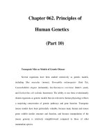
Fig. 1. ALS and communica-
tion. Progression of ALS may
impose severe communication-
al problems. Dysarthria and in-
ability to speak can be com-
pensated in some patients with
computer devices, such as spe-
cial keyboards and a mouse
442
Typically, mentation, extraocular movements, bowel and bladder functions,
and sensation are spared in ALS. Ophthalmoplegia (ocular apraxia) has been
reported. Dementia is observed in 1–2% of patients. Nearly one third of ALS
patients report urgent and obstructive micturition.
Over time, muscles become atrophied and patients complain of fatigue.
As ALS affects both upper and lower motor neurons, most (80%) of patients
show both upper and lower motor neuron signs. There is usually a combination
of spasticity, hyperreflexia, and progressive muscle weakness and wasting.
A small percentage of patients will only show lower motor neuron signs and
symptoms. On the other hand, there are rare instances where patients only have
upper motor neuron disease. There is currently debate as to whether this
condition, called Primary Lateral Sclerosis (PLS), is a separate entity. The
diagnostic procedures and treatments for PLS are currently identical to those for
ALS.
Most cases of ALS (at least 80%) are sporadic. A smaller number are attributable
to autosomal dominant familial ALS (FALS). The cause of sporadic ALS is
currently unknown, although proposed etiologies include glutamate neurotox-
icity, abnormal accumulation of neurofilaments, altered neurotrophism, and
toxicity from oxygen radicals or environmental sources.
The genetic cause of most FALS is unknown, but 20% of FALS cases show a
mutation in the protein cytosolic copper-zinc superoxide dismutase (SOD1),
found on chromosome 21q. SOD1 detoxifies superoxide anions, which can
lead to cell death when they accumulate and oxidize proteins and lipids. FALS,
whether caused by SOD1 mutations or not, is indistinguishable clinically from
sporadic ALS; thus, there is reason to believe that oxidative damage to neurons
is a common mechanism underlying all forms of ALS.
The El Escorial World Federation of Neurology criteria for the diagnosis of ALS
divides the body into four regions: bulbar (face, jaw, tongue, palate, larynx),
cervical (neck, arm, hand, diaphragm), thoracic (back, abdomen), and lum-
bosacral (back, abdomen, leg, and foot). Upper and lower motor signs must be
present in the bulbar region and two of the spinal regions, or in all three spinal
regions. A patient with signs in two spinal regions is diagnosed with probable
ALS. A diagnosis of possible ALS is given in cases where only one region is
affected, or if only lower motor neuron signs are present in two regions, or if
regions with lower motor neuron signs occur rostrally to regions with upper
motor neuron signs.
Genetic testing can be done to determine if a case of FALS is due to an SOD1
mutation.
EMG and nerve conduction studies with repetitive stimulation are used to
confirm lower motor neuron degeneration.
Imaging can be used to confirm that anatomy is normal, and exclude other
pathology.
Laboratory tests used to exclude other conditions that may resemble ALS
include: CBC and routine chemistries, serum VDRL, creatine kinase, thyroid
studies, serum protein electrophoresis, serum immunoelectrophoresis, ANA,
rheumatoid factor, and sedimentation rate.
Signs
Pathogenesis
Diagnosis
443
Neuroimaging and laboratory tests can be used to rule out the following
conditions: syringomyelia, syringobulbia, paraneoplastic motor neuronopathy,
polyradiculopathy with myelopathy, post-polio syndrome, multifocal motor
neuropathy, motor neuron disease with paraproteinemia, hexoseaminidase-A
deficiency, and heavy metal intoxication.
Riluzole (2-amino-6-(trifluormethoxy)benzothiazole) is the only targeted treat-
ment available. Riluzole blocks glutamate release, which may slow disease if
glutamate toxicity is contributing to motor neuron loss. Riluzole is given 50 mg
twice daily and may cause nausea and asthenia, but is generally tolerated well.
Symptomatic treatment may be indicated for spasticity, cramps, excessive
drooling, and pseudobulbar symptoms. Physical therapy, braces, and ambula-
tory supports are helpful. As speech becomes difficult, alternative communica-
tion devices are needed (Fig. 1). A severely dysphagic patient may choose to
have a gastric feeding tube placed. Bilevel positive airway pressure ventilation
is helpful for the respiratory symptoms of patients.
Prognosis for ALS is poor and the progression of the disease is generally
relentless. The average 5-year survival is 25%. The mean duration of disease
from onset of symptoms to death is 27 to 43 months, with median duration of
23–52 months.
Primary lateral sclerosis progresses much more slowly, with a mean duration of
224 months.
Benditt JO, Smith TS, Tonelli MR (2001) Empowering the individual with ALS at the end of
life: disease specific advance care planning. Muscle Nerve 24: 1706–1709
Hand CK, Rouleau GA (2002) Familial amyotrophic lateral sclerosis. Muscle Nerve 25:
135–159
Mitsumoto H, Chad DA, Pioro EP (1998) Amyotrophic lateral sclerosis. FA Davis, Philadel-
phia
Willson CM, Grace GM, Munoz DG, et al (2001) Cognitive impairment in sporadic ALS.
A pathologic continuum underlying a multisystem disorder. Neurology 57: 651–657
De Carvalho M, Swash M (2000) Nerve conduction studies in amyotrophic lateral sclero-
sis. Muscle Nerve 23: 344–352
Differential diagnosis
Therapy
Prognosis
References
444
Spinal muscular atrophies
Genetic testing NCV/EMG Laboratory Imaging Biopsy
+++ + +
Fig. 2. SMA. Marked general-
ized muscle atrophy due to
slowly progressive disease.
Symmetric atrophy of the trape-
zoid muscles A, mild winging B
of the medial borders of the
scapula
445
The spinal muscular atrophies (SMAs) are hereditary motor neuron diseases that
cause the loss of alpha motor neurons in the spinal cord. At autopsy, the spinal
cord is atrophied, showing loss of motor neurons and gliosis. The ventral roots
are also atrophied. Muscle atrophy is accompanied with signs of denervation
and reinnervation.
The onset and severity of symptoms depends upon the type of SMA the patient
has.
SMA1 (Werdnig-Hoffmann disease) is the most severe form, with symptoms
appearing in utero, or up to 3 months post-partum. Infants have severe diffuse
weakness that eventually leads to fatal loss of respiration.
SMA2 (late infantile SMA) causes weakness that appears between 18–24
months. Although less severe, these children may not be able to stand or walk,
and develop scoliosis and respiratory failure.
SMA3 (Kugelberg-Welander disease) has the mildest symptoms, and may not
present until the teenage years. These patients have proximal, symmetric
weakness but can still stand and walk. Deterioration of muscle function is slow
and mild.
Signs of lower motor neuron loss (hypotonia, reduced or absent reflexes,
fasciculations atrophy as shown in Figs. 2. and 3) are apparent, depending upon
the severity of disease.
SMA is caused by mutations in one of two copies of the survival motor neuron
(SMN) gene on chromosome 5q13. Loss of exons 7 and 8 in the telomeric copy
of the SMN gene leads to SMA1, the most severe form of the disease. Mutations
Anatomy
Symptoms
SMA1
SMA2
SMA3
Signs
Pathogenesis
Fig. 3. Spinal atrophy. Distal at-
rophy of lower legs, foot defor-
mity
446
that convert the telomeric copy of the gene to the centromeric copy cause the
less severe forms, SMA2 and 3. SMA is also associated with deletions in the
neuronal apoptosis inhibitor protein (NAIP) gene. These mutations occur in up
to 65% of SMA patients and may modify the severity of the disease. Both genes
are believed to suppress neuronal apoptosis, and thus the loss of motor neurons
may be the result of misregulated apoptosis.
Genetic testing in patients with appropriate signs and symptoms can reveal
SMN deletions in 95% of patients. Carrier testing is available.
EMG and muscle biopsy show signs of denervation. Nerve conduction studies
are normal. While these tests are often done early in the diagnosic process, they
are unnecessary if a genetic diagnosis has been established.
Cerebrospinal fluid analysis and serum creatine kinase are normal.
Infantile botulism must be ruled out in possible cases of SMA1. In botulism,
impairment is detected using EMG with high frequency nerve stimulation. Stool
examination for botulism can also confirm the diagnosis.
SMA2 and 3 can be distinguished from chronic inflammatory demyelinating
polyneuropathy by the presence of normal nerve conduction and cerebrospinal
fluid protein studies.
SMA3 may resemble hereditary motor sensory neuropathies (Charcot-Marie-
Tooth disease), but again the nerve conduction studies are normal in SMA.
There is no treatment for these diseases, although physical therapy and braces
are helpful for SMA2 and 3 patients. Surgery may be indicated to correct
scoliosis.
Half of infants with SMA1 die from respiratory failure by 7 months; 95% die by
17 months. Respiratory failure also shortens the life span of children with
SMA2, although not as early as in SMA1. SMA3 patients survive to adulthood
and typically maintain ambulatory function. It is not clear whether SMA3
affects lifespan.
Dubowitz V (1995) Disorders of the lower motor neurone: the spinal muscular atrophies.
In: Muscle disorders in childhood, 2nd edn. Saunders, London, pp 325–369
Wang CH, Carter TA, Gilliam TC (1997) Molecular and genetic basis of the spinal muscular
atrophies. In: Rosenberg RN, Pruisner SB, DiMauro S, Barchi RL (eds) The molecular and
genetic basis of neurological disease, 2nd edn. Butterworth-Heinemann, Boston, pp 787–
796
Diagnosis
Differential diagnosis
Therapy
Prognosis
References
447
Poliomyelitis is a viral infection that causes the death of motor neurons in the
spinal cord and brainstem. During the acute phase of the infection, the virus
may infect the cortex, thalamus, hypothalamus, reticular formation, brainstem
motor and vestibular nuclei, cerebellar nuclei, and motor neurons of the
anterior and lateral horns of the spinal cord, causing an inflammatory reaction.
Death of motor neurons may result, leading to muscle atrophy. The motor
neurons that survive recover fully and may reinnervate denervated muscle.
Paralytic poliomyelitis is characterized by an initial period of muscle pain and
spasms, followed by muscle weakness that peaks in severity by one week after
the onset of symptoms. Patients do not experience sensory impairment, but may
complain of paresthesias.
Bulbar symptoms occur in some patients and include dysphagia, dysarthria,
hiccups, and respiratory weakness leading to anxiety and restlessness. In adults,
bulbar disease is found in conjunction with spinal disease, but children (espe-
cially those without tonsils or adenoids) may present with a pure bulbar
poliomyelitis.
Urinary retention is common during the acute phase. Patients may also com-
plain of neck and back stiffness and pain, from meningeal inflammation.
Muscle weakness is asymmetric and typically proximal. Lumbar segments are
usually more severely affected, with trunk muscles being largely spared. Ten-
don reflexes may be initially brisk, but become diminished or absent. Muscles
progressively and permanently atrophy over a period of 2–3 months.
Loss of bulbar motor neurons occurs in some patients and can lead to paralysis
of the facial muscles (unilaterally or bilaterally), pharynx, larynx, tongue, and
mastication muscles.
If infection strikes the reticular formation, severe respiratory and autonomic
impairment may result. Breathing and swallowing difficulties, as well as loss of
vasomotor control, are serious risks for mortality and warrant intensive life
support.
Acute poliomyelitis is caused by infection with one of three forms of entero-
virus, a single-stranded, encapsilated RNA virus in the picornavirus family.
Enteroviruses spread by fecal-oral transmission. Rare cases have been attribut-
Poliomyelitis
Anatomy
Genetic testing NCV/EMG Laboratory Imaging Biopsy
+ +++ +
Symptoms
Signs
Pathogenesis
Acute poliomyelitis
448
ed to live attenuated virus in the polio vaccine. The replication phase takes
place 1–3 weeks post-infection in the pharynx and lower gastrointestinal tract.
Secretion of the virus occurs in the saliva and feces. The severity of infection is
variable, and can be classified into several categories:
Most patients (95%) are asymptomatic, or exhibit pharyngitis or gastroenteritis.
After this initial phase, up to 5% of infected patients may show signs of nervous
system involvement.
Nervous system involvement is preceded by a flu-like set of symptoms, includ-
ing fever, headache, muscle aches, pharyngitis, anorexia, nausea, and vomit-
ing. Neurological signs and symptoms include restlessness, irritability, and
signs of meningitis (back/neck stiffness, Brudzinski and Kernig signs). This
situation may then proceed to paralytic poliomyelitis.
Paralytic poliomyelitis develops in only 1–2% of infected patients, anywhere
from 4 days to 5 weeks following initial infection. Factors believed to predis-
pose a patient to paralytic disease include muscle damage from recent strenu-
ous exercise or muscle injections, increased age, tonsillectomy, weakened
B-cell function, and pregnancy. Acute paralytic poliomyelitis causes fatal respi-
Minor or abortive
poliomyelitis
Non-paralytic or pre-
paralytic poliomyelitis
Paralytic poliomyelitis
Fig. 4. Postpolio syndrome,
with polio in early infancy. A
and B Foot deformity reveas ear-
ly onset. C Very often involve-
ment of the lower limbs is asym-
metric (om this case right calf is
more atrophic than left)
449
ratory or cardiovascular problems in 5–10% of cases, or as high as 60% of cases
with bulbar involvement.
Encephalitic poliomyelitis is extremely rare and has a high mortality associated
with autonomic dysfunction. Patients present with confusion and agitation,
which may progress to stupor and coma.
Post-polio syndrome (PPS) occurs 10 years or longer after the initial polio
infection, and is characterized by slowly progressive, asymmetric increases in
weakness and muscle atrophy (Fig. 4). Patients may complain of joint and
muscle pain, and fatigue. PPS is not caused by the virus itself. It is believed that
surviving motor neurons that have reinnervated muscle fibers become incapa-
ble of maintaining all the connections in their enlarged motor units, and begin
to lose some connections. Some clinicians have suggested that excessive
exercise aimed at keeping diseased muscles strong leads to this “burn-out”, but
studies show that the primary associative factor for PPS is the severity of disease
during the acute phase of the infection. PPS may lead to weakness in muscle
groups previously thought to be unaffected, but typically these muscles were
originally affected and the patient developed sufficient strength and adaptation
to mask the deficits until the onset of PPS.
Laboratory:
Virus recovery from stool cultures during the first 2–3 weeks of disease is
considered diagnostic for poliomyelitis. Virus may also be detected in throat
washings, and occasionally from CSF or blood.
CBC may show increased white count.
CSF pressure may be increased. Neutrophils, and then lymphocytes, may be
found in the CSF prior to neurological impairment. Slight to severe protein
elevation with normal glucose may be detected.
EMG:
Early on, there is decreased recruitment and interference, with decreased motor
unit action potential amplitudes. In 2–4 weeks, fibrillations will develop, with
possible fasciculations. Over time, reinnervation will lead to polyphasic motor
units.
Nerve conduction velocities and sensory studies are normal.
Imaging:
Inflammation of the anterior spinal cord may be detected with MRI.
Post-polio syndrome:
The diagnosis of PPS is by exclusion of other conditions and demonstration of
progressive weakness over time.
Encephalitis caused by echovirus or coxsackie virus
Meningitis
Guillain-Barre syndrome
Motor polyneuropathies
Acute transverse myelitis
Encephalitic poliomyelitis
Post-polio syndrome
Diagnosis
Differential diagnosis
450
Vaccination programs have tremendously decreased the incidence of poliomy-
elitis in developed countries. However, rare cases are still reported in countries
with good vaccine programs, frequently in isolated cultures that reject modern
medical care. In countries without adequate vaccination, poliomyelitis is still
common.
Once a patient has poliomyelitis, the only treatment is supportive therapy. This
includes physical therapy to prevent contractures and joint ankylosis, prosthet-
ic devices, and respiratory/swallowing therapy to minimize pulmonary compli-
cations like aspiration and atelectasis. Some clinicians recommend that pa-
tients with PPS minimize their activity, but studies suggest that exercise is
beneficial for PPS, too.
Respiratory failure can be caused by central depression, weakness of the
respiratory muscles, or other complications (pneumonia, edema, etc.) associat-
ed with airway obstruction. Cardiovascular collapse may also occur from
infection of the brainstem. These situations require intensive care with artificial
ventilation.
During the acute phase of polio paralysis, the mortality rate is fairly low
(5–10%). Patients requiring ventilation during this period usually recover over
a period of several months, during which the respiratory muscles become
reinnervated and hypertrophic. Continued dependence on artificial ventilation
is uncommon. In general, the prognosis for polio patients is good.
Patients that later develop PPS will experience slowly worsening weakness.
This does not usually cause increased disability or mortality, although deterio-
ration of respiratory function is a rare possibility.
Dalakas MC (1995) The post-polio syndrome as an evolved clinical entity. Definition and
clinical description. Annals of the New York Academy of Science 753: 68–80
Mulder DW (1995) Clinical observations on acute poliomyelitis. Annals of the New York
Academy of Science 753: 1–10
Price RW, Plum F (1978) Poliomyelitis. In: Handbook of clinical neurology, vol. 32, pp
2091–2092
Rowland LP (2000) Viral infections of the nervous system: syndrome of acute anterior
poliomyelitis. In: Merritt´s neurology, 10th edn. pp 764–767
Trojan DA, et al (1994) Predictive factors for post-poliomyelitis syndrome. Arch Phys Med
Rehab 75: 770–777
Therapy
Prognosis
References
451
Bulbospinal muscular atrophy (BSMA), or Kennedy’s syndrome, affects the
lower (alpha) motor neurons found in the brainstem cranial nerve motor nuclei
and the anterior horns of the spinal cord. On autopsy, patients with BSMA show
mild atrophy of the brainstem and spinal cord. Muscle atrophy is also present,
with signs of denervation and reinnervation.
The mean onset for BSMA is 30 years (range, 15–60 years). Patients exhibit
symmetrical weakness that progresses slowly over many years, and typically do
not need canes or walkers until they are in their fifties or sixties. Facial, tongue,
and proximal weakness are typical at presentation. Dysphagia, dysarthria, and
masseter weakness are commonly observed.
As BSMA only affects lower motor neurons, there are no upper motor neuron
signs. Tendon reflexes are reduced or absent. Fasciculations are common in the
face (Fig. 5B). Vibratory sensation may be reduced, and patients often show a
mild postural tremor. Gynecomastia occurs in 50% of patients (Fig. 5A).
BSMA is an X-linked recessive disorder, caused by a tri-nucleotide repeat
expansion in the first exon of the androgen receptor gene on chromosome
Xq11–12. It is unknown how disruption of the androgen receptor in this way
leads to specific loss of lower motor neurons, as there are other mutations in
Bulbospinal muscular atrophy (Kennedy’s syndrome)
Anatomy
Genetic testing NCV/EMG Laboratory Imaging Biopsy
+++ + + +
Symptoms
Signs
Pathogenesis
Fig. 5. Kennedy’s syndrome. A
Pt with gynecomastia. B Pres-
ence of tonque atrophy