Case Files Neurology - part 10 ppsx
Bạn đang xem bản rút gọn của tài liệu. Xem và tải ngay bản đầy đủ của tài liệu tại đây (917.67 KB, 44 trang )
❖
CASE 52
A 43-year-old right-handed woman presents to the office with hearing loss,
facial paralysis, and headache. Her history began 1 month ago with a sudden
decrease in hearing in her right ear. One week prior to this visit she began to
notice weakness of the right face, which has now progressed to complete
paralysis. Over the last 3 months she has had intermittent right occipital
headache, and clumsiness and imbalance if she turns quickly. She denies any
change in her voice or difficulty with swallowing or swallowing difficulty. Her
past medical history is unremarkable. She is not on any medications except
birth control pills. Her physical examination shows a 43-year-old woman that
has an obvious right facial paralysis. Her pulse is 62 beats/min; blood pressure,
118/62 mmHg; and temperature, 36.7°C (98.6°F). The head and face have no
lesions. Her voice is normal, but her speech is slightly distorted because of the
facial paralysis. Her extra-ocular movements are normal. Her eye grounds do
not show any papilledema. Her ears have normal tympanic membranes. The
Weber tuning fork lateralizes to the left ear. Air conduction is louder than bone
conduction in both ears. There is no neck lymphadenopathy or other masses.
There are no cerebellar signs. The remaining physical examination, including
the neurologic examination, is normal. An audiogram shows a mild sen-
sorineural hearing loss in the right ear; the left ear has normal hearing. An
auditory brainstem response (ABR) is abnormal for the right ear; it is normal
for the left ear.
◆
What is the most likely neuroanatomic etiology and diagnosis?
◆
What is the next diagnostic step?
434 CASE FILES: NEUROLOGY
ANSWERS TO CASE 52: Meningioma of the Acoustic Nerve
Summary: A 43-year-old woman has a history of headache, hearing loss, and
facial paralysis.
◆
Neuroanatomic Etiology and Diagnosis: Cerebellopontine angle
tumor, with the most common tumors being acoustic neuroma and
meningioma
◆
Next diagnostic step: MRI with gadolinium
Analysis
Objectives
1. Learn the most common tumors that occur in the cerebellopontine
angle.
2. Learn the most common imaging features of these tumors.
3. Learn the available treatment options for these tumors.
Considerations
This 43-year-old woman has symptoms of hearing loss, facial paralysis,
and headache. She also has symptoms of imbalance and disequilibrium.
The most common cause of facial nerve paralysis is Bell palsy; however,
this patient also has hearing loss, balance issues, and headache, which
point to a central rather than peripheral disorder. Patients that present
with the combination of hearing loss and facial paralysis demand eval-
uation by diagnostic imaging. This patient’s symptoms strongly suggest
an abnormality in the cerebellopontine angle. Modern imaging techniques
have revolutionized the evaluation of this area. MRI with contrast can
readily differentiate the various pathologic processes that occur in this area
(Table 52-1).
CLINICAL CASES 435
APPROACH TO CEREBELLOPONTINE ANGLE
TUMORS
Definitions
Acoustic neuroma: A benign tumor that rises from Schwann cells on the
vestibular nerve also called vestibular schwannoma. This is the most
common tumor found in the cerebellopontine angle.
Table 52–1
MRI CHARACTERISTICS OF COMMON PATHOLOGY IN THE
CEREBELLOPONTINE ANGLE
Gadolinium Special
Tumor Type T1 Appearance* T2 Appearance* Enhancement Features
Schwannoma Isointense Intermediate ++++ Can be cystic,
inside or
centered on
the IAC
Meningioma Isointense or Hyperintense +++ Dural tail,
slightly to hypointense eccentric to the
hypointense IAC, can have
calcification
Epidermoid Hypointense Isointense None Internal
stranding
Glomus tumor Hypointense Isointense +++ “salt and
(Paraganglioma) pepper
appearance”
Arachnoid cyst Hypointense Hyperintense None Homogenous
contents
Lipoma Hyperintense Hypointense None Intensity
disappears with
fat suppression
Cholesterol Hyperintense Hyperintense None Located within
cysts the petrous
apex
IAC, internal auditory canal.
*Intensity relative to brain.
+ Minimal enhancement.
+++ moderate enhancement.
++++ Maximal enhancement.
Auditory brainstem response (ABR): An electrical evoked hearing test. In
this test, electrodes are placed on each ear lobe and on the forehead. A
stimulus sound (either a click or tone burst) is delivered into the test ear
at a specified loudness; an attached computer captures the electrical brain
activity that results from this stimulus and filters out background noise.
Bell palsy: Idiopathic facial weakness.
Cerebellopontine angle: The anatomic space between the cerebellum, pons,
and temporal bone. This space contains cranial nerves V through XI.
Conductive hearing loss: A form of hearing loss that results from a defect
in the sound collecting mechanism of the ear. These structures include
the ear canal, tympanic membrane, middle ear, and the ossicles.
Epidermoid tumor: A benign tumor composed of squamous epithelial ele-
ments thought to arise from congenital rests.
Glomus tumor: The common name for paraganglioma. This highly vascular
tumor arises from neuroepithelial cells. These tumors are further named
by the structures that they arise from: glomus tympanicum (middle ear),
glomus jugulare (jugular vein), glomus vagale (vagus nerve), and carotid
body tumor (carotid artery). A rule of 10% is associated with this tumor:
approximately 10% of these tumors produce a catecholamine-like sub-
stance, approximately10% of these tumors are bilateral, approxi-
mately10% are familial, and approximately 10% are malignant (i.e.,
potential to metastasize).
Meningioma: Common benign extra-axial tumors of the coverings of the
brain. The cell of origin is probably from arachnoid villi. Several histo-
logic subtypes are described: syncytial, transitional, fibroblastic,
angioblastic, and malignant.
Sensorineural hearing loss: A form of hearing loss that results from an
abnormality in the cochlea or auditory nerve.
Clinical Approach
Meningiomas
Meningiomas are usually benign tumors, of mesodermic origin, attached to the
dura. They commonly are located along the sagittal sinus, over the cerebral con-
vexities, and in the cerebellar-pontine angle. Grossly, they are gray, sharply
demarcated, and firm. Microscopically, the cells are uniform with round or elon-
gated nuclei, and a characteristic tendency to whorl around each other.
Meningiomas tend to affect women more than men in the middle age. The typ-
ical clinical presentation is the slow onset of a neurologic deficit or a focal
seizure; an unexpected finding on a brain imaging is also a common presenta-
tion. MRI usually reveals a dural-based mass with dense homogeneous contrast
enhancement. Surgical therapy is optimal, and complete resection is curative.
For lesions not amenable to surgery, local or stereotactic radiotherapy can ame-
liorate symptoms. Small asymptomatic lesions in older patients can be observed.
436
CASE FILES: NEUROLOGY
Rarely, meningiomas can be more aggressive and have malignant potential;
these tumors tend to have higher mitosis and cellular and nuclear atypia.
Surgical therapy followed by radiotherapy should be used in these instances.
Approach to Facial Paralysis
Facial paralysis is a relatively common disorder. In its most common presen-
tation, facial paralysis occurs as a sudden sporadic cranial mononeuropathy. It
is not associated with hearing loss; rather, it might be associated with hypera-
cusis. This form of facial paralysis, also called Bell palsy, is not associated
with middle ear disease, parotid tumor, Lyme disease or any other known
cause of facial paralysis. Essentially, Bell palsy is a diagnosis of exclusion.
Generally, a pointed history and detailed physical examination will eliminate
most of the differential diagnosis. Likewise, the various causes of hearing loss
can be eliminated by a careful physical examination. Disease processes, such
as otitis media, cholesteatoma, and otosclerosis, can be eliminated by careful
history and physical examination with tuning fork tests. However, to know the
type and degree of hearing loss, an audiogram is necessary.
Although it requires patient cooperation, the audiogram will give the clini-
cian a very accurate measure of the patient’s hearing level. The audiogram can
distinguish between sensorineural and conductive hearing loss. Occasionally,
patients have mixed hearing loss, or a combination of conductive and sen-
sorineural losses in a single ear. Furthermore, the audiogram can give a clue
regarding the presence of retrocochlear hearing loss or hearing loss caused by
diseases proximal to the cochlea. Tests that might indicate retrocochlear
pathology include speech discrimination, acoustic reflexes, and reflex decay.
Diagnosis
Sensorineural hearing loss can be further evaluated by auditory brainstem
response (ABR). This test measures the electrical activity within the auditory
pathway; and as such, this test helps to evaluate retrocochlear causes of hear-
ing loss. The ABR has five waves that are numbered I through V, and these are
correlated to major neural connections in the auditory pathway. These waves
have expected morphologies and occur at predictable latencies. Waves that are
absent or delayed are indicative of pathology at that point in the auditory path-
way. The interwave latencies (such as I to III, III to V, or I to V) can be com-
pared to the opposite side or to standard norms. Abnormalities on ABR need
to be further evaluated by imaging studies.
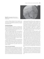
MRI provides excellent definition of the structures within the posterior
fossa. Gadolinium contrast allows additional differentiation of various
pathologies. Additionally, newer technology, such as fat suppression and dif-
fusion weighted imaging can help to identify pathology (Fig. 52–1). The MRI
appearances of the most common tumors in the posterior fossa are indicated in
the Table 52–1.
CLINICAL CASES 437
Although MRI with gadolinium contrast gives excellent resolution for
brain, nerve and soft tissues, CT scanning is necessary for bony imaging.
Often, both imaging modalities are combined to understand the full extent of
the disease process within the skull base.
Treatment
A treatment plan must be created once a tumor in the cerebellopontine angle is
diagnosed. Many factors must be considered when approaching these tumors. The
patient’s age, overall health status, tumor size and location, degree of hearing loss,
and other neurologic signs are all factors to be taken into account. The various
available treatment options must be discussed with the patient; the final decision
of treatment course must be decided between the patient and the physician.
At least three options should be considered in managing tumors in the pos-
terior fossa: observation and serial imaging, stereotactic radiosurgery, or
conventional surgery. Some of these options might be unavailable or unwise
for certain tumor types or tumor size. Clearly, the patient that has a large tumor
that is producing brainstem compression or obstructive hydrocephalus should
not be observed over time and serially imaged. These findings demand imme-
diate attention.
Surgery can provide several benefits to the patient. Removal of tumor
allows for final pathologic diagnosis, might correct neurologic deficits, and
might prevent further complications caused by continued tumor growth. These
benefits can come at a price of new neurologic deficits, meningitis, infection,
438
CASE FILES: NEUROLOGY
Figure 52–1. Post-gadolinium T1 MRI with fat suppression. Cerebellopontine
angle meningioma. (With permission from Fischbein NJ, Ong KC, Radiology.
In: Lalwani A. Current Diagnosis and Treatment in Otolaryngology Head &
Neck Surgery, New York: McGraw-Hill; 2004, p 158.)
CLINICAL CASES 439
stroke, or even death. The patient’s underlying health status must be consid-
ered because these surgical procedures are often lengthy. Patients with low
overall health status might not tolerate such a procedure.
A relatively new (although more than 20 years experience) type of therapy
involves the use of directed, focus radiation beam to the tumor. Several dif-
ferent proprietary devices have been developed to destroy or at least prevent
growth of these types of tumors. The experience with stereotactic radiotherapy
is probably greatest with acoustic neuroma, because that tumor is the most
common mass found in the cerebellopontine angle. Stereotactic radiother-
apy has been found to be very effective at managing small to medium sized
tumors (up to 3 cm). In these tumors, the complication rate for stereotactic
radiotherapy is at least as low as that from conventional surgery; and with this
type of therapy, a long hospital stay or recovery period is not required. The dis-
advantage with stereotactic radiotherapy is the potential for continued growth,
and this growth does occur in a significant number of patients. Unfortunately,
surgery following stereotactic radiotherapy is technically more difficult, and
surgical results are not as good as from surgery alone.
Stereotactic radiotherapy does have limitations. It is not useful for certain
tumor types (meningiomas and epidermoids). Of course, stereotactic radio-
therapy cannot provide pathologic specimens for study, and it should never be
used when the pathologic diagnosis is in doubt.
Comprehension Questions
[52.1] A 45-year-old painter is found to have ataxia. An MRI scan shows a
tumor of the cerebellopontine angle. What is the most likely tumor in
the location?
A. Epidermoid tumor
B. Paraganglioma
C. Meningioma
D. Acoustic neuroma
E. Lipoma
[52.2] What is the best test to evaluate unilateral sensorineural hearing loss?
A. Otoacoustic emissions
B. Auditory brainstem response
C. MRI of the internal auditory canals with gadolinium
D. Electronystagmography
E. Detailed physical examination
[52.3] What is the most common cause of unilateral facial paralysis?
A. Idiopathic
B. Otitis media
C. Parotid malignancy
D. Acoustic neuroma
E. Lyme disease
440 CASE FILES: NEUROLOGY
Answers
[52.1] D. By far, the most common tumor in the cerebellopontine angle is the
acoustic neuroma.
[52. 2] C. Although ABR is used to evaluate unilateral sensorineural hearing
loss, its limitation is a lack of specificity for diagnosis. Otoacoustic
emissions can measure the degree of hearing loss, but it cannot shed
light on a pathologic cause. Electronystagmography is a test that meas-
ures the vestibular ocular reflex. Detailed physical examination is an
important prerequisite before any diagnostic tests are ordered. Only
MRI with contrast enhancement can elucidate the cause of unilateral
sensorineural hearing loss.
[52.3] A. The most common form of facial paralysis is idiopathic. It is also
called Bell palsy. Recent evidence suggests that the cause of Bell palsy
is probably recrudescence of herpes simplex virus. Every patient
should have a careful examination to rule out other causes of facial
paralysis, such as those diagnoses listed. Where indicated, this exami-
nation might require an audiogram or MRI imaging.
CLINICAL PEARLS
❖ Idiopathic facial paralysis (also called Bell palsy) is the most com-
mon cause of unilateral facial weakness.
❖ Bell palsy is a diagnosis of exclusion, and patients with facial paral-
ysis require a careful otologic and cranial nerve examination.
❖ Patients that present with a complaint related to one cranial nerve
require evaluation of all cranial nerves.
❖ Acoustic neuromas are the most common tumor of the cerebello-
pontine angle.
❖ Unilateral sensorineural hearing loss should be further evaluated by
MRI with gadolinium contrast.
REFERENCES
Fan G, Curtin H. Imaging of the lateral skull base. In: Jackler R, Brackmann D, eds.
Neurotology, 2nd ed. Philadelphia, PA: Elsevier; 2004, pp 383–418.
Lo W, Hovsepian M. Imaging of the cerebellopontine angle. In: Jackler R,
Brackmann D, eds. Neurotology, 2nd ed. Philadelphia, PA: Elsevier; 2005.
pp 349–382.
❖
CASE 53
A 59-year-old retired bartender presents with the complaint of headaches and
difficulty concentrating over the past 6 weeks. He has been healthy all of his
life and presents yearly for an annual checkup. He describes the headaches as
occurring primarily over the right frontal temporal region and describes it as
“dull” in nature. He has experienced occasional nausea but no vomiting with
the headaches. Additionally, he has had difficulty focusing and concentrating
on tasks at hand, such as reading the newspaper or playing cards. His wife
states that he has been more irritable, moody, and “not himself” for 1 month.
There is no history of alcohol abuse or exposure to toxins. He admits to a
30-pack-a-year smoking history. The review of systems is significant for
weight loss and productive cough.
His examination reveals that he is afebrile with a blood pressure of 124/72
mmHg and a heart rate of 78 beats/min. His general examination is normal. He
is oriented to person, time, location, and situation, although he becomes upset
during the examination. Cranial nerve and sensory examination findings are
unremarkable. Motor strength testing is normal except for questionable weak-
ness in the left finger extensors. The deep tendon reflexes are normal except
for a Babinski sign present on the left. With ambulation, he has less arm swing
on the left than the right.
◆
What is the most likely diagnosis?
◆
What is the next diagnostic step?
◆
What is the next step in therapy?
ANSWERS TO 53: Metastatic Brain Tumor
Summary: A 59-year-old healthy man presents with a 6-week history of right
frontal temporal headaches associated with difficulty concentrating, weight loss,
and coughing. His headaches are often associated with nausea and are dull in
nature. His wife reports personality changes and the patient himself recognizes
mood disturbances. His examination is notable for decreased arm swing on the
left, questionable weakness of the left finger extensors, and a left Babinski sign.
◆
Most likely diagnosis: Metastatic brain tumor affecting the right
cerebral hemisphere.
◆
Next diagnostic step: MRI of the brain with and without gadolinium
and chest x-ray.
◆
Next step in therapy: Corticosteroids and anticonvulsants are started
immediately while waiting for surgical evaluation.
Analysis
Objectives
1. Know the clinical presentation and diagnostic approach to metastatic
brain tumor.
2. Be familiar with the differential diagnosis of metastatic brain tumor.
3. Describe the treatment for metastatic brain tumor.
Considerations
This 59-year-old otherwise healthy man presents with unilateral dull headaches
associated with nausea and personality changes. Additionally there is a history of
difficulty concentrating, weight loss, and cough. His physical examination sug-
gests mild left-sided weakness most likely from a right hemispheric lesion given
the left Babinski sign. Based on the history and examination the most likely diag-
nosis is a right hemispheric mass lesion. Taking it one step further the history of
weight loss and cough are concerning for a lung cancer. With this in mind,
metastatic lung cancer should be considered. A chest x-ray will reveal that he has
a large right upper-lobe mass lesion highly suggestive of lung cancer. An MRI of
the brain will show a right frontal temporal well-circumscribed lesion at the gray-
white junction with hemorrhage and surrounding edema. Evidence of midline
shift or impending herniation should be evaluated. Corticosteroids such as dex-
amethasone should be started as this reduces edema and capillary permeability.
Prophylaxis with anticonvulsants in individuals with metastatic tumors that
have not experienced a seizure is controversial. Approximately 40% of
patients with metastatic brain tumors will experience a seizure. Only 20% of
patients with metastatic brain tumors present with seizures. In this particular
case the patient has a hemorrhage, which is known to be epileptogenic. Most
442
CASE FILES: NEUROLOGY
CLINICAL CASES 443
physicians would begin anticonvulsants. Caution should be taken in patients who
are receiving both anticonvulsants and corticosteroids as the latter can signifi-
cantly reduce anticonvulsant levels. Neurosurgical consultation should be
obtained as should an oncology consultation.
APPROACH TO METASTATIC BRAIN TUMORS
Definitions
Metastatic brain tumors: Tumors that arise from metastasis of systemic
neoplasm to the brain parenchyma.
Babinski sign: Extension of the big toe followed by abduction of the other
toes when the lateral sole of the foot is stimulated. It is performed by
stroking the foot at the heel and moving the stimulus toward the toes. It
is a sensitive and reliable sign of cortical spinal tract disease. It is also
known as the plantar reflex.
Midline shift: Movement of a cerebral hemisphere to the opposite side sec-
ondary to intracranial swelling. This can cause compression of the lat-
eral ventricles and contribute to further elevated intracranial pressure.
Herniation: Downward displacement of the cerebral hemisphere from
increased intracranial pressure.
Clinical Approach
Metastatic brain tumors can arise from primary systemic cancers that spread to
the leptomeninges, brain parenchyma, calvaria, or dura. Brain metastases are 10
times more common than primary brain tumors. In the United States roughly
150,000 new cases per year of metastatic brain tumors are reported. Men have a
slightly higher incidence than females at a ratio of 1.4:1. Approximately 66% of
metastatic brain tumors go to the parenchyma with almost 50% of these being a
solitary lesion. The most common tumors that metastasize to the brain are listed
in the Table 53–1, with lung cancer being most common.
Table 53–1
METASTATIC TUMOR AND FREQUENCY
Tumor Type Cases (%)
Lung cancer 50%
Breast cancer 20%
Melanoma 10%
Unknown primary 10%
Others: thyroid and sarcoma Unknown
444 CASE FILES: NEUROLOGY
Tumors metastasize to the brain most commonly by entering the systemic
circulation known as hematogenous spread. The distribution of tumor parallels
blood flow to the brain with approximately 82% metastasizing supratento-
rially, 15% spreading to the cerebellum, and 3% affecting the brain stem.
Metastatic brain tumors are commonly located at the gray-white junction
and arterial border zones, locations that have narrowed blood vessels that
can trap tumor cells.
Clinical features of metastatic brain disease are varied and can depend on
location. Neurologic symptoms occur from direct tumor infiltration, hemor-
rhage, edema, or even hydrocephalus. Table 53–2 illustrates the most common
clinical features of brain metastases.
The differential diagnosis for metastatic brain tumors includes brain
abscess, demyelinating diseases, radiation necrosis, cerebral vascular acci-
dents, intracranial bleed, and primary brain tumors. Approximately 60% of
those without any known primary tumor that present with brain metas-
tasis have a primary lung cancer.
The clinical evaluation in patients with unknown primary cancer is focused
and includes an MRI of the brain with gadolinium. Gadolinium or contrast
is critical as it will show enhancement around the lesions. Given the fact
that lung cancer is the most common type to metastasis to brain, a chest x-ray
followed by a CT scan of the chest should be performed. If these studies are
Table 53–2
CLINICAL FEATURES OF BRAIN TUMORS
Patients Presenting with
Clinical Features Features (%)
Headaches dull and associated with nausea 45–50%
Visual disturbances including blurred vision;
unilateral on side of tumor and more commonly
associated with posterior fossa metastases
Cognitive impairment including personality 33%
changes, mood and memory problems
New onset seizure; more frequently associated 10–20%
with frontal, temporal or multiple metastases
Stroke-like syndrome 5–10%
Papilledema 10% (at time of presentation)
Other nonspecific neurologic findings 20–40%
CLINICAL CASES 445
unrevealing, than an abdominal or pelvic CT scan should be performed. Careful
attention should be placed to the prostate, testicles, breasts, and rectum during
clinical examination. A guaiac examination should be performed to evaluate for
occult blood. This will help evaluate for gastrointestinal cancers.
Unfortunately an MRI of the brain cannot diagnose the type of tumor in
patients with unknown primary malignancy. One exception is malignant
melanoma, which has been shown to be hyperintense on T1-weighted images
and hypointense on T2-weighted images. A brain biopsy can be necessary if
a primary tumor cannot be found. Patients with signs of severe increased
intracranial pressure can benefit from surgery.
Treatment with corticosteroids such as dexamethasone is important in
reducing intracranial pressure and edema. Commonly a dose of 10 mg of dex-
amethasone, either orally or intravenously, followed by 4 mg every 6 hours is
given. As previously discussed, it is controversial as to whether or not anti-
convulsants are necessary in patients who have not experienced seizures.
However those individuals that have presented or developed a seizure warrant
anticonvulsant therapy.
The decision as to whether or not patients should undergo surgery is
dependent on the number of brain metastases, location, the size, the likeli-
hood of response to treatment, and the patient’s overall health status. The
most important factor when considering surgery is the tumor burden located
outside the brain. Improved survival and quality of life has been shown in
patients with single lesions when they have been treated with whole brain
radiotherapy and surgery. Those that do better following this treatment are
individuals that present at a younger age, absence of extracranial disease,
and increased time to developing brain metastasis. Radiation therapy has
been shown to decrease the mortality from neurologic dysfunction. The most
common regimen is given over a period of 2 weeks using 30 Gy in 10 frac-
tions. Radiation therapy improves neurologic symptoms in 50% to 93% of
patients. Complications from radiotherapy include brain necrosis, brain atro-
phy, cognitive deterioration, leukoencephalopathy, and neuroendocrine dys-
function. Stereotactic radiation via the gamma knife, linear particle
accelerators, or charged particles can also be used. This has been found to
decrease toxicity to healthy tissue and minimize side effects. Stereotactic
radiation is often used in tumors that are surgically inaccessible; complica-
tions from stereotactic radiation include seizures, headaches, nausea, hem-
orrhage, and radiation necrosis. For the most part chemotherapy is not used
for brain metastasis.
Favorable prognostic factors include being less than 60 years of age,
two or less brain metastasis, good baseline function, and accessible to sur-
gical resection. Individuals with single brain metastasis who receive all brain
radiation plus surgery have a median survival of 10 to 16 months. Patients who
have metastasis to infratentorial regions of the brain carry a worse prognosis
than those with supratentorial metastasis.
446 CASE FILES: NEUROLOGY
Comprehension Questions
[53.1] A 56-year-old man who is complaining of confusion and motor deficits
is noted to have multiple lesions to the brain. A metastatic tumor is sus-
pected. Which of the following is the most common tumor causing
brain metastasis?
A. Breast
B. Melanoma
C. Renal
D. Lung
E. Thyroid
[53.2] A 50-year-old man is noted to have some symptoms suggestive of a
brain tumor. Which of the following is the most commonly found
symptom for brain tumors?
A. Seizures
B. Headaches
C. Papilledema
D. Personality changes
E. Ataxia
[53.3] A 45-year-old man with a history of smoking presents after experiencing
a generalized tonic-clonic seizure. He has been experiencing dull left-
sided headaches over the past 2 months. His examination reveals hyper-
reflexia on the right with mild weakness of the right iliopsoas and finger
extensor muscles. The MRI of the brain shows a large 7 cm × 10 cm
lesion over the left frontal region with associated midline shift. A chest
x-ray shows a left lower lobe mass. What is the next step?
A. Consult neurosurgery for immediate brain biopsy and debulking
B. Start dexamethasone at a dose of 10 mg followed by 4 mg every
6 hours. Concomitantly begin an anticonvulsant medication
C. Start dexamethasone at a dose of 100 mg followed by 4 mg every
6 hours and hold off on starting anticonvulsant medication
D. Consult the oncology service to assist you in deciding on
chemotherapy
E. Start whole brain radiation therapy
Answers
[53.1] D. Lung cancer is the most common tumor metastasizing to brain,
accounting for approximately 50% of all cases.
[53.2] B. Headache is the most commonly found symptom associated with
brain tumors and is found in approximately half of cases.
CLINICAL CASES 447
[53.3] B. Patients with brain metastasis that present with seizures should be
started on anticonvulsant therapy in addition to dexamethasone. In this
particular case there is associated midline shift that warrants immedi-
ate medical management.
CLINICAL PEARLS
❖ Metastatic malignancies account for the majority of brain tumors in
adults.
❖ Enhancing brain lesions on MRI that are located at the gray-white
junction are likely to be metastatic brain tumors.
❖ Most metastatic MRI brain lesions are nonspecific. Melanoma is an
exception, being consistently hyperintense on T1-weighted
images and hypointense on T2-weighted images.
❖ Patients that present with new onset headaches, personality
changes, and mood disorders need to be evaluated for brain
tumors.
REFERENCES
Kaye AH, Laws ER. Brain tumors, an encyclopedic approach, 2nd ed. Philadelphia,
PA: Churchill Livingstone; 2001.
Dorland’s Illustrated Medical Dictionary, 27th ed. Philadelphia, PA: WB Saunders;
1988.
Nathoo N, Toms SA, Barnett GH. Metastases to the brain: current management.
Expert Rev Neurother 2004;4:4, 633–640. Online publication updated: July 1,
2004.
Sawaya R, Ligon BL, Bindal RK. Management of metastatic brain tumors. Ann
Surg Oncol 1994;1(2):169–178.
This page intentionally left blank
SECTION III
Listing of Cases
Listing by Case Number
Listing by Disorder (Alphabetical)
Copyright © 2008 by the McGraw-Hill Companies, Inc. Click here for terms of use.
This page intentionally left blank
LISTING OF CASES 451
LISTING BY CASE NUMBER
CASE NO. DISEASE CASE PAGE
1 Essential Tremor 18
2 Huntington Disease 24
3 Dystonia 33
4 Parkinson Disease 40
5 Ataxia, Spinocerebellar 48
6 Tardive Dyskinesia 58
7 Spinal Cord Injury, Traumatic 64
8 Epidural/Subdural Hematoma 72
9 Delirium from Head Trauma 80
10 Cerebral Contusion 88
11 Acute Cerebral Infarct 96
12 Subarachnoid Hemorrhage 104
13 Stroke in a Young Patient 112
14 New Onset Seizure: Adult 118
15 Absence Versus Complex Partial Seizure 126
16 Cardiogenic Syncope Related to Bradycardia 136
17 Pseudoseizure 144
18 Migraine Headache 150
19 Chronic Headache 162
20 Alzheimer Dementia 170
21 Dementia (Lewy Body) 182
22 Subacute Combined Degeneration of Spinal 190
Cord
23 Optic Neuritis 198
24 Multiple Sclerosis 206
25 Acute Disseminated Encephalomyelitis 216
26 Viral Meningitis 222
27 Infantile Botulism 230
28 HIV-Associated Dementia 236
29 Sporadic Creutzfeldt-Jakob Disease 242
30 Tabes Dorsalis 252
31 Intracranial Lesion (Toxoplasmosis) 260
32 Unreactive Pupil 268
33 Papilledema 276
34 Sixth Nerve Palsy (Ischemic Mononeuropathy) 284
35 Facial Paralysis 292
36 Ptosis (Myasthenia Gravis) 300
37 Vertigo, Benign Paroxysmal Positional 310
38 Chronic Inflammatory Demyelinating 322
Polyneuropathy
39 Guillain-Barré Syndrome 330
40 Dermatomyositis 338
41 Amyotrophic Lateral Sclerosis 350
42 Median Nerve Mononeuropathy 360
43 Foot Drop 368
CASE NO. DISEASE CASE PAGE
44 New Onset Seizure, Child 374
45 Febrile Seizures 380
46 Pediatric Headache 386
47 Duchenne Muscular Dystrophy 394
48 Tourette Syndrome 403
49 Benign Rolandic Epilepsy 410
50 Lissencephaly 418
51 Autism 424
52 Meningioma 432
53 Metastatic Brain Tumor 440
LISTING BY DISORDER (ALPHABETICAL)
CASE NO. DISEASE CASE PAGE
15 Absence Versus Complex Partial Seizure 126
11 Acute Cerebral Infarct 96
25 Acute Disseminated Encephalomyelitis 216
20 Alzheimer Dementia 170
41 Amyotrophic Lateral Sclerosis 350
5 Ataxia, Spinocerebellar 48
51 Autism 424
49 Benign Rolandic Epilepsy 410
16 Cardiogenic Syncope Related to Bradycardia 136
10 Cerebral Contusion 88
19 Chronic Headache 162
38 Chronic Inflammatory Demyelinating 322
Polyneuropathy
9 Delirium from Head Trauma 80
21 Dementia (Lewy Body) 182
40 Dermatomyositis 338
47 Duchenne Muscular Dystrophy 394
3 Dystonia 33
8 Epidural/Subdural Hematoma 72
1 Essential Tremor 18
35 Facial Paralysis 292
45 Febrile Seizures 380
43 Foot Drop 368
39 Guillain-Barré Syndrome 330
28 HIV-Associated Dementia 236
2 Huntington Disease 24
27 Infantile Botulism 230
31 Intracranial Lesion (Toxoplasmosis) 260
50 Lissencephaly 418
42 Median Nerve Mononeuropathy 360
452 CASE FILES: NEUROLOGY
CASE NO. DISEASE CASE PAGE
52 Meningioma 432
53 Metastatic Brain Tumor 440
18 Migraine Headache 150
24 Multiple Sclerosis 206
14 New Onset Seizure: Adult 118
44 New Onset Seizure, Child 374
23 Optic Neuritis 198
33 Papilledema 276
4 Parkinson Disease 40
46 Pediatric Headache 386
17 Pseudoseizure 144
36 Ptosis (Myasthenia Gravis) 300
34 Sixth Nerve Palsy (Ischemic Mononeuropathy) 284
7 Spinal Cord Injury, Traumatic 64
13 Stroke in a Young Patient 112
22 Subacute Combined Degeneration of Spinal 190
Cord
12 Subarachnoid Hemorrhage 104
29 Sporadic Creutzfeldt-Jakob Disease 242
30 Tabes Dorsalis 252
6 Tardive Dyskinesia 58
48 Tourette Syndrome 403
32 Unreactive Pupil 268
26 Viral Meningitis 222
37 Vertigo, Benign Paroxysmal Positional 310
LISTING OF CASES
453
This page intentionally left blank
❖
INDEX
Note: Page numbers followed by a t or f indicate that the entry is included in a table
or figure.
A
abdominal examination, 6, 329, 375
abdominal migraines, 159
abortive therapy, for migraines,
155–157, 314t, 388, 391
absence seizures (petit mal)
vs. complex partial seizures, 129t
considerations, 127, 380
definition, 127
valproic acid for, 131
acetaminophen, for pediatric
headaches, 391
acoustic neuromas, 432, 433, 437, 438
acquired Creutzfeldt-Jakob disease, 243
acquired epileptic aphasia
(Landau-Kleffner syndrome),
120
activated partial thromboplastin time
(aPTT) assessment, 74
acute communicating hydrocephalus, 108
acute disseminated encephalomyelitis
(ADEM)
clinical pearls, 219
considerations, 216
etiologies/clinical presentation, 217
multifactorial etiopathogenesis, 219
prognosis, 217
symptoms, 215, 217
treatment options, 218
acute inflammatory demyelinating
polyneuropathy (AIDP). See
Guillain-Barré syndrome (GBS)
acute necrotizing hemorrhagic
encephalomyelitis (ANHE),
216
adult onset seizures
associated definitions, 119
clinical pearls, 122
clinical presentation, 120–121
considerations, 118–119
etiologies
complex partial seizures, 119
generalized seizures, 119
idiopathic epilepsy syndromes,
120
simple partial seizures, 119
symptomatic epilepsy syndromes,
120
symptoms, 117
treatment options, 121t
afferent pupillary defect, 200f
agnosia, 98
AIDS dementia complex (ADC), 240
alcohol
and cardiogenic syncope, 140
and dementia, 176
and tremors, 18, 20, 22
alprazolam (Xanax), for tremors, 21
Alzheimer disease. See also dementia
associated definitions, 172
clinical pearls, 178
considerations, 170–171
vs. diffuse Lewy body dementia, 184
MRI brain images, 176f
Copyright © 2008 by the McGraw-Hill Companies, Inc. Click here for terms of use.
Alzheimer disease (Cont.):
photomicrograph, amyloid
plaque/neurofibrillary tangle,
175f
symptoms, 169
treatment options, 177
amantadine
for multiple sclerosis, 211
for Parkinson disease, 44
amyloidosis, 362
amyotrophic lateral sclerosis (ALS)
associated definitions, 351
vs. cervical myelopathy, 356
clinical features/epidemiology, 352
clinical pearls, 356
considerations, 350–351
diagnosis, 353
epidemiology, 352
etiology/pathogenesis, 352
symptoms, 349
treatment/management
nutrition assistance, 354
riluzole (Rilutek), 353
speech therapy, 354
ventilatory assistance, 355
anemia
macrocytic, 191, 192, 195
megaloblastic, 192
pernicious, 176, 191, 194, 195
aneurysms
angiogram, 107f
risk factors, 105–106
ruptured saccular/berry, 105
angiography
for aneurysms, 108
for chronic daily headaches, 164
for ischemic stroke, 98
for ptosis, 303
for subarachnoid hemorrhage, 104,
106, 107f, 108
ankle-foot orthosis, for foot drop, 372
anterior cord syndrome, 65
anterior optic neuritis (papillitis), 198
antibiotics
for new onset seizures, children, 378
for respiratory infection, in MD, 398
for toxoplasmosis, 264
for viral meningitis, 222, 223, 225
anticholinergics
for dystonia, 37
for multiple sclerosis, 211
for Parkinson disease, 44
anticholinesterase medications
for Alzheimer disease, 178
for diffuse Lewy body dementia, 186
for myasthenia gravis, 306
anticoagulant treatment, for strokes,
114
anticonvulsants
for benign rolandic epilepsy, 412
for chronic headaches, 165
for Duchenne/Becker muscular
dystrophy, 398
for migraine headaches, 157
for pediatric headaches, 391
antidepressants
for chronic headaches, 165
for migraine headaches, 157
for multiple sclerosis, 211
for pediatric headaches, 391
anti-Jo-1 antibody, 339
anti-MuSK antibodies, 301, 305
antipyretics, for febrile seizures,
384, 385
anti-seizure medications, 21, 22, 131
antitoxin, for infantile botulism, 233
aphasia, 98
apraxia, 98
constructional, 183
ideomotor, 183
aPTT. See activated partial
thromboplastin time (aPTT), 74
arboviruses, 224
Arenaviruses, 224
Argyll Robertson pupils, 251, 252
Arteriogram, see also angiography
for cerebral infarction, 98
internal carotid artery (ICA)
dissection, 114f
for stroke, young patient, 112, 114f
arteriovenous malformations, 112
Asperger syndrome, 426
aspirin
for carpal tunnel syndrome, 364
causative for BPPV, 319
for chronic daily headache, 165
for stroke prevention, 99
vs. warfarin, 114
456 INDEX
asthma
avoidance of beta-blockers, 21
in pseudoseizures, 147
ataxia
autosomal dominant spinocerebellar,
50t–51t
clinical approach, 49, 52–53
considerations, 48–49
definitions, 49
and HIV-associated dementia, 237
symptoms, 47
treatment options, 53–54
occupational therapy, 53
speech therapy, 53
athetosis, 25
atrial fibrillation, 98, 99–100
attention deficit hyperactivity disorder
(ADHD), 404, 405, 406
audiogram
for facial paralysis, 292, 294, 311,
435
for vertigo, 311, 312
Auditory Brainstem Evoked Responses
test, 425
auditory brainstem response (ABR),
434, 435
auras
with migraine headaches, 150, 151
with pediatric headaches, 390
with simple partial seizures, 126,
128, 377
autism. See also epilepsy
associated definitions, 425–426
clinical approach, 426–428
clinical pearls, 429
considerations, 424–425
management, 427–428
prognosis, 428
symptoms, 423, 426–427
autism spectrum disorder (ASD)/
pervasive developmental
disorder (PDD), 403, 426
autosomal dominant spinocerebellar
ataxias, 50t–51t
B
Babinski sign, 81, 221, 441
back/spine examination, 6
baclofen
for dystonia, 36, 37
for tardive dyskinesia, 59
bacterial meningitis, 223, 226
basilar artery migraines, 159
Bell palsy, 292, 296, 434, 435, 438
benign epilepsy with centrotemporal
spikes. See benign rolandic
epilepsy (BRE)
benign family tremors, 20
benign paroxysmal positional vertigo
(BPPV)
associated definitions, 310–311
clinical approach, 311–313
clinical evaluation, 313
clinical pearls, 319
considerations, 310
differential diagnosis,
314t–316t
symptoms, 309, 319
testing for, 317
benign rolandic epilepsy (BRE)
approach, 411–413
clinical pearls, 414
considerations, 380, 410–411
diagnosis, 412–413
symptoms, 409
treatment options, 412
benzodiazepines
for Alzheimer disease, 177
for dystonia, 36, 37
for febrile seizures, 384
for tardive dyskinesia, 59
berry aneurysms, 105
beta-blockers
for cardiogenic syncope, 137
for chronic headaches, 165
for essential tremor, 21, 22
for migraine headaches, 157–158
for pediatric headaches, 391
side effects, 137, 158
bilateral facial paralysis, 295
binocular diplopia, 284, 285,
286–287, 289
biopsy
of brain
for CJD, 247
for migraine headaches, 152
for toxoplasmosis, 263
for tumors, 443
INDEX
457