Digital histology an interactive cd atlas with review text phần 5 pot
Bạn đang xem bản rút gọn của tài liệu. Xem và tải ngay bản đầy đủ của tài liệu tại đây (366.85 KB, 22 trang )
Classification of Skin—Based on the Thickness of
the Epidermis
➢ Thin skin
᭹
Covers entire body except palms and soles; 0.5mm thick on the
eyelid, 5mm thick on the back and shoulders
᭹
Epidermis is thin, 0.075–0.15mm thick, but the dermis can be quite
thick.
᭹
Possesses hair with sebaceous glands
᭹
Sweat glands are present.
➢ Thick (glabrous) skin
᭹
Located on palms of the hands and soles of the feet; 0.8–1.5mm
thick
᭹
Epidermis is 0.4–0.6mm thick.
᭹
Hairless and, thus, possesses no sebaceous glands
᭹
Sweat glands are present.
Epidermis
➢ Cell types
᭹
Keratinocytes. Keratinizing epidermal cells, major cell type in the
epidermis
᭹
Melanocytes. Melanin pigment-producing cells
᭹
Langerhans cells. Macrophages, antigen-presenting cells
94
Digital Histology
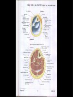
FIGURE 11.1. Structure of thin and thick skin.
➢ Layers of the epidermis and keratinization
᭹
The epidermis is a stratified squamous, keratinized (dry) epithe-
lium. It is continually renewed every 15–30 days. Rapid cell pro-
liferation occurs in the deepest layer (stratum germinativum) and
daughter cells differentiate as they migrate toward the surface.
This differentiation involves a process called keratinization, which
results in a variably thick layer of nonliving cellular husks at the
surface of the epidermis. All cells in the epidermis that undergo
the keratinization process are called keratinocytes.
᭹
Layers of the epidermis
᭜
Stratum germinativum—(basale). A single layer of cuboidal to
columnar shaped cells that rest on the basement membrane and
undergo rapid cell proliferation. These cells contain intermedi-
ate filaments composed of keratin.
᭜
Stratum spinosum. “Prickle-cell” or spiny cell layer; 3–10 cells
thick. This layer is so-called because the cells are attached to one
another by desmosomes, and the cellular shrinkage resulting
from fixation produces the spine-like structures. These cells
accumulate bundles of keratin filaments called tonofibrils.
᭜
Stratum granulosum: two to four cells thick; cells synthesize
basophilic, keratohyalin granules, which associate with the
tonofibrils. Cells also accumulate lamellar bodies, which contain
a lipid material that is secreted and serves as a sealant and
penetration barrier between cells. Cells also begin to lose other
organelles.
11.
Skin 95
FIGURE 11.2. Comparison of epidermal layers in thick and thin skin.
᭜
Stratum lucidum. A clear layer of non-nucleated, flattened cells
that is only visible as a distinct layer in thick skin. In this region,
the proteins contained in the keratohyalin granules mediate the
aggregation of bundles of keratin filaments (tonofilaments). This
process occurs whether or not a distinct stratum lucidum is
visible.
᭜
Stratum corneum. Variably thick layer of extremely flattened,
cornified scales containing aggregated tonofibrils surrounded
by a thickened plasma membrane. These cell remnants are
sloughed off without damage to the underlying, living epider-
mal cells.
➢ Other cell types
᭹
Melanocytes
᭜
Present in stratum germinativum and stratum spinosum
᭜
Rounded cell bodies with numerous “dendrite-like” processes
that insinuate themselves between the keratinocytes
᭜
Synthesize melanin, a dark brown pigment that is packaged into
melanosomes and injected into keratinocytes
᭜
Melanin caps the nucleus, reducing damage from solar radiation.
᭹
Langerhans cells. Macrophages that function in immunological
skin reactions
᭹
Merkel’s cells. Touch receptors.
➢ Epidermal-dermal junction
᭹
Scalloped margin at the interface of the epidermis and dermis,
formed by interdigitations of:
᭜
Epidermal pegs. Downward projections of the epidermis
᭜
Dermal papillae. Upward, finger-like protrusion of connective
tissue from the dermis
᭹
This junction strengthens the attachment of the epidermis to the
underlying dermis.
Dermis
➢ Composition
᭹
Papillary layer
᭜
Located immediately beneath the basement membrane of the
epidermis, forming the dermal papillae
᭜
Thin layer composed of loose connective tissue
᭜
Contains small blood vessels, nerves, lymphatics, and the
sensory receptors, Meissner’s corpuscles
96
Digital Histology
᭹
Reticular layer
᭜
Located between the papillary layer and the hypodermis
᭜
Thick layer composed of dense, irregular connective tissue
᭜
Contains larger nerves and blood vessels, glands, hair follicles,
and the sensory receptors, Pacinian corpuscles and Ruffini end
organs
➢ Vasculature of the dermis
᭹
Papillary plexus located in the dermal papillae
᭹
Cutaneous plexus located in the reticular layer of the dermis
᭹
Arteriovenous anastomoses allow shunting of blood between papil-
lary and cutaneous plexuses for temperature regulation.
Hypodermis
➢ Not technically part of the integument
➢ Composed of loose connective tissue and adipose tissue, which can
accumulate in large fatty deposits
➢ Provides anchorage for skin to the underlying tissues
➢ May contain the bases of sweat glands and hair follicles
➢ Many sensory receptors, especially Pacinian corpuscles, are present.
Structures Associated with the Skin
➢ Glands
᭹
Sweat glands
᭜
Simple, coiled tubular glands
᭜
Contain myoepithelial cells, which are specialized cells that con-
tract to aid in the expulsion of the sweat
᭜
Types of sweat glands
Merocrine or eccrine. Located in all regions of the body except
the axillary and anal regions; produce a watery secretion that
empties onto the surface of the epidermis
Apocrine. Restricted to the axillary, areolar, and anal regions;
much larger than eccrine sweat glands with a broader lumen.
Produce a viscous secretion that empties into the hair follicle.
Do not secrete by the apocrine mode.
᭹
Sebaceous glands
᭜
Simple, branched acinar glands
᭜
Usually secrete into a hair follicle
11.
Skin 97
᭜
Produce sebum, an oily secretory product, released by the
holocrine mode of secretion
᭜
Absent from thick skin
➢ Hair follicles
᭹
Invaginations of the epidermis
᭹
Consist of a bulb at the base of the follicle that is located in the
hypodermis or in the deep layers of the dermis. Internal and exter-
nal sheaths surround the growing hair shaft as it passes though the
dermis and epidermis.
᭹
An arrector pili muscle attaches a hair follicle to the papillary layer
of the dermis. Contraction provides elevation of the hair, forming
“goose-bumps.”
➢ Nails
᭹
Keratinized epithelial cells on the dorsal surface of the fingers and
toes
᭹
Consist of a nail plate that corresponds to the stratum corneum
of the epidermis. This plate rests on the nail bed, consisting of
cells corresponding to the stratum spinosum and stratum
germinativum.
➢ Sensory structures
᭹
Nonencapsulated. Free nerve endings in the epidermis, responsive
to touch, pressure, heat, cold, and pain
᭹
Encapsulated pressure receptors
᭜
Meissner’s corpuscle
Located at the apex of a dermal papilla
Consists of a coil of endoneurial cells around a nerve terminal
Responds to light touch
᭜
Pacinian corpuscle
Located in the dermis and hypodermis
Consists of concentric layers of endoneurial cells around a
nerve terminal
Responds to deep pressure
᭜
Ruffini ending
Located in the dermis
Consists of groups of nerve terminals surrounded by a thin
connective tissue capsule
Responds to touch and pressure
98
Digital Histology
11.
Skin 99
Thick skin
Thin skin
Epidermis
Epidermal pegs
Keratin filaments
Keratinocyte
Stratum corneum
Stratum germinativum
Melanocyte
Melanin granules
(melanosome)
Stratum granulosum
Stratum lucidum
Stratum spinosum
Dermis
Collagen bundles
Dermal papillae
Sensory papilla
Meissner’s corpuscle
Vascular papilla
Elastic fibers
Papillary layer
Reticular layer
Hypodermis
Adipose connective tissue
Hair follicle
Bulb
Papilla
Follicle sheath
Hair shaft
Pacinian corpuscle
Perineurial cells
Axon
Sebaceous gland
Sweat gland
Duct
Secretory portion
Myoepithelial cell
Secretory granules
Structures Identified in This Section
Oral Cavity
Components
➢ Vestibule. Bounded anteriorly and laterally by the lips and cheeks;
bounded medially by teeth and gingiva
➢ Oral cavity proper. Bounded anteriorly and laterally by the lingual sur-
faces of the teeth and gingiva, superiorly by the hard and soft palate,
inferiorly by the tongue and floor of the mouth, and posteriorly by
the pillars of the fauces leading to the pharynx
Oral Mucosa
➢ Oral mucosa, the mucous membrane lining the oral cavity, is con-
tinuous with external skin and with the mucous membrane of the
pharynx.
➢ Composition
᭹
Epithelium. Stratified squamous keratinized or nonkeratinized
depending on location
᭹
Lamina propria
᭹
Muscularis mucosae is not present.
CHAPTER
12
Digestive System
101
Digital Histology: An Interactive CD Atlas with Review Text, by Alice S. Pakurar and
John W. Bigbee
ISBN 0-471-64982-1 Copyright © 2004 John Wiley & Sons, Inc.
᭹
Although not part of the oral mucosa, a submucosa of dense
connective tissue, containing the minor salivary glands, underlies
much of the oral mucosa.
➢ Regional variations
᭹
Masticatory mucosa
᭜
Located where mucosa is exposed to forces of mastication, such
as gingiva and hard palate
᭜
Composition
Stratified squamous epithelium, keratinized
Underlying submucosa is lacking in some locations.
᭹
Lining mucosa
᭜
Located where mucosa is not exposed to forces of mastication,
such as lining of lips and cheeks, soft palate, alveolar mucosa,
undersurface of tongue, and floor of mouth
᭜
Epithelium. Stratified squamous epithelium, nonkeratinized
(moist)
᭹
Specialized mucosa
᭜
Named “specialized” due to the presence of taste buds
᭜
Located on the dorsum of the tongue, where it forms
papillae
᭜
Epithelium
Stratified squamous keratinized, modified to form filiform
papillae that facilitate the movement of food posteriorly
Stratified squamous moist, covering fungiform and circum-
vallate papillae
Tongue
➢ The subdivisions of the tongue are based on embryologic origins:
anterior two-thirds (body) and posterior one-third (root) are
separated by the sulcus terminalis.
➢ Composition
᭹
Mucosa. Dorsum of the tongue is lined by a specialized oral
mucosa, modified to form papillae. (See “Specialized mucosa”
above.) The ventral surface of the tongue is lined by a lining
mucosa.
᭹
The submucosa possesses minor salivary glands that are mucus-
secreting except for those associated with the circumvallate
papillae, which are serous-secreting.
102
Digital Histology
➢ Papillae. Each consists of a connective tissue core covered by a
stratified squamous epithelium.
᭹
Filiform
᭜
Most numerous; cover body of tongue
᭜
Cone-shaped protrusions angled so that they aid in movement
of food toward the pharynx
᭹
Fungiform
᭜
Less numerous than filiform but also located on anterior
two-thirds of tongue
᭜
Mushroom shaped, possess taste buds on superior surface
᭹
Circumvallate
᭜
Eight to twelve papillae located just anterior to the sulcus
terminalis
᭜
Mushroom shaped and surrounded by a narrow moat; lateral
wall of papilla possesses taste buds
᭜
Serous glands of von Ebner open into the base of the moat and
flush the moat for reception of new tastes.
᭹
Foliate. Parallel folds on the posterolateral surface of the tongue;
not well developed in humans
➢ Taste buds are onion-shaped structures embedded in the surface of
the fungiform and circumvallate papillae. Taste buds contain taste-
receptor cells that communicate with the surface of the papilla
through a taste pore. Depolarization of the taste cells leads to the stim-
ulation of gustatory nerve fibers and the discrimination of sweet,
salty, bitter, and sour sensations.
➢ Intrinsic tongue muscles. Skeletal muscle bundles are arranged in three
separate planes, with connective tissue bands from the lamina
propria separating the bundles and firmly anchoring the muscle to
the mucous membrane.
Teeth
➢ Overview of the teeth
᭹
Anatomic crown. The portion of the tooth covered by enamel.
᭹
Anatomic root. The portion of the tooth covered by cementum.
᭹
Cervix. Region where enamel abuts cementum
᭹
Pulp cavity is the central core of a tooth and is divided into a pulp
chamber in the crown and a root canal in the root. An apical
foramen at the tip of the root allows passage of nerves and blood
vessels into and out of the pulp cavity.
12.
Digestive System 103
᭹
Gingiva. Oral mucosa encircling the cervical region of the tooth and
providing support for the tooth
➢ Components
᭹
Enamel is the hardest tissue in the body, consisting of a mineral-
ized tissue that is 96% hydroxyapatite. Enamel covers the anatomic
crown of the tooth. During tooth development, enamel deposition
by ameloblasts begins on the surface of dentin and progresses
away from this dentinoenamel junction. No additional enamel
can be formed after the tooth erupts, as the ameloblasts die on
exposure to the oral cavity.
᭹
Dentin
᭜
Comprises the bulk of the tooth, underlying both enamel and
cementum; dentin is a connective tissue that is 70% mineralized
with hydroxyapatite.
᭜
Dentin is formed continuously throughout life by odontoblasts
whose cell bodies line the pulp cavity.
᭜
Odontoblast processes extend through the dentin in S-shaped
dentinal tubules radiating from the odontoblasts toward the
dentinoenamel or dentinocemental junctions.
᭹
Cementum, a connective tissue mineralized with 50% hydroxyap-
atite, covers the anatomic root of the tooth. Cementum is formed
continuously throughout life by activity of cementoblasts lying on
the surface of the root at the interface of the cementum with the
periodontal ligament.
᭹
The pulp cavity is lined by odontoblasts and filled with loose
connective tissue, blood vessels, nerves, and lymphatics.
᭹
The periodontal ligament, collagen fiber bundles interconnecting
cementum with the surrounding alveolar bone, suspends and
supports each tooth in its alveolar socket.
Major Salivary Glands
➢ Overview
᭹
All major salivary glands are compound, exocrine glands, and all
open into the oral cavity.
᭹
Functions
᭜
Produce saliva to wet, lubricate, and buffer the oral cavity and
its contents
᭜
Produce amylase for the initial digestion of carbohydrates
᭜
Produce lysozyme to control bacteria in the oral cavity
104
Digital Histology
➢ Major cell types
᭹
Serous cells
᭜
Synthesize, store, and release a thin, protein-rich secretion
containing digestive enzymes, primarily amylase
᭜
Are pyramidal in shape and possess all organelles necessary for
protein production and secretion (e.g., basal RER, Golgi, and
apical secretory granules)
᭜
Are arranged into either
Acini (singular, acinus) or alveoli (singular, alveolus). Flask-
shaped sacs with tiny lumens
Serous demilunes. Half moon–shaped caps positioned over the
ends of mucous tubules
᭹
Mucous cells
᭜
Synthesize, store, and release mucus, a viscous, thick, glyco-
protein secretion that protects and lubricates epithelia
᭜
Have flattened nuclei that are located at the bases of the cells
along with the RER. Abundant mucigen droplets are located in
the apex of each cell, giving it a frothy, vacuolated appearance.
᭜
Are organized in test tube–shaped tubules with relatively wide
lumens
᭹
Myoepithelial cells are stellate-shaped epithelial cells with contrac-
tile functions that lie between the secretory or duct cells and the
basement membrane. These cells contract to aid in movement of
the secretory product.
➢ Duct system conducts secretions to oral cavity.
᭹
Ducts are more numerous with serous acini than with mucous
tubules because the tubules can act as their own ducts.
᭜
Intralobular ducts
Intercalated ducts exit from secretory acini and are smaller in
diameter than the acini they drain. These ducts are lined by
simple cuboidal epithelia.
Striated ducts are larger in diameter than the secretory
units they drain. They are lined by simple columnar epithe-
lia. Numerous mitochondria and infoldings of the plasma
membrane in the basal region of the cells give the duct a
striated periphery. Striated ducts alter the content and con-
centration of the saliva.
᭜
Interlobular ducts are located in the connective septa between
lobules and are lined with simple columnar to stratified colum-
nar epithelia.
12.
Digestive System 105
106
Digital Histology
Oral Cavity
Lip
Connective tissue
Connective tissue papillae
Hair
Labial glands
Labial glands, ducts
Mucosa, lining
Orbicularis oris
Sebaceous glands
Skeletal muscle
Skin
Epithelium, stratified squamous
moist
Epithelium, stratified squamous
keratinized
Vermilion zone
Tooth
Cementum
Dentin
Dentinal tubules
Dentino-cementum junction
Dentino-enamel junction
Enamel
Enamel stria
Pulp cavity
Tongue
Capillaries
Connective tissue core
Dorsal surface
Epithelium, keratinized
Furrow (moat)
Papillae, circumvallate
Papillae, filiform
Papillae, fungiform
Serous glands of von Ebner
Serous glands of von Ebner,
ducts
Skeletal muscle
Taste buds
Taste pores
Ventral surface
᭜
The main excretory duct(s) is lined by a stratified epithelium that
becomes stratified squamous moist just prior to its junction with
the epithelium of the oral cavity.
➢ Major salivary glands
᭹
Parotid glands
᭜
Compound acinar glands producing only serous products; their
secretions account for 25% of the saliva
᭜
Possess the most highly developed duct system of the major
salivary glands
᭹
Submandibular glands
᭜
Compound tubulo-acinar glands producing both serous and
mucous products, although serous acini predominate. Their
secretions account for 70% of the saliva.
᭜
Serous cells are present as both acini and serous demilunes.
᭹
Sublingual glands secrete approximately 5% of the saliva. These
are compound tubulo-acinar glands, producing both mucous and
serous products, although mucous tubules predominate.
Structures Identified in This Section
12.
Digestive System 107
Major salivary glands
Connective tissue septa
Ducts, intercalated
Ducts, interlobular
Ducts, striated
Lobules
Secretory units
Parotid gland
Acinar lumens
Artery, small
Capillaries
Ducts, intercalated
Ducts, interlobular
Ducts, intralobular
Ducts, striated
Epithelium, stratified columnar
Interlobular connective tissue
Lobules
Peripheral nuclei
Secretory granules
Serous acini
Vein, small
Submandibular gland
Acinar lumens
Ducts, intercalated
Ducts, interlobular
Ducts, intralobular
Ducts, striated
Mucous tubules
Secretory granules
Serous acini
Tubular lumens
Sublingual gland
Ducts, intercalated
Ducts, interlobular
Ducts, intralobular
Lobules
Mucous cells
Mucous tubules
Serous acini
Serous cells
Serous demilune
Tubular Digestive System
Components
➢ Pharynx
➢ Esophagus
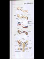
FIGURE 12.1. Organs comprising the tubular digestive tract.
➢ Stomach
➢ Small intestine
➢ Large intestine
Basic Histological Organization
➢ Layers
᭹
Mucosa (mucous membrane). Innermost layer facing the lumen
᭜
Epithelium. Either a stratified squamous moist or a simple
columnar epithelium
᭜
Lamina propria. Loose connective tissue; usually possesses
digestive glands
᭜
Muscularis mucosae of smooth muscle is usually present.
᭹
Submucosa. Denser connective tissue than the lamina propria. The
submucosa possesses Meissner’s nerve plexus that supplies inner-
vation to the muscularis mucosae and to digestive glands in the
mucosa and submucosa. The submucosa possesses glands in the
esophagus and duodenum.
᭹
Muscularis externa of smooth muscle is usually arranged into inner
circular and outer longitudinal layers. Auerbach’s nerve plexus is
located between the two muscle layers and provides innervation
to this smooth muscle.
᭹
Serosa (serous membrane) is present if the organ protrudes into
the peritoneal cavity, or an adventitia (only the connective tissue
portion of the serosa) is present if the organ is retroperitoneal.
108
Digital Histology
FIGURE 12.2. Overview of the layers and components of the tubular digestive tract.
➢ Glands
᭹
Exocrine glands, aiding in digestion and/or lubrication, are
located in:
᭜
Epithelium (e.g., goblet cells throughout the intestines)
᭜
Lamina propria (e.g., gastric glands)
᭜
Submucosa (e.g., Brunner’s glands in the duodenum)
᭜
Glands located external to the digestive tract that open into the
system (e.g., liver and pancreas)
᭹
Endocrine and paracrine cells, belonging to the diffuse neuroen-
docrine system (DNES), are located throughout the mucosa of the
gastrointestinal tract, influencing the secretion of glands and the
motility of the gut.
Variations That Distinguish Each Organ from the
Basic Organizational Plan
➢ Esophagus
᭹
Epithelium. Stratified squamous nonkeratinized epithelium
᭹
Lamina propria possesses esophageal cardiac glands that are
mucus-secreting and are particularly prominent near the junction
of the esophagus with the stomach.
᭹
Submucosa has mucus-secreting, esophageal glands.
᭹
Muscularis externa is composed of striated muscle in the upper
portion of the esophagus, skeletal, and smooth muscle in the
middle portion, and smooth muscle in the lower portion.
᭹
Adventitia. Composed of loose connective tissue.
➢ Stomach
᭹
Structures present throughout the stomach
12.
Digestive System 109
FIGURE 12.3. Cross-section of the esophagus.
᭜
Surface epithelium
Simple columnar epithelium facing the lumen is modified so
that all cells secrete mucus, forming a sheet gland that protects
the stomach from its acidic environment.
Gastric pit. A channel formed by the invagination of the
surface epithelium into the underlying lamina propria; con-
nects the sheet gland with the gastric glands. The length of
the gastric pit varies with each stomach region.
᭜
Gastric glands
Simple, branched tubular glands begin at a gastric pit and
extend through the lamina propria to the muscularis
mucosae.
The region of the gland that attaches to the gastric pit is called
the neck region; the base region of the gland is located adja-
cent to the muscularis mucosae.
Secretory cells in these glands vary in each region of the
stomach.
᭜
Muscularis externa. Subdivisions of this layer frequently
interdigitate, making it difficult to distinguish one layer from
another.
Internal oblique layer
Middle circular layer that is modified in the pyloric region to
form the pyloric sphincter
Outer longitudinal layer is separated from the inner circular
layer by Auerbach’s plexus, nerve fibers from the autonomic
nervous system that supply muscularis externa.
110
Digital Histology
FIGURE 12.4. Cross-section of the stomach.
᭜
Serosa
᭜
Rugae. Longitudinal folds of the mucosa and submucosa in the
undistended stomach allow for expansion.
᭹
Variations specific to the cardiac region (narrow region adjacent to
the esophagus)
᭜
Abrupt transition of epithelium from stratified squamous
moist of the esophagus to a sheet gland lining the cardiac
stomach
᭜
Length of gastric pits is about equal to the length of cardiac
glands.
᭜
Cardiac glands primarily secrete mucus, although other products
are also produced. Glands are frequently coiled.
᭜
Cardiac glands of the stomach extend into the lower esophagus,
forming the esophageal cardiac glands.
᭹
Variations specific to the fundic and body regions (Glands in both
regions are called fundic glands.)
᭜
Fundic glands are about twice as long as their gastric pits.
᭜
Cell types present:
Stem cells replenish both the surface epithelial cells and cells
of the glands. Stem cells are located in the neck region.
Mucous neck cells are irregular in shape and stain basophili-
cally. They secrete mucus and are located in the neck region.
Parietal cells are large, spherical, eosinophilic cells that secrete
hydrogen and chloride ions and gastric intrinsic factor.
They possess numerous mitochondria. An umbrella-shaped
canaliculus indents the luminal surface, increasing surface
area. Although present throughout the gland, parietal cells
are more numerous in the upper regions.
Chief or zymogen cells, typical protein-producing cells,
predominate in the bases; stain blue with hematoxylin and
secrete pepsinogen.
Enteroendocrine cells (part of the diffuse neuroendocrine
system, DNES) are located on the basement membrane and
do not usually reach the lumen of the gland. This population
of cells secretes a variety of hormones with endocrine and
paracrine influences on digestive activity. Secretory granules
cluster toward the basement membrane for their subsequent
release into the lamina propria. Most common at the bases of
the glands.
᭹
Variations specific to the pyloric region
12.
Digestive System 111
᭜
Pits are longer in pylorus than in the cardiac region.
᭜
Pyloric glands, not as coiled as in the cardiac region; primarily
secrete mucus.
᭜
Enteroendocrine cells are also present here.
᭜
Circular layer of muscularis externa is greatly thickened to form
the pyloric sphincter.
➢ Small intestine
᭹
Subdivided into duodenum, jejunum, and ileum
᭹
Common features of the small intestine
᭜
Structures that increase the surface area of the small intestine
Microvilli. Increase surface area of absorptive cells and,
collectively, form a brush or striated border
Villi. Finger-like protrusions of the lamina propria and
overlying epithelium into the lumen
– Villi assume different shapes in each of the three intestinal
subdivisions.
– A lacteal (blind-ending lymphatic capillary) is located in
the center of each villus to absorb digested fat.
– Individual smooth muscle cells lie parallel to the long
axis of each villus, “milking” the lacteal contents to the
periphery.
Plicae circulares. Permanent circular folds formed by an up-
welling of the submucosa and its overlying mucosa into the
lumen. Villi protrude from the plicae.
112
Digital Histology
FIGURE 12.5. Longitudinal section through the duodenum (left) and the jejunum/
ileum (right). Note the orientation of the layers of muscularis externa when sectioned
longitudinally.
᭜
Mucosal epithelium is composed of:
Absorptive cells, forming a simple columnar epithelium with
microvilli, absorb digested food
Goblet cells (unicellular glands) are interspersed among
absorptive cells and secrete mucus. These cells increase in
number from duodenum to rectum.
᭜
Intestinal glands (crypts of Lieberkuhn) are simple tubular glands
that begin at the bases of the villi in the mucosa and extend
through the lamina propria to the muscularis mucosae. Possess:
Absorptive cells
Goblet cells
Paneth cells possess large, eosinophilic granules whose con-
tents digest bacterial-cell walls.
Enteroendocrine cells
᭜
Muscularis externa of inner circular and outer longitudinal
layers with an intervening Auerbach’s nerve plexus
᭜
Serosa covers all of small intestine except for the beginning
of the duodenum, which is retroperitoneal and possesses an
adventitia.
᭹
Variations specific to the intestinal subdivisions
᭜
Brunner’s glands in the submucosa are present only in the duo-
denum. These compound tubular glands open into the bases of
the intestinal glands and secrete an alkaline mucus to neutral-
ize the acidity of the stomach contents.
᭜
Peyer’s patches are clusters of 10–200 lymphoid nodules located
primarily in the lamina propria of the ileum. Each cluster is posi-
tioned on the side of the intestine away from the mesentery and
forms a bulge that may protrude into the lumen as well as into
the submucosa.
➢ Large intestine (colon)
᭹
Mucosal epithelium:
᭜
Absorptive cells form a simple columnar epithelium with
microvilli.
᭜
Goblet cells increase in number toward the rectum and provide
lubrication.
᭜
A reduced number of enteroendocrine cells is present.
᭹
Intestinal glands (crypts of Lieberkuhn) are very straight in the
large intestine.
᭹
No villi or plicae circulares are present in the large intestine.
12.
Digestive System 113
᭹
Muscularis externa
᭜
Inner circular layer is intact.
᭜
Outer longitudinal layer is segregated into three longitudinal
bands, the taeniae coli, that are placed equidistantly around the
tube. The contraction of the taenia produces permanent saccu-
lations in the large intestine, termed haustrae.
᭹
Either an adventitia or a serosa is present, depending on the
particular portion of the large intestine.
᭹
The appendix resembles the large intestine except that the outer
longitudinal smooth muscle layer is intact. Additionally, abundant
lymphoid tissue is present in the lamina propria to protect against
invading microorganisms.
᭹
Rectum is a 12-cm-long tube continuing from the sigmoid colon.
The mucosa of the rectum is similar to that of the majority of the
large intestine. The rectum narrows abruptly to become the anal
canal.
᭹
Anal canal. The terminal portion of the intestinal tract is about
4cm long.
᭜
The intestinal glands disappear and the epithelium undergoes
an abrupt transition from simple columnar to stratified squa-
mous with sebaceous and apocrine sweat glands.
᭜
The inner circular portion of the muscularis externa expands to
form the internal anal sphincter. The external anal sphincter is
composed of skeletal muscle.
114
Digital Histology
FIGURE 12.6. Cross-section of the large intestine.
12.
Digestive System 115
Overview of tubular digestive tract
Adventitia
Epithelium
Esophagus
Glands
Lamina propria
Large intestine
Mucosa
Muscularis externa
Muscularis mucosae
Plicae circulares
Serosa
Small intestine
Stomach
Submucosa
Esophagus
Adventitia
Capillaries
Cardiac glands
Cardiac stomach
Epithelium, stratified
squamous moist
Esophageal glands
Gastric pits
Gastric pits, openings
Gastroesophageal junction
Lamina propria
Mucosa
Muscularis externa
Muscularis mucosae
Skeletal muscle (ls and xs)
Smooth muscle
Squamous cells
Submucosa
Stomach
Auerbach’s plexus
Brunner’s glands
Cardiac glands
Chief cells
Collagen fibers
Enteroendocrine cells
Fundic glands
Gastric glands
Gastric glands, bodies
Gastric glands, branching
Gastric glands, necks
Gastric pits
Gastroduodenal junction
Intestinal glands
Lamina propria
Lymphoid nodule
Mast cells
Meissner’s plexus
Mucosa
Mucous neck cells
Mucigen
Muscularis externa
Muscularis externa, middle
circular layer
Muscularis externa, outer
longitudinal layer
Muscularis mucosae
Neuron cell body
Parietal cells
Peripheral nerves
Plasma cells
Pyloric glands
Pyloric sphincter
Secretory granules
Serosa
Sheet gland
Sheet gland, stem of cell
Stomach, cardiac
Stomach, fundic and body
Stomach, pyloric
Submucosa
Villi
Small intestine
Absorptive cells
(enterocytes)
Brunner’s glands
Brunner’s glands, ducts
Duodenum
Enteroendocrine cells
Structures Identified in This Section
116
Digital Histology
Epithelium, simple columnar
with microvilli
Epithelium, simple squamous
Germinal center
Goblet cells
Intestinal glands
Jejunum/ileum
Junctional complex
Lacteal
Lamina propria
Lumen
Lymphoid nodule
Mesentery
Microvilli
Mucosa
Muscularis externa
Muscularis mucosae
Paneth cells
Peyer’s patch
Plasma cells
Plicae circulares
Serosa
Submucosa
Villi
Large intestine
Anal canal
Anal sphincter, external
Anal sphincter, internal
Anus
Apocrine sweat glands
Diffuse lymphoid tissue
Hair follicle
Intestinal glands
Lymphatic nodules
Mucosa
Muscularis externa
Muscularis externa, inner
circular layer
Muscularis externa, outer
longitudinal layer
Recto-anal junction
Rectum
Serosa
Submucosa
Taeniae coli
Major Digestive Glands
Pancreas
Overview
➢ Located in the abdomen in the curve of the duodenum and divided
into a head, body, and tail
➢ Is both an exocrine and an endocrine gland
᭹
The exocrine portion produces an alkaline secretion containing
digestive enzymes that empties into the duodenum.
᭹
The endocrine portion secretes insulin, glucagon, and somatostatin
that regulate blood glucose levels.
Microscopic Anatomy
➢ Exocrine pancreas
᭹
Compound acinar gland; the acinar cells secrete numerous diges-
tive enzymes that breakdown proteins, carbohydrates, lipids, and
nucleic acids.