Digital histology an interactive cd atlas with review text phần 7 potx
Bạn đang xem bản rút gọn của tài liệu. Xem và tải ngay bản đầy đủ của tài liệu tại đây (300.35 KB, 22 trang )
Inner zone (paracortex or deep cortex). Filled with diffuse lym-
phoid tissue composed of T lymphocytes
Sinuses in cortex. Loose network of macrophages and reticu-
lar fibers through which lymph percolates
– Subcapsular sinus lies immediately beneath the capsule and
receives incoming lymph fluid from afferent lymphatic
vessels that enter through the capsule.
– Intermediate sinuses. Lie adjacent to the trabeculae. Receive
lymph from the subcapsular sinus and continue as
medullary sinuses
᭜
Medulla. Composed of:
Medullary cords of B lymphocytes that extend from the inner
cortex into the medulla
Medullary sinuses. Continuations of the intermediate sinuses
in the cortex. Lymph flows from medullary sinuses into the
efferent lymph vessels that exit at the hilum of the node.
᭹
Blood supply. Small arteries enter at the hilum to supply a capil-
lary plexus in the outer cortex. The capillaries anastomose to form
HEVs in the paracortex and small veins that exit at the hilum.
᭹
Filter and provide immune surveillance for lymph
➢ Spleen
᭹
Encapsulated, intraperitoneal organ located in upper left quadrant
of the abdominal cavity
᭹
Structure
᭜
Capsule surrounds organ, sending trabeculae into the spleen.
Larger blood vessels enter through the trabeculae.
᭜
Subdivisions
White pulp appears white in fresh specimens and is composed
of:
– Periarterial lymphoid sheath (PALS). A sleeve of T lympho-
cytes that surrounds a central arteriole as soon as it exits
from a trabecula
– Lymphoid nodules, composed of B lymphocytes, are ran-
domly located along and embedded in the PALS.
Red pulp appears red in fresh specimens because of the abun-
dant venous sinuses it possesses.
– Splenic cords (of Billroth). Cords of lymphocytes (T and B),
macrophages, plasma cells, and other lymphoid cells sus-
pended in a reticular connective tissue stroma. Surrounded
by:
140
Digital Histology
– Splenic sinuses. Venous sinuses separating splenic cords.
These sinuses are lined by endothelial cells and surrounded
by reticular fibers.
᭜
The spleen filters and provides immune surveillance for the
blood percolating through it. The spleen also phagocytoses aged
and abnormal erythrocytes and stores blood.
᭹
Blood flow through the spleen
᭜
Splenic artery enters at the hilum of the spleen and branches
into arteries that lie in the trabeculae.
᭜
Arteries exit from the trabeculae as central arterioles and are
immediately surrounded by the PALS. The central arteriole
becomes eccentrically located when it is displaced by a lym-
phoid nodule. Branches from the central arterioles supply the
PALS, including forming marginal sinuses at the perimeter of
the white pulp.
᭜
Central arterioles lose their PALS ensheathment and form a
series of smaller arterioles in the red pulp. These arterioles
either:
Open directly into a splenic sinus (closed circulation)
Open into a splenic cord where the blood percolates through
the cells of the cord before entering a splenic sinus (open
circulation)
᭜
Trabecular veins are formed by splenic sinuses anastomosing
and then entering a trabecula. Trabecular veins anastomose to
form the splenic vein.
᭜
The splenic vein exits at the hilum of the spleen.
➢ Thymus
᭹
Thymus is a primary lymphoid organ that receives immature
lymphocytes (thymocytes) from the bone marrow. These cells
mature in the thymus and are carried to secondary lymphoid
structures/organs via the blood vascular system.
᭹
The thymus is located in the superior mediastinum under the
sternum. The thymus involutes after puberty.
᭹
Structure
᭜
A connective tissue capsule surrounds the thymus and extends
into the thymus, dividing it into lobules.
᭜
The stroma is formed by a network of reticular cells of
endodermal, rather than the usual mesodermal, origin and are
called, therefore, epithelial reticular cells. These cells do not form
fibers.
᭜
Each lobule contains an:
14.
Lymphoid System 141
142
Digital Histology
Overview
Artery
Bone marrow
Capillary
Diffuse lymphoid tissue
Mucosa
Epithelial barrier
Lamina propria
Mucosal glands
Muscularis mucosae
Large intestine
Lymph capillary
Lymph nodes
Lymph vessels
Lymphoid nodules
MALT
Microbes
Peripheral nerve
Small intestine
Spleen
Thymus
Tonsils
Valve
Vein
Lymphoid tissues
Adventitia
Arteriole
Capsule
Crypt epithelium
Crypts
Dense connective tissue capsule
Diffuse lymphoid tissue
Dome
Endothelium
Epithelium, stratified squamous
Germinal center
High endothelial venules
Lymph
Lymphatic vessel
Lymphoblasts
Lymphoid nodules
Macrophages
Mitotic figures
Muscularis mucosae
Nodular lymphoid tissue
Primary nodule
Reticular cells
Secondary lymphoid nodule
Septa
Skeletal muscle
Small lymphocytes
Smooth muscle
Solitary lymphoid nodule
Tonsils
Valve flap
Outer cortex that is densely packed with thymocytes, the
developing T lymphocytes. These cells mature in the cortex,
then migrate into the medulla where they enter the blood
stream for transport to secondary lymphoid structures and
organs.
Inner medulla has fewer thymocytes and, therefore, stains
more palely than does the cortex. Hassall’s corpuscles are the
degenerating remains of the epithelial reticular cells with
their keratin granules and are diagnostic for the thymus.
᭹
A blood-thymic barrier is formed around capillaries in the cortex,
so that the developing lymphocytes are not exposed to circulating
antigens.
Structures Identified in This Section
14.
Lymphoid System 143
Venule
Lymph nodes
Afferent lymphatics
Arteriole
Artery and vein
Capsule
Cortex
Outer cortex
Paracortex
Cortical sinuses
Deep cortex
Efferent lymphatic
Epithelium, simple squamous
High endothelial venules
Hilum
Lymphocytes
Lymphoid nodules
Macrophages
Medulla
Medullary cords
Medullary sinuses
Reticular cells
Sinuses
Subcapsular sinus
Trabeculae
Valve
Venule
Spleen
Capsule
Endothelial cells
Macrophages
Red pulp
Red pulp arterioles
Splenic cords
Splenic sinuses
Circulation, closed
Circulation, open
Reticular fibers
Splenic veins
Trabeculae
Trabecular artery
Venous drainage
White pulp
Central arteriole
Germinal centers
Lymphoid nodules
Marginal zone
PALS
White pulp vasculature
Thymus
Blood vessels
Capsule
Cortex
Epithelial reticular cells
Hassall’s corpuscles
Keratohyaline
Lobule
Lymphoblasts
Medulla
Septa
Thymic lymphocytes
Thymocytes
Components
➢ Kidneys. Contain the uriniferous tubules, which consist of nephrons
and a system of collecting ducts; filter blood and produce urine
➢ Ureters. Muscular tubes that collect urine output from the kidney and
carry it to the urinary bladder
➢ Urinary bladder. Hollow muscular organ that stores urine
➢ Urethra. Tube that drains urine from urinary bladder to the exterior
Functions of the Urinary System
➢ Excretion of waste products of metabolism
➢ Regulation and maintenance of the fluid volume of the body
➢ Regulation of acid-base balance
➢ Regulation of salt concentrations and other compounds in body
fluids
➢ Production of renin, an enzyme that influences blood pressure
Macroscopic Organization of the Kidney
➢ Cortex. Broad outer zone of kidney
CHAPTER
15
Urinary System
145
Digital Histology: An Interactive CD Atlas with Review Text, by Alice S. Pakurar and
John W. Bigbee
ISBN 0-471-64982-1 Copyright © 2004 John Wiley & Sons, Inc.
᭹
Subdivisions
᭜
Labyrinth. “True” cortical tissue
᭜
Medullary rays. Medullary tissue located in the cortex
᭹
Consists of renal corpuscles, portions of renal tubules, and col-
lecting ducts
➢ Medulla. Deep to cortex
᭹
Subdivisions
᭜
Renal pyramids. Inverted cones whose bases are adjacent to the
cortex; send “stripes” of medullary tissue into the cortex
forming the medullary rays
᭜
Renal columns. Extensions of cortical tissue between renal
pyramids
᭹
Consists of portions of renal tubules and collecting ducts
➢ Renal lobulations
᭹
Renal lobe. A medullary pyramid, the surrounding renal column
extending to the interlobar vessels, and the overlying cortical
tissue
᭹
Renal lobule. A central medullary ray and the adjacent cortical
labyrinth extending to the interlobular vessels
➢ Extrarenal passageways
᭹
Minor calyx. Funnel-shaped structure (one for each pyramid) into
which the point (apex) of a pyramid projects; urine flows from the
pyramid into a minor calyx and several minor calyces unite to form
a major calyx.
146
Digital Histology
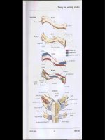
FIGURE 15.1. Extrarenal passageways and vascular supply of the kidney.
᭹
Major calyx. Four or five per kidney; formed by the confluence of
minor calyces
᭹
Renal pelvis. Structure formed by the uniting of the major calyces;
forms the expanded upper portion of the ureter
The Nephron
➢ 1.5–2 million per kidney
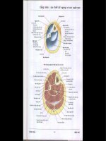
➢ Renal corpuscle
᭹
Located in the cortical labyrinth
᭹
Components
᭜
Glomerulus. A tuft of fenestrated capillaries, whose pores lack
diaphragms; filter blood. Formed by an afferent arteriole, the
glomerulus indents into Bowman’s capsule like a baseball fits
into a baseball glove. Blood leaves the glomerulus via the effer-
ent arteriole.
᭜
Bowman’s capsule. Double-walled, epithelial capsule with central
space called Bowman’s space; surrounds the glomerulus and
receives the fluid filtered from the blood
Parietal layer. Outer layer, simple squamous epithelium which
is reflected at the vascular pole of the renal corpuscle to
become the visceral layer; continuous with the proximal
tubule at the urinary pole
15.
Urinary System 147
FIGURE 15.2. The nephron, collecting tubule, and associated blood supply.
Visceral layer. Inner layer surrounding the glomerulus. Con-
sists of a single layer of modified epithelial cells called
podocytes. The radiating foot processes of these cells give rise
to many secondary processes called pedicels. Pedicels of adja-
cent podocytes interdigitate and surround the glomerular
capillaries. The slits (filtration slits) between the pedicels are
bridged by slit diaphragms.
᭜
Filtration barrier. Barrier between blood in glomerular capillary
and space of Bowman’s capsule
Fenestrated endothelium of glomerular capillary
Thick, fused basal laminae of the podocytes and the glomeru-
lar endothelial cells
Slit diaphragms between pedicels of visceral layer of
epithelium
᭜
Poles of the glomerulus
Vascular pole. Where afferent and efferent arterioles enter and
leave the renal corpuscle, respectively
Urinary pole. Where the parietal layer of Bowman’s capsule is
continuous with the proximal convoluted tubule
➢ Renal tubule
᭹
The glomerular filtrate of the blood continues from Bowman’s
space into the renal tubule, which meanders first through the
cortex, then the medulla, then back to the cortex, and finally enters
the collecting duct.
᭹
Regions of the renal tubule
᭜
Listed in order are regions of the renal tubule through which
urine passes
Proximal convoluted tubule
Proximal straight tubule
Thin limbs
Distal straight tubule
Distal convoluted tubule
᭜
Proximal tubule, convoluted portion
Located in labyrinth of cortex; highly convoluted
Interconnects parietal epithelium of Bowman’s capsule with
straight portion of proximal tubule
Composed of a simple cuboidal epithelium with microvilli;
cells possess numerous infoldings of the basal plasma mem-
brane and many mitochondria
148
Digital Histology
Absorption of glucose, amino acids, and the majority of salt
and water occur here.
᭜
Loop of Henle. Located in medullary tissue (i.e., medullary ray
and medulla)
Proximal tubule, straight portion (thick descending limb of loop of
Henle)
– Located either in medullary ray (in cortex) or in medulla
– Interconnects proximal convoluted tubule with thin limb of
Henle’s loop
– Histology is identical to that of the proximal convoluted
tubule
– Absorption of same substances as in proximal convoluted
tubule
Thin segment
– Found in medulla
– Interconnects proximal straight tubule with distal straight
tubule
– Frequently makes the “loop” in the loop of Henle
– Composed of a simple squamous epithelium
– Actively pumps out chloride, with sodium following pas-
sively, to produce a hypertonic urine
Distal tubule, straight portion (thick ascending limb of Henle’s
loop).
– Located either in medulla or in medullary ray (in cortex)
– Interconnects thin segment with distal convoluted tubule
– Composed of a simple cuboidal epithelium with inconsis-
tent microvilli. The cytoplasm is less acidophilic and the
lumen is wider than in the proximal tubule. The basal
plasma membrane is extensively infolded with numerous
mitochondria between the folds.
᭜
Distal tubule, convoluted portion
Located in the labyrinth portion of cortex; highly
convoluted
Interconnects the distal straight tubule with collecting tubule
Histology is identical with the distal straight tubule
Returns to a glomerulus to form part of the juxtaglomerular
apparatus
Major site of salt and water control in the body
15.
Urinary System 149
᭹
Juxtaglomerular (JG) apparatus
᭜
Located at the vascular pole of a nephron; helps regulate blood
pressure
᭜
JG cells. Modified smooth muscle cells in wall of an afferent
arteriole
᭜
Macula densa. Cluster of modified cells in the wall of a distal con-
voluted tubule adjacent to the JG cells. The clustering of cells,
and therefore of their nuclei, gives the appearance of a “dense
spot” in the wall of the distal convoluted tubule.
᭜
Monitors the tonicity of the urine in the distal tubule. The
macula densa affects the adjacent JG cells to adjust their pro-
duction of renin, a hormone that aids in regulating blood
pressure.
Excretory Tubules and Ducts and
Extrarenal Passages
➢ Separate embryological origin from the nephron
➢ Components
᭹
Collecting tubule
᭜
Composed of simple cuboidal to simple columnar cells; usually
displays distinct lateral boundaries between cells
150
Digital Histology
FIGURE 15.3. The renal corpuscle and associated structures.
᭜
Drains urine from the distal convoluted tubule of many
nephrons in the cortical labyrinth, enters the medullary ray in
the cortex and descends into the medulla
᭜
Joins with other collecting tubules to form the papillary ducts
(of Bellini)
᭜
Aids in concentrating the urine
᭹
Papillary ducts. Located deep in the medullary pyramid near the
minor calyces; composed of a tall, pale, simple columnar epithe-
lium. Empty into the minor calyx at the area cribosa at the apex of
each pyramid
᭹
Minor and major calyces. Transport urine to the renal pelvis and into
the ureter; lined by transitional epithelium
᭹
Renal pelvis. Expanded origin of the ureter, lined by transitional
epithelium; formed by the union of major calyces
᭹
Ureter. Muscular tube connecting the renal pelvis and the urinary
bladder, lined by transitional epithelium; two layers of smooth
muscle in the upper two-thirds, inner longitudinal and outer cir-
cular, with the addition of a third outer longitudinal layer in the
lower one-third
᭹
Urinary bladder. Lined by a transitional epithelium, a stratified
cuboidal epithelium specialized to provide for distension of the
organ; a thick muscular wall contains three interlacing layers of
smooth muscle.
Blood Supply of the Kidney
➢ Renal artery. A branch of the aorta, enters the kidney at the hilus;
branches to form the interlobular arteries
➢ Interlobar arteries. Lie between adjacent pyramids in renal columns
and branch into arcuate arteries
➢ Arcuate arteries. Arch between medulla and cortex; give rise to inter-
lobular arteries
➢ Interlobular arteries. Branch perpendicular to the arcuate artery in the
cortex and lie between adjacent lobules; supply a number of afferent
arterioles
➢ Afferent arterioles supply the glomerulus, entering at the vascular pole
of the renal corpuscle
➢ An efferent arteriole exits from the glomerulus and forms either per-
itubular capillaries, which nourish the convoluted tubules, or the vasa
recta. The vasa recta parallel the straight portions of the renal tubule
into the medulla and play an important role is concentrating the
urine.
15.
Urinary System 151
152
Digital Histology
Blood vessels
Afferent arteriole
Arcuate vessels
Interlobular arteries
Peritubular capillaries
Vasa recta
Kidney
Cortex
Convoluted portion (cortical
labyrinth)
Medullary rays
Medulla
Area cribosa
Medullary pyramid
Minor calyx
Renal papilla
Nephron and collecting ducts
Basal lamina
Bowman’s space
Collecting ducts
Distal convoluted tubules
Endotheial cell
Glomerulus
Juxtaglomerular apparatus
Juxtaglomerular cells
Macula densa
Medullary collecting duct
Mesangial cells
Parietal and visceral layers of
Bowman’s capsule
Pedicles of podocytes
Podocytes (visceral layer of
Bowman’s capsule)
Proximal convoluted tubules
Basal membrane infoldings
Brush border (microvilli)
Mitochondria
Renal corpuscles
Straight portions of proximal and
distal tubules
Thin limb of the loop of Henle
Urinary pole
Vascular pole
Ureter
Adventitia
Smooth muscle layers
(muscularis externa)
Inner longitudinal
Middle circular
Outer longitudinal
Transitional epithelium
Urinary bladder
Muscularis externa
Transitional epithelium with
dome cells
Structures Identified in This Section
General Concepts
➢ Unlike exocrine glands, which release their products onto the epithe-
lial surface from which the glands were formed, endocrine glands
lose contact with their epithelial origin and release their products,
called hormones, into the extracellular space around the endocrine
cells. From here, hormones can affect adjacent cells (paracrine
secretion) or diffuse into capillaries to be transported in the
blood (endocrine secretion). Hormones act only on selected cells, called
target cells, which express specific receptors to mediate the hormone
signal.
➢ The endocrine system consists of organs (pituitary, thyroid, parathy-
roid, adrenal and pineal glands), clusters of cells (pancreatic islets of
Langerhans, theca interna in the ovary and interstitial cells in the
testis) and individual cells (enteroendocrine cells in the digestive
tract that belong to the belong to the diffuse neuroendocrine system,
DNES). In addition, numerous organs, which are not exclusively
endocrine, also secrete hormones including the kidney, heart, liver,
thymus and placenta.
➢ Endocrine cells and organs have diverse structures, functions and
embryological origins. Their hormones can be steroids, (cortisol,
CHAPTER
16
Endocrine System
153
Digital Histology: An Interactive CD Atlas with Review Text, by Alice S. Pakurar and
John W. Bigbee
ISBN 0-471-64982-1 Copyright © 2004 John Wiley & Sons, Inc.
testosterone), amino acid derivatives (thyroxine, epinephrine) or pep-
tides and proteins (insulin, growth hormone).
➢ Endocrine organs are highly vascular and most have fenestrated cap-
illaries which facilitate the entry of the hormone into the blood
stream. The cells are usually arranged in plates or cords to maximize
surface contact with blood vessels. The organelles of the secretory
cells do not show polarity as seen in cells of exocrine glands. Major
exceptions to this feature are the follicle cells of the thyroid and
individual endocrine cells which are contained in an epithelium, e.g.
enteroendocrine cells in the digestive tract.
➢ Together, the nervous and endocrine systems coordinate functions of
all body systems and are functionally integrated as the neuroen-
docrine system. In fact, the secretory products of some neurons are
not neurotransmitters, but rather are neurohormones, because they
are released into the blood stream.
➢ While the nervous and endocrine systems combine to regulate body
functions, there are notable differences in the manner in which they
do so. Nervous impulses produce their effects within a few milli-
seconds in contrast to hormones which may require minutes to hours
to produce an effect. Furthermore, the effect of a nerve impulse is
local whereas hormones often work at a distance and may have
diffuse targets.
Pituitary Gland (Hypophysis)
Origins of the Pituitary Gland
➢ The pituitary gland consists of two different glands, the adeno-
hypophysis and the neurohypophysis, which are derived embry-
ologically from two distinct tissues.
᭹
Adenohypophysis
᭜
The adenophypophysis develops from a hollow evagination,
Rathke’s pouch, an outgrowth of stomadeal ectoderm from the
roof of the mouth.
᭜
Rathke’s pouch loses its connection with the oral cavity and
ascends toward the base of the brain where it contacts the
neurohypophysis.
᭜
Subdivisions
Pars distalis. Largest subdivision; forms from the anterior
wall of Rathke’s pouch, constituting >95% of the
adenophypophysis
154
Digital Histology
Pars tuberalis. Forms a collar of cells around the infundibulum
of the neurohypophysis.
Cystic remnants of Rathke’s pouch. Small cysts persisting from
the original cavity of Rathke’s pouch
Pars intermedia. Forms from the posterior wall of Rathke’s
pouch at the interface of the adenohypophysis with the pars
nervosa of the neurohypophysis; these cells also surround
small cystic remnants of Rathke’s pouch; this subdivision is
rudimentary in humans.
᭹
Neurohypophysis
᭜
The neurohypophysis develops as an outgrowth from the
hypothalamus of the diencephalon of the brain, and retains its
connection with the brain, abutting the posterior wall of
Rathke’s pouch.
᭜
The subdivisions of the neurohypophysis consist of the
infundibulum and the pars nervosa.
᭹
Pituitary terminology
16.
Endocrine System 155
Terminology based on Pituitary subdivisions Clinical terminology
embryonic origin
Pars distalis
Anterior lobe of pituitary
Adenohypophysis Pars tuberalis
Pars intermedia
Pars nervosa Posterior lobe of pituitary
Neurohypophysis
Infundibulum
Adenohypophysis
➢ Cell types
᭹
Chromophils
᭜
Acidophils. Hormone-containing granules in the cytoplasm stain
with acidic dyes, e.g., eosin
Somatotropes. Secrete somatotropin, (growth hormone, GH)
which promotes growth (anabolic)
Mammotropes. Secrete prolactin which stimulates milk
production
᭜
Basophils. Hormone-containing granules in the cytoplasm of
these cells stain with basic dyes, e.g., hematoxylin
Thyrotropes. Secrete thyroid stimulating hormone (TSH) which
stimulates thyroid hormone synthesis and release
Gonadotropes. Secrete luteinizing hormone (LH) and
follicle stimulating hormone (FSH); both hormones are present
in males; however, in males, LH can be referred to as intersti-
tial cell stimulating hormone (ICSH); regulate egg and sperm
maturation and sex hormone production.
Adrenocorticotropes. Secrete adrenocorticotropic hormone (ACTH)
which regulates glucocorticoid secretion by adrenal gland
᭹
Chromophobes
᭜
Cells with sparse granule content that do not stain with either
hematoxylin or eosin
᭜
May be degranulated cells or reserve, undifferentiated cells
156
Digital Histology
Hormone(s) General Cell Type Specific Cell Type
GH Acidophil Somatotrope
Prolactin Acidophil Mammotrope
TSH Basophil Thyrotrope
FSH/LH Basophil Gonadotrope
ACTH Basophil Adrenocorticotrope
➢ Distribution of cell types in the adenohypophysis
᭹
Pars distalis contains all five cell types
᭹
Pars tuberalis contains gonadotropes only
᭹
Pars intermedia contains basophils; however, their function in
humans is unclear.
➢ Regulation of adenohypophyseal secretion
᭹
Adenohypophyseal hormone secretion is regulated by factors
produced by neurons in the hypothalamus. These factors either
stimulate or inhibit hormone secretion from their target cells in
the adenohypophysis.
᭹
The releasing or inhibitory factors (neurohormones) are transported
down their axons which terminate in a capillary bed located at
the base of the hypothalamus in a region called the median
eminence. Activity in these neurons causes release of the neurohor-
mones from the terminals and their uptake into the capillaries.
᭹
The capillaries anastomose into the hypophyseal portal vessels which
travel down the infundibulum and end in a second capillary
network within the adenohypophysis.
᭹
Hypothalamic factors exit this second capillary plexus and either
stimulate or inhibit the secretion of hormones from their target
acidophil or basophil cells.
Neurohypophysis
➢ Components
᭹
Infundibulum (hypophyseal stalk)
᭜
Extension from the hypothalamus; continuous with the pars
nervosa
᭜
Contains the hypothalamo-hypophyseal tract which consists
of axons from neurons whose cell bodies are located in the
supraoptic and paraventricular nuclei of the hypothalamus
᭹
Pars nervosa
᭜
Contains axons and axon terminals of the neurons forming the
hypothalamo-hypophyseal tract
᭜
Herring bodies. Expanded axon terminals which accumulate
secretory granules containing oxytocin or antidiuretic hormone
(vasopressin)
16.
Endocrine System 157
FIGURE 16.1. Comparison of the structure and regulation of secretion of pituitary
gland subdivisions.
Oxytocin causes smooth muscle and myoepithelial cell
contraction.
Antidiuretic hormone (ADH) acts on the kidney tubules to
prevent water loss.
᭜
Also contains “astrocyte-like” cells, called pituicytes; no
secretory cells are present.
➢ Regulation of neurohypophyseal secretion
᭹
Oxytocin and vasopressin are synthesized by neurons in the
hypothalamus, transported down the axons and stored in axons
terminals (Herring bodies) in the pars nervosa.
᭹
Activity in these neurons, in response to physiological signals,
causes hormone release (neurosecretion) in a manner similar to
release of neurotransmitters.
Thyroid Gland
➢ The thyroid gland consists of two unique structural and func-
tional subdivisions, the thyroid follicles and the parafollicicular
cells.
➢ Thyroid follicles
᭹
Spheres composed of a single layer of follicle cells; the follicle
cells form an epithelium (follicular epithelium) and, thus, these
cells have apical and basal surfaces and demonstrate cellular
polarity.
᭹
Follicle cells secrete thyroglobulin, a glycoprotein that is stored in
the center of the follicle.
᭹
Thyroglobulin contains modified tyrosine amino acids that con-
stitute the thyroid hormones, thyroxine (tetraiodothyronine, T
4
) and
triiodothyronine (T
3
). Follicle cells take up the stored thyroglobulin
and release the hormones into the blood stream.
᭹
Thyroid hormones regulate the basal metabolic rate.
➢ Parafollicular cells (C cells, clear cells)
᭹
Occur within the follicular epithelium and in small clusters
between follicles
᭹
Possess secretory granules containing the hormone calcitonin,
which acts to inhibit bone resorption, lowering calcium levels
᭹
Belong to the diffuse neuroendocrine system (DNES)
158
Digital Histology
Synthesis and Release of Thyroid Hormones
➢ Follicle cells synthesize and secrete thyroglobulin from their apical
surfaces into the follicle lumen where it is stored. The follicle lumen
is an extracellular compartment and, thus, secretion of thyroglobulin
constitutes the exocrine secretion of the follicle cells and accounts for
the polarity of the cells.
➢ The tyrosines of thyroglobulin are iodinated in the follicle lumen
and rearranged to form the thyroid hormones (T
3
and T
4
), which are
modified tyrosines that are retained in the primary structure of
thyroglobulin.
➢ The iodinated thyrogobulin is resorbed by pinocytosis into the
follicle cells where it is hydrolyzed, liberating T
3
and T
4
.
➢ T
3
and T
4
are released from the basolateral surfaces of the follicle cell
and enter the blood stream.
➢ Active and inactive follicles
᭹
Active follicle. Follicle cells are cuboidal to columnar and are
involved with both secretion and resorption of thyroglobulin.
᭹
Inactive follicle. Follicle cells are squamous, reflecting the paucity of
secretory organelles and the lack of synthetic and uptake
activity.
Parathyroid Glands
➢ The parathyroid glands are four small, spherical glands that are
embedded in the posterior surface of the thyroid gland.
➢ Cell types
᭹
Chief cell
᭜
Major cell type, arranged in cords or clumps
᭜
Small polyhedron-shaped cells with secretory granules visible
only with electron microscope
᭜
Secrete parathyroid hormone (PTH) which increases blood calcium
levels, primarily by increasing osteoclast activity
᭹
Oxyphil cell
᭜
Large cell may appear singly or in clumps
᭜
Heterochromatic nucleus and abundant eosinophilic cytoplasm,
due to numerous mitochondria
᭜
No secretory granules
᭜
Function is unknown.
16.
Endocrine System 159
Adrenal Glands
Structure
➢ Paired glands, each located at the superior pole of a kidney; consist
of two distinct subdivisions with different embryological origins
➢ Subdivisions
᭹
Cortex. Derived from mesoderm and constitutes the major steroid-
producing gland
᭹
Medulla. Derived from neural crest and is a major source of
epinephrine and norepinephrine neurohormones
➢ Surrounded by a dense capsule
Cortex
➢ Features of steroid-secreting cells
᭹
Abundant smooth endoplasmic reticulum
᭹
Mitochondria with tubular cristae in the zona fasciculata and the
zona reticularis; shelf-like cristae in the zona glomerulosa
᭹
Numerous lipid droplets filled with cholesterol, precursor for
steroid hormones
᭹
Secretion is by diffusion, with no hormone storage.
➢ Zona glomerulosa
᭹
Located immediately beneath the capsule
᭹
Cells arranged in round clusters
᭹
Secretes mineralocorticoids, e.g., aldosterone
➢ Zona fasciculata
᭹
Middle layer, largest cortical zone
᭹
Cells arranged in rows perpendicular to the capsule with alternat-
ing wide-diameter, fenestrated capillaries
᭹
Secretes glucocorticoids and androgens
➢ Zona reticularis
᭹
Occupies deepest layer of the cortex
᭹
Cells arranged as anastomosing cords
᭹
Same secretions as zona fasciculata, glucocorticoids and
androgens
Adrenal Medulla
➢ Composed of chromaffin cells
160
Digital Histology
᭹
Modified adrenergic neurons without axons or dendrites;
represent sympathetic ganglion cells
᭹
Polyhedral cells containing abundant dense-core, secretory
granules
➢ Chromaffin cells synthesize and release epinephrine and
norepinephrine.
Pineal Gland (Epiphysis Cerebri)
Structure
➢ Conical-shaped gland, 5–8mm in length and 3–5mm in width; devel-
ops from the roof of the diencephalon and remains attached by a
short pineal stalk
➢ Surrounded by a capsule composed of pia mater
᭹
Connective tissue septa derived from the pia mater penetrate the
gland and subdivide it into indistinct lobules.
᭹
Sympathetic axons and blood vessels enter the gland with the
septa.
➢ Cells
᭹
Pinealocytes
᭜
Major cell type, represent modified neurons
᭜
Euchromatic nucleus, spherical to ovoid, with a prominent
nucleolus
᭜
Cytoplasm not evident with conventional stains; however, silver
staining reveals that the cell generally has two or more exten-
sions similar to neuronal processes.
᭜
Processes end in association with capillaries.
᭜
Secrete melatonin, an indoleamine hormone
᭹
Interstitial cells
᭜
Minor cell type, similar to astrocytes in the brain
᭜
Nucleus is elongated and more heterochromatic than that of
pinealocytes.
᭜
Possess long processes with intermediate filaments
᭜
Located among groups of pinealocytes and in the connective
tissue septae
➢ Corpora araneacea (brain sand)
᭹
Globular, basophilic accumulations of calcium phosphates and
carbonates in the interstitial space
16.
Endocrine System 161
162
Digital Histology
Pituitary gland (hypophysis)
Adenohypophysis
Pars distalis
Acidophils
Basophils
Capillary
Chromophobes
Pars intermedia
Basophils
Colloid
Remnants of Rathke’s pouch
Pars tuberalis
Basophils
Neurohypophysis
Infundibulum
Pars nervosa
Axons
Capillary
Herring bodies
Pituicyte
Thyroid gland
Capillary
Colloid
Follicle
Active follicle
Inactive follicle
Follicle cells
Parafollicular cell (clear cell)
Secretory granules
Stroma
Parathyroid gland
Capillary
Oxyphil cell
Principal cell
Adrenal gland
Adrenal cortex
Adrenal medulla
Capsule
Capillaries
Chromaffin cell
Secretory granule
Veins
Zona fasciculata
Zona glomerulosa
Zona reticularis
Pineal gland
Blood vessels
Capsule
Connective tissue septum
Interstitial cell
Pia mater
Pinealocyte
᭹
Radio-opaque and, thus, often used as indicators of midline deflec-
tion of the brain resulting from pathological conditions
Secretion
➢ Major hormone secreted is melatonin which regulates diurnal (circa-
dian) light-dark cycles and seasonal rhythms.
➢ Melatonin is secreted during darkness; secretion is inhibited by light.
➢ Retinal stimulation by light is relayed to the pineal via sympathetic
innervation from the superior cervical ganglion.
Structures Identified in this Section