Báo cáo y học: " Expansion of the human mitochondrial proteome by intra- and inter-compartmental protein duplication" ppsx
Bạn đang xem bản rút gọn của tài liệu. Xem và tải ngay bản đầy đủ của tài liệu tại đây (302.24 KB, 12 trang )
Genome Biology 2009, 10:R135
Open Access
2009Szklarczyk and HuynenVolume 10, Issue 11, Article R135
Research
Expansion of the human mitochondrial proteome by intra- and
inter-compartmental protein duplication
Radek Szklarczyk and Martijn A Huynen
Address: Centre for Molecular and Biomolecular Informatics, NCMLS, Radboud University Medical Centre, 6500 HB Nijmegen, The
Netherlands.
Correspondence: Martijn A Huynen. Email:
© 2009 Szklarczyk et al.; licensee BioMed Central Ltd.
This is an open access article distributed under the terms of the Creative Commons Attribution License ( which
permits unrestricted use, distribution, and reproduction in any medium, provided the original work is properly cited.
Mitochondrial proteome expansion<p>The human mitochondrial proteome is shown to have expanded due to duplication of protein encoding genes and re-localization of these duplicated proteins.</p>
Abstract
Background: Mitochondria are highly complex, membrane-enclosed organelles that are essential
to the eukaryotic cell. The experimental elucidation of organellar proteomes combined with the
sequencing of complete genomes allows us to trace the evolution of the mitochondrial proteome.
Results: We present a systematic analysis of the evolution of mitochondria via gene duplication in
the human lineage. The most common duplications are intra-mitochondrial, in which the ancestral
gene and the daughter genes encode mitochondrial proteins. These duplications significantly
expanded carbohydrate metabolism, the protein import machinery and the calcium regulation of
mitochondrial activity. The second most prevalent duplication, inter-compartmental, extended the
catalytic as well as the RNA processing repertoire by the novel mitochondrial localization of the
protein encoded by one of the daughter genes. Evaluation of the phylogenetic distribution of N-
terminal targeting signals suggests a prompt gain of the novel localization after inter-compartmental
duplication. Relocalized duplicates are more often expressed in a tissue-specific manner relative to
intra-mitochondrial duplicates and mitochondrial proteins in general. In a number of cases, inter-
compartmental duplications can be observed in parallel in yeast and human lineages leading to the
convergent evolution of subcellular compartments.
Conclusions: One-to-one human-yeast orthologs are typically restricted to their ancestral
subcellular localization. Gene duplication relaxes this constraint on the cellular location, allowing
nascent proteins to be relocalized to other compartments. We estimate that the mitochondrial
proteome expanded at least 50% since the common ancestor of human and yeast.
Background
Mitochondria, next to their widely recognized function in res-
piration and ATP production, also play a role in key cellular
processes such as lipid metabolism, synthesis of steroid hor-
mones, regulation of apoptosis [1] and calcium signaling [2].
Instrumental to mitochondrial function is the proteome of
the organelle, consisting of an estimated 1,500 proteins in
human [3]. Recently, owing to advanced proteomics tech-
niques, major progress has been made in elucidating the con-
tent of the mammalian mitochondrial proteome. The
integration of many types of experimental data and computa-
tional predictions resulted in a list of mitochondrial proteins
Published: 24 November 2009
Genome Biology 2009, 10:R135 (doi:10.1186/gb-2009-10-11-r135)
Received: 9 June 2009
Revised: 9 October 2009
Accepted: 24 November 2009
The electronic version of this article is the complete one and can be
found online at /> Genome Biology 2009, Volume 10, Issue 11, Article R135 Szklarczyk and Huynen R135.2
Genome Biology 2009, 10:R135
approaching saturation, with a reasonably small false discov-
ery rate of 10% [4]. At the same time analyses of the list of pro-
teins revealed that only a minor fraction of the present day
mitochondrial proteome, less than 20%, shows convincing
evidence of having originated from the alpha-proteobacterial
ancestor [5-7]. This brings the origin of the large majority of
mitochondrial proteins into question and suggests that other
cellular compartments may have been a source for new mito-
chondrial proteins. We can examine this hypothesis by com-
paring organellar proteomes between species.
Detailed, large-scale studies of the inter-species evolution of
subcellular localization have begun only recently and have
shown conservation between Schizosaccharomyces pombe
and Saccharomyces cerevisiae [8]. There are a number of
specific discoveries that indicate that present-day localiza-
tions for mitochondrial enzymes and complete pathways do
not necessarily reflect their evolutionary origin and there is
evidence for the relocalization of multiple metabolic path-
ways between subcellular compartments. For example, a cit-
rate synthase has been relocalized from mitochondria to the
peroxisome in S. cerevisiae [9], and most of the proteins that
were derived from the ancestor of the mitochondria are not
mitochondrial in present day species [6]. It has been observed
that Frataxin and Isu1P, which are involved in the iron-sulfur
cluster assembly in mitochondria, are localized mainly in the
cytosol of the microsporidian species Trachipleistophora
hominis [10]. After the whole genome duplication event in the
ancestor of S. cerevisiae a great majority of duplicated genes
were purged from the genome [11]. Of those retained, at least
25% functionally diversified via a localization change, altering
their amino acid composition, interaction partners and level
of expression [12]. But what are the quantitative trends in the
evolution of mitochondria in the lineage leading to human?
The composition of the human and mammalian mitochon-
drial proteome has received great attention in the past years
[13-17]. Most recently, probabilistic integrative strategies,
which are less plagued with false discoveries specific to any
single approach, have allowed the estimation of the mamma-
lian mitochondrial proteome at a level nearing saturation [4].
Next to the human mitochondrion, a wealth of data is availa-
ble specifically on the localization of mitochondrial proteins
in various species: S. cerevisiae [18,19], Arabidopsis thaliana
[20] and Tetrahymena thermophila [21]. More than 500 pro-
teins have been found in the mitochondria of the ciliate T.
thermophila and the estimate for yeast reaches approxi-
mately 1,000 proteins [19]. The mammalian mitochondrion
is larger still and leads to the question: which biological proc-
esses and molecular functions of proteins were introduced to
the organelle? Furthermore, how and when were these inte-
grated? We examine the evolutionary history of gene families
that contain mitochondrial proteins to answer these ques-
tions.
The phylogenomic evidence indicates that the mitochondrial
proteome expanded not only by duplications of mitochon-
drial proteins, but also by relocalizations of paralogs to the
organelle, when a copy of a non-mitochondrial protein
became targeted to the mitochondrion. We also found that
the dates of the appearance of mitochondrial targeting signals
indicate that the relocalization of proteins followed promptly
after gene duplication.
Results
Human nuclear-encoded mitochondrial proteins were col-
lected from MitoCarta, the state-of-the-art compendium for
the mammalian mitochondrial proteome, created using a
combination of experimental identification, bioinformatics
analysis, and literature curation [4]. The mitochondrial pro-
teome of S. cerevisiae, containing published experimental
data [18,22-24] was obtained from the MitoP2 database [25]
together with the most comprehensive yeast mitochondrial
proteome dataset to date [19]. For the dataset of non-mito-
chondrial proteins required for our analysis, we used proteins
known to localize to 1 of 24 other subcellular compartments
(see Materials and methods for details).
Conservation of mitochondrial localization among
one-to-one orthologs
We first ask to what extent mitochondrial localization is con-
served between man and yeast for unambiguous one-to-one
orthologs that have not been duplicated since the common
ancestor of the two species. Mitochondrial localization
appears to be very well conserved, with a few notable excep-
tions. From 143 one-to-one orthologous pairs localized to
mitochondria in either of the two species, we find that 124
proteins (87%) are found in this organelle in both species and
only 19 proteins localize to mitochondria in one species, but
not the other (13%; Table S1 in Additional data file 1). Of the
19 differentially localized proteins, 17 are localized to mito-
chondria in human and not in yeast, with experimental evi-
dence supporting the localization for all but one protein
(Table S1 in Additional data file 1). The two remaining yeast
proteins (SEN2 and DNM1), unlike the 17 human mitochon-
drial proteins, do not enter the yeast mitochondrion, but
instead attach to the outer membrane [26,27]. We can infer
the ancestral localization of the human mitochondrial pro-
teins by using the A. thaliana mitochondrial proteome. Of all
143 unambiguous human-yeast orthologs, 27 proteins were
found in plant mitochondria in a liquid chromatography-tan-
dem mass spectrometry study [20], a number that includes
only 1 of the 19 differentially localized proteins. With this lack
of corroborated mitochondrial localization in the outgroup
species, we propose that a gain of mitochondrial localization
in the human lineage, rather than a loss in the yeast lineage,
has been the main cause of this disparate localization.
A search for a discernible functional coherence among the
retargeted proteins revealed the relocalization of a multi-pro-
Genome Biology 2009, Volume 10, Issue 11, Article R135 Szklarczyk and Huynen R135.3
Genome Biology 2009, 10:R135
tein functional module in human. Three enzymes participat-
ing in ornithine metabolism can be found in mitochondria in
human and ureotelic mammals, but not in yeast: OTC,
CPSase I and P5CS. Of these, OTC and CPSase I are part of the
urea cycle whose evolutionary relocalization has been
reported extensively [28,29].
At least 8 of the 17 proteins relocalized in human were con-
comitantly found in other subcellular compartments of the
mammalian cell as indicated in the published literature based
on small-scale experiments (Table S2 in Additional data file
1). It should therefore be noted that complete relocalizations
to the mitochondria that also involve the loss of the ancestral
localization are even more rare than proteins that gain mito-
chondrial localization without the loss of the ancestral one.
Apparently, a protein tends to gain a novel localization with-
out losing the ancestral subcellular localization - for example,
by adding a mitochondrial targeting signal to one of its iso-
forms, as in the case of dUTP pyrophosphatase (DUT) and
peroxiredoxin-5 [30,31]. Although interesting in themselves,
these observations emphasize that relocalizations of products
of single copy genes between subcellular compartments are
rare and limited to a relatively small set of cellular functions.
Increase of the human mitochondrial proteome via
intra-mitochondrial protein duplication
Investigations of the subcellular localization of one-to-one
orthologs do not explain the expansion of the mitochondrial
proteome. We therefore examined the evolutionary history of
duplicated genes containing mitochondrial paralogs. We ana-
lyzed eukaryotic gene trees reconciled with the species phyl-
ogeny to identify gene duplications that followed the
divergence of human and yeast (see Materials and methods
for details). We observed two prevailing ways in which gene
duplications contributed to the expansion of the metazoan
mitochondrial proteome (Table 1). In the first mode, 65 dupli-
cations of nuclear genes encoding mitochondrial proteins
gave rise to a set of 118 mitochondrial proteins, with up to four
proteins per family as in the case of pyruvate dehydrogenases
or ADP/ATP translocases (see Table S3 in Additional data file
1 for the list of proteins). With all human paralogs and the
yeast ortholog localized to mitochondria, the ancestral pro-
tein was most likely targeted to this organelle as well, which is
confirmed by the presence of approximately 50% orthologous
proteins in plant mitochondria in the study [20]. Figure 1
shows the specific cellular functions performed by intra-mito-
chondrial protein duplications. A Gene Ontology (GO) analy-
sis reveals enrichment of proteins involved in carbohydrate
metabolism ([GO:5975], P < 2e-4) and various components of
transport ([GO:6810], P < 6e-4, amino acid transport, ion
transport and protein transport complexes embedded in the
inner and outer membranes). Additionally, 11 out of 23 cal-
cium ion binding proteins [GO:5509] originate from intra-
mitochondrial duplications (P < 7e-4; see Table S5 in Addi-
tional data file 1 for the list of all categories). These functional
gene classes are significantly overrepresented relative to the
composition of the whole mitochondrial proteome, and there-
fore reflect a specific characteristic of intra-mitochondrial
duplications.
Increase of the human mitochondrial proteome via
inter-compartmental protein duplication
The second most common type of duplication associated with
increasing the mitochondrial proteome is characterized by
human mitochondrial proteins with a human non-mitochon-
drial paralog (Table 1; Table S6 in Additional data file 1). For
those gene families that have a non-mitochondrial ortholog in
yeast, the most parsimonious scenario suggests a non-mito-
chondrial localization in the common ancestor of human and
yeast, and a subsequent gain of mitochondrial localization.
We hypothesized that these proteins can constitute gains of
mitochondrial localization in the human lineage. To validate
this hypothesis, we inspected the localization of plant
orthologs of inter-compartmental duplications, identifying
only two mitochondrial proteins among 29 orthologs in A.
thaliana. This suggests that the majority of mitochondrial
proteins with a non-mitochondrial paralog were ancestrally
non-mitochondrial and represent gains of mitochondrial
localization in the lineage leading to human. A detailed GO
analysis of the entire set of inter-compartmental duplications
reveals enrichment among biological processes responsible
for molecular functions, such as cofactor binding (P < 2e-3,
[GO:48037]), intramolecular oxidoreductase (P < 5e-3,
[GO:16863]), ceramide kinase (P < 4e-4, [GO:1729]), cata-
lytic activity in general (P < 2e-3, [GO:3824]), but also the
process of 12S rRNA methylation (P < 4e-3, [GO:154]; Table
S7 in Additional data file 1) necessary for the stability of the
small ribosomal subunit [32].
Table 1
Duplications in gene families with products localized to the mitochondria
Human localization of gene family Yeast localization of gene family Number of families Number of human proteins
Mitochondrial Mitochondrial 53 118
Mitochondrial and non-mitochondrial Non-mitochondrial 26 101
Other Other 25 55
'Mitochondrial' denotes mitochondrial localization for all genes from this family in a species; 'non-mitochondrial' indicates a known localization to
another subcellular compartment; 'mitochondrial and non-mitochondrial' indicates families with both mitochondrial and non-mitochondrial paralogs.
See also Table S4 in Additional data file 1 for other duplication classes.
Genome Biology 2009, Volume 10, Issue 11, Article R135 Szklarczyk and Huynen R135.4
Genome Biology 2009, 10:R135
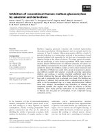
Contribution of different duplication types to the function of the mitochondriaFigure 1
Contribution of different duplication types to the function of the mitochondria. Classes significantly overrepresented compared to the mitochondrial
proteome are shown. The height of a bar represents the fraction of proteins that is annotated with a specific category. Three datasets are shown: the
whole mitochondrial proteome (MitoCarta proteins [4]; yellow), intra-mitochondrial (blue) and inter-compartmental (red) duplications. Protein functional
classes are defined by GO functional classification [68]. Benjamini and Hochberg false discovery rate correction was used to derive statistically significant
categories. See Tables S5 and S7 in Additional data file 1 for the full list.
Transport
Ion transport
Oxidoreductase activity
Carbohydrate metabolism
Calcium ion binding
Mitochondrial membrane
Ceramide kinase activity
rRNA methyltransferase
Cofactor binding
Catalytic activity
0
0.1
0.2
0.3
0.4
0.5
0.6
0.7
0.8
Mitochondrial proteome Intra-mitochondrial Inter-compartmental
Functional classes
Fraction
Timing of gene duplications of mitochondrial proteinsFigure 2
Timing of gene duplications of mitochondrial proteins. The solid blue line represents duplicating mitochondrial proteins, while the solid red line
corresponds to duplications of genes followed by relocalization of one of the proteins to the mitochondria. The dashed line denotes protein duplications
in other cellular compartments, outside the mitochondria (all proteins are listed in Table S9 in Additional data file 1).
< Bilateria
Coelomata
Chordata
Euteleostomi
Tetrapoda
0
0.1
0.2
0.3
0.4
0.5
0.6
0.7
Intra-mitochondrial Inter-compartmental Outside mitochondria
Duplication time
Fraction of duplications
_
Genome Biology 2009, Volume 10, Issue 11, Article R135 Szklarczyk and Huynen R135.5
Genome Biology 2009, 10:R135
The assumption that we can use the non-mitochondrial local-
ization in yeast as a proxy for the ancestral localization ena-
bles us to recognize protein retargeting events between
mitochondria and other subcellular compartments, including
the nucleus (8 out of 29 proteins; Table S8 in Additional data
file 1), peroxisome (6 out of 29) and endoplasmic reticulum (5
out of 29 proteins). Four of the six peroxisomal relocalization
events encode proteins responsible for fatty acid beta-oxida-
tion in yeast (PCD1, ECI1, DCI1, POX1) and their duplicated
orthologs are found in human mitochondria.
Relocalized proteins often originate from ancient, pre-
metazoan duplications
Using phylogenetic trees of genes that encode the modern
human mitochondrion, we inferred the timing of duplications
(see Materials and methods). Around 80% of duplications are
equally divided between two evolutionary stages: before the
divergence of bilateria and before the divergence of verte-
brates (Figure 2). Intra-mitochondrial gene duplications were
found to be representative of the general duplication trends
across the whole genome (no statistical difference with the
genome-wide duplication trend, P > 0.6 Fisher exact test). By
contrast, the duplications associated with relocalizations to
the mitochondria happened predominantly in the earlier
stage of evolution, before the divergence of bilateria. At this
evolutionary time point they significantly exceed the genome-
wide fraction of duplications (P < 0.003). Following the mas-
sive duplication events before the radiation of vertebrates
(the 2R hypothesis [33,34]; although alternative hypotheses
exist [35]), mitochondrial protein content continued to evolve
as exemplified by the recent duplication of glutamate dehy-
drogenase [36]. And even though the reference mitochondrial
proteome used in this study is derived from mouse tissues,
and therefore the accurate protein localization data for pri-
mate-specific duplications is limited, we encountered 16 gene
duplications of mitochondrial proteins in primates (Table S11
in Additional data file 1).
Relocalizations promptly follow duplications
An unmentioned assumption in the analysis of inter-com-
partmental protein duplications is that the protein relocaliza-
tion followed shortly after the gene duplication. Even though
the pre-sequence mitochondrial import pathway is only one
of four presently recognized means of protein import
(reviewed in [37]), many mitochondrial proteins contain a
short, amino-terminal localization sequence that is indicative
of this pathway. This sequence feature is amenable to compu-
tational methods [38]. For proteins imported to the mito-
chondria via the pre-sequence pathway, the gain of a novel
localization may be caused by the acquisition of an amino-ter-
minal targeting signal. Indeed, when examining all proteins
with a novel mitochondrial localization, a potential mito-
chondrial targeting signal can be identified in 50% of the pro-
teins, five times more often than in their non-mitochondrial
human paralogs (P < 0.00005, Fisher exact test). Assuming
that in these proteins the targeting signal is responsible for
the mitochondrial localization, we examined whether its
appearance in evolution coincides with the gene duplication,
and thus whether the duplication was concomitant with a
gain of mitochondrial localization.
Among human mitochondrial proteins with a non-mitochon-
drial paralog we find 12 proteins with a recognizable short,
amino-terminal targeting sequence. Despite the limitations
of computational targeting sequence prediction (for example,
[20]) in 9 out of the 12 gene families the phylogenetic analysis
indicates that the mitochondrial targeting signal was gained
in the same era as, or shortly after, the gene duplication
(Table 2).
Tissue-specific expression of novel mitochondrial
proteins
Using mass spectrometry total peak intensity data available
for 14 different mouse tissues [4], we performed quantitative
analysis of tissue-specific protein expression by counting the
Table 2
Dating of gene duplication of mitochondrial proteins compared to time when the mitochondrial targeting signal appeared in the protein
sequence
Paralogs Duplication before the divergence of Targeting signal found in
TOP1MT, TOP1 Vertebrates Vertebrates (Gallus gallus)
TFB2M, DIMT1L Animals Animals (Anopheles gambiae)
NUDT8, NUDT7 Animals Animals (Drosophila melanogaster)
SIRT3, SIRT2 Coelomata to chordata* Vertebrates (Danio rerio)
HTRA2, HTRA1 Vertebrates Vertebrates (Danio rerio)
PDE12, CNOT6 Animals Chordates (Ciona intestinalis)
PECI, CDYL Animals Animals (Caenorhabditis elegans)
HINT2, HINT1 Animals Animals (Drosophila melanogaster)
GOT2, GOT1 Animals Animals (Drosophila melanogaster)
The names of genes encoding mitochondrial proteins are highlighted in bold. *While species overlap suggests a duplication at the root of chordata,
the species distribution within the SIRT2 and SIRT3 branches suggest an earlier duplication and multiple losses in many evolutionary branches.
Genome Biology 2009, Volume 10, Issue 11, Article R135 Szklarczyk and Huynen R135.6
Genome Biology 2009, 10:R135
number of tissues in which the protein was detected (specifi-
cally, the number of tissues with log
10
peak intensities of at
least 7). A typical mitochondrial protein is abundantly
expressed and detectable in 12 (median value) out of 14 tis-
sues (Table S12 in Additional data file 1). Only proteins that
underwent inter-compartmental duplications are expressed
in significantly fewer tissues (median 5; P < 0.01 using a two-
sided Wilcoxon rank sum test performed pairwise with other
datasets). These novel mitochondrial proteins (proteins that
possess a non-mitochondrial paralog and a non-mitochon-
drial yeast ortholog) more often exhibit a tissue specific
expression pattern with 45% expressed in three tissues or
fewer (compared to the mitochondrial average of 23%), and
are more rarely widely expressed (in more than 10 tissues;
28% novel mitochondrial proteins compared to 55% on aver-
age) (Figure 3).
Subcellular differentiation via independent gene
duplications
While tracing the history of duplications that extend the mito-
chondrial proteome, one can imagine, in the most drastic sce-
nario, that independent duplications in unrelated lineages
with subsequent parallel relocalizations to mitochondria
could lead to a convergent evolution in the mitochondrial
protein content. Several paralogs present this unusual evolu-
tionary pattern (Table 3). For example, branched-chain-
amino-acid aminotransferase underwent duplication at the
root of vertebrates, in addition to an independent event in
yeast as a result of whole genome duplication. In both species
one copy is targeted to the mitochondria (BCAT2 in human),
the other is cytosolic (BCAT1). In the case of this gene family,
the analysis of distant orthologs for the presence/absence of
the targeting signal sheds light on the likely ancestral locali-
zation. Using MitoProt II [39] and TargetP [38] the signal can
be detected in the fly sequence as well as Leishmania major
orthologs, suggesting that the ancestral BCAT protein was
part of the mitochondrial proteome in the ancestor of human
and yeast (Figure 4).
The growth of the mitochondrial proteome by gene
duplication
Knowing the homology of proteins with a determined locali-
zation in human and yeast, we reconstructed the (partial)
protein complement of mitochondria of the common ancestor
of human and yeast, comprising circa 200 proteins in total.
Starting with this ancestral proteome, we counted 128 dupli-
cations of mitochondrial proteins in the human lineage,
including intra-mitochondrial duplications and proteins
novel to the mitochondria (relocalizations following the
duplication of non-mitochondrial proteins). As not all types
of evolutionary events allow us to easily infer the ancestral
localization, this puts a lower bound on the protein count,
concluding that the metazoan mitochondrion in the human
lineage expanded by 64% (128 out of 200) by means of gene
duplication and relocalization since the evolutionary split
with the yeast lineage (see Materials and methods for details).
These counts are likely to be an underestimate of a real mito-
chondrial proteome expansion, as we disregard proteins
without recognizable orthologs in S. cerevisiae that appeared
in the metazoan lineage.
Discussion and conclusions
Our investigation reveals a dynamic mitochondrial proteome
and paints a picture of a eukaryotic organelle with a func-
tional repertoire evolving by gene duplication. In the absence
of gene duplication, we find little room for functional diversi-
fication of the mitochondrial proteome by relocalization of
proteins. The subcellular localization of proteins that did not
duplicate since the divergence of human and yeast is almost
always conserved in evolution, with a few notable exceptions.
In the presence of duplication events the mitochondrion
expanded via two major modes. In the first, more conserva-
tive mode, intra-mitochondrial duplications expanded the
mitochondrial proteome by duplication of proteins that were
already localized to mitochondria. In the second and a more
radical mode of proteome growth, inter-compartmental
duplications expanded the metazoan and human mitochon-
drial proteome by the duplication of non-mitochondrial pro-
teins and redirecting the newly arisen gene products to the
mitochondria.
The two modes of proteome expansion comprise different
functional protein classes. Duplications of genes responsible
for carbohydrate metabolism, calcium ion binding and vari-
ous forms of transport appear to be specific to intra-mito-
chondrial protein duplications, whereas cofactor binding,
intramolecular oxidoreductases, ceramide kinase and rRNA
Expression profiles of mitochondrial proteins across a range of mouse tissuesFigure 3
Expression profiles of mitochondrial proteins across a range of mouse
tissues. The number of tissues with a detectable protein mass-
spectrometry signal (up to 14 tissues investigated in [4]) is shown. The
height of the bar represents the fraction of proteins - of intra-
mitochondrial or inter-compartmental duplication origins - expressed in a
tissue-specific manner (up to three tissues), widely expressed (in more
than ten tissues) or expressed in a moderate number of tissues. Figure S2
in Additional data file 1 presents the data in more detail.
4 < t < 10 t >11
0
0.1
0.2
0.3
0.4
0.5
0.6
0.7
Mitochondrial
proteome
Intra-mitochondrial
Inter-compartmental
Number of tissues
Fraction
t < 3
_
_
_
_
Genome Biology 2009, Volume 10, Issue 11, Article R135 Szklarczyk and Huynen R135.7
Genome Biology 2009, 10:R135
methylation functions are more often associated with dupli-
cates that have novel mitochondrial localization.
Intra-mitochondrial duplications that expanded the reper-
toire of transport proteins are exemplified by two duplica-
tions of TIMM8A/B and TIMM17A/B proteins. Expression of
both paralogs leads to distinct variants of the intermembrane
complexes TIMM8-TIMM13 and Tim23 embedded in the
inner membrane [40-42] (Additional data file 1). The Pyru-
vate dehydrogenase (PDH) complex, which participates in
carbohydrate metabolism (a functional class significantly
enriched among intra-mitochondrial duplications), under-
went intra-mitochondrial duplications at various points in
evolution (E1-beta subunit duplicated before the divergence
of bilateria; E2 subunit duplicated before the divergence of
chordates; E1-alpha subunit duplicated before the divergence
Table 3
Independent duplications and parallel relocalizations in the human and yeast lineages have happened multiple times during evolution
Human Yeast
Family Mitochondrial Non-mitochondrial Mitochondrial Non-mitochondrial
Thioredoxins TXN, TXN2 TXNDC2 TRX3 TRX1, TRX2
Glutaredoxins GLRX2 GLRX, GLRXL GRX2 GRX1 (nucleus)
Isocitrate dehydrogenases [NADP] IDH2 IDH1 IDP1 IDP2, IDP3 (peroxisome)
Branched-chain-amino-acid aminotransferases BCAT2 BCAT1 BAT1 BAT2
Serine hydroxymethyltransferases SHMT2 SHMT1 SHM1 SHM2
Rows show proteins targeted to mitochondria and their paralogs targeted to other subcellular compartments in both human and yeast. Non-
mitochondrial homologs (both human and yeast) are cytosolic if not indicated otherwise.
Evolution of mitochondrial localization for the branched-chain-amino-acid aminotransferases familyFigure 4
Evolution of mitochondrial localization for the branched-chain-amino-acid aminotransferases family. (a) Gene tree generated by PhyML [70] for
vertebrates, yeast and outgroup species that speciated before duplication events. Bootstrap values (100 repetitions) are shown on the internal branches.
Proteins surrounded by an oval are localized to mitochondria; loss of the mitochondrial localization is marked by a cross. (b) Clustal W [71] alignment of
the amino-terminal region of orthologs. The predicted targeting sequences are highlighted in blue. Abbreviations: hs. Homo sapiens; mm, Mus musculus; dm,
Drosophila melanogaster; dr, Dano rerio; lm, L. major; sc, S. cerevisiae.
BCAT-lm
BAT2-sc
BAT1-sc
100
BCAT2-dr
BCAT2-mm
BCAT2-hs
100
60
BCAT1-mm
BCAT1-hs
99
BCAT1-dr
96
85
CG1673-dm
89
0.1
(a)
(b)
100
89
85
96
99
100
60
mitochondrial
B
CAT1-hs
B
CAT2-hs
B
AT1-sc
B
AT2-sc
B
CAT-lm
-MKDCSNG CSAECTGEGGSKEVVGTFKAKDL I VTPAT I LKEKPDPNN- LVFGTVFTDHMLTVEWSSEFGW
- MAAAALGQ I WARKLL SVPWLLCGPRRYASSSFKAADLQLEMTQKPHKKPGPGEPLV FGKT FTDHMLMVEWN- DKGWM A A A A L GQ I WA R K L L S V PW L L C G P R R Y
MLQRHSLK LGKFSIRTLATGAPLDASKLKITRNPNP-SKPRPNEELVFGQTFTDHMLTIPWSAKEGW
M L Q R H S L K L GK F S I R T L
MTLAPLDASKVKITTTQHA-SKPKPNSELVFGKSFTDHMLTAEWTAEKGW
MLLSRRWH QASAARGSRAPVVSFTAAALTKTLVADPPPLP-PMKGVAFGTLFTPHMVIIDFN-DGRWM L L S R RWH
Genome Biology 2009, Volume 10, Issue 11, Article R135 Szklarczyk and Huynen R135.8
Genome Biology 2009, 10:R135
of eutheria). The duplication pattern of post-translational
regulators of the PDH complex differs from that of the com-
plex itself. The inactivating phosphorylation of the PDH com-
plex is carried out by four paralogs of PDH kinase, and all
duplication events occurred before the divergence of the ver-
tebrates. Prior to the catalytic activation, PDH must be
dephosphorylated by one of the two paralogous proteins:
PDP1 (PPM2C) and PDP2. PDP1, in contrast to its paralog, is
activated by calcium ions and, therefore, might mediate the
effects of calcium-mobilizing hormones [43]. It is difficult to
establish the evolutionary origin of a domain responsible for
the binding with Ca
2+
, as the binding site is created upon the
formation of a complex with the E2 subunit of the PDH com-
plex and requires the lipoyl groups of E2 [44]. Nevertheless,
the calcium-dependence of PDP1 is consistent with a trend
present in mitochondrial proteins. We identify duplications
of Ca
2+
-binding mitochondrial solute carriers [45], as well as
proteins responsible for calcium-sensitive mitochondrial
trafficking along microtubules [46,47]. Overall, 11 out of 23 of
the calcium ion binding proteins originate from intra-mito-
chondrial duplications that occurred at the root of vertebrates
(P < 7e-4, [GO:5509]).
In general, it appears that the regulation of cellular complexes
is more evolutionarily recent than the complexes they control.
That the duplications of the PDH complex occurred before
the vertebral duplications of their regulators, kinases and
phosphatases, is not a unique case. Also, the soluble mito-
chondrial matrix deacetylase SIRT3 has a relatively recent
origin, and was shown to augment Complex I activity by bind-
ing with the 39 kDa subunit of Complex I, NDUFA9 [48]. It is
known that the growth of many mitochondrial protein com-
plexes occurred early in evolution, with mitochondrial Com-
plex I and the mitochondrial ribosome expanding
significantly at the root of eukaryotes [49-51]. Interestingly,
regulators of activity of the complexes via phosphorylation
and dephosphorylation (as for PDH) or deacetylation (Com-
plex I) did not appear concomitantly in evolution and were
not adapted from existing regulators, but emerged long after
the metazoan diversification.
When analyzing duplications of proteins that expanded the
mitochondrial proteome, it would be interesting to know the
selective forces driving duplication events. We show that the
novel mitochondrial localization that is detectable at the
sequence level has been gained rapidly after the duplication
event. On the one hand, we know that only a small fraction of
duplicated genes is retained in the genome in the long term,
and this holds also for large-scale genomic events such as
whole genome duplication [52]. On the other hand, the acqui-
sition of an amino-terminal targeting signal coinciding with
the gene duplication event could provide the rationale for the
retention of the duplicated gene. As the change of localization
alters the role of a protein in the cell, it could be accompanied
by further functional diversification. This diversification may
be extensive, even for relatively recent duplications, as in the
case of HTRA2 protease (Table 2). The membrane-bound
HTRA2, unlike its secreted paralogs, promotes or induces cell
apoptosis through caspase-dependent and -independent
pathways [53] and its loss of function mutations cause neuro-
degeneration and Parkinson's disease [54].
Analysis of the timing of duplication events reveals that the
majority of inter-compartmental duplications occurred fur-
ther back in time than the genomic trend would suggest and
that they contributed little to the expansion of the mitochon-
drial proteome in the vertebrate lineage. The fact that most
inter-compartmental duplications occurred before animals
diverged suggests that cellular differentiation is partly
responsible for inter-compartmental duplications. We pro-
pose that the inter-compartmental duplicated proteins could
have helped to satisfy the variable energy demands that
emerging metazoan tissues presented. There is some anecdo-
tal evidence that could support this hypothesis. For example,
the pattern of tissue-specific expression of TOP1MT (Table 2)
has adapted to meet the requirements for higher mitochon-
drial activity in specific organs - for example, skeletal muscle,
heart, and brain [55]. Additionally, we observed that inter-
compartmental duplications/relocalizations are character-
ized by a more narrow, tissue-specific expression than aver-
age mitochondrial proteins (see Table S12 and Figure S2 in
Additional data file 1).
Our quantitative results of the evolution of the mitochondrial
proteome match anecdotal evidence for the role of inter-com-
partmental duplications in the expansion of the proteomes of
other eukaryotic organelles. Some pathways and key enzymes
were known to have duplicated between plastids and other
cellular compartments [56], as observed in the case of sulfate
assimilation and cysteine biosynthesis found in the chloro-
plasts, cytosol and mitochondria of plants [57]. In addition,
the evolutionary history of 12 Calvin cycle enzymes shows
that plant proteins encoded by the nucleus have relocalized to
alternative compartments, regardless of their origin, cyano-
bacterial or otherwise [58].
With 87% of mitochondrial proteins preserving their ances-
tral compartment between human and yeast, a gene duplica-
tion event appears to be a necessary prerequisite to release
the localization constraint, allowing nascent proteins to be
retargeted to distinct compartments. We therefore conclude
that non-mitochondrial protein duplications followed by the
gain of a novel mitochondrial localization comprise a qualita-
tively and quantitatively important mode of expansion of the
mitochondrial proteome.
Materials and methods
Mitochondrial proteomes
Mammalian nuclear-encoded mitochondrial proteins were
downloaded from MitoCarta, the state-of-the-art compen-
dium of the human mitochondrial proteome established
Genome Biology 2009, Volume 10, Issue 11, Article R135 Szklarczyk and Huynen R135.9
Genome Biology 2009, 10:R135
using combination of experimental identification, bioinfor-
matic analysis, and literature curation [4]. We mapped 1,001
human orthologous proteins onto Ensembl identifiers using
human-mouse ortholog lists from Ensembl v44 (April 2007)
[59] and Mouse Genome Database [60]. For yeast, to assure
specificity of its mitochondrial proteome, a reference set was
downloaded from the MitoP2 database [61]. This set of 545
proteins contains published experimental data based on var-
ious studies [18,22-24] and was subsequently manually
curated. To exclude non-confirmed mitochondrial proteins,
for which a mitochondrial localization was only predicted or
derived from early high-throughput studies, we also required
mitochondrial proteins to be present among 851 proteins
from the most comprehensive dataset of the yeast mitochon-
drial proteome to date [19]. The proteomes selected as
described assure few false positive proteins, but do not com-
pletely cover mitochondrial protein content. Because of the
incomplete coverage, the absence of evidence for mitochon-
drial localization cannot be taken as evidence for the absence
of mitochondrial localization. For the non-mitochondrial
proteins set, only proteins localized to other eukaryotic sub-
cellular compartments were taken into account. This
included proteins explicitly assigned to 24 non-mitochondrial
compartments as annotated in GO of human genes (see Table
S10 in Additional data file 1 for the full list of the compart-
ments), analogous to the non-mitochondrial reference data-
set from [62].
Gene trees of mitochondrial proteins
To take into account the evolutionary history of every protein,
including gene losses and duplications, we performed analy-
sis of individual gene trees reconciled with the species phylog-
eny, as provided by the Ensembl team [59]. The
phylogenomic Ensembl pipeline provides a dataset of gene
trees across multiple species, constructed using both dS, dN
(substitution rates), nucleotide and protein distance meas-
ures [63]. These data, together with the standard species tree,
informs the gene tree construction performed by the Tree-
BeST program [64] (L Heng, AJ Vilella, E Birney, R Durbin,
in preparation). First, all protein coding genes are queried
using WUBLASTP against the whole protein database. Subse-
quently, a graph of proteins is constructed, with edges created
for best reciprocal hits or when score(P1, P2)/max(score(P1,
P1), score(P2, P2)) >0.33. Connected components of the
graph are extracted and aligned subsequently with MUSCLE
[65]. The back-translated multiple alignment is passed to the
tree constructing program, TreeBeST, together with the spe-
cies tree for the reconciliation and the duplication calls on
internal nodes, as the coverage of genomes in the Ensembl
database provides topologically based timings in order to
label duplication events [63]. All human gene trees with a
mitochondrial gene product (mitochondrial proteins in either
human or yeast) were downloaded from Ensembl database
v44 [59]. When integrating datasets from human and yeast
for 50% human genes and 46% yeast proteins, we did not
detect homologs in the other species, representing a likely
gene loss or gain in one of these lineages.
Unambiguous one-to-one orthologs between human
and yeast
The trees for gene families were separated at the speciation
branches into opisthokont orthogroups and the number of
paralogs in human and yeast lineages was counted. One-to-
one unambiguous orthologs were represented by trees with a
single gene in both lineages.
Gene duplications
For each gene family of n genes, we infer n-1 duplications,
each duplication corresponding to an internal tree node. The
dating of the duplication was inferred from the analysis of the
tree topology, as annotated by the Ensembl team. We use
rooted trees of homologous genes, where branching points
are labeled with the inferred time of duplication. For exam-
ple, a gene tree ((GeneA, GeneB):Euteleostomi,(GeneC,
GeneD):Euteleostomi):Chordata yields a single chordate
duplication that is followed by two vertebrate duplications.
For the inter-compartmental duplication a divergence time of
a mitochondrial and a closest non-mitochondrial paralog was
inferred from the internal node giving rise to the duplication.
To asses the quality of gene duplication calls, we used the
duplication consistency score [63]. The score measures the
intersection of the number of species post-duplication over
the union; one expects that most duplications should have the
gene persisting in an equally likely manner in subsequent lin-
eages [63]. All of the three duplication datasets (intra-mito-
chondrial, inter-compartmental or duplications outside
mitochondria) had similar, high consistency scores, with
median values of 0.85, 0.86, 0.85, respectively (Figure S1 in
Additional data file 1). The datasets tested with two-sided
Wilcoxon rank sum test do not exhibit statistically significant
differences (P-value > 0.65).
Differential localization
Of the differentially localized one-to-one orthologs, we find 17
proteins localized to mitochondria only in human and 16 of
these are either reference mitochondrial proteins known
from the literature or were experimentally verified in the
Pagliarini et al. study [4]. For families with gene duplications
and differentially localized human paralogs, localization was
predicted computationally for only three mitochondrial pro-
teins, with the remaining proteins validated experimentally
in the Pagliarini et al. study by either green fluorescent pro-
tein marker (4 proteins), proteomics approaches (7 proteins)
or being part of a mammalian mitochondrial reference set
based on the literature curation (15 proteins).
A. thaliana orthologs
Of the one-to-one human-yeast orthologs, 104 possess an
ortholog in plants (determined using the homologene data-
base [66] and 27 were found in mitochondria in Heazlewood
et al. [20]. With regard to intra-mitochondrial duplications,
Genome Biology 2009, Volume 10, Issue 11, Article R135 Szklarczyk and Huynen R135.10
Genome Biology 2009, 10:R135
47 plant orthologs were found, 23 of which are in the mito-
chondria.
Estimation of the expansion of the mitochondrial proteome
We identified 122 unambiguous one-to-one nuclear encoded
gene products with a reliable mitochondrial localization in
human and yeast (Table S1 in Additional data file 1), with 17
differentially localized orthologs likely to be mitochondrial
gains in the human lineage (see Results). Genes that under-
went duplications originated from at least 66 ancestral
opisthokont genes (for which we can find at least one protein
from the family in mitochondria of both human and yeast;
family counts are 53 + 8 + 4 + 1 from Table S4 in Additional
data file 1, with each family stemming from a single ancestral
gene), or 78 if we add families with uncertain common ances-
try (mitochondrial only in human; an additional 12 families).
This, together with one-to-one orthologs, gives 188 to 200
ancestral proteins. Given the present human mitochondrial
protein compendium, restricted to proteins with an ortholog
in yeast with a known localization, we arrive at 128 to 140
mitochondrial acquisitions in the human lineage. Given 188
to 200 ancestral mitochondrial proteins and 128 to 140 gains
in the metazoan evolutionary branch, we estimate an expan-
sion of the mitochondrial proteome between 64% (128/200)
and 74% (140/188).
Dating mitochondrial relocalization
For the prediction of the amino-terminal targeting signal in
the protein sequences, Target P was used [67] for all known
isoforms of a given gene. It is important to mention that the
pre-sequence analysis programs do not use homology to
known mitochondrial proteins or mitochondria-specific
domains as an indicator of presence/absence of targeting sig-
nal.
Gene Ontology analysis
GO [68] analysis was performed using the BiNGO package
[69] using Benjamini and Hochberg false discovery rate cor-
rection; corrected P-values are specified in Additional data
file 1.
Abbreviations
GO: Gene Ontology; PDH: pyruvate dehydrogenase.
Authors' contributions
RS and MH conceived the study. RS carried out the analysis
and wrote the manuscript. All authors read and approved the
final manuscript.
Additional data files
The following additional data are available with the online
version of this paper: supplementary text, Tables S1-S12, and
Figures S1 and S2 (Additional data file 1).
Additional data file 1Supplementary text, Tables S1-S12, and Figures S1 and S2Supplementary text, Tables S1-S12, and Figures S1 and S2.Click here for file
Acknowledgements
We thank the Ensembl team, including A Vilella and B Overduin for helping
us with tree analysis, I Duarte, U Kudla, and T Cuypers for stimulating dis-
cussions, and J Parmley for the critical reading of the manuscript. We also
thank anonymous reviewers for suggestions. This work was supported by
the Netherlands Genomics Initiative (Horizon Programme).
References
1. Green DR, Reed JC: Mitochondria and apoptosis. Science 1998,
281:1309-1312.
2. Berridge MJ, Lipp P, Bootman MD: The versatility and universal-
ity of calcium signalling. Nat Rev Mol Cell Biol 2000, 1:11-21.
3. Meisinger C, Sickmann A, Pfanner N: The mitochondrial pro-
teome: from inventory to function. Cell 2008, 134:22-24.
4. Pagliarini DJ, Calvo SE, Chang B, Sheth SA, Vafai SB, Ong S, Walford
GA, Sugiana C, Boneh A, Chen WK, Hill DE, Vidal M, Evans JG, Thor-
burn DR, Carr SA, Mootha VK: A mitochondrial protein com-
pendium elucidates complex I disease biology. Cell 2008,
134:112-123.
5. Karlberg O, Canbäck B, Kurland CG, Andersson SG: The dual ori-
gin of the yeast mitochondrial proteome. Yeast 2000,
17:170-187.
6. Gabaldón T, Huynen MA: Reconstruction of the proto-mito-
chondrial metabolism. Science 2003, 301:609.
7. Gabaldón T, Huynen MA: From endosymbiont to host-control-
led organelle: the hijacking of mitochondrial protein synthe-
sis and metabolism. PLoS Comput Biol 2007, 3:e219.
8. Matsuyama A, Arai R, Yashiroda Y, Shirai A, Kamata A, Sekido S,
Kobayashi Y, Hashimoto A, Hamamoto M, Hiraoka Y, Horinouchi S,
Yoshida M: ORFeome cloning and global analysis of protein
localization in the fission yeast Schizosaccharomyces pombe.
Nat Biotechnol 2006, 24:841-847.
9. Gabaldón T, Snel B, van Zimmeren F, Hemrika W, Tabak H, Huynen
MA: Origin and evolution of the peroxisomal proteome. Biol
Direct 2006, 1:8.
10. Goldberg AV, Molik S, Tsaousis AD, Neumann K, Kuhnke G, Delbac
F, Vivares CP, Hirt RP, Lill R, Embley TM: Localization and func-
tionality of microsporidian iron-sulphur cluster assembly
proteins. Nature 2008, 452:624-628.
11. Wolfe KH, Shields DC: Molecular evidence for an ancient dupli-
cation of the entire yeast genome. Nature 1997,
387:708-713.
12. Marques A, Vinckenbosch N, Brawand D, Kaessmann H: Functional
diversification of duplicate genes through subcellular adapta-
tion of encoded proteins. Genome Biol 2008, 9:R54.
13. Taylor SW, Fahy E, Zhang B, Glenn GM, Warnock DE, Wiley S, Mur-
phy AN, Gaucher SP, Capaldi RA, Gibson BW, Ghosh SS: Charac-
terization of the human heart mitochondrial proteome. Nat
Biotechnol 2003, 21:281-286.
14. Forner F, Foster LJ, Campanaro S, Valle G, Mann M: Quantitative
proteomic comparison of rat mitochondria from muscle,
heart, and liver. Mol Cell Proteomics 2006, 5:608-619.
15. Johnson DT, Harris RA, French S, Blair PV, You J, Bemis KG, Wang
M, Balaban RS: Tissue heterogeneity of the mammalian mito-
chondrial proteome. Am J Physiol Cell Physiol 2007, 292:C689-697.
16. Foster LJ, de Hoog CL, Zhang Y, Zhang Y, Xie X, Mootha VK, Mann
M: A mammalian organelle map by protein correlation pro-
filing. Cell 2006, 125:187-199.
17. Kislinger T, Cox B, Kannan A, Chung C, Hu P, Ignatchenko A, Scott
MS, Gramolini AO, Morris Q, Hallett MT, Rossant J, Hughes TR, Frey
B, Emili A: Global survey of organ and organelle protein
expression in mouse: combined proteomic and transcrip-
tomic profiling. Cell 2006, 125:173-186.
18. Sickmann A, Reinders J, Wagner Y, Joppich C, Zahedi R, Meyer HE,
Schönfisch B, Perschil I, Chacinska A, Guiard B, Rehling P, Pfanner N,
Meisinger C: The proteome of Saccharomyces cerevisiae mito-
chondria. Proc Natl Acad Sci USA 2003, 100:13207-13212.
19. Reinders J, Zahedi RP, Pfanner N, Meisinger C, Sickmann A: Toward
the complete yeast mitochondrial proteome: multidimen-
sional separation techniques for mitochondrial proteomics.
J Proteome Res 2006, 5:1543-1554.
20. Heazlewood JL, Tonti-Filippini JS, Gout AM, Day DA, Whelan J, Millar
AH: Experimental analysis of the Arabidopsis mitochondrial
Genome Biology 2009, Volume 10, Issue 11, Article R135 Szklarczyk and Huynen R135.11
Genome Biology 2009, 10:R135
proteome highlights signaling and regulatory components,
provides assessment of targeting prediction programs, and
indicates plant-specific mitochondrial proteins. Plant Cell 2004,
16:241-256.
21. Smith DGS, Gawryluk RMR, Spencer DF, Pearlman RE, Siu KWM,
Gray MW: Exploring the mitochondrial proteome of the cili-
ate protozoon Tetrahymena thermophila: direct analysis by
tandem mass spectrometry. J Mol Biol 2007, 374:837-863.
22. Pflieger D, Le Caer J, Lemaire C, Bernard BA, Dujardin G, Rossier J:
Systematic identification of mitochondrial proteins by LC-
MS/MS. Anal Chem 2002, 74:2400-2406.
23. Ohlmeier S, Kastaniotis AJ, Hiltunen JK, Bergmann U: The yeast
mitochondrial proteome, a study of fermentative and respi-
ratory growth. J Biol Chem 2004, 279:3956-3979.
24. Prokisch H, Scharfe C, Camp DG, Xiao W, David L, Andreoli C, Mon-
roe ME, Moore RJ, Gritsenko MA, Kozany C, Hixson KK, Mottaz HM,
Zischka H, Ueffing M, Herman ZS, Davis RW, Meitinger T, Oefner PJ,
Smith RD, Steinmetz LM: Integrative analysis of the mitochon-
drial proteome in yeast. PLoS Biol 2004, 2:e160.
25. Prokisch H, Andreoli C, Ahting U, Heiss K, Ruepp A, Scharfe C,
Meitinger T: MitoP2: the mitochondrial proteome database
now including mouse data. Nucleic Acids Res 2006, 34:D705-711.
26. Yoshihisa T, Ohshima C, Yunoki-Esaki K, Endo T: Cytoplasmic
splicing of tRNA in Saccharomyces cerevisiae. Genes Cells 2007,
12:285-297.
27. Otsuga D, Keegan BR, Brisch E, Thatcher JW, Hermann GJ, Bleazard
W, Shaw JM: The dynamin-related GTPase, Dnm1p, controls
mitochondrial morphology in yeast. J Cell Biol 1998,
143:333-349.
28. Casey CA, Anderson PM: Submitochondrial localization of argi-
nase and other enzymes associated with urea synthesis and
nitrogen metabolism, in liver of Squalus acanthias
. Comp Bio-
chem Physiol B 1985, 82:307-315.
29. Walsh : Subcellular localization and biochemical properties of
the enzymes of carbamoyl phosphate and urea synthesis in
the batrachoidid fishes Opsanus beta, Opsanus tau and Porich-
thys notatus. J Exp Biol 1995, 198:755-766.
30. Ladner RD, McNulty DE, Carr SA, Roberts GD, Caradonna SJ: Char-
acterization of distinct nuclear and mitochondrial forms of
human deoxyuridine triphosphate nucleotidohydrolase. J Biol
Chem 1996, 271:7745-7751.
31. Knoops B, Clippe A, Bogard C, Arsalane K, Wattiez R, Hermans C,
Duconseille E, Falmagne P, Bernard A: Cloning and characteriza-
tion of AOEB166, a novel mammalian antioxidant enzyme of
the peroxiredoxin family. J Biol Chem 1999, 274:30451-30458.
32. Metodiev MD, Lesko N, Park CB, Cámara Y, Shi Y, Wibom R,
Hultenby K, Gustafsson CM, Larsson N: Methylation of 12S rRNA
is necessary for in vivo stability of the small subunit of the
mammalian mitochondrial ribosome. Cell Metab 2009,
9:386-397.
33. Lundin LG: Evolution of the vertebrate genome as reflected in
paralogous chromosomal regions in man and the house
mouse. Genomics 1993, 16:1-19.
34. Dehal P, Boore JL: Two rounds of whole genome duplication in
the ancestral vertebrate. PLoS Biol 2005, 3:e314.
35. Hughes AL, Friedman R: 2R or not 2R: testing hypotheses of
genome duplication in early vertebrates. J Struct Funct Genomics
2003, 3:85-93.
36. Rosso L, Marques AC, Reichert AS, Kaessmann H: Mitochondrial
targeting adaptation of the hominoid-specific glutamate
dehydrogenase driven by positive Darwinian selection. PLoS
Genet 2008, 4:e1000150.
37. Bolender N, Sickmann A, Wagner R, Meisinger C, Pfanner N: Multi-
ple pathways for sorting mitochondrial precursor proteins.
EMBO Rep 2008, 9:
42-49.
38. Emanuelsson O, von Heijne G: Prediction of organellar targeting
signals. Biochim Biophys Acta 2001, 1541:114-119.
39. Claros MG, Vincens P: Computational method to predict mito-
chondrially imported proteins and their targeting
sequences. Eur J Biochem 1996, 241:779-786.
40. Bauer MF, Gempel K, Reichert AS, Rappold GA, Lichtner P, Gerbitz
KD, Neupert W, Brunner M, Hofmann S: Genetic and structural
characterization of the human mitochondrial inner mem-
brane translocase. J Mol Biol 1999, 289:69-82.
41. Bömer U, Rassow J, Zufall N, Pfanner N, Meijer M, Maarse AC: The
preprotein translocase of the inner mitochondrial mem-
brane: evolutionary conservation of targeting and assembly
of Tim17. J Mol Biol 1996, 262:389-395.
42. Ewing RM, Chu P, Elisma F, Li H, Taylor P, Climie S, McBroom-Cera-
jewski L, Robinson MD, O'Connor L, Li M, Taylor R, Dharsee M, Ho
Y, Heilbut A, Moore L, Zhang S, Ornatsky O, Bukhman YV, Ethier M,
Sheng Y, Vasilescu J, Abu-Farha M, Lambert J, Duewel HS, Stewart II,
Kuehl B, Hogue K, Colwill K, Gladwish K, Muskat B, et al.: Large-
scale mapping of human protein-protein interactions by
mass spectrometry. Mol Syst Biol 2007, 3:89.
43. Huang B, Gudi R, Wu P, Harris RA, Hamilton J, Popov KM: Isoen-
zymes of pyruvate dehydrogenase phosphatase. DNA-
derived amino acid sequences, expression, and regulation. J
Biol Chem 1998, 273:17680-17689.
44. Turkan A, Gong X, Peng T, Roche TE: Structural requirements
within the lipoyl domain for the Ca2+-dependent binding and
activation of pyruvate dehydrogenase phosphatase isoform 1
or its catalytic subunit. J Biol Chem 2002, 277:14976-14985.
45. del Arco A, Satrústegui J: Identification of a novel human sub-
family of mitochondrial carriers with calcium-binding
domains. J Biol Chem 2004, 279:24701-24713.
46. Saotome M, Safiulina D, Szabadkai G, Das S, Fransson A, Aspenstrom
P, Rizzuto R, Hajnóczky G: Bidirectional Ca2+-dependent con-
trol of mitochondrial dynamics by the Miro GTPase. Proc Natl
Acad Sci USA 2008, 105:20728-20733.
47. Macaskill AF, Rinholm JE, Twelvetrees AE, Arancibia-Carcamo IL,
Muir J, Fransson A, Aspenstrom P, Attwell D, Kittler JT: Miro1 is a
calcium sensor for glutamate receptor-dependent localiza-
tion of mitochondria at synapses. Neuron 2009, 61:541-555.
48. Ahn B, Kim H, Song S, Lee IH, Liu J, Vassilopoulos A, Deng C, Finkel
T: A role for the mitochondrial deacetylase Sirt3 in regulat-
ing energy homeostasis. Proc Natl Acad Sci USA 2008,
105:14447-14452.
49. Gabaldón T, Rainey D, Huynen MA: Tracing the evolution of a
large protein complex in the eukaryotes, NADH:ubiquinone
oxidoreductase (Complex I). J Mol Biol 2005, 348:857-870.
50. Huynen MA, de Hollander M, Szklarczyk R: Mitochondrial pro-
teome evolution and genetic disease. Biochim Biophys Acta 2009,
1792:1122-1129.
51. Smits P, Smeitink JAM, Heuvel LP van den, Huynen MA, Ettema TJG:
Reconstructing the evolution of the mitochondrial ribos-
omal proteome. Nucleic Acids Res 2007, 35:4686-4703.
52. Nadeau JH, Sankoff D: Comparable rates of gene loss and func-
tional divergence after genome duplications early in verte-
brate evolution. Genetics 1997, 147:1259-1266.
53. Vande Walle L, Lamkanfi M, Vandenabeele P: The mitochondrial
serine protease HtrA2/Omi: an overview. Cell Death Differ
2008, 15:453-460.
54. Strauss KM, Martins LM, Plun-Favreau H, Marx FP, Kautzmann S, Berg
D, Gasser T, Wszolek Z, Müller T, Bornemann A, Wolburg H, Down-
ward J, Riess O, Schulz JB, Krüger R: Loss of function mutations
in the gene encoding Omi/HtrA2 in Parkinson's disease. Hum
Mol Genet 2005, 14:2099-2111.
55. Zhang H, Barceló JM, Lee B, Kohlhagen G, Zimonjic DB, Popescu NC,
Pommier Y: Human mitochondrial topoisomerase I. Proc Natl
Acad Sci USA 2001, 98:10608-10613.
56. Lunn JE: Compartmentation in plant metabolism. J Exp Bot
2007, 58:35-47.
57. Lunn JE, Droux M, Martin J, Douce R: Localization of ATP Sulfu-
rylase and O-Acetylserine(thiol)lyase in Spinach Leaves.
Plant Physiol 1990, 94:1345-1352.
58. Martin W, Schnarrenberger C: The evolution of the Calvin cycle
from prokaryotic to eukaryotic chromosomes: a case study
of functional redundancy in ancient pathways through endo-
symbiosis. Curr Genet 1997, 32:1-18.
59. Hubbard TJP, Aken BL, Beal K, Ballester B, Caccamo M, Chen Y,
Clarke L, Coates G, Cunningham F, Cutts T, Down T, Dyer SC, Fit-
zgerald S, Fernandez-Banet J, Graf S, Haider S, Hammond M, Herrero
J, Holland R, Howe K, Howe K, Johnson N, Kahari A, Keefe D,
Kokocinski F, Kulesha E, Lawson D, Longden I, Melsopp C, Megy K,
et al.: Ensembl 2007. Nucleic Acids Res 2007, 35:D610-617.
60. Bult CJ, Eppig JT, Kadin JA, Richardson JE, Blake JA: The Mouse
Genome Database (MGD): mouse biology and model sys-
tems. Nucleic Acids Res 2008, 36:D724-728.
61. Perocchi F, Jensen LJ, Gagneur J, Ahting U, von Mering C, Bork P,
Prokisch H, Steinmetz LM: Assessing systems properties of
yeast mitochondria through an interaction map of the
organelle. PLoS Genet 2006, 2:e170.
62. Calvo S, Jain M, Xie X, Sheth SA, Chang B, Goldberger OA, Spinazzola
A, Zeviani M, Carr SA, Mootha VK: Systematic identification of
human mitochondrial disease genes through integrative
Genome Biology 2009, Volume 10, Issue 11, Article R135 Szklarczyk and Huynen R135.12
Genome Biology 2009, 10:R135
genomics. Nat Genet 2006, 38:576-582.
63. Vilella AJ, Severin J, Ureta-Vidal A, Heng L, Durbin R, Birney E:
EnsemblCompara GeneTrees: Complete, duplication-aware
phylogenetic trees in vertebrates. Genome Res 2009,
19:327-335.
64. TreeSoft: Softwares for Phylogenetic Trees [http://
treesoft.sourceforge.net/treebest.shtml]
65. Edgar RC: MUSCLE: multiple sequence alignment with high
accuracy and high throughput. Nucleic Acids Res 2004,
32:1792-1797.
66. Wheeler DL, Barrett T, Benson DA, Bryant SH, Canese K,
Chetvernin V, Church DM, DiCuccio M, Edgar R, Federhen S, Geer
LY, Kapustin Y, Khovayko O, Landsman D, Lipman DJ, Madden TL,
Maglott DR, Ostell J, Miller V, Pruitt KD, Schuler GD, Sequeira E,
Sherry ST, Sirotkin K, Souvorov A, Starchenko G, Tatusov RL, Tatus-
ova TA, Wagner L, Yaschenko E: Database resources of the
National Center for Biotechnology Information. Nucleic Acids
Res 2007, 35:D5-12.
67. Emanuelsson O, Nielsen H, Brunak S, von Heijne G: Predicting sub-
cellular localization of proteins based on their N-terminal
amino acid sequence. J Mol Biol 2000, 300:1005-1016.
68. Ashburner M, Ball CA, Blake JA, Botstein D, Butler H, Cherry JM,
Davis AP, Dolinski K, Dwight SS, Eppig JT, Harris MA, Hill DP, Issel-
Tarver L, Kasarskis A, Lewis S, Matese JC, Richardson JE, Ringwald M,
Rubin GM, Sherlock G: Gene ontology: tool for the unification
of biology. The Gene Ontology Consortium. Nat Genet 2000,
25:25-29.
69. Maere S, Heymans K, Kuiper M: BiNGO: a Cytoscape plugin to
assess overrepresentation of gene ontology categories in
biological networks. Bioinformatics 2005, 21:3448-3449.
70. Dereeper A, Guignon V, Blanc G, Audic S, Buffet S, Chevenet F,
Dufayard J, Guindon S, Lefort V, Lescot M, Claverie J, Gascuel O:
Phylogeny.fr: robust phylogenetic analysis for the non-spe-
cialist. Nucleic Acids Res 2008,
36:W465-469.
71. Thompson JD, Higgins DG, Gibson TJ: CLUSTAL W: improving
the sensitivity of progressive multiple sequence alignment
through sequence weighting, position-specific gap penalties
and weight matrix choice. Nucleic Acids Res 1994, 22:4673-4680.