Báo cáo y học: "Tandem repeats modify the structure of human genes hosted in segmental duplications" docx
Bạn đang xem bản rút gọn của tài liệu. Xem và tải ngay bản đầy đủ của tài liệu tại đây (1.51 MB, 12 trang )
Genome Biology 2009, 10:R137
Open Access
2009De Grassi and CiccarelliVolume 10, Issue 12, Article R137
Research
Tandem repeats modify the structure of human genes hosted in
segmental duplications
Anna De Grassi and Francesca D Ciccarelli
Address: Department of Experimental Oncology, European Institute of Oncology, IFOM-IEO Campus, Via Adamello, 20139 Milan, Italy.
Correspondence: Francesca D Ciccarelli. Email:
© 2009 De Grassi and Ciccarelli; licensee BioMed Central Ltd.
This is an open access article distributed under the terms of the Creative Commons Attribution License ( which
permits unrestricted use, distribution, and reproduction in any medium, provided the original work is properly cited.
Gene structure modification by tandem repeats<p>Internal tandem repeats are shown to modify the gene structure of human primate-specific paralogs.</p>
Abstract
Background: Recently duplicated genes are often subject to genomic rearrangements that can
lead to the development of novel gene structures. Here we specifically investigated the effect of
variations in internal tandem repeats (ITRs) on the gene structure of human paralogs located in
segmental duplications.
Results: We found that around 7% of the primate-specific genes located within duplicated regions
of the genome contain variable tandem repeats. These genes are members of large groups of
recently duplicated paralogs that are often polymorphic in the human population. Half of the
identified ITRs occur within coding exons and may be either kept or spliced out from the mature
transcript. When ITRs reside within exons, they encode variable amino acid repeats. When located
at exon-intron boundaries, ITRs can generate alternative splicing patterns through the formation
of novel introns.
Conclusions: Our study shows that variation in the number of ITRs impacts on recently
duplicated genes by modifying their coding sequence, splicing pattern, and tissue expression. The
resulting effect is the production of a variety of primate-specific proteins, which mostly differ in
number and sequence of amino acid repeats.
Background
The completion of the human genome and recent advances in
sequencing technologies have revealed the presence of
recently duplicated genomic segments with high degrees of
sequence identity. Some of these regions have reached fixa-
tion during primate evolution and are known as segmental
duplications (SDs) [1]. Other segments are still polymorphic
and represent copy number variants (CNVs) within the
human population [2-4]. Duplicated blocks are usually
enriched in genes, thus providing raw material for the evolu-
tion of novel gene families [5-7].
Newly duplicated paralogs undergo rearrangements that usu-
ally cause their non-functionalization [8]. Sporadically, these
modifications lead to advantageous events, such as the devel-
opment of a novel function (neo-functionalization) or the
repartition of the original function between paralogs (sub-
functionalization). Under these circumstances, the new genes
are rapidly preserved and fixed into the population [9-14].
Rapid divergence of paralogs immediately after gene duplica-
tion is a consequence of the relaxed evolutionary pressure
that favors the retention and the propagation of the mutated
Published: 2 December 2009
Genome Biology 2009, 10:R137 (doi:10.1186/gb-2009-10-12-r137)
Received: 23 July 2009
Revised: 8 October 2009
Accepted: 2 December 2009
The electronic version of this article is the complete one and can be
found online at /> Genome Biology 2009, Volume 10, Issue 12, Article R137 De Grassi and Ciccarelli R137.2
Genome Biology 2009, 10:R137
alleles [8,15]. In this particular context, errors in DNA repli-
cation act as a major source of evolutionary innovation.
One of the most frequent replication errors involves internal
tandem repeats (ITRs), which are short genomic regions that
undergo homologous unequal crossing-over and replication
slippage. ITRs are very frequent in eukaryotic genomes [16]
and show a positive correlation with genome size in metazo-
ans [17,18]. The biological role of ITRs has been a matter of
long-standing debate. In a gene-centric view of genome evo-
lution, these regions have been often tagged as junk DNA,
particularly when they localize in intergenic segments and
within introns. However, growing evidence has shown that
ITRs are important for the evolution of eukaryotic genomes
because they act as potential source of genetic variation owing
to their 'mutator properties' [19]. Several examples support-
ing this role have been accumulating over the years, including
cell adhesion in yeast [20], morphological modifications in
dogs [21], social behaviors in voles [22], and differences in
sexual behavior between primates [23]. In coding exons, rep-
etitions mostly involve trinucleotides due to selection against
frameshift [24]. In this context, ITRs, and particularly short
repeats or microsatellites (< 10 bp), are highly polymorphic
within the human population. Polymorphic trinucleotides are
often associated with human genetic diseases, one of the best-
known examples being the expansion of polyglutamine traits
in Huntington disease and various other spinocerebellar
ataxias (for recent reviews, see [25,26]). The association
between trinucleotide polymorphisms and genetic diseases
might lead to the conclusion that repeat variations are always
evolutionarily deleterious. However, this is not true: the CAG
repeat of the SCA2 gene is under positive selection within the
CEU population, although the biological reasons for this
selection are still unknown [27].
We have recently shown other possible evolutionary out-
comes deriving from ITR variations. Namely, we described
the structural modifications occurring in PRDM7, a primate-
specific member of the PRDM gene family, where the repeat
contributes to the acquisition of a complex pattern of splicing
variants and tissue-specific expression [14]. In the present
study, we extend the analysis to all variable ITRs in human
paralogs lying in SDs with the aim of verifying whether ITR-
driven modifications represent a widespread mechanism for
the evolution of novel genes.
Results
ITRs modify around 7% of human genes hosted in
segmental duplications
SDs are regions of the human genome longer than 1 kb, with
at least 90% sequence identity, and that underwent duplica-
tions during the last 25 million years of primate evolution
[28]. We grouped all human genes hosted in SDs into 2,948
discrete gene loci, each composed of all overlapping mRNAs
lying on the same DNA strand (Figure 1a). This definition of
gene loci allowed the identification of human genes hosted
within SDs by using directly the genome annotation rather
than pre-compiled collections of genes. We performed an all-
against-all alignment between all exons in these gene loci to
extract the 2,008 loci that are associated through nearly iden-
tical exons (Figure 1b). We only considered for further analy-
sis the exon alignments with a variable number of ITRs
between the aligned exons (Figure 1c). By looking at the exon-
intron structure of the corresponding genes, we identified 102
alignments with variable ITRs retained within both exons and
162 alignments with ITRs occurring at exon-intron bounda-
ries (Figure 1c). After manual inspection to eliminate false
positives (see Materials and methods), we identified 53 exon
and 27 intron modifications (Figure 1d). We carefully ana-
lyzed the transcription evidence supporting each of these
modifications to exclude that they were artifacts of RNAs with
multiple matches on highly identical genomic sequences,
such as SDs. Strikingly, almost all loci with variable ITRs (>
96%) are supported by RNAs with unique or best matches,
and only less than 4% are associated with ambiguous tran-
scription evidence (Table 1). This result confirms that ITR-
driven gene modifications are real events and not artifacts of
genome mapping.
Due to the multiple rounds of duplications during primate
evolution, the same modification could be found in several
gene loci. Overall, the 80 modifications were detectable in
210 gene loci, lying within 496 SDs (Table 1; Additional file 1).
Table 1
Gene loci, segmental duplications and transcription evidence associated with internal tandem repeat-driven modifications
RNA support
Modifications Associated gene loci Associated SDs Unique match Best match Multiple match
Exon (53) 180 474 48 5 2
Intron (27) 106 370 21 4 1
Both (18) 76 154 - - -
Total (80) 210 496 68 9 3
In 18 cases, the same variable ITR can result in both exon and intron modifications. RNAs supporting the modification can have a unique or best
match in that locus, or multiple matches in the genome (see Materials and methods).
Genome Biology 2009, Volume 10, Issue 12, Article R137 De Grassi and Ciccarelli R137.3
Genome Biology 2009, 10:R137
Variable ITRs therefore affect 7% (210 out of 2,498) of the
human genes hosted in primate-specific duplications.
Variable ITRs occur in large groups of recently
duplicated paralogs
To better characterize the genes that undergo ITR-driven
modifications, we compared the paralogs of the 210 loci with
variable ITRs with those of the remaining 1,798 nearly identi-
cal loci. We counted the number of paralogs of the 210 loci
that were associated through any nearly identical exon, and
through only ITR-containing exons. Both comparisons
showed that the 210 loci with variable ITRs are significantly
enriched in larger groups of paralogs (P-value < 10
-3
, Wil-
coxon test; Figure 2a, b). The same trend is detectable also
when exon and intron modifications are analyzed separately
(P-value < 10
-3
; Figure S1 in Additional file 2). These data sug-
gest that ITR-driven modifications have occured in genes that
underwent several rounds of duplications. This is not surpris-
ing as these genes had higher chances to undergo modifica-
tions and likely experienced periods of relaxed evolutionary
pressure due to functional redundancy [8].
Interestingly, SDs with variable ITRs tend to occur within the
same chomosome more frequently than all other SDs (70.4%
and 39.3%, respectively, P-value < 10
-3
, chi-squared test).
Since intrachromosomal SDs are known to be recent duplica-
tions [29], this enrichment may suggest that ITR-driven gene
modifications occurred recently during primate evolution. To
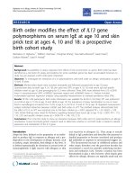
Genome-wide detection of ITR-driven gene modificationsFigure 1
Genome-wide detection of ITR-driven gene modifications. (a) The set of human gene loci within human SDs was retrieved. Each locus is composed of
transcripts that overlap on the same strand. (b) After an all-against-all alignment between exons, only loci that share at least one exon with 95% coverage
and 90% sequence identity were kept. The alignments could involve complete exons (blue) or portions of adjacent exons (pink). (c) From this dataset,
alignments with a variable number of ITR units were extracted. (d) The effect of the variable ITR on the gene structure was manually checked to remove
false positives and discriminate between exon and intron modifications.
Genome Biology 2009, Volume 10, Issue 12, Article R137 De Grassi and Ciccarelli R137.4
Genome Biology 2009, 10:R137
be able to date the appearance of the loci with variable ITRs
during primate evolution, we relied on the percentage of iden-
tity between pairs of SDs, which returns an indication of when
the duplication occurred in time [30,31]. When compared to
the rest, the 496 SDs hosting the loci with variable ITRs are
enriched in recent SDs (Figure 2c). In particular, 161 of them
(32.5%) share more than 98% sequence identity and hence
underwent duplication during or after the speciation between
human and chimpanzee [30]. This percentage is significantly
higher compared to all SDs (11.4%) and increases to 46.4%
when, for each of the 80 modifications, only the 168 SDs with
the longest version of the repeat are considered (P-value < 10
-
3
, chi-squared test; Figure 2c). Genes with variable ITRs, and
especially those with the longest ITR version, have formed
through recent duplications.
Genes with variable ITRs lie in polymorphic regions of
the human genome
The results reported above may suggest that the 496 SDs
bearing variable ITRs continue to undergo further rearrange-
ments and fixation in the human population. We therefore
measured the co-occurrence of SDs with variable ITRs and
human CNVs, which are large polymorphic regions (> 1 kb) of
the human genome accounting for a large portion of human
variation [3,32]. We observed the expected general trend
[30,33,34] in which recent SDs tend to undergo variation
within the human population (Figure 2d). However, when
only SDs with variable ITRs are considered, they are signifi-
cantly more represented within human CNVs, independent of
the age of the SD. Also in this case, the signal is still detectable
when only SDs with the longest version of the repeat are con-
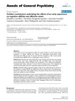
Number of paralogs and age of the loci with variable ITRsFigure 2
Number of paralogs and age of the loci with variable ITRs. (a) Comparison between the number of paralogs of the 210 loci with variable ITRs and the
remaining 1,798 nearly identical loci. The former are enriched in large groups of paralogs. (b) The same trend is observed when only the paralogs directly
hosting ITR-containing exons are compared to the rest. (c) Sequence identity between all pairs of SDs (25,914), pairs of SDs with variable ITRs (496), and
pairs of SDs with the longest version of each ITR (168). The last two are enriched in highly identical SDs. (d) Overlap between SDs and human CNVs.
Both SDs with variable ITRs and SDs with longer versions of each ITR tend to overlap with human CNVs. *P-value < 0.01 (chi-squared test between the
corresponding fraction of SDs and all SDs in that bin of sequence identity).
(a)
(b)
0 20406080
2−5 6−9 10−13 14−17 >=18
Loci (%)
Number of Paralogs
Rest of associated loci (1798)
Loci with variable ITRs (210)
0 20406080
2−5 6−9 10−13 14−17 >=18
Loci (%)
Number of Paralogs
(c)
(d)
90-92 92-94 94-96 96-98 98-100
Sequence identity (%)
Pairs of SDs (%)
010 30 50
90-92 92-94 94-96 96-98 98-100
Sequence identity (%)
Overlap with CNVs (%)
60 80 100
*
*
*
*
**
*
*
*
All SDs
SDs with the long ITR (168)
SDs with variable ITRs (496)
* = P-val <0.01
Genome Biology 2009, Volume 10, Issue 12, Article R137 De Grassi and Ciccarelli R137.5
Genome Biology 2009, 10:R137
sidered (Figure 2d). This observation suggests that ITR-
driven modifications preferentially occur in evolutionarily
dynamic regions of the genome that are still undergoing mod-
ification within the human population.
The fact that SDs with the longest version of the ITRs are par-
ticularly enriched in recent SDs as well as in human CNVs
may indicate that the direction of ITR modifications within
the primate lineage is towards expansion more than contrac-
tion, possibly through replication slippage or unequal crosso-
ver. To further verify this, we counted the number of ITRs in
the orthologous exons of two outgroup species, mouse and
dog. For 11 out of 80 modifications we could detect no orthol-
ogous sequence (Additional file 1), suggesting that the exon
itself originated in primates. For the remaining 69 ITR mod-
ifications, at least one ortholog was recovered in mouse or
dog. In all cases but two (ZNF100 and FOXD4L) the number
of ITRs was higher in human than in the other species. This
result confirms that variable ITRs in SDs mostly expanded in
the primate lineage, resulting in exon and intron elongations.
ITR-driven modifications are due to expansion of
minisatellites
Variable ITRs responsible for gene modifications are com-
posed, on average, of 30-bp units that are repeated 4 times for
a total length of 160 bp (Table S1 in Additional file 2). When
compared to all ITRs within exonic and non-exonic regions
hosted in SDs as well as in the whole human genome, variable
ITRs affecting the gene structure are significantly longer (Fig-
ure 3a) as a consequence of longer units (Figure 3b) rather
then of higher numbers of repetitions (Figure 3c). Therefore,
ITR-driven modifications of genes hosted in SDs are prefer-
entially mediated by minisatellites. This result can be
explained by different and concomitant reasons. First, it
partly reflects the fact that we focused on almost identical
regions, thus favoring the detection of longer repeat units. As
a general trend, ITRs lying in SDs have, on average, repeat
units significantly longer than ITRs dispersed in the rest of
human genome (Figure 3b). Second, long repeats are more
variable than short repeats probably because they enlarge the
target sequence for slippage or unequal crossover [35].
Finally, the absence of variable ITRs with repeat units shorter
than 9 bp (Figure 3b) suggests a preferential retention of
repeats that can significantly diversify the sequence of the
encoded proteins.
Fifty percent of variable ITRs modify protein sequences
In agreement with an active role in modifying protein
sequences, we found that 50% of the detected ITRs occur in
the coding sequence of genes lying in SDs (Table 2). This is
different, for example, from smaller ITRs in housekeeping
genes, which preferentially occur within untranslated regions
[36]. We manually analyzed all these 40 modifications in
order to verify the effect of the repeats on the resulting pro-
teins. In the majority of cases, the reading frame of the origi-
nal protein is preserved and variable ITRs cause the
elongation of low complexity regions in between globular
domains as well as of amino acid repeats, such as zinc fingers
and protein-specific repeats (Table S2 in Additional file 2).
Often these modifications occur in polymorphic human pro-
teins, such as the keratin-associated proteins, the VCX/Y pro-
teins, the nuclear pore interacting proteins, and the prostate-
ovary-testis-endometrium proteins. In this latter case, the
formation of an amino acid repeat is involved in the modifica-
tion of the protein's cellular localization [37].
In seven cases variable ITRs introduce frame shifts with the
formation of novel amino acid sequences (Table S2 in Addi-
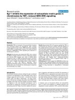
Length of variable ITRs compared to all ITRs in SDs and in the human genomeFigure 3
Length of variable ITRs compared to all ITRs in SDs and in the human genome. Compared are (a) the total length of the repeats, (b) the length of the
repeat unit, and (c) the number of repeat units between the variable ITRs that modify the gene structure (grey) and all other exonic and non-exonic ITRs
in SDs (pink) and in the rest of the human genome (light-blue). ITR modifications occur preferentially through the repetition of minisatellites and are
depleted in short repeats.
0
0
0
40 80 120
(b)
Unit length (bp)
10 20 30 40 50
(c)
Repeat units (n)
200 400 600
(a)
Repeat length (bp)
variable ITRs in
SDs
* = P-val <0.01
all ITRs in SDs
all ITRs in the
human genome
exonic non-
exonic
exonic non-
exonic
exonic non-
exonic
* *
*
*
Genome Biology 2009, Volume 10, Issue 12, Article R137 De Grassi and Ciccarelli R137.6
Genome Biology 2009, 10:R137
tional file 2). Although no specific functional assignment has
been made so far for any of these new sequences, they repre-
sent an innovation in terms of amino acid composition within
the primate lineage. In all seven cases ITR modifications
occur in the last coding exon, thus producing an accretion of
the protein sequences without affecting the original composi-
tion. Moreover, this also suggests that the resulting mRNAs
are potentially able to escape nonsense-mediated mRNA
decay [38] and produce functional proteins.
ITRs are involved in the diversification of Morpheus
paralogs
One of the most complex cases of ITR-driven modification
that we identified involves the paralogs of morpheus, a pri-
mate-specific gene under strong positive selection in homi-
noids [11]. To investigate the role of variable ITRs in the
diversification of potentially important genes in human evo-
lution, we manually analyzed and reconstructed the structure
of the ITR-containing exons in this family. Overall, we identi-
fied 26 paralogous loci, most of which have uniquely or best
mapping transcripts that encode nuclear pore interacting
proteins (NPIPs; Table 3). All these genes but one are hosted
in a region of human chromosome 16 that underwent several
rounds of duplications during primate evolution [39-41]. The
paralogous exons can host two different ITRs, namely type 1
and type 2 repeats (Figure 4a). Type 1 repeats are associated
with two different units of 57 bp and 69 bp, respectively,
which in turn can be translated into two different frames. As
a result, type 1 repeats can produce four distinct amino acid
sequences. Type 2 ITRs are much simpler repeats with a sin-
gle 87-bp unit and a unique reading frame. Depending on the
percentage of identity between exons, morpheus paralogs can
be assembled into two groups (G29 and G40; Additional file
1), which also reflect a different degree of expansion of the
ITR units. Members of G29 host a maximum of four repeats
of type 1 and two repeats of type 2, while members of G40
show a large repeat expansion, with up to 39 copies of type 1,
and 4 repeats of type 2 (Table 3). Not all variable ITRs present
at the genomic level are also found in the mature transcripts
and often the same locus is associated with transcripts that
differ in the number of ITRs (Figure 4b). However, rather
than being due to a complex pattern of alternative splicing,
this variability seems the result of the high structural poly-
morphism of these regions in the human population. All mor-
pheus loci but two overlap with human CNVs, and in at least
three cases the polymorphic region corresponds to the
repeats (Table 3). Interestingly, there are only two paralogs
with less than three ITRs of type 1 and in none are the ITRs in
coding exons, suggesting that at least three units of type 1
repeat are required for protein function.
The reason for the high variability in the number of ITRs in
the morpheus family is currently unknown. Together with
positive selection occurring at the upstream exons 2 and 4 of
morpheus [11], it could be a sign of the fixation process that
the entire family is currently undergoing in hominoids. Inter-
estingly, the protein encoded by morpheus localizes at the
nuclear membrane, where it interacts with the nuclear pore
complex [11]. According to several secondary structure pre-
dictors [42-46], morpheus hosts a transmembrane segment
in its amino-terminal part, followed by a helical portion
before the repeats (Figure 4c). Variation in amino acid
repeats is a known mechanism to vary the surface of interac-
tion with different targets [47]. This could be the case also for
the different paralogs of morpheus, which in this way could
adapt and fine-tune their binding to different interactors.
Discussion
Tandem repetitions of short sequences represent an effective
case of 'dynamic mutations' in which a secondary event can
easily occur after the primary duplication [48]. As a conse-
quence of such high dynamism, it is not surprising that ITRs
highly differ between lineages [49,50], species [51], and even
individuals [52]. In this study we show that long and variable
ITRs are involved in the modification of 7% of human genes
hosted in primate-specific SDs. These genes are very recent
and therefore likely to be still in the process of formation and
fixation. Confirming their evolutionary dynamism, genes
with variable ITRs are enriched in human variant regions, in
spite of the overall paucity of CNVs that overlap with RefSeq
genes [53]. At least 50% of variable ITRs contribute to the
modification of coding sequences, mostly leading to the elon-
gation of amino acid repeats in the encoded proteins. When
Table 2
Occurrence of variable internal tandem repeats within coding and non-coding transcripts
Coding sequences
Modification Non-coding RNAs UTRs ITR unit (bp) In-frame Out of frame
Exon (53) 10 13 < 40 (9-39) 14 3
> 60 (63-228) 9 4
Intron (27) 9 8 < 40 (9-30) 3 0
> 60 (62-126) 6 1
The number of ITR modifications localized in non-coding mRNAs, untranslated regions (UTRs), and coding sequences is reported. For ITRs lying
within coding sequences, the length of the ITR unit in base pairs and the effect on the reading frame of the encoded proteins are also shown.
Genome Biology 2009, Volume 10, Issue 12, Article R137 De Grassi and Ciccarelli R137.7
Genome Biology 2009, 10:R137
variable ITRs occur at the exon-intron boundary, they may
cause the formation of novel introns (Table S3 in Additional
file 2). The majority of such intron modifications (66% of the
total; Table 1) also show support for the alternative transcript,
in which the repeat is retained within the exon. Alternative
transcripts occur less frequently in exon modifications (34%,
P-value = 0.01, chi-squared test), suggesting that the forma-
tion of novel introns is a more complex event that requires
further rearrangements to generate novel splice sites [54].
For some of the reported cases, variable ITRs cause the acti-
vation of cryptic splice sites and the formation of novel
introns [55]. This model of intron formation has so far been
invoked only very seldom [14,56,57], likely because the fast
divergence of intronic sequences makes the identification of
Table 3
Features of variable internal tandem repeats present in paralogs of the human morpheus gene
ITRs in genome ITRs in mRNA
Group ID Genomic locus of exon 8 Exon length (bp) Type1 Type2 CNVs Associated
proteins or
transcripts
Exon
length
(aa)
Type1 Type2
G29 chr.16:68567795-68568136 342 1 0 1 NR_003610,
PDXC2 * (U)
-10
chr.18:11928734-11929237 504 2 2 0 - - - -
chr.16:14952973- 408 3 0 4 BC023572 * (M) - 2 0
14953380 NP_008916,
morpheus (B)
136 3 0
chr.16:15365020-15365427 408 3 0 2 - - - -
chr.16:11609511-11609918 408 3 1 0 - - - -
chr.16:28261432-28262010 579 3 2 1 - - - -
chr.16:15105678-15106116 439 4 0 1 - - - -
chr.16:16351450-16351914 465 4 0 3 BAC85871 (U) 155 4 0
chr.16:16394814-16395312 465 4 0 3 NP_848636 (M) 155 4 0
chr.16:18319329-18319793 465 4 0 5 NP_848636 (M) 155 4 0
chr.16:18359482-18359946 465 4 0 5 NP_848636 (M) 155 4 0
chr.16:28375249-28375848 600 4 2 2 - - - -
chr.16:28690975-28691574 600 4 2 4 - - - -
chr.16:28576850-28577449 600 4 2 5 - - - -
chr.16:72982790-72983476 687 4 2 5 AAI60029,
NPIPL2 (U)
229 4 2
chr.16:28970894-28971553 660 4 2 1 - - - -
G40 chr.16:22452448-22455204 2757 35 4 12 NP_001129337
(U)
919 35 4
AAH94882 (B) 544 18 4
NP_569731,
NPIPL3 (B)
464 14 4
chr.16:29300271-29303111 2841 37 3 2
†‡
chr.16:30141785-30144625 2841 37 4 2 BAC87606 (B) 794 29 4
chr.16:21321092-21324115 3024 39 4 5
†§
BAG65049 (U) 970 39 3
BAG64593 (U) 849 31 4
NP_569731 (B) 464 14 4
chr.16:21753543-21756566 3024 39 4 10
†
BAA13210 (B) 840 31 4
The paralogous sequences of morpheus exon 8 were detected and characterized for the presence and number of type 1 and 2 ITRs. Group IDs refer
to Additional file 1. Exon length is reported in base pairs and in amino acids (aa) only when supported by coding mRNAs. Unique (U), best (B) or
multiple (M) transcript support is reported for each locus. CNVs that overlap the genomic locus of exon 8 are indicated. *ITRs residing in non-
coding RNAs.
†
CNVs overlapping exclusively with ITRs, and corresponding to identifiers
‡
30774,
§
30757 and 30761 of the Database of Genomic
Variants.
Genome Biology 2009, Volume 10, Issue 12, Article R137 De Grassi and Ciccarelli R137.8
Genome Biology 2009, 10:R137
intron gains very challenging and often questionable [58-61].
Because our analysis focuses on recent events, it enables the
capture of signs of intron formation before they disappear as
a result of sequence divergence. None of the putative intron
gains reported in our study had been previously identified,
likely because our approach does not limit the search for
repeated regions to intron boundaries [62] but extends it to
the entire ITRs contained within exons. This approach also
reduces the chance of false positives due to RT-PCR artifacts
[63]. When no canonical splice sites can be identified (Table
S3 in Additional file 2), the discrepancy between the number
of putative ITRs between genomic and transcribed sequences
can be better explained by structural polymorphisms more
than by intron gains. This is the case of morpheus paralogs,
where both ITRs associated with these genes undergo copy
number variation within the human population (Table 3).
While microsatellites can be involved in CNV formation
[31,53], we found that the repetition of minisatellites seems to
play a role especially in the diversification of recently
acquired paralogs. How do these modifications affect the
function of these genes? The general paucity of functional
information on primate-specific genes prevents us from fully
addressing this issue. Some hints can be derived, however,
from the functional enrichment and the tissue expression of
genes with variable ITRs. As expected for recent paralogs,
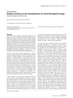
Effect of variable ITRs on the coding sequence of nuclear pore interacting proteinsFigure 4
Effect of variable ITRs on the coding sequence of nuclear pore interacting proteins. (a) Amino acid sequences encoded by the ITR units of the human
morpheus paralog BAG65049 were taken as representatives. Type 1 repeats are associated with two different units of 57 bp and 69 bp, respectively, and
are translated into two different frames. This results in four distinct amino acid repeats (1a, 1b, 1c and 1d). Type 2 repeats are much simpler and only
produce one sequence. Amino acids are highlighted according to the Clustal color scheme [71]. (b) Representation of the protein sequences encoded by
the human paralogs of exon 8 of morpheus. Only proteins associated with transcripts shown in Table 3 are reported. (c) Cartoon of the possible three-
dimensional structural organization of morpheus, based on secondary structure predictions (see text). These predictions were confirmed also for the other
RefSeq transcripts. The representation is not to scale.
Genome Biology 2009, Volume 10, Issue 12, Article R137 De Grassi and Ciccarelli R137.9
Genome Biology 2009, 10:R137
genes with variable ITRs are significantly more expressed in
skin and testis. Accordingly, the encoded proteins bind kera-
tin filaments and are involved in spermatogenesis (Figure 5;
Additional file 3). In agreement with previous reports [35],
other over-represented functional categories are DNA bind-
ing, regulation of transcription and mismatch repair (Figure
5; Additional file 3). In these cases, the variable ITRs are pref-
erentially located within the untranslated regions and thus
probably involved in the regulation of transcription (G11 and
G12; Additional file 1). Furthermore, variable ITRs can influ-
ence the tissue expression [14] and localization of the
encoded protein [37], thus confirming that their presence
actively modifies the gene function.
Conclusions
In this study we show that some variable ITRs underlie recent
changes in the structure of coding sequences, as well as
changes in exon-intron boundaries. These modifications
could constitute a mechanism for the evolution of novel gene
arrangements. They especially occur in large groups of
recently duplicated genes, which are also polymorphic in the
human population. These ITRs are biased towards expansion
of long units that can modify sequence, tissue expression and
splicing patterns of newly formed paralogs. The focus on very
recent modifications allows the observation of events that are
usually very hard to detect, such as the formation of novel
introns through activation of cryptic splice sites, and protein
sequence accretion through the repetitions of long units.
Materials and methods
Identification of gene loci and detection of ITR-driven
modifications
The genomic coordinates of 51,809 alignments between
30,833 primate-specific SDs [28] were recovered from the
assembly of the human genome (hg18, March 2006) at UCSC
[64]. Starting from 11,118 GenBank human RNAs lying within
SDs for at least 95% of their sequence (UCSC mRNA track,
frozen at May 2007), 2,948 discrete gene loci were derived by
merging all RNAs mapping in the same locus and on the same
strand. All exons within SDs were aligned using all-against-all
BLAT [65]. Only pairwise alignments with at least 90% iden-
tity and covering at least 95% of the shortest exon were
retained. Alignments between isoforms of the same exon
and/or between unrelated SDs were discarded. From the
resulting set, alignments bearing a different number of ITRs
were extracted. ITRs were recovered from the UCSC sim-
pleRepeat annotation track generated using Tandem Repeat
Finder (TRF) [66]. As reported in the UCSC website, TRF was
run under the following parameters: 2,7,7 = weights for
match, mismatch and indels used in the Smith-Waterman
local alignment; .80, .10 = matching and indel probability; 50
= minimum alignment score; 2000 = maximum size of the
repeat unit. Applied to the entire sequence of the human
genome, TRF is able to detect ITRs with a total length of 20 to
100,000 bp. Alignments were divided into two groups,
according to the position of the variable ITRs in respect to the
gene structure. ITR variation could occur either within exons,
resulting in exon modification, or at exon-intron boundaries,
leading to intron modification. For each modification, repre-
sentative alignments were manually checked to eliminate
false positives due to errors of TRF and/or incorrect align-
ment between repeats. The 524 RNAs associated with ITR
modifications were carefully analyzed to verify the robustness
of the transcription support for the new gene structure
arrangements. Transcripts were classified as: unequivocally
associated with the locus, if their only genomic match in
terms of sequence identity and coverage corresponded to the
locus with variable ITR; best associated with the locus, if the
best genomic match corresponded to the locus with variable
ITR; or multiply associated if they had multiple best matches
on the genome.
Groups of paralogs, age of SDs and overlap with human
CNVs
For each of the 2,008 gene loci, the number of paralogs was
defined as the number of loci associated by at least one nearly
identical exon (> 95% coverage, > 90% identity). The result-
ing distributions of paralogs between the 210 loci with varia-
ble ITRs and the remaining 1,798 loci were compared, using
the non-parametric Wilcoxon test. The percentage of identity
between pairs of SDs was recovered directly from UCSC. A
non-redundant set of 25,914 pair-wise alignments between
SDs was derived, 496 of which involve the 210 loci with vari-
able ITRs. The genomic coordinates of 21,178 human CNVs
grouped in 6,558 non-overlapping CNV loci were recovered
from the Database of Genomic Variants [67,68] (version 7,
March 2009).
Functional enrichment of genes with variable ITRsFigure 5
Functional enrichment of genes with variable ITRs. Tissue specificity
(green) and functional enrichment (blue) of genes with variable ITRs
compared to all other genes in SDs. The color gradient reflects the P-value
of the chi-squared analysis.
Genome Biology 2009, Volume 10, Issue 12, Article R137 De Grassi and Ciccarelli R137.10
Genome Biology 2009, 10:R137
Orthology assignment, tissue expression and
functional enrichment
Orthologous regions corresponding to the human ITR-con-
taining exons were extracted from the pair-wise BlastZ align-
ments between human and mouse (mm9) and human and
dog (canFam2) using Galaxy [69]. The human/dog align-
ments were screened only in case the alignment between
human and mouse was not available. The portions of the
alignments corresponding to variable ITRs were manually
checked. For 377 out of the 524 mRNAs with variable ITRs
and for the 5,256 out of 8,638 mRNAs in 2,008 loci it was
possible to extract information on the tissue type directly
from GenBank. Tissues that were represented by at least 15
mRNAs with variable ITRs (> 3%) were selected for chi-
squared comparison between transcripts with variable ITRs
and other transcripts in SDs. Forty-nine percent of genes with
variable ITRs can be associated with functional categories
according to the Gene Ontology [70]. The functional enrich-
ment was measured in comparison to other genes hosted in
SDs. The functional terms in common between the two
groups at levels 3 to 9 of the Gene Ontology hierarchy were
compared using the chi-squared test and P-values were
adjusted using the Bonferroni correction for multiple testing.
Abbreviations
CNV: copy number variant; ITR: internal tandem repeat; SD:
segmental duplication; TRF: Tandem Repeat Finder.
Authors' contributions
FDC conceived and designed the study; ADG performed the
experiments; ADG and FDC analyzed the data and wrote the
paper.
Additional files
The following additional data are available with the online
version of this paper: an Excel file containing genomic coor-
dinates and transcription evidence supporting ITR-driven
gene modifications (Additional file 1); a Word file containing
properties of variable ITRs, their effect on coding sequences
and groups of paralogs associated with exon and intron mod-
ifications (Additional file 2); an Excel file providing func-
tional analysis of genes with variable ITRs (Additional file 3).
Additional file 1Genomic coordinates and transcription evidence supporting ITR-driven gene modificationsFor each group of paralogs, the genomic coordinates of exons, loci and repeats are reported, together with the transcriptional support.Click here for fileAdditional file 2Properties of variable ITRs, their effect on coding sequences and groups of paralogs associated with exon and intron modificationsTable S1: features of variable ITRs associated with modifications of the gene structure. Table S2: effect of variable ITRs on coding sequences. Table S3: effect of variable ITRs on introns. Figure S1: number of paralogs associated with intron and exon modifications.Click here for fileAdditional file 3Functional analysis of genes with variable ITRsFor each over-represented category of the three main Gene Ontol-ogy classes (biological process, molecular function, and cellular component), the number and percentage of genes with variable ITRs and other genes in SDs are reported.Click here for file
Acknowledgements
This work was supported by AIRC Start-Up grant and by the Italian Ministry
of Health to FDC.
References
1. Bailey JA, Gu Z, Clark RA, Reinert K, Samonte RV, Schwartz S, Adams
MD, Myers EW, Li PW, Eichler EE: Recent segmental duplica-
tions in the human genome. Science 2002, 297:1003-1007.
2. Korbel JO, Urban AE, Affourtit JP, Godwin B, Grubert F, Simons JF,
Kim PM, Palejev D, Carriero NJ, Du L, Taillon BE, Chen Z, Tanzer A,
Saunders AC, Chi J, Yang F, Carter NP, Hurles ME, Weissman SM,
Harkins TT, Gerstein MB, Egholm M, Snyder M: Paired-end map-
ping reveals extensive structural variation in the human
genome. Science 2007, 318:420-426.
3. Redon R, Ishikawa S, Fitch KR, Feuk L, Perry GH, Andrews TD, Fie-
gler H, Shapero MH, Carson AR, Chen W, Cho EK, Dallaire S, Free-
man JL, Gonzalez JR, Gratacos M, Huang J, Kalaitzopoulos D, Komura
D, MacDonald JR, Marshall CR, Mei R, Montgomery L, Nishimura K,
Okamura K, Shen F, Somerville MJ, Tchinda J, Valsesia A, Woodwark
C, Yang F, et al.: Global variation in copy number in the human
genome. Nature 2006, 444:444-454.
4. Wong KK, deLeeuw RJ, Dosanjh NS, Kimm LR, Cheng Z, Horsman
DE, MacAulay C, Ng RT, Brown CJ, Eichler EE, Lam WL: A compre-
hensive analysis of common copy-number variations in the
human genome. Am J Hum Genet 2007, 80:91-104.
5. Eichler EE: Recent duplication, domain accretion and the
dynamic mutation of the human genome. Trends Genet 2001,
17:661-669.
6. Zhang L, Lu HHS, Chung W-y, Yang J, Li W-H: Patterns of segmen-
tal duplication in the human genome. Mol Biol Evol 2005,
22:135-141.
7. Bailey JA, Eichler EE: Primate segmental duplications: crucibles
of evolution, diversity and disease. Nat Rev Genet 2006,
7:552-564.
8. Lynch M, Conery JS: The evolutionary fate and consequences of
duplicate genes. Science 2000, 290:1151-1155.
9. Long M, Betran E, Thornton K, Wang W: The origin of new genes:
glimpses from the young and old. Nat Rev Genet 2003,
4:865-875.
10. Ciccarelli FD, von Mering C, Suyama M, Harrington ED, Izaurralde E,
Bork P: Complex genomic rearrangements lead to novel pri-
mate gene function. Genome Res 2005, 15:343-351.
11. Johnson ME, Viggiano L, Bailey JA, Abdul-Rauf M, Goodwin G, Rocchi
M, Eichler EE: Positive selection of a gene family during the
emergence of humans and African apes. Nature 2001,
413:514-519.
12. Birtle Z, Goodstadt L, Ponting C: Duplication and positive selec-
tion among hominin-specific PRAME genes. BMC Genomics
2005, 6:120.
13. Semple C, Rolfe M, Dorin J: Duplication and selection in the evo-
lution of primate beta-defensin genes. Genome Biol 2003, 4:R31.
14. Fumasoni I, Meani N, Rambaldi D, Scafetta G, Alcalay M, Ciccarelli FD:
Family expansion and gene rearrangements contributed to
the functional specialization of PRDM genes in vertebrates.
BMC Evol Biol 2007, 7:187.
15. Taylor JS, Raes J: Duplication and divergence: the evolution of
new genes and old ideas. Annu Rev Genet 2004, 38:615-643.
16. Ellegren H: Microsatellites: simple sequences with complex
evolution. Nat Rev Genet 2004, 5:435-445.
17. Katti MV, Ranjekar PK, Gupta VS: Differential distribution of sim-
ple sequence repeats in eukaryotic genome sequences. Mol
Biol Evol 2001, 18:1161-1167.
18. Toth G, Gaspari Z, Jurka J: Microsatellites in different eukaryo-
tic genomes: survey and analysis. Genome Res 2000, 10:967-981.
19. Kashi Y, King DG: Simple sequence repeats as advantageous
mutators in evolution. Trends Genet 2006, 22:253-259.
20. Verstrepen KJ, Jansen A, Lewitter F, Fink GR: Intragenic tandem
repeats generate functional variability. Nat Genet 2005,
37:986-990.
21. Fondon JW, Garner HR: Molecular origins of rapid and contin-
uous morphological evolution. Proc Natl Acad Sci USA 2004,
101:18058-18063.
22. Hammock EA, Young LJ: Microsatellite instability generates
diversity in brain and sociobehavioral traits. Science 2005,
308:1630-1634.
23. Jensen-Seaman MI, Li WH: Evolution of the hominoid semeno-
gelin genes, the major proteins of ejaculated semen. J Mol Evol
2003, 57:261-270.
24. Metzgar D, Bytof J, Wills C: Selection against frameshift muta-
tions limits microsatellite expansion in coding DNA. Genome
Res 2000, 10:72-80.
25. Gatchel JR, Zoghbi HY: Diseases of unstable repeat expansion:
mechanisms and common principles. Nat Rev Genet 2005,
6:743-755.
26. Usdin K: The biological effects of simple tandem repeats: les-
sons from the repeat expansion diseases. Genome Res 2008,
18:1011-1019.
27. Yu F, Sabeti PC, Hardenbol P, Fu Q, Fry B, Lu X, Ghose S, Vega R,
Genome Biology 2009, Volume 10, Issue 12, Article R137 De Grassi and Ciccarelli R137.11
Genome Biology 2009, 10:R137
Perez A, Pasternak S, Leal SM, Willis TD, Nelson DL, Belmont J, Gibbs
RA: Positive selection of a pre-expansion CAG repeat of the
human SCA2 gene. PLoS Genet 2005, 1:e41.
28. Bailey JA, Yavor AM, Massa HF, Trask BJ, Eichler EE: Segmental
duplications: organization and impact within the current
human genome project assembly. Genome Res 2001,
11:1005-1017.
29. She X, Liu G, Ventura M, Zhao S, Misceo D, Roberto R, Cardone MF,
Rocchi M, Green ED, Archidiacano N, Eichler EE: A preliminary
comparative analysis of primate segmental duplications
shows elevated substitution rates and a great-ape expansion
of intrachromosomal duplications. Genome Res 2006,
16:576-583.
30. Cheng Z, Ventura M, She X, Khaitovich P, Graves T, Osoegawa K,
Church D, DeJong P, Wilson RK, Paabo S, Rocchi M, Eichler EE: A
genome-wide comparison of recent chimpanzee and human
segmental duplications. Nature 2005, 437:88-93.
31. Kim PM, Lam HY, Urban AE, Korbel JO, Affourtit J, Grubert F, Chen
X, Weissman S, Snyder M, Gerstein MB: Analysis of copy number
variants and segmental duplications in the human genome:
Evidence for a change in the process of formation in recent
evolutionary history. Genome Res 2008, 18:1865-1874.
32. Levy S, Sutton G, Ng PC, Feuk L, Halpern AL, Walenz BP, Axelrod N,
Huang J, Kirkness EF, Denisov G, Lin Y, MacDonald JR, Pang AW,
Shago M, Stockwell TB, Tsiamouri A, Bafna V, Bansal V, Kravitz SA,
Busam DA, Beeson KY, McIntosh TC, Remington KA, Abril JF, Gill J,
Borman J, Rogers YH, Frazier ME, Scherer SW, Strausberg RL, et al.:
The diploid genome sequence of an individual human. PLoS
Biol 2007, 5:e254.
33. Fortna A, Kim Y, MacLaren E, Marshall K, Hahn G, Meltesen L, Bren-
ton M, Hink R, Burgers S, Hernandez-Boussard T, Karimpour-Fard A,
Glueck D, McGavran L, Berry R, Pollack J, Sikela JM: Lineage-spe-
cific gene duplication and loss in human and great ape evolu-
tion. PLoS Biol 2004, 2:E207.
34. Tuzun E, Sharp AJ, Bailey JA, Kaul R, Morrison VA, Pertz LM, Haugen
E, Hayden H, Albertson D, Pinkel D, Olson MV, Eichler EE: Fine-
scale structural variation of the human genome. Nat Genet
2005, 37:727-732.
35. Legendre M, Pochet N, Pak T, Verstrepen KJ: Sequence-based esti-
mation of minisatellite and microsatellite repeat variability.
Genome Res 2007, 17:1787-1796.
36. Lawson MJ, Zhang L: Housekeeping and tissue-specific genes
differ in simple sequence repeats in the 5'-UTR region. Gene
2008, 407:54-62.
37. Das S, Ise T, Nagata S, Maeda H, Bera TK, Pastan I: Palmitoylation
of POTE family proteins for plasma membrane targeting.
Biochem Biophys Res Commun 2007, 363:751-756.
38. Maquat LE: Nonsense-mediated mRNA decay: splicing, trans-
lation and mRNP dynamics. Nat Rev Mol Cell Biol 2004, 5:89-99.
39. Loftus BJ, Kim UJ, Sneddon VP, Kalush F, Brandon R, Fuhrmann J,
Mason T, Crosby ML, Barnstead M, Cronin L, Deslattes Mays A, Cao
Y, Xu RX, Kang HL, Mitchell S, Eichler EE, Harris PC, Venter JC,
Adams MD: Genome duplications and other features in 12 Mb
of DNA sequence from human chromosome 16p and 16q.
Genomics 1999, 60:295-308.
40. Martin J, Han C, Gordon LA, Terry A, Prabhakar S, She X, Xie G,
Hellsten U, Chan YM, Altherr M, Couronne O, Aerts A, Bajorek E,
Black S, Blumer H, Branscomb E, Brown NC, Bruno WJ, Buckingham
JM, Callen DF, Campbell CS, Campbell ML, Campbell EW, Caoile C,
Challacombe JF, Chasteen LA, Chertkov O, Chi HC, Christensen M,
Clark LM, et al.: The sequence and analysis of duplication-rich
human chromosome 16. Nature 2004, 432:988-994.
41. Johnson ME, Cheng Z, Morrison VA, Scherer S, Ventura M, Gibbs RA,
Green ED, Eichler EE: Recurrent duplication-driven transposi-
tion of DNA during hominoid evolution. Proc Natl Acad Sci USA
2006, 103:17626-17631.
42. Bagos PG, Liakopoulos TD, Hamodrakas SJ: Algorithms for incor-
porating prior topological information in HMMs: application
to transmembrane proteins. BMC Bioinformatics 2006, 7:189.
43. Claros MG, von Heijne G: TopPred II: an improved software for
membrane protein structure predictions. Comput Appl Biosci
1994, 10:685-686.
44. Krogh A, Larsson B, von Heijne G, Sonnhammer EL: Predicting
transmembrane protein topology with a hidden Markov
model: application to complete genomes. J Mol Biol 2001,
305:567-580.
45. Tusnady GE, Simon I: The HMMTOP transmembrane topology
prediction server. Bioinformatics 2001, 17:849-850.
46. Bryson K, McGuffin LJ, Marsden RL, Ward JJ, Sodhi JS, Jones DT: Pro-
tein structure prediction servers at University College Lon-
don. Nucleic Acids Res 2005, 33:W36-38.
47. Andrade MA, Perez-Iratxeta C, Ponting CP: Protein repeats:
structures, functions, and evolution. J Struct Biol 2001,
134:117-131.
48. Richards RI, Sutherland GR: Dynamic mutations: a new class of
mutations causing human disease. Cell 1992, 70:709-712.
49. Gibbs RA, Weinstock GM, Metzker ML, Muzny DM, Sodergren EJ,
Scherer S, Scott G, Steffen D, Worley KC, Burch PE, Okwuonu G,
Hines S, Lewis L, DeRamo C, Delgado O, Dugan-Rocha S, Miner G,
Morgan M, Hawes A, Gill R, Celera , Holt RA, Adams MD, Amanati-
des PG, Baden-Tillson H, Barnstead M, Chin S, Evans CA, Ferriera S,
Fosler C, et al.: Genome sequence of the Brown Norway rat
yields insights into mammalian evolution. Nature 2004,
428:493-521.
50. Waterston RH, Lindblad-Toh K, Birney E, Rogers J, Abril JF, Agarwal
P, Agarwala R, Ainscough R, Alexandersson M, An P, Antonarakis SE,
Attwood J, Baertsch R, Bailey J, Barlow K, Beck S, Berry E, Birren B,
Bloom T, Bork P, Botcherby M, Bray N, Brent MR, Brown DG, Brown
SD, Bult C, Burton J, Butler J, Campbell RD, Carninci P, et al.: Initial
sequencing and comparative analysis of the mouse genome.
Nature 2002, 420:520-562.
51. Webster MT, Smith NG, Ellegren H: Microsatellite evolution
inferred from human-chimpanzee genomic sequence align-
ments. Proc Natl Acad Sci USA 2002, 99:8748-8753.
52. Ellegren H: Heterogeneous mutation processes in human mic-
rosatellite DNA sequences. Nat Genet 2000, 24:400-402.
53. Conrad DF, Pinto D, Redon R, Feuk L, Gokcumen O, Zhang Y, Aerts
J, Andrews TD, Barnes C, Campbell P, Fitzgerald T, Hu M, Ihm CH,
Kristiansson K, MacArthur DG, MacDonald JR, Onyiah I, Pang AWC,
Robson S, Stirrups K, Valsesia A, Walter K, Wei J, Tyler-Smith C,
Carter NP, Lee C, Scherer SW, Hurles ME: Origins and functional
impact of copy number variation in the human genome.
Nature 2009 in press.
54. Catania F, Lynch M: Where do introns come from? PLoS Biol
2008, 6:e283.
55. Rogers JH: How were introns inserted into nuclear genes?
Trends Genet 1989, 5:213-216.
56. Venkatesh B, Ning Y, Brenner S: Late changes in spliceosomal
introns define clades in vertebrate evolution. Proc Natl Acad Sci
USA 1999, 96:10267-10271.
57. Knowles DG, McLysaght A: High rate of recent intron gain and
loss in simultaneously duplicated Arabidopsis genes. Mol Biol
Evol 2006, 23:1548-1557.
58. Roy SW, Gilbert W: The evolution of spliceosomal introns: pat-
terns, puzzles and progress. Nat Rev Genet 2006, 7:211-221.
59. Roy SW, Penny D: Smoke without fire: most reported cases of
intron gain in nematodes instead reflect intron losses. Mol
Biol Evol 2006, 23:2259-2262.
60. Roy SW, Fedorov A, Gilbert W: Large-scale comparison of
intron positions in mammalian genes shows intron loss but
no gain. Proc Natl Acad Sci USA 2003, 100:7158-7162.
61. Coulombe-Huntington J, Majewski J: Characterization of intron
loss events in mammals. Genome Res 2007, 17:23-32.
62. Zhuo D, Madden R, Elela SA, Chabot B: Modern origin of numer-
ous alternatively spliced human introns from tandem arrays.
Proc Natl Acad Sci USA 2007, 104:882-886.
63. Roy SW, Irimia M: When good transcripts go bad: artifactual
RT-PCR 'splicing' and genome analysis. Bioessays 2008,
30:601-605.
64. UCSC Genome Bioinformatics [ />65. Kent WJ: BLAT - the BLAST-like alignment tool.
Genome Res
2002, 12:656-664.
66. Benson G: Tandem repeats finder: a program to analyze DNA
sequences. Nucleic Acids Res 1999, 27:573-580.
67. Database of Genomic Variants [ />]
68. Iafrate AJ, Feuk L, Rivera MN, Listewnik ML, Donahoe PK, Qi Y,
Scherer SW, Lee C: Detection of large-scale variation in the
human genome. Nat Genet 2004, 36:949-951.
69. Galaxy [ />70. Ashburner M, Ball CA, Blake JA, Botstein D, Butler H, Cherry JM,
Davis AP, Dolinski K, Dwight SS, Eppig JT, Harris MA, Hill DP, Issel-
Tarver L, Kasarskis A, Lewis S, Matese JC, Richardson JE, Ringwald M,
Rubin GM, Sherlock G: Gene ontology: tool for the unification
of biology. The Gene Ontology Consortium. Nat Genet 2000,
25:25-29.
Genome Biology 2009, Volume 10, Issue 12, Article R137 De Grassi and Ciccarelli R137.12
Genome Biology 2009, 10:R137
71. Larkin MA, Blackshields G, Brown NP, Chenna R, McGettigan PA,
McWilliam H, Valentin F, Wallace IM, Wilm A, Lopez R, Thompson
JD, Gibson TJ, Higgins DG: Clustal W and Clustal X version 2.0.
Bioinformatics 2007, 23:2947-2948.