Báo cáo y học: "Genome-wide DNA demethylation in mammals" docx
Bạn đang xem bản rút gọn của tài liệu. Xem và tải ngay bản đầy đủ của tài liệu tại đây (308.6 KB, 4 trang )
In mammalian genomes, DNA methylation is found at
cytosine residues that are followed by guanines. is
epigenetic modification is essential for the repression of
retrotransposons and other elements of foreign origin; it
regulates developmental genes, including the pluri po-
tency genes OCT4 and NANOG, and is crucial for
genomic imprinting. CpG methylation undergoes dramatic
global changes at specific stages of mammalian develop-
ment. ese include acquisition of new methylation
patterns early in development, genome-wide removal of
DNA methylation in the primordial germ cells (PGCs),
and, following fertilization, removal of DNA methylation
from the sperm-derived genome ([1] and references
therein). Whereas the acquisition of DNA methylation is
now well understood, the mechanisms involved in global
DNA demethylation in PGCs and the zygote had
remained elusive. Two exciting recent studies [2,3] now
show that the cytidine deaminase AID contributes to
active DNA demethylation in mammals. Another
remarkable study has discovered that components of the
elongator complex are involved in the process as well [4].
After fertilization, the sperm-derived pronucleus
undergoes a rapid, global loss of DNA methylation, which
occurs independently of DNA replication. Some genes,
however, including imprinted genes, show resistance to
this active demethylation process. e maternal pro nucleus
is also resistant, but undergoes passive, replication-
dependent, demethylation during the first few cell cycles
of development. Consequently, by the blastocyst stage,
both the parental genomes have acquired low levels of
methylation. At a later developmental stage, during and
following implantation of the embryo, there is extensive
acquisition of de novo DNA methylation, so that
eventually, 70% or more of all CpGs are methylated [2]. A
second round of methylation reprogramming in mammals
occurs in the early PGCs of the embryo, between 10.5
and 13.5 days post-coitum (d.p.c.) in the mouse. is
wave of DNA demethylation affects the entire genome,
although certain sequence elements, including intra-
cisternal A particles (IAPs), are resistant [1]. e removal
of DNA methylation in PGCs affects both the parental
genomes and, apparently, no genes escape this essential
process, which serves to wipe the genome clean of marks
so that the germ cells acquire the capacity to support
post-fertilization development.
e enzymes that control the acquisition of new DNA
methylation are well known and have been studied in
detail. Whereas the de novo DNA methyltransferases
(DNMTs) DNMT3A and DNMT3B establish new methy-
lation on DNA, DNMT1 maintains patterns of methyla-
tion in a replication-dependent manner in all somatic cells
([1] and references therein). In contrast, the enzy matic
machineries involved in the active removal of CpG
methylation had remained enigmatic in mammals. Impor-
tant conceptual insights were obtained from flowering
plants, though, in which DNA demethylation is mediated
by 5-methylcytosine glycosylases. e best studied
example of such glycosylases is DEMETER, which
mediates the DNA demethylation involved in genomic
imprinting in the endosperm, the extra-embryonic part of
the developing seed [5]. In mammals, however, this
specific class of 5-methylcytosine glyco sylases seems not
to exist and, therefore, most attention has been focused on
cytidine deaminases of the APOBEC family, particularly
on Activation-Induced cytidine Deaminase (AID). AID
was known to act as a single-strand DNA deaminase in
developing B cells, in which it is required for somatic
hypermutation and class switch recombination at
immunoglobulin genes. In B cells, the deamination of
cytosine residues leads to U-G mis matches which can be
processed to give rise to double-strand breaks involved in
recombination at the immunoglobulin genes.
New roles for AID and components of the elongator
complex
Since AID was found to be expressed in PGCs and early
embryos, it was suggested that it might be involved in
global DNA demethylation [6]. Christian Popp
Abstract
The cytidine deaminase AID and elongator-complex
proteins contribute to the extensive removal of DNA
methylation in mammalian primordial germ cells and
in the paternal pronucleus of the zygote.
© 2010 BioMed Central Ltd
Genome-wide DNA demethylation in mammals
Lionel A Sanz, Satya K Kota and Robert Feil*
R ES E ARC H HIG HLI GHT
*Correspondence:
Institute of Molecular Genetics, CNRS UMR-5535 and University of Montpellier,
1919 route de Mende, 34293 Montpellier, France
Sanz et al. Genome Biology 2010, 11:110
/>© 2010 BioMed Central Ltd
and co-workers [2] tested this intriguing possibility by
explor ing DNA methylation in PGCs obtained from Aid
-/-
embryos. In their technically challenging study (mamma-
lian PGCs can be obtained only in small numbers) an
unbiased approach was taken that combined bisulphite
treatment of genomic DNA with next-generation
sequencing. is allowed the authors to assess global
levels of DNA methylation. ey combined this approach
with locus-specific studies in which bisulphite-converted
DNA was amplified by PCR followed by methylation
analyses by mass spectrometry. In agreement with earlier
studies, wild-type PGCs were found to have very low
levels of global DNA methylation at 13.5 d.p.c., par ticularly
in female PGCs, which showed less than 10% of
methylation globally. e lowest levels of methylation were
observed within introns, intergenic regions and repeat
elements. PGCs purified from AID-deficient embryos, in
contrast, showed higher levels of DNA methylation at
these sequences, and globally, demonstrating that AID
contri butes to the genome-wide demethylation in
primordial germ cells.
is novel discovery nicely complements a recent study
by Bhutani et al. [3] on heterokaryons made by artificially
fusing mouse embryonic stem (ES) cells with human
fibroblasts. In these heterokaryons, DNA methylation
was rapidly removed from the NANOG and OCT4 genes
in the fibroblast-derived genome. By using a small
interfering RNA (siRNA) approach, the authors showed
that this active demethylation requires AID. Concor-
dantly, AID was found to be targeted specifically to the
(methylated) NANOG and OCT4 promoters. In combina-
tion, the two new studies demonstrate a novel role for
AID in mammals: active demethylation of genomic DNA
(Figure 1a).
Intriguingly, however, the Aid
-/-
PGCs still attained low
levels of methylation compared with ES cells and somatic
tissues, which indicated that considerable demethylation
had occurred even in the absence of AID. Not
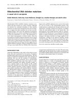
Figure 1. Active DNA demethylation in mammals. (a) The action of AID on 5-methylcytosine residues (white circles) in DNA (thick black line)
gives rise to deaminated 5-methylcytosine, which can be bound by the repair glycosylase MBD4. Through yet-unknown further repair mechanisms,
there is conversion into unmethylated cytosines, as shown by the disappearance of the white circles on the lower diagram. The canonical histones
found in nucleosomes are colored in blue. In the accessibility model presented here, the presence (green circles) or absence of specic histone tail
modications and/or histone variants (pink spheres) guide the recruitment of the enzymes and other factors involved in the DNA demethylation.
It is not yet known whether the requirement for elongator complex proteins is direct or whether they aect DNA demethylation indirectly,
by a mechanism unrelated to chromatin. (b) Protection against active DNA demethylation could be linked to the presence of specic histone
modications (red circles). Non-histone proteins could be involved in this process as well.
Zygote: paternal genome demethylated
(b)
Elongator
complex
proteins
Elongator
complex
proteins
AID
MBD4
(a)
AID
MBD4
Protection against DNA demethylation
?
Zygote: maternal genome protected
Imprinting control regions protected
PGCs: IAP elements protected
PGCs: both parental genomes demethylated
Sanz et al. Genome Biology 2010, 11:110
/>Page 2 of 4
surprisingly, therefore, Popp et al. [2] did not observe
pronounced developmental defects in the offspring of the
Aid
-/-
parent mice. e methylation phenotype in the
absence of AID indicates that other factors must also be
contributing to the DNA demethylation process.
e question of which other protein factors could be
involved was addressed in the third recent study, by Yuki
Okada and co-workers [4]. In their elegant study, these
authors used the global demethylation in the zygote’s
paternal pronucleus as a model. rough careful siRNA-
mediated knockdown experiments, they tested several
candidate proteins. Rather unexpectedly, they discovered
that a component of the elongator complex, elongator
protein 3 (ELP3), to be required for the removal of DNA
methylation in the zygote. e elongator complex was
first described as a component of RNA polymerase II
holoenzyme in transcriptional elongation, and has histone
acetyltransferase activity ([7], and references herein). In
particular, a live-cell imaging system allowed these
authors to follow global methylation states in zygotes,
and showed that knockdown of Elp3 prevented paternal
DNA demethylation. Subsequently, the authors showed
the same to be true for two other components of the
elongator complex, ELP1 and ELP4. ese remarkable
findings could signify that the whole elongator complex is
involved (Figure 1a). Its mode of action in DNA
demethylation remains to be discovered.
As is often the case with exciting new discoveries, the
recent studies raise many questions. Could transcription
be somehow linked to the removal of DNA methylation?
Little is known about whether there is actually
transcription through genomic regions in PGCs and in
the zygote. Embryonic transcription at many genes starts
only after the first cell division, but what about
transcription across other, non-genic, regions? Could the
involvement of elongator proteins be linked to one of
their transcription-independent roles, which include
modifi cation of tRNAs [7]. Although the RNA poly-
merase II complex has been shown to interact with AID
in B cells, it is not known whether such an interaction
could be involved in removing DNA methylation in
PGCs and zygotes.
How, in mammals, deamination of 5meC leads bio-
chemically to DNA demethylation remains unclear.
However, a recent study in Zebrafish provides interesting
clues [8]. Also in Zebrafish, AID deaminates 5meC
leading to the formation of thymine residues, and hence
G:T mismatches. Mismatched Ts are thought to be
replaced by cytosines by base excision repair (BER).
Methyl binding domain protein 4 (MBD4) is one of the
known thymine glycocylases in vertebrates and it
recognizes specifically the product of deamination at
methylated CpG dinucleotides [9]. Over-expression of
MBD4 together with AID in Zebrafish embryos led to
partial demethylation of injected methylated DNA
fragments. Mbd4 knockdown, in contrast, caused re-
methy lation of DNA [8]. us, in Zebrafish, the mismatch-
specific thymine glycosylase MBD4 contributes to the
demethylation process involving AID. It should be most
interesting to explore whether the same is true in
mammals.
Is there a correlation between DNA demethylation
and histone modifications?
Irrespective of the precise biochemical conversions
involved, the new studies raise the question of why
certain chromosomal regions lose their DNA methylation
and others not. AID and elongator complex proteins are
widely expressed, but global DNA demethylation occurs
specifically in PGCs and in the zygote. Furthermore, in
the zygote the sperm-derived genome undergoes active
DNA demethylation, but the maternal genome is
resistant. Demethylation of the paternal genome appears
to occur after the sperm’s protamines have been replaced
by histones [10]. At this early time point, however, the
newly formed chromatin in the male pronucleus is clearly
different from the chromatin of the maternal genome.
e histone H3 variant H3.3 is incorporated onto the
paternal genome (independent of DNA replication),
whereas the maternal genome is already packaged with
nucleosomes containing mostly the canonical histone
H3.1. At this stage, the histones on the paternal genome
show little lysine methylation compared with histones on
the maternal genome. e paternal genome is negative
for H3 lysine 9 di- and trimethylation, and H3 lysine 27
trimethylation, marks that are present in the maternal
pronucleus [10]. One idea, therefore, could be that
histone modifications and histone variants determine
whether the DNA demethylation machinery (including
AID) can access the genomic DNA (Figure 1b).
In early PGCs, there is extensive loss of histone
methylation together with the appearance of chaperone
proteins that could be involved in incorporating histone
variants into chromatin [11]. e nucleosomes and
histones are modified around the time that DNA de-
methy lation occurs, so these changes could well be
involved in recruiting the DNA demethylation
machinery. Certain IAP elements, however, are protected
against DNA demethylation in PGCs and it would be
interesting to explore the organization of chromatin at
these regions.
Research on mammalian DNA demethylation is
gaining momentum and, undoubtedly, new players and
mechanisms will be revealed during the coming years.
Together with the novel discoveries on AID and elongator
complex proteins reviewed above, this could provide
opportunities to further unravel the biological roles of
DNA demethylation in PGCs and in the early embryo.
Sanz et al. Genome Biology 2010, 11:110
/>Page 3 of 4
Published: 16 March 2010
References
1. Sasaki H, Matsui Y: Epigenetic events in mammalian germ-cell
development: reprogramming and beyond. Nat Rev Genet 2008, 9:129-140.
2. Popp C, Dean W, Feng S, Cokus SJ, Andrews S, Pellegrini M, Jacobsen SE, Reik
W: Genome-wide erasure of DNA methylation in mouse primordial germ
cells is affected by AID deficiency. Nature 2010, 463:1101-1105.
3. Bhutani N, Brady JJ, Damian M, Sacco A, Corbel SY, Blau HM: Reprogramming
towards pluripotency requires AID-dependent DNA demethylation.
Nature 2010, 463:1042-1047.
4. Okada Y, Yamagata K, Hong K, Wakayama T, Zhang Y. A role for the elongator
complex in zygotic paternal genome demethylation. Nature 2010,
463:554-558.
5. Gehring M, Huh JH, Hsieh TF, Penterman J, Choi Y, Harada JJ, Goldberg RB,
Fisher RL: DEMETER DNA glycosylase establishes MEDEA polycomb gene
self-imprinting by allele specific demethylation. Cell 2006, 124:495-506.
6. Morgan HD, Dean W, Coker HA, Reik W, Petersen-Mahrt SK: Activation-
induced cytidine deaminase deaminates 5-methylcytosine in DNA and is
expressed in pluripotent tissues: implications for epigenetic
reprogramming. J Biol Chem 2004, 279:52353-52360.
7. Svejstrup JQ: Elongator complex: how many roles does it play? Curr Opin
Cell Biol 2007, 19:331-336.
8. Rai K, Huggins IJ, James SR, Karpf AR, Jones DA, Cairns BR: DNA
demethylation in Zebrafish involves the coupling of a deaminase, a
glycosylase, and Gadd45. Cell 2008,135:1201-1212.
9. Hendrich B, Hardeland U, Ng HH, Jiricny J, Bird A: The thymine glycosylase
MBD4 can bind to the product of deamination at methylated CpG sites.
Nature 1999, 401:301-304.
10. Santos F, Peters AH, Otte AP, Reik W, Dean W: Dynamic chromatin
modifications characterise the first cell cycle in mouse embryos. Dev Biol
2005, 280:225-236.
11. Hajkova P, Ancelin K, Waldmann T, Lacoste N, Lange UC, Cesari F, Lee C,
Almouzni G, Schneider R, Surani MA: Chromatin dynamics during
epigenetic reprogramming in the mouse germ line. Nature 2008,
452:877-881.
doi:10.1186/gb-2010-11-3-110
Cite this article as: Sanz LA, et al.: Genome-wide DNA demethylation in
mammals. Genome Biology 2010, 11:110.
Sanz et al. Genome Biology 2010, 11:110
/>Page 4 of 4