Báo cáo y học: " The yin and yang of chromatin spatial organization" ppt
Bạn đang xem bản rút gọn của tài liệu. Xem và tải ngay bản đầy đủ của tài liệu tại đây (1.18 MB, 8 trang )
Long-range chromatin interactions can occur over many
megabases, either between regions of the same chromo-
some (cis) or between different chromosomes (trans).
Many chromatin clustering events involve preferential
inter actions between genomic loci and are cell type
specific, indicating a functional role of genome organiza-
tion in regulating gene expression. Many mechanisms are
involved in establishing global organization, including
transcription by specific sets of transcription factors or
gene repression among similar epigenetically marked
domains. Here, we discuss several examples of specific
spatial organization patterns from transcriptionally active
and silent chromatin and the potential mechanisms
involved in their establishment.
Long-range chromatin interactions influence
function
A growing number of specific long-range chromatin
interactions have been identified, indicating that the
three-dimensional organization of chromatin within the
nucleus is not random. ese interactions have been
found using tools such as RNA and DNA fluorescence in
situ hybridization (FISH) and the chromatin proximity-
ligation assay chromosome conformation capture (3C)
and its derivatives [1]. In 3C, genomic regions in spatial
proximity are cross-linked and digested with a restriction
enzyme while in the nucleus. After nuclear lysis, the
cross-linked chromatin complexes are diluted and ligated
such that ends of restriction fragments in the same cross-
linked complex form novel ligation junctions that can be
detected by various methods. Numerous studies using
these tools have shown that the three-dimensional
organization of chromatin within the nucleus is not
random. One of the best known and studied long-range
interactions occurs between the erythroid-specific β-
globin gene and its long-range enhancer, the distal locus
control region (LCR). e mammalian β-globin LCR
consists of five DNase I hypersensitive sites (HS1-HS5)
distributed over 15 kb, located approximately 50 kb
upstream of the β-globin gene. e LCR regulates β-
globin gene transcription during erythroid development
by physically interacting with the β-globin gene, leaving
the intervening 50 kb of DNA looped out [2,3]
(Figure1a). Deletion of the LCR, or ablation of specific
transcription factors or cofactors required for the
interaction, leads to dramatic decreases in β-globin gene
transcription levels, highlighting the functional signifi-
cance of the interaction [4-8].
Long-range interactions are also required for the
processes of T cell receptor and V(D)J recombination in
T cells and B cells. V(D)J recombination involves the
selec tion of one of each gene from the V, D and J gene
families of the immunoglobulin gene locus. A single V
gene is selected from over 190 different V genes distri-
buted over 2.5 Mb and is brought into close spatial
proximity and physically linked to a previously recom-
bined (D)J gene, creating a functional immunoglobulin
gene [9]. ese findings show that chromatin or genes
distally arranged on the same chromosome can interact
in close physical proximity in three-dimensional space.
Interchromosomal or trans interactions have also been
proposed to regulate gene activity. In murine naïve
Tcells the T helper cell 2 (T
H
2) LCR on chromosome 11
interacts with the interferon-γ (IFN-γ) promoter located
on chromosome 10 [10,11]. Following differentiation to
effector T
H
1 or T
H
2 cells, these trans interactions are lost
in favor of cis interactions: T
H
1 cells have interactions
between the IFN-γ promoter and regulator elements
located upstream to promote high levels of IFN-γ
expression, whereas in T
H
2 cells the T
H
2 LCR interacts
with three nearby interleukin (IL) genes, IL-4, IL-5 and
IL-13, to enhance their expression (Figure 1b). In another
example, the H19 imprinting control region, located on
chromosome 7 in mice, drives the silencing of the
maternally inherited insulin-like growth factor 2 receptor
(Igf2r) allele and has been shown to interact in trans with
Abstract
Spatial organization of the genome is non-random.
Preferential chromatin interactions, both in cis and in
trans and between transcriptionally active and silent
regions, inuence organization.
© 2010 BioMed Central Ltd
The yin and yang of chromatin spatial organization
Nathan F Cope, Peter Fraser and Christopher H Eskiw*
R E V IE W
*Correspondence:
Laboratory of Chromatin and Gene Expression, The Babraham Institute, Babraham
Research Campus, Cambridge CB22 3AT, UK
Cope et al. Genome Biology 2010, 11:204
/>© 2010 BioMed Central Ltd
(a)
up to four different chromosomes in embryonic tissue
[12].
In the examples of the T
H
2 LCR and H19 imprinting
control region mentioned above, deletion of genetic
elements on one chromosome affected the expression of
interacting genes on other chromosomes, indicating the
functional significance of interchromosomal interactions.
In contrast, conflicting reports surround the function of
the mouse homology (H) enhancer, which engages in cis
and trans interactions with odorant receptor genes. e
H enhancer is located within the MOR28 odorant
receptor gene cluster on mouse chromosome 14, while
other odorant receptor gene clusters are scattered on
multiple chromosomes. It has been proposed [13] that
the choice of expression of a single mouse odorant
receptor gene in a sensory neuron is determined by an
interaction in cis or trans between the H enhancer and a
single odorant receptor gene. However, two later reports
[14,15] showed that deletion of the H enhancer abolished
expression of three flanking odorant receptor genes in
the MOR28 cluster with no demonstrable effect on
odorant receptor gene expression in trans.
Trans interactions may also be indirectly linked to
diseases resulting from chromosomal translocations [16].
e Myc and IgH loci (encoding a transcription factor
and an immunoglobulin, respectively), which are located
on different mouse chromosomes, are frequent break-
points in chromosomal translocations, in which two
different chromosomes are fused together through
inappro priate DNA repair. In mouse B cells, Myc and IgH
are found in close proximity in the nucleus only when
transcribed, suggesting that transcriptional organization
could affect their frequency of translocation [17]. is
finding is analogous to recent data indicating that, for
androgen-receptor-regulated genes, a combination of
irradiation-induced DNA breakage and transcription-
induced proximity synergistically increases their chromo-
somal translocation frequency [18].
Architecture of association
Examination of nucleolar structure and function provides
some of the first evidence for how clustering of specific
genes in three-dimensional space could be achieved.
Nucleoli are assembled through association of the nucle-
olar organizing regions (NORs) and various nucleolar
proteins. Each of the five human NORs is composed of
many tandemly repeated rRNA genes located on the
acrocentric chromosomes 13, 14, 15, 21 and 22 (Figure2).
As cells exit mitosis, NORs are bound by the essential
transcription protein upstream binding factor (UBF) [19]
and coalesce into between one and four nucleolar
structures. e NORs that are transcriptionally quiescent
are not bound by UBF and are excluded from nucleoli,
indicating that this transcription factor may be funda-
mental in the organization of these structures [20].
Transcription is also fundamental to the organization of
nucleoli. Inhibition of the nucleolar RNA polymerase
(RNAPI) with actinomycin D (which intercalates into
DNA that is being transcribed and immobilizes the
polymerase) results in the formation of ‘mini-nucleoli’
when cells exit mitosis [21]. Mini-nucleoli contain NORs,
but other nucleolar components are distributed to
discrete structures, or ‘caps’, on the mini-nucleolar
surface. Removal of actinomycin D and the initiation of
RNAPI transcription restores nucleolar morphology,
showing that transcription itself has an important role in
the organization of nuclear architecture. e nucleolus
may represent the first observed specialized ‘trans crip-
tion factory’ that can form a trans interaction network
with a specific function.
RNA polymerase II (RNAPII)-transcribed genes, which
represent the majority of protein coding genes, also
engage in long-range transcription-dependent associa-
tions [22,23]. Transcriptionally active genes, such as
those genes involved with globin synthesis and regula-
tion, have been shown to colocalize with shared RNAPII
foci [22,24] (Figure3a). Co-regulated genes in cis and in
trans share RNAPII foci with each other at higher
frequencies than they do with other transcribed genes,
suggesting the presence of large-scale transcription
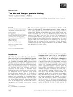
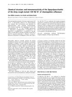
Figure 1. Intra- and inter-chromosomal interactions. The
β-globin gene, located approximately 50 kb downstream of the locus
control region (LCR), is activated during erythropoiesis. The β-globin
gene interaction with the LCR ensures high and ecient β-globin
transcription, with the intervening sequence looping out. (b) Naïve T
cells show a trans association between the T
H
2 LCR, on chromosome
11, and the IFN-γ promoter, on chromosome 10. This interaction is
lost in favor of specic intra-chromosomal interactions following
dierentiation into T
H
1 or T
H
2 eector cells.
Chromosome 11
territory
T
H
2 LCR
Naïve T cells
IFN-γ
promoter
Chromosome 11
territory
Chromosome 10
territory
Chromosome 10
territory
Inter-chromosomal (trans) interaction
Intra-chromosomal (cis) interaction
LCR β−Globin gene
50 kb
Enhanced
β-Globin
transcription
(a)
(b)
Differentiated T
H
1
or T
H
2 cells
Differentiaton
Cope et al. Genome Biology 2010, 11:204
/>Page 2 of 8
networks [24]. ese preferential interactions occur at
nuclear subcompartments containing high local concen-
trations of hyperphosphorylated RNAPII, called trans-
crip tion factories. Described as protein rich structures of
about 10 MDa with an average diameter of about 87 nm,
transcription factories contain multiple active RNAPII
complexes at one time [25-27]. Gene interactions at
transcription factories rely on active transcription: heat-
shock treatment, which blocks initiation and elongation,
resulted in release of genes from factories and disruption
of their long-range associations [23]. Treatment with
5,6-dichloro-β--ribofuranosylbenzimidazole (DRB), which
interferes with phosphorylation of the carboxy-terminal
domain of RNAPII and thus inhibits transcriptional
elongation but not initiation, did not affect the frequency
of gene co-associations [23]. Transcription initiation is
therefore critical for the long-range association of genes
that are being transcribed. Transcription factories remained
after heat shock, consistent with previous results
suggesting that factories are meta-stable structures [28].
ese findings indicate that the structure and function of
transcription factories are fundamental to long-range
interactions between genes being transcribed.
Gene clustering through specialized transcription
factories
e idea of transcription factories being specialized to
transcribe a specific subset of genes in order to achieve
high-level gene transcription seems logical and reason-
able, because no two regions within the nucleus will
contain the same genes or proteins. Early investigations
in human cells into the spatial distribution of certain
transcription factors (glucocorticoid receptor, Oct1 and
E2f-1) revealed only a slight overlap with RNAPII and
sites of transcription [29,30], which the authors [29,30]
argued as evidence against transcription factory speci-
aliza tion. Contrary to this, the Oct1/PTF/transcription
(OPT) domain was the first example of a nuclear
compartment to be shown to contain high concentrations
of interacting transcription factors (PTF1 and Oct1) at a
transcription factory, which specifically recruited regions
from human chromosomes 6 or 7 in early G1 phase [31].
is suggests that specialization of transcription factories
could provide a level of control over genome organization
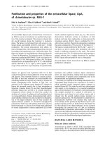
Figure 2. NORs cluster as cells exit mitosis. (a) The short arms of
acrocentric chromosomes 13, 14, 15, 21 and 22 contain NORs, which
are separated during mitosis. (b) As cells exit mitosis and the nuclear
membrane begins to reform, chromosomes begin to decondense.
(c)Loops of chromatin may extend away from the core of the
territory. (d) As G1 phase is established and nucleoli form, loops of
NOR-containing chromatin co-associate with the other components
of the nucleolus and ribosomal DNA gene transcription is initiated.
Chromosome territory
Nucleolus
NOR
(a)
(b) (c)
(d)
2221151413
Key:
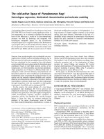
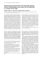
Figure 3. Colocalization of like-regulated genes and specialized
transcription factories.(a) Quadruple-label RNA immuno-FISH of
three genes that are being transcribed and their association with
RNAPII transcription factories. RNAPII staining is shown on the left
and an overlay of the RNAPII staining showing the contributions
of the genes is on the right. The side panels show the enlarged
images of colocalizing FISH signals, showing that transcription
factories can simultaneously transcribe at least three genes, located
on dierent chromosomes. (b) Immunouorescence detection of
Klf1 (red) and RNAPII transcription factories (green), showing the
selective and specialized nature of transcription factories. (c) Triple-
label RNA immuno-FISH for Hbb and Epb4.9, showing association of
these genes at Klf1 foci. All images show denitive erythroid cells
and the scale bar in each panel represents 2 µm. Reproduced, with
permission, from [24].
Hbb
Trfc HbaRNAPII
(a)
Klf1 RNAPII
Klf1
Hbb Epb4.9
(b) (c)
Cope et al. Genome Biology 2010, 11:204
/>Page 3 of 8
by encouraging specific genes to reside in the same
factory. is, along with other studies, gives strong
evidence in favor of transcription factory specialization.
Examination of cotransfected plasmids in COS7 monkey
cells showed that constructs with identical promoters
colocalized to the same transcription factory to a higher
degree than those with heterologous promoters [32].
Furthermore, the finding that the erythroid transcription
factor Klf1 mediates preferential co-associations of Klf1-
regulated genes at Klf1-specialized transcription factories
provided the first functional evidence that transcription
factors could be responsible for the organization of a
specific subset of genes at transcription factories [24]
(Figure 3b,c).
Despite recent demonstrations of spatial clustering in
three dimensions by 3C-based methods and RNA and
DNA FISH [12,24,33,34], it is still unclear whether
association influences gene transcriptional output. Hu et
al. [35] noted the appearance of larger RNA FISH signals
in primary human breast epithelial cells from spatially
associated genes induced by estrogen receptor (ER)a,
suggesting increased transcriptional output from clus-
tered alleles. In addition, long-range association of
transcription factor binding sites or co-regulated genes
correlated with an increased probability of transcriptional
activity of the clustered alleles, suggesting that clustered
alleles were more likely to show higher transcriptional
activity [24,36].
Spatial organization of silent chromatin
ere are obvious potential incentives to cluster specific
genes and chromatin regions. For example, clustering of
co-regulated genes in specialized factories may be more
efficient in terms of the machinery needed for their
expression. e clustering of silent chromatin in the
nucleus could also decrease the amount of machinery
needed for maintenance. Indeed, heterochromatin has
long been observed to form clusters that are distinct from
euchromatin within the nucleoplasm. For example,
centro meres cluster into chromocenters, visualized by
staining with the DNA stain 4',6-diamidino-2-phenyl-
indole (DAPI) or immuno-labeling of centromeric proteins.
Clustering of centromeres is unusually pronounced in
rodent rod cells, where these regions are gathered in the
center of the nucleus surrounded by heterochromatin,
which is suggested to reduce diffraction and permit more
efficient passing of photons [37]. is clustering
demonstrates an extraordinary spatial organization of
chromatin for a specific function. Silenced genes have
also been observed clustering with pericentromeric
hetero chromatin [38]. For example, the non-functional,
rearranged IgH locus is recruited to centromeres
concurrent with transcriptional silencing of its V genes in
B cells [39,40]. is relocalization correlates with dramatic
deacetylation of the locus [41], but it is currently unclear
whether this deacetylation occurs before or after
localization to chromocenters. Telomeres are regions of
transcriptionally silent chromatin and have been reported
to cluster throughout the nucleoplasm [42]. However,
human telomeres with NORs located in their short
acrocentric arms cluster separately at the perinucleolar
compartment [43], again highlighting spatial localization.
Chromatin clustering may also be mediated through
long non-coding RNAs (lncRNAs) such as Xist, Air and
Kcnq1ot1, which range in size from 17 to 108 kb. e
most studied of these lncRNAs is Xist. Transcription of
Xist [43,44] from one of the two X chromosomes results
in the inactivation of that X chromosome in female
mammals. e Xist RNA (about 17 kb in length) interacts
with the future inactive X chromosome to create a
nuclear domain devoid of RNAPII and basal transcription
factors such as TFIIH and TFIIF. X-linked genes are
recruited into this nuclear domain, correlating with their
transcriptional silencing [45]. is internal repositioning
of previously active genes is the first structural change
following Xist accumulation. Intriguingly, genes that
escape X-inactivation are located on the periphery of, or
outside the Xist domain [45], presumably interacting
with RNAPII and various transcription factors.
lncRNAs have also been implicated in the regulation of
imprinted gene clusters. Imprinted genes show effects
specific to the parent of origin, in which a single allele
(maternal or paternal) is epigenetically silenced during
development. Imprinted repression of a selected allele
may occur in a similar mechanism to that of Xist. For
example, the murine Air (antisense to Igf2r) lncRNA is
essential for imprinted allele-specific silencing of the cis-
linked solute carrier genes Slc22a3 and Slc22a2 together
with Igf2r from the paternal chromosome 17 [46]. e
Air RNA forms a cloud within nuclei and interacts, by an
unknown mechanism, with the Slc22a3 promoter. Air is
also required to target the histone H3 lysine 9 histone
methyltransferase G9a to the Slc22a3 promoter [47]. It
seems plausible that the Air cloud recruits specific genes
into the volume it occupies to induce silencing. Unlike
Xist, which induces silencing over the entire X chromo-
some, Air’s influence is restricted to a cluster of genes
spanning a 300 kb region immediately adjacent to the Air
gene. e structural aspects to how Air functions or what
restricts the size of the Air compartment remains unclear.
is effect is mirrored by the Kcnq1ot1 lncRNA, which
also seems to create a repressive domain that is respon-
sible for repression of a variable number of cis-linked
genes in embryonic and placental tissues [48-51]. Kcnq1ot1
is an imprinted 50 kb lncRNA transcribed in the
antisense direction from within the potassium voltage-
gated channel gene, Kcnq1, on mouse chromosome 7.
e Kcnq1ot1 repressive domain is larger in placental
Cope et al. Genome Biology 2010, 11:204
/>Page 4 of 8
tissue than in embryonic tissue, and this may be
correlated with a higher number of silenced genes in the
placenta [49,50].
lncRNA repression may also occur in trans. e 2.2 kb
HOTAIR ncRNA, expressed from the HOXC locus on
chromosome 12 in humans, has been shown to be
necessary for repression of the HOXD locus, present on
chromosome 2 [52]. Although loss of the HOTAIR
lncRNA results in the reactivation of the HOXD locus,
indicating a potential trans mechanism of gene repression
[52], no direct interaction between HOTAIR and the
HOXD locus has been observed.
Establishing spatial organization
Spatial genome organization implies movement. e
tissue-specific clustering of specific genomic elements
requires that at some stage chromatin regions must move
towards each other, in either a directed or a passive way.
As cells exit mitosis and chromosomes decondense,
large-scale movements of chromatin domains have been
observed [53,54]; these may result in the repositioning of
chromosomal and sub-chromosomal regions to their
generalized relative positions. Constrained diffusion [55]
or chromatin movements mediated by nuclear actin and
myosin [35,56-58] may have a role in refining these
positions throughout interphase (Figure4).
e organization of the genome as it is transcribed is
achieved to a large extent through interactions of genes
with transcription factories. Although it is not known
how factories form, the pulsatile nature of individual
gene transcription during interphase [59,60], which seems
to involve dynamic gene associations with factories
[17,22], suggests two possible models to describe how
specialized factories are established. In a deterministic
factory model, specific key transcription factors (such as
Klf1) are directed to or become concentrated at a subset
of factories. Genes requiring that particular factor for
transcription would then need to move to those factories
to become active. In the second model, referred to as the
self-organization model, genes and their bound regu-
latory factors stochastically engage factories in their local
environment. Specialization may occur when several
similarly regulated genes associate with the same factory
simultaneously. is may stabilize their presence at the
shared factory through factor sharing, in other words the
increased local concentration of specific regulatory
factors may increase occupancy at key regulatory sites on
the clustered genes, thus promoting their reinitiation and
stabilizing their co-association. ere is little evidence in
favor of either model at the moment. e deterministic
model requires some mechanism to direct specific factors
to a subset of factories, suggesting that differences in
factories must precede their specialization. In the self-
organization model, all factories may start out being
equal but then may become specialized, perhaps
transiently by character of the transcription units
engaged there.
Evidence in favor of the self-organization model can be
seen in a population of virally infected cells: the quickest
cells to respond by producing IFN-β are those in which
the IFN-β gene is in close physical proximity with other
genetic loci that bind the NF-κB transcription factor [36].
NF-κB induces the formation of the enhanceosome
multiprotein complex, which binds upstream of the IFN-
β promoter and interacts with the transcriptional
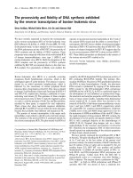
Figure 4. Schematic summary of some of the processes
and structures that inuence the spatial organization of
the genome. Although not exhaustive, the gure depicts:
(a)chromosome territories; (b) nucleoli and genomic regions
clustering through nucleolar organizing regions (NOR); (c) the
Xchromosome and Xist RNA; (d) regulatory proteins such as CTCF,
transcription factors and Polycomb repressive complexes (PRCs)
that can induce loops between genomic elements; (e) transcription
factories (blue) and specialized transcription factories (red); (f) the
potential role of nuclear actin in mediating long-range chromatin
movement; and (g) the interactions of chromatin regions with the
nuclear lamina. These processes, along with others described in
this article and many more, are likely be important in dynamically
shaping the spatial environment and organization of the genome.
Specialized transcription
factory
Transcription factory
Regulatory protein
Nuclear actin
Nuclear lamin
Chromosome
territory
NOR
Chromatin
Xist RNA
Nucleolus
(g)
(f)
(d)
(e)
(b)
(a)
(c)
Key:
Cope et al. Genome Biology 2010, 11:204
/>Page 5 of 8
machinery necessary for the induction of the IFN-β gene.
e formation of the enhanceosome at the IFN-β promo-
ter is more likely to occur if one NF-κB-dependent gene is
in close physical proximity to another NF-κB-depen dent
gene, thereby enabling these loci to establish an
environment that favors transcription [36]. is supports
a role for transcription factors mediating chromosomal
interactions specific for the tissue and stimulus involved.
Such transcriptional organization of genes may also be
mediated by other proteins that are not part of the core
transcriptional apparatus, such as the CCCTC-binding
factor (CTCF) and Polycomb repressive complexes
(PRCs).
Some proteins may have a structural role in main te-
nance of genome conformation. CTCF is a highly
conserved vertebrate transcriptional regulator that has
been reported to bind at many thousands of sites in
multiple genomes [61-65]. is binding does not seem to
correlate to specific networks of genes, but CTCF has
been suggested to mediate chromatin interactomes [66].
Indeed, CTCF binding has been suggested to silence the
mater nally inherited Igf2 allele [67], form active
chromatin hubs [68], and establish cytokine-induced
loops within the human MHC class II locus [69].
Furthermore, CTCF interacts with a large number of
nuclear proteins ranging from transcription factors to
structural proteins [70]. Cohesin, which is a key
component for holding sister chromatids together and
which is implicated in several diseases, has been shown to
bind to about 70% of all CTCF sites in the human genome
[71]. Specifically, CTCF mediates cohesin binding [72],
and this interaction has been suggested to impart cell-
type-specific intra chromosomal interactions at the
developmentally regu lated human cytokine locus IFN-γ
[72] and the apo lipo protein A1/C3/A4/A5 gene region on
human chromo some 11 [73]. ese processes suggest a
multifunctional role of CTCF in the organization of the
genome, adding another organizational layer of
complexity.
Repressive domains and complexes may also provide a
structural component for establishing long-range inter-
actions and organizing the genome. For example,
genome-wide studies have revealed that PRCs associate
with promoter regions of some developmentally regu-
lated and silenced genes [74,75]. Evidence to support
long-range interactions through PRCs comes from
studies investigating Polycomb response elements (PREs),
which allow the recruitment of PRCs to target genes
through DNA binding proteins [76]. Fab-7 is a Drosophila
regulatory element containing a PRE that contributes to
regulated spatial transcription of the Abdominal-B gene
of the Drosophila bithorax complex [77,78]. e endoge-
nous Fab-7 PRE has been shown to interact with
transgenic Fab-7 elements inserted at heterologous sites
[79], highlighting specific long-range PRE-mediated chro-
ma tin interactions. Similarly, Mcp, another PRE contain-
ing regulatory element from the Drosophila bithorax
complex, can interact with other remote copies of Mcp
elements in the genome [80]. ese results provided
direct evidence that regulatory elements can promote
sequence-specific long-range chromosomal interactions,
suggesting that PRCs are likely to provide another
mechanism for organizing the genome.
Recently, the roles of nuclear actin and myosin have
generated considerable interest in the organization of the
mammalian genome. Data strongly indicate that nuclear
actin is involved in gene transcription by all three
polymerases [81]. Long-range directed interphase chro-
ma tin movement seems to require actin polymeriza tion,
as the expression of mutant actin that cannot poly merize
prevents chromatin relocation [56,57]. Nuclear actin and
nuclear myosin I have also been implicated in mediating
interchromosomal interactions between the ERα-
dependent genes [35] and in repositioning of selected
chromosomes during serum starvation [58].
Spatial organization and the future
Here, we have focused on the relationships between trans-
cription, silencing and the three-dimensional organi za-
tion of the genome (Figure 4). is is at the expense of
other structures that also contribute to the genome’s
organization, such as the nuclear lamina [82,83]. In
summary, it is apparent that the genome is arranged in a
non-random, cell- and tissue-specific manner that is
suited for various nuclear functions. Highly expressed
housekeeping genes are often organized in the linear
genome in RIDGES (regions of increased gene expression)
[84], but linear clustering of tissue-specific genes is not
evident [85]. Although clustering of housekeeping genes
may be favored in a two-dimensional arrangement along
the chromosome, clustering of tissue-specific genes is
evident only in three dimensions across the nucleus
[12,24,33], presumably reflecting transcrip tional and
other regulatory requirements. It is clear that the local
folding of chromatin, for example between a gene and
long-range enhancer or between PREs, is a critical
determinant of gene expression. e way these regions
interact with other regions of the same chromo some,
some of which may be similarly regulated, also seems to
be important for function. Similarly, the way these
chromosomal regions interact with regions on other
chromosomes will undoubtedly affect spatial genome
organization, but it may also be important in contributing
to tissue-specific gene expression programs. It is likely
that three-dimensional organization is an important
missing link in understanding how the genome is
regulated; unraveling this organization is a major
challenge for the future.
Cope et al. Genome Biology 2010, 11:204
/>Page 6 of 8
Acknowledgements
We thank all members of the Laboratory of Chromatin and Gene Expression
for their help and advice, and also thank Lyubomira Chakalova, Claire Joyce
and Nicole Shoaf for critical reading of the manuscript. This work was
supported by the Medical Research Council and the Biotechnology and
Biological Sciences Research Council, UK.
Published: 29 March 2010
References
1. Simonis M, Kooren J, de Laat W: An evaluation of 3C-based methods to
capture DNA interactions. Nat Methods 2007, 4:895-901.
2. Carter D, Chakalova L, Osborne CS, Dai YF, Fraser P: Long-range chromatin
regulatory interactions in vivo. Nat Genet 2002, 32:623-626.
3. Tolhuis B, Palstra RJ, Splinter E, Grosveld F, de Laat W: Looping and
interaction between hypersensitive sites in the active beta-globin locus.
Mol Cell 2002, 10:1453-1465.
4. Pevny L, Simon MC, Robertson E, Klein WH, Tsai S-F, D’Agati V, Orkin SH,
Costantini F: Erythroid differentiation in chimaeric mice blocked by a
targeted mutation in the gene for transcription factor GATA-1. Nature 1991,
349:257-260.
5. Starck J, Sarkar R, Romana M, Bhargava A, Scarpa AL, Tanaka M, Chamberlain
JW, Weissman SM, Forget BG: Developmental regulation of human gamma-
globin and beta-globin genes in the absence of the locus-control region.
Blood 1994, 84:1656-1665.
6. Drissen R, Palstra RJ, Gillemans N, Splinter E, Grosveld F, Philipsen S, de Laat W:
The active spatial organization of the beta-globin locus requires the
transcription factor EKLF. Genes Dev 2004, 18:2485-2490.
7. Song SH, Hou CH, Dean A: A positive role for NLI/Ldb1 in long-range beta-
globin locus control region function. Mol Cell 2007, 28:810-822.
8. Kim SI, Bultman SJ, Kiefer CM, Dean A, Bresnick EH: BRG1 requirement for
long-range interaction of a locus control region with a downstream
promoter. Proc Natl Acad Sci USA 2009, 106:2259-2264.
9. Bolland DJ, Wood AL, Corcoran AE: Large-scale chromatin remodeling at
the immunoglobulin heavy chain locus: a paradigm for multigene
regulation. Adv Exp Med Biol 2009, 650:59-72.
10. Spilianakis CG, Flavell RA: Long-range intrachromosomal interactions in the
T helper type 2 cytokine locus. Nat Immunol 2004, 5:1017-1027.
11. Spilianakis CG, Lalioti MD, Town T, Lee GR, Flavell RA: Interchromosomal
associations between alternatively expressed loci. Nature 2005, 435:637-645.
12. Zhao Z, Tavoosidana G, Sjolinder M, Gondor A, Mariano P, Wang S, Kanduri C,
Lezcano M, Sandhu KS, Singh U, Pant V, Tiwari V, Kurukuti S, Ohlsson R:
Circular chromosome conformation capture (4C) uncovers extensive
networks of epigenetically regulated intra- and interchromosomal
interactions. Nat Genet 2006, 38:1341-1347.
13. Lomvardas S, Barnea G, Pisapia DJ, Mendelsohn M, Kirkland J, Axel R:
Interchromosornal interactions and olfactory receptor choice. Cell 2006,
126:403-413.
14. Fuss SH, Omura M, Mombaerts P: Local and cis effects of the H element on
expression of odorant receptor genes in mouse. Cell 2007, 130:373-384.
15. Nishizumi H, Kurnasaka K, Inoue N, Nakashima A, Sakano H: Deletion of the
core-H region in mice abolishes the expression of three proximal odorant
receptor genes in cis. Proc Natl Acad Sci USA 2007, 104:20067-20072.
16. Rowley JD: Chromosomal translocations: revisited yet again. Blood 2008,
112:2183-2189.
17. Osborne CS, Chakalova L, Mitchell JA, Horton A, Wood AL, Bolland DJ,
Corcoran AE, Fraser P: Myc dynamically and preferentially relocates to a
transcription factory occupied by IgH. PLoS Biol 2007, 5:e192.
18. Lin C, Yang L, Tanasa B, Hutt K, Ju BG, Ohgi K, Zhang J, Rose DW, Fu XD, Glass
CK, Rosenfeld MG: Nuclear receptor-induced chromosomal proximity and
DNA breaks underlie specific translocations in cancer. Cell 2009,
139:1069-1083.
19. Prieto JL, McStay B: Nucleolar biogenesis: the first small steps. Biochem Soc
Trans 2005, 33:1441-1443.
20. Kalmarova M, Smirnov E, Masata M, Koberna K, Ligasova A, Popov A, Raska I:
Positioning of NORs and NOR-bearing chromosomes in relation to
nucleoli. J Struct Biol 2007, 160:49-56.
21. Dousset T, Wang C, Verheggen C, Chen DY, Hernandez-Verdun D, Huang S:
Initiation of nucleolar assembly is independent of RNA polymerase I
transcription. Mol Biol Cell 2000, 11:2705-2717.
22. Osborne CS, Chakalova L, Brown KE, Carter D, Horton A, Debrand E,
Goyenechea B, Mitchell JA, Lopes S, Reik W, Fraser P: Active genes
dynamically colocalize to shared sites of ongoing transcription. Nat Genet
2004, 36:1065-1071.
23. Mitchell JA, Fraser P: Transcription factories are nuclear subcompartments
that remain in the absence of transcription. Genes Dev 2008, 22:20-25.
24. Schoenfelder S, Sexton T, Chakalova L, Cope NF, Horton A, Andrews S,
Kurukuti S, Mitchell JA, Umlauf D, Dimitrova DS, Eskiw CH, Luo Y, Wei CL, Ruan
Y, Bieker JJ, Fraser P: Preferential associations between co-regulated genes
reveal a transcriptional interactome in erythroid cells. Nat Genet 2010,
42:53-61.
25. Eskiw CH, Rapp A, Carter DRF, Cook PR: RNA polymerase II activity is located
on the surface of protein-rich transcription factories. J Cell Sci 2008,
121:1999-2007.
26. Iborra FJ, Pombo A, Jackson DA, Cook PR: Active RNA polymerases are
localized within discrete transcription ‘factories’ in human nuclei. J Cell Sci
1996, 109:1427-1436.
27. Pombo A, Hollinshead M, Cook PR: Bridging the resolution gap: imaging
the same transcription factories in cryosections by light and electron
microscopy. J Histochem Cytochem 1999, 47:471-480.
28. Kimura H, Sugaya K, Cook PR: The transcription cycle of RNA polymerase II
in living cells. J Cell Biol 2002, 159:777-782.
29. Grande MA, vanderKraan I, deJong L, vanDriel R: Nuclear distribution of
transcription factors in relation to sites of transcription and RNA
polymerase II. J Cell Sci 1997, 110:1781-1791.
30. vanSteensel B, vanBinnendijk EP, Hornsby CD, vanderVoort HTM, Krozowski
ZS, deKloet ER, vanDriel R: Partial colocalization of glucocorticoid and
mineralocorticoid receptors in discrete compartments in nuclei of rat
hippocampus neurons. J Cell Sci 1996, 109:787-792.
31. Pombo A, Cuello P, Schul W, Yoon JB, Roeder RG, Cook PR, Murphy S: Regional
and temporal specialization in the nucleus: a transcriptionally-active
nuclear domain rich in PTF, Oct1 and PIKA antigens associates with
specific chromosomes early in the cell cycle. EMBO J 1998, 17:1768-1778.
32. Xu M, Cook PR: Similar active genes cluster in specialized transcription
factories. J Cell Biol 2008, 181:615-623.
33. Fullwood MJ, Liu MH, Pan YF, Liu J, Xu H, Mohamed YB, Orlov YL, Velkov S, Ho
A, Mei PH, Chew EG, Huang PY, Welboren WJ, Han Y, Ooi HS, Ariyaratne PN,
Vega VB, Luo Y, Tan PY, Choy PY, Wansa KD, Zhao B, Lim KS, Leow SC, Yow JS,
Joseph R, Li H, Desai KV, Thomsen JS, Lee YK, et al.: An oestrogen-receptor-
alpha-bound human chromatin interactome. Nature 2009, 462:58-64.
34. Simonis M, Klous P, Splinter E, Moshkin Y, Willemsen R, de Wit E, van Steensel
B, de Laat W: Nuclear organization of active and inactive chromatin
domains uncovered by chromosome conformation capture-on-chip (4C).
Nat Genet 2006, 38:1348-1354.
35. Hu Q, Kwon YS, Nunez E, Cardamone MD, Hutt KR, Ohgi KA, Garcia-Bassets I,
Rose DW, Glass CK, Rosenfeld MG, Fu XD: Enhancing nuclear receptor-
induced transcription requires nuclear motor and LSD1-dependent gene
networking in interchromatin granules. Proc Natl Acad Sci USA 2008,
105:19199-19204.
36. Apostolou E, Thanos D: Virus infection induces NF-kappa B-dependent
interchromosomal associations mediating monoallelic IFN-beta gene
expression. Cell 2008, 134:85-96.
37. Solovei I, Kreysing M, Lanctot C, Kosem S, Peichl L, Cremer T, Guck J, Joe B:
Nuclear architecture of rod photoreceptor cells adapts to vision in
mammalian evolution. Cell 2009, 137:356-368.
38. Brown KE, Guest SS, Smale ST, Hahm K, Merkenschlager M, Fisher AG:
Association of transcriptionally silent genes with Ikaros complexes at
centromeric heterochromatin. Cell 1997, 91:845-854.
39. Skok JA, Brown KE, Azuara V, Caparros ML, Baxter J, Takacs K, Dillon N, Gray D,
Perry RP, Merkenschlager M, Fisher AG: Nonequivalent nuclear location of
immunoglobulin alleles in B lymphocytes. Nat Immunol 2001, 2:848-854.
40. Roldan E, Fuxa M, Chong W, Martinez D, Novatchkova M, Busslinger M, Skok
JA: Locus ‘decontraction’ and centromeric recruitment contribute to allelic
exclusion of the immunoglobulin heavy-chain gene. Nat Immunol 2005,
6:31-41.
41. Chowdhury D, Sen R: Transient IL-7/IL-7R signaling provides a mechanism
for feedback inhibition of immunoglobulin heavy chain gene
rearrangements. Immunity 2003, 18:229-241.
42. Ramirez MJ, Surralles J: Laser confocal microscopy analysis of human
interphase nuclei by three-dimensional FISH reveals dynamic
perinucleolar clustering of telomeres. Cytogenet Genome Res 2008,
122:237-242.
Cope et al. Genome Biology 2010, 11:204
/>Page 7 of 8
43. Avner P, Heard E: X-chromosome inactivation: counting, choice and
initiation. Nat Rev Genet 2001, 2:59-67.
44. Cohen DE, Lee JT: X-chromosome inactivation and the search for
chromosome-wide silencers. Curr Opin Genet Dev 2002, 12:219-224.
45. Chaumeil J, Le Baccon P, Wutz A, Heard E: A novel role for Xist RNA in the
formation of a repressive nuclear compartment into which genes are
recruited when silenced. Genes Dev 2006, 20:2223-2237.
46. Sleutels F, Zwart R, Barlow DP: The non-coding Air RNA is required for
silencing autosomal imprinted genes. Nature 2002, 415:810-813.
47. Nagano T, Mitchell JA, Sanz LA, Pauler FM, Ferguson-Smith AC, Feil R, Fraser P:
The air noncoding RNA epigenetically silences transcription by targeting
G9a to chromatin. Science 2008, 322:1717-1720.
48. Fitzpatrick GV, Soloway PD, Higgins MJ: Regional loss of imprinting and
growth deficiency in mice with a targeted deletion of KvDMR1. Nat Genet
2002, 32:426-431.
49. Pandey RR, Mondal T, Mohammad F, Enroth S, Redrup L, Komorowski J,
Nagano T, Mancini-DiNardo D, Kanduri C: Kcnq1ot1 antisense noncoding
RNA mediates lineage-specific transcriptional silencing through
chromatin-level regulation. Mol Cell 2008, 32:232-246.
50. Redrup L, Branco MR, Perdeaux ER, Krueger C, Lewis A, Santos F, Nagano T,
Cobb BS, Fraser P, Reik W: The long noncoding RNA Kcnq1ot1 organises a
lineage-specific nuclear domain for epigenetic gene silencing.
Development 2009, 136:525-530.
51. Terranova R, Yokobayashi S, Stadler MB, Otte AP, van Lohuizen M, Orkin SH,
Peters A: Polycomb group proteins Ezh2 and Rnf2 direct genomic
contraction and imprinted repression in early mouse embryos. Dev Cell
2008, 15:668-679.
52. Rinn JL, Kertesz M, Wang JK, Squazzo SL, Xu X, Brugmann SA, Goodnough LH,
Helms JA, Farnham PJ, Segal E, Chang HY: Functional demarcation of active
and silent chromatin domains in human HOX loci by noncoding RNAs. Cell
2007, 129:1311-1323.
53. Walter J, Schermelleh L, Cremer M, Tashiro S, Cremer T: Chromosome order in
HeLa cells changes during mitosis and early G1, but is stably maintained
during subsequent interphase stages. J Cell Biol 2003, 160:685-697.
54. Thomson I, Gilchrist S, Bickmore WA, Chubb JR: The radial positioning of
chromatin is not inherited through mitosis but is established de novo in
early G1. Curr Biol 2004, 14:166-172.
55. Chubb JR, Boyle S, Perry P, Bickmore WA: Chromatin motion is constrained
by association with nuclear compartments in human cells. Curr Biol 2002,
12:439-445.
56. Chuang CH, Carpenter AE, Fuchsova B, Johnson T, de Lanerolle P, Belmont AS:
Long-range directional movement of an interphase chromosome site. Curr
Biol 2006, 16:825-831.
57. Dundr M, Ospina JK, Sung MH, John S, Upender M, Ried T, Hager GL, Matera
AG: Actin-dependent intranuclear repositioning of an active gene locus in
vivo. J Cell Biol 2007, 179:1095-1103.
58. Mehta IS, Amira M, Harvey AJ, Bridger JM: Rapid chromosome territory
relocation by nuclear motor activity in response to serum removal in
primary human fibroblasts. Genome Biol 2010, 11:R5.
59. Chubb JR, Trcek T, Shenoy SM, Singer RH: Transcriptional pulsing of a
developmental gene. Curr Biol 2006, 16:1018-1025.
60. Raj A, Peskin CS, Tranchina D, Vargas DY, Tyagi S: Stochastic mRNA synthesis
in mammalian cells. PLoS Biol 2006, 4:e309.
61. Xie XH, Mikkelsen TS, Gnirke A, Lindblad-Toh K, Kellis M, Lander ES: Systematic
discovery of regulatory motifs in conserved regions of the human
genome, including thousands of CTCF insulator sites. Proc Natl Acad Sci USA
2007, 104:7145-7150.
62. Kim TH, Abdullaev ZK, Smith AD, Ching KA, Loukinov DI, Green RD, Zhang
MQ, Lobanenkov VV, Ren B: Analysis of the vertebrate insulator protein
CTCF-binding sites in the human genome. Cell 2007, 128:1231-1245.
63. Jothi R, Cuddapah S, Barski A, Cui K, Zhao K: Genome-wide identification of
in vivo protein-DNA binding sites from ChIP-Seq data. Nucleic Acids Res
2008, 36:5221-5231.
64. Cuddapah S, Jothi R, Schones DE, Roh TY, Cui KR, Zhao KJ: Global analysis of
the insulator binding protein CTCF in chromatin barrier regions reveals
demarcation of active and repressive domains. Genome Res 2009, 19:24-32.
65. Chen X, Xu H, Yuan P, Fang F, Huss M, Vega VB, Wong E, Orlov YL, Zhang W,
Jiang J, Loh YH, Yeo HC, Yeo ZX, Narang V, Govindarajan KR, Leong B, Shahab
A, Ruan Y, Bourque G, Sung WK, Clarke ND, Wei CL, Ng HH: Integration of
external signaling pathways with the core transcriptional network in
embryonic stem cells. Cell 2008, 133:1106-1117.
66. Phillips JE, Corces VG: CTCF: Master weaver of the genome. Cell 2009,
137:1194-1211.
67. Kurukuti S, Tiwari VK, Tavoosidana G, Pugacheva E, Murrell A, Zhao ZH,
Lobanenkov V, Reik W, Ohlsson R: CTCF binding at the H19 imprinting
control region mediates maternally inherited higher-order chromatin
conformation to restrict enhancer access to Igf2. Proc Natl Acad Sci USA
2006, 103:10684-10689.
68. Splinter E, Heath H, Kooren J, Palstra RJ, Klous P, Grosveld F, Galjart N, de Laat
W: CTCF mediates long-range chromatin looping and local histone
modification in the beta-globin locus. Genes Dev 2006, 20:2349-2354.
69. Majumder P, Gomez JA, Chadwick BP, Boss JM: The insulator factor CTCF
controls MHC class II gene expression and is required for the formation of
long-distance chromatin interactions. J Exp Med 2008, 205:785-798.
70. Zlatanova J, Caiafa P: CTCF and its protein partners: divide and rule? J Cell
Sci 2009, 122:1275-1284.
71. Parelho V, Hadjur S, Spivakov M, Leleu M, Sauer S, Gregson HC, Jarmuz A,
Canzonetta C, Webster Z, Nesterova T, Cobb BS, Yokomori K, Dillon N, Aragon
L, Fisher AG, Merkenschlager M: Cohesins functionally associate with CTCF
on mammalian chromosome arms. Cell 2008, 132:422-433.
72. Hadjur S, Williams LM, Ryan NK, Cobb BS, Sexton T, Fraser P, Fisher AG,
Merkenschlager M: Cohesins form chromosomal cis-interactions at the
developmentally regulated IFNG locus. Nature 2009, 460:410-413.
73. Mishiro T, Ishihara K, Hino S, Tsutsumi S, Aburatani H, Shirahige K, Kinoshita Y,
Nakao M: Architectural roles of multiple chromatin insulators at the
human apolipoprotein gene cluster. EMBO J 2009, 28:1234-1245.
74. Schuettengruber B, Chourrout D, Vervoort M, Leblanc B, Cavalli G: Genome
regulation by polycomb and trithorax proteins. Cell 2007, 128:735-745.
75. Schwartz YB, Pirrotta V: Polycomb complexes and epigenetic states. Curr
Opin Cell Biol 2008, 20:266-273.
76. Muller J, Kassis JA: Polycomb response elements and targeting of Polycomb
group proteins in Drosophila. Curr Opin Genet Dev 2006, 16:476-484.
77. Gyurkovics H, Gausz J, Kummer J, Karch F: A new homeotic mutation in the
Drosophila bithorax complex removes a boundary separating 2 domains
of regulation. EMBO J 1990, 9:2579-2585.
78. Busturia A, Bienz M: Silencers in abdominal-B, a homeotic Drosophila gene.
EMBO J 1993, 12:1415-1425.
79. Bantignies F, Grimaud C, Lavrov S, Gabut M, Cavalli G: Inheritance of
Polycomb-dependent chromosomal interactions in Drosophila. Genes Dev
2003, 17:2406-2420.
80. Vazquez J, Muller M, Pirrotta V, Sedat JW: The Mcp element mediates stable
long-range chromosome-chromosome interactions in Drosophila. Mol Biol
Cell 2006, 17:2158-2165.
81. Percipalle P: The long journey of actin and actin-associated proteins from
genes to polysomes. Cell Mol Life Sci 2009, 66:2151-2165.
82. Pickersgill H, Kalverda B, de Wit E, Talhout W, Fornerod M, van Steensel B:
Characterization of the Drosophila melanogaster genome at the nuclear
lamina. Nat Genet 2006, 38:1005-1014.
83. Guelen L, Pagie L, Brasset E, Meuleman W, Faza MB, Talhout W, Eussen BH, de
Klein A, Wessels L, de Laat W, van Steensel B: Domain organization of human
chromosomes revealed by mapping of nuclear lamina interactions. Nature
2008, 453:948-951.
84. Caron H, van Schaik B, van der Mee M, Baas F, Riggins G, van Sluis P, Hermus
MC, van Asperen R, Boon K, Voûte PA, Heisterkamp S, van Kampen A, Versteeg
R: The human transcriptome map: clustering of highly expressed genes in
chromosomal domains. Science 2001, 291:1289-1292.
85. Lercher MJ, Urrutia AO, Hurst LD: Clustering of housekeeping genes
provides a unified model of gene order in the human genome. Nat Genet
2002, 31:180-183.
doi:10.1186/gb-2010-11-3-204
Cite this article as: Cope NF, et al.: The yin and yang of chromatin spatial
organization. Genome Biology 2010, 11:204.
Cope et al. Genome Biology 2010, 11:204
/>Page 8 of 8