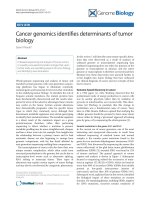
Báo cáo y học: "Cancer genomics identifies determinants of tumor biology" ppt
Bạn đang xem bản rút gọn của tài liệu. Xem và tải ngay bản đầy đủ của tài liệu tại đây (859.49 KB, 8 trang )
Whole-genome sequencing and analysis of tumor and
matched normal genomes with next-generation sequen-
cing platforms has begun to illuminate commonly
mutated genes and transcript-level events that contribute
to the underlying tumor biology. To elucidate the role of
frequent somatic mutations, the mutant proteins have
been biochemically characterized and the results inter-
preted in terms of the selective advantages these variants
may confer on the tumor. Certain somatic alterations
have demonstrable prognostic value for specific tumor
types in which they commonly occur, although their
down stream metabolic signatures may obviate geno typing
to identify their mutational status. e metabolic signature
is a direct result of the mutation’s impact on a given
protein/enzyme; therefore, rather than performing
sequencing to detect whether a mutation is present,
metabolic profiling may be more straightforward, cheaper,
and have a lower error rate, for example. New insights into
the relationship between a primary tumor and its fatal
metastatic disease are also beginning to emerge from
genomic comparisons, with the fine detail afforded by
next-generation sequencing enabling these comparisons.
e transcriptomes of cancer cells also have their own
unique somatic complexities, which often result from
structural perturbations to the genome, but can be due to
transcription-level events such as alternative splicing,
RNA editing or transcript fusion. ese types of
alterations may explain certain aspects of tumor biology
and may also be corroborated by cytogenetic phenomena.
In this review, I will describe some tumor-specific altera-
tions that were discovered as a result of analyses of
unbiased genome or transcriptome sequencing data
(unbiased sequencing does not select for portions of the
genome or transcriptome in advance, and the entire
genome or transcriptome is therefore surveyed) and then
illustrate how these discoveries were pursued further to
reveal insights into tumor biology that have enhanced
our clinical diagnosis of cancer and our concepts of how
best to treat it.
Genome-based discovery in cancer
In a 1956 paper [1], Otto Warburg observed that the
predominant mode of energy production in cancer cells
was by aerobic glycolysis rather than by oxidation of
pyruvate in mitochondria, as in normal cells. is obser-
vation led Warburg to postulate that this change in
metabolism was a fundamental cause of cancer. Years
later, in 1986, Renato Dulbecco opined that studying the
cellular genome should be pursued to learn more about
cancer, either by taking a ‘piecemeal’ approach of looking
gene by gene, or by sequencing the whole genome [2].
Somatic mutations in the genes IDH1 and IDH2
In the current era of cancer genomics, one of the most
interesting and unexpected discoveries to result from
unbiased sequencing of matched tumor and normal
samples is the somatic point mutations found in the
genes for two isocitrate dehydrogenase isoenzymes, IDH1
and IDH2. First discovered by sequencing the exome (the
exons collectively) of the glial brain tumor glioblastoma
multiforme (GBM) [3], mutated IDH1 was found in 12%
of tumors analyzed. e general approach to exome
capture and analysis is shown in Figure 1. Subsequent
focused re-sequencing refined the occurrence of muta-
tions at arginine 132 (R132) of IDH1, which are found in
more than 80% of secondary GBMs (GBMs that initially
present as low-grade astrocytomas (tumors of
astrocytes)), and less than 10% of primary GBMs [4-6].
Subsequent work, explained later in this review, identified
the biological impact of these mutations on enzyme
function. On examining gliomas, including GBMs, negative
for IDH1 mutations, recurrent somatic muta tions of
Abstract
Unbiased sequencing and analysis of human tumors
is revealing unsuspected somatic changes that, upon
further study, are elucidating aspects of tumor biology
and identifying new biomarkers.
© 2010 BioMed Central Ltd
Cancer genomics identifies determinants of tumor
biology
Elaine R Mardis*
R E VI E W
*Correspondence:
The Genome Center, Washington University School of Medicine, St Louis, MO
63108, USA
Mardis Genome Biology 2010, 11:211
/>© 2010 BioMed Central Ltd
IDH2 at the analogous R172 residue were identified [4,7].
Not only are the IDH1 and IDH2 mutations frequent, but
studies by several laboratories have established that the
mutation in IDH1 occurs early in glioma progression [8].
Notably, the mutations affect only one allele at the given
locus (of the two alleles of either IDH1 or IDH2, but not
both in the same tumor), which is puzzling considering
the evidence that they are selected for early in tumori-
genesis. Analysis of the correlation between mutation in
IDH1 or IDH2 and various clinical features has revealed
interesting asso ciations between the presence of mutation
and an early age of disease onset and overall longer
survival time in GBMs and in anaplastic astrocytomas
(another type of glial tumor, distinct from GBMs) [4].
Figure 1. General schema for targeted exome capture, whole genome sequencing, and transcriptome sequencing. (a) In exome capture,
a random library of genomic fragments, each containing platform-specic adapters on each end, is combined with a set of probes that dene
the human exome. Following hybridization, the probe:genomic library fragment hybrids are captured using magnetic beads and isolated from
solution by the application of a magnet, or by solid phase capture. Denaturing conditions are used to elute the captured genomic library fragment
population from the hybrids, and prepared for sequencing. (b) In whole genome sequencing, the same random fragment library is constructed as
in (a), but the resulting fragments are sequenced directly without a capture step. (c) In transcriptome sequencing, the RNA is converted to cDNA,
the resulting cDNAs are fragmented, and the library adapters are ligated to the resulting fragments, followed by sequencing. Panel (a) reproduced
with permission from [27].
Target capture > 100,000 exons
Adapter modified shotgun library
B
B
B
B
B
B
B
B
B
B
Solution
hybridization
Bead capture
Array capture
AAAAAAAA
Poly T + adapter based reverse
transcription
TTTTTTTTT
AAAAAAAA
AAAAAAAA
TTTTTTTTT
TTTTTTTTT
AAAAAAAA
TTTTTTTTT
Adapter based paired-end
sequencing of cDNA library
cDNA library
Poly-A mRNA pool
(a) Exome sequencing (c) Transcriptome sequencing
(b) Whole genome shotgun sequencing
Whole genome shotgun
using paired end reads
Align reads to human genome reference.
Analyze alignments to identify point mutations, focused insertion/deletion
changes and large structural rearrangements
Adaptor modified shotgun library
B
B
B
B
Mardis Genome Biology 2010, 11:211
/>Page 2 of 8
e IDH enzymes play a key role in cellular metabo-
lism, catalyzing the conversion of isocitrate to α-keto-
glutarate (α-KG) and generating NADPH from NADP
+
in
the process (Figure 2). e crystal structure of IDH1 [9]
predicts that the amino acid substitutions found at the
R132 position will impair the interaction of the enzyme
with its isocitrate substrate, and functional and bio-
chemical studies of the mutant proteins by several groups
have provided critical insights into this [4,10,11]. Zhao
and colleagues [10] evaluated the in vitro enzymatic
activities of three tumor-derived IDH1 mutants - R132H,
R132C and R132S - by expressing mutant constructs in
transformed human embryonic kidney 293T cells. ey
observed all three mutants to have a more than 80%
reduction in the ability to convert isocitrate to α-KG
compared with the wild-type enzyme, and further kinetic
analyses revealed a dramatically reduced affinity for
isocitrate in all three mutants. As IDH1 functions as a
homodimeric complex, Zhao et al. [10] isolated IDH1
dimers expressed from the R132H mutant and wild-type
genes introduced into Escherichia coli. ree dimer
combinations were identified, the wild-type:R132H
heterodimer exhibited only 4% of the wild-type dimer
enzyme activity, while R132H:R132H homodimers were
almost completely inactive.
What are the metabolic consequences of IDH1 muta-
tions? Using the U-87MG human glioblastoma cell line,
Zhao et al. [10] demonstrated a concomitant reduction in
Figure 2. Impact of IDH1/2 mutations on tumor cell biology. (a) In normal cells, the role of IDH1 and IDH2 enzymes is to convert isocitrate
to α-ketoglutarate (α-KG), converting NADP+ to NADPH. The presence of α-KG regulates prolylhydroxylases (PHD) that, in turn, promote the
degradation of hypoxia-inducible factor 1α (HIF-1α). HIF-1α is a transcription factor that regulates the expression of genes related to glucose
metabolism, angiogenesis, and other signaling pathways by sensing low cellular oxygen levels. The mutant IDH enzymes convert α-KG to
2-hydroxyglutarate (2-HG), leading to the build up of this oncometabolite. (b) Comparison of metabolomic proling of IDH wild-type (upper panel)
and mutant (lower panel) cells, indicating the increased levels of 2-HG associated with the mutation. 2-HG is absent in IDH wild-type cells. Panel (b)
reproduced with permission from [15].
NADP
NADP
NADPH
NADPH
(a) (b)
IDH1
IDH2
IDH2 R172K / R140Q
AML with IDH2
AML with IDH2 R140Q
α-KG
)
Isocitrate
2-HG
PHD
α-KG
OH -
Hydroxylation
Degradation
HIF-1α
Glucose metabolism
associated genes
Angiogenesis
associated genes
1.00
0.80
0.60
0.40
0.20
0
30.5 31.0 31.5 32.0 32.5 33.0 33.5 34.0
Asp
Glu
Metabolite abundanceMetabolite abundance
Elution time (min)
1.00
0.80
0.60
0.40
0.20
0
30.5 31.0 31.5 32.0 32.5 33.0 33.5 34.0
Asp
Glu
Elution time (min)
2HG
IDH1 R132 H/C/S
+
+
Mardis Genome Biology 2010, 11:211
/>Page 3 of 8
cellular α-KG levels after knocking down endogenous
IDH1, and because α-KG is required by prolyl hydroxy-
lases, enzymes that hydroxylate and promote the degra-
dation of hypoxia-inducible factor 1α (HIF-1α), the intra-
cellular levels of HIF-1α were also characterized. Zhao et
al. showed that when wild-type IDH1 is knocked down
by RNA interference using short hairpin RNA, HIF-1α is
elevated, and when IDH1 is overexpressed, HIF-1α levels
are reduced. HIF-1α is a component of HIF-1, a
transcription factor that regulates the expression of genes
related to glucose metabolism, angiogenesis, and other
signaling pathways by sensing low cellular oxygen levels.
By performing quantitative PCR to measure the trans-
cripts of three known HIF-1 target genes - glucose
transporter 1 (Glut1), vascular endothelial growth factor
(VEGF), and phosphoglycerate kinase (PGK1) - Zhao et
al. demonstrated induced expression of these genes as a
consequence of either the knockdown of wild-type IDH1
or the expression of the IDH1 R132H mutant. On
staining glioma samples for HIF-1α, those tumors with
previously identified R132H mutations showed a
statistically stronger staining signal than those without
mutations. us, this body of evidence has demonstrated
that when IDH1 is mutated, its function is reduced and
the downstream impact of that reduced function (the
consequential upregulation of HIF-1α) contributes to the
cell’s progression to cancer, thereby indicating that a
likely function of IDH1 is that of a tumor suppressor
gene. Further experimentation is needed to support the
claim that IDH2 may be a tumor suppressor gene also.
Building on the initial characterizations of IDH1 muta-
tions in gliomas, Dang et al. [11] took a metabolomics-
based approach to identify further changes in associated
metabolite levels when an IDH1 mutation is present [11].
ey found 2-hydroxyglutarate (2-HG) to be the only
metabolite with significantly increased abundance in cells
expressing R132H mutant IDH1. In a clever series of
experiments, the increase in 2-HG was shown to result
from the NADPH-dependent reduction of α-KG by
mutant IDH1, a new function that is enabled by the
mutation at R132. e authors demonstrated a similar
gain of function for the R132C, R132L and R132S muta-
tions. eir X-ray crystallographic studies showed that
the R132H mutation in IDH1 results in the formation of
an active site distinct from that of the wild-type enzyme.
With the aim of improving diagnostic efficacy, Dang et al.
examined 12 GBM tumors with various R132 mutations
in IDH1, and found 2-HG levels 100-fold greater or more
than in tumors with wild-type IDH1; the measured
decrease in α-KG was, however, not statistically different
in mutant versus wild-type IDH1 tumors. is finding
indicates that in the clinic, detecting patients with
increased 2-HG levels would identify GBMs with IDH1
mutations, predicting an overall longer survival time.
Indeed, since secondary GBMs develop from lower-grade
gliomas, therapeutic inhibition of 2-HG production
might slow the transition time to GBM development,
offering an improved survival benefit as a result.
In our laboratory we have been using a whole-genome
shotgun approach to sequence tumor genomes. In the
second case of acute myeloid leukemia (AML) we
sequenced, we discovered an IDH1 R132 mutation that
was subsequently found in about 8% of our 187 banked
AML patient samples, showing that this mutation is not
restricted to gliomas [12]. In these tumor genomes, we
detected R132C, R132H and R132S substitutions, with
R132C being most common (8 of 16). PCR assays designed
to detect IDH2 R172 mutations in the 188 AML samples
did not reveal any. Correlation analyses of clinical data and
mutational status for AML patients with IDH1 mutations
indicated that the presence of IDH1 mutations, in cases
with normal cytogenetics and with wild-type nucleo-
phosmin-1 (NPM1), a commonly mutated gene in AML
that has been known for some time, predicted a worse
prognosis, although we did not reach statistical
significance with our cohort. Soon after, Schnittger et al.
[13] reported an analysis of a cohort of 999 AML patients,
finding that IDH1 mutations were frequent (in 9.3% of
samples), that the R132C mutant was the most common,
and that in the presence of wild-type NPM1 and inter-
mediate cytogenetics (a cytogenetic evaluation of the
leukemia cells revealing no clues as to the patient’s
prognosis), patients with IDH1 mutations had a signifi-
cantly unfavorable prognosis (P = 0.038).
A subsequent study by Gross et al. [14] examined an
additional 145 AML biopsies, identifying 11 IDH1 R132
mutant samples [14]. Four IDH1-mutant primary
samples had relapse samples that also carried the IDH1
mutation. AML cells carrying the R132 mutant of IDH1
were found by gas chromatography-mass spectrometry
to have 2-HG levels around 50-fold greater than in
samples with wild-type IDH1. Similarly, higher 2-HG
levels were detected in sera from patients positive for the
IDH1 R132 mutation. Two wild-type IDH1 samples had
elevated 2-HG levels and were found to be carrying IDH2
R172 mutations, the first report of these in AML.
Importantly, this paper reinforced the fact that metabolite
screening rather than mutational screening can be an
important diagnostic approach for the detection of
elevated 2-HG levels and, by inference, IDH1/2
mutations. Because of the apparent predominance in
AML of the IDH1 R132C mutation over R132H (which is
more predominant in gliomas), Gross et al. [14] looked at
the kinetics of the R132C mutant enzyme. e R132C
enzyme showed a dramatic loss of affinity for isocitrate
(resulting in a reduction in K
M
) and a drop of more than
six orders of magnitude in net efficiency (K
cat
/K
M
) of
isocitrate metabolism.
Mardis Genome Biology 2010, 11:211
/>Page 4 of 8
In another recent study, our understanding of IDH
mutations and their detection has been extended. Ward
et al. [15] have determined that the gain of function seen
in the IDH1 R132 mutants (that is, the ability to reduce
α-KG) is also found in the IDH2 R172K mutant.
Metabolic profiling of cells expressing IDH2 R172K
revealed an approximately 100-fold increase in intra-
cellular 2-HG compared with cells overexpressing wild-
type IDH2, and this finding was extended to leukemia
cells carrying the IDH2 R172K mutation. Ward et al. [15]
also screened AML samples with normal cytogenetics
but unknown IDH mutational status for increased levels
of 2-HG, and then evaluated the mutational status based
on the result of the screening assay. In this test, 2-HG
measurement was found to predict mutational status
with high accuracy. In addition, a new IDH2 mutation,
R140Q, was identified in five samples. In a second
evaluation of 78 AML samples, IDH2 R140Q mutations
were found to be more frequent than either IDH1 R132
mutations or IDH2 R172K [15].
Despite some differences among sample sets, this body
of work, aiming to characterize the impact of IDH
mutations on tumor cell biology has led to the conclusion
that all mutations discovered so far enable a gain of
function in α-KG reduction with a concomitant increase
in the tumor-specific metabolite, or oncometabolite, 2-
HG. Although the contribution of 2-HG to tumor cell
biology remains speculative, Ward et al. noted that all
IDH mutation-containing tumor types identified so far
(leukemias and gliomas) are distinguished by prolifera-
tion of a relatively undifferentiated cell popula tion, and in
this context the effect of 2-HG in the tumor and its
microenvironment is to block cellular differentiation [15].
Whole-genome comparisons of matched primary
and metastatic cancers
One intriguing aspect of cancer genomics, for which
published examples are few, involves comparing genome-
wide alterations between the matched primary and
metastatic cancer genomes from the same patient as a
way of elucidating both their inter-relationships and the
metastatic process. e first such study, by Shah et al.
[16], focused whole-genome and whole-transcriptome
sequencing on a metastatic tumor genome from an
estrogen-receptor-positive, invasive lobular breast cancer
that occurred 9 years after the patient’s initial diagnosis
and treatment. After a combined analysis to identify
somatic mutations in both the genomic and trans-
criptomic data, the primary tumor taken 9 years earlier
and the matched blood normal genomes were queried in
the light of these findings. e aim was to search the
primary tumor genome for the 30 mutations that had
been found and validated as tumor-associated in the
metastatic genome, and to establish the somatic or
germline nature of the variants by comparing their
occurrence in blood cells. Variants that were found in the
primary tumor data were deeply sampled by sequencing
to provide an estimate of the allele frequency for the
somatic mutation in the primary tumor. Because of the
9-year interval between diagnosis of the primary tumor
and metastasis, significant differences in mutational load
were present; only 3 of the 28 tested mutations were
prevalent in the primary cancer cells, 6 had an allele
frequency of 1 to 13%, and 19 were not detected and were
therefore metastasis-specific. Genes found in this
analysis to have somatic point mutations were tested for
frequency of mutation in 192 breast cancers, revealing
that both the gene for the receptor kinase HER2 (3 of
192) and for the HAUS augmin-like complex, subunit 3
(HAUS3); 2 of 192) were mutated [16]. A second
interesting finding from this study was the detection of
two nonsynonymous variants that were introduced by
RNA editing, perhaps the first description of this
phenomenon from next-generation sequencing data and
a strong testament to the importance of transcriptomic
data in broadening the range of variant discovery from
cancer genomics.
A recent study by our laboratory used next-generation
whole-genome resequencing, analysis and comparison of
the genomes of a matched primary breast tumor, meta-
static brain tumor, and blood normal from an African-
American patient with basal subtype breast cancer [17]
is estrogen-receptor-negative tumor represents one
the most aggressive types of breast cancer, and in this
patient only 8 months elapsed between diagnosis of the
primary tumor and a diagnosis of metastatic disease. We
also sequenced the genome of a human-in-mouse
xenograft [18] passage of the patient’s primary tumor,
taken by core biopsy procedure before adjuvant
chemotherapy and placed into the fat pad of a NOD/
SCID female mouse. Briefly, we found 48 mutations
shared across all 3 tumors, and only 2 metastasis-specific
point mutations. Two mutations, identified in this
analysis, in the kinase JAK2 (affecting the JAK-STAT
signaling pathway) and in the gene CSMD1 (CUB and
Sushi multiple domains 1; loss of CSMD1 expression is
associated with poor survival in invasive ductal breast
carcinoma [19]) were also found mutated in other types
of breast cancer samples. Interestingly, we established
that the frequency of 16 mutations in the primary tumor
cell population was lower than 10% but rose to very high
frequency in both the metastatic and xenograft samples.
In addition to point mutations, 7 inter-chromosomal
translocations, 6 inversions and 28 large deletion events
were detected and validated as tumor-associated. One of
the large deletions was particularly interesting, and nicely
illustrates the exquisite resolution afforded by next-
generation sequencing as well as emphasizing the
Mardis Genome Biology 2010, 11:211
/>Page 5 of 8
impor tant role of large structural rearrangements in
tumor biology. A large (more than 500 kb) biallelic
deletion, shown in Figure 3, was detected by the
BreakDancer algorithm [20], which identifies read pair
sequences that map to the reference genome at an
unexpected distance or orientation relative to one
another, and hence identifies a putative site of structural
variation. e assembly of paired reads defining the
deleted region resulted in two contigs with distinctly
different breakpoints, both of which were confirmed by
PCR and sequencing. Annotation of the region indicated
that the gene CTNNA1 was completely deleted on both
alleles. CTNNA1 encodes an α-catenin, loss of which has
been shown to lead to global loss of cell adhesion in
human breast cancer cells [21]. Figure 3 shows that there
are an increasing number of cells containing the bi-allelic
deletion in the transition from primary to metastatic
disease, and that the xenograft tumor cells carry only the
bi-allelic deletion in their genomes - trends reminiscent
of the somatic point mutations mentioned in the previous
paragraph.
Monitoring tumor-specific DNA biomarkers
At the interface between variant discovery and the
transition from primary to metastatic disease lies the
possibility of identifying personalized biomarkers that
might enable an oncologist to monitor the progression or
remission of a cancer. is approach has been elegantly
developed by Leary et al. [22], who utilized long-distance
mate-pair sequencing (a variation of paired end
sequencing involving circularization of long DNA
fragments (>1 kb) and ligation to an internal adapter
DNA of known sequence) of an approximately 1.4 kbp
region on the SOLiD platform to detect and characterize
tumor-specific structural variations in two colon and two
breast tumors [22]. Each sample was found to carry at
least four validated somatic rearrangements that were
then used to design PCR assays by which they could be
detected. Patients’ sera were assayed by PCR and this
revealed that the amount of DNA circulating in plasma
was sufficient to detect the tumor DNA rearrangements.
In one case, serum samples were taken before and after
tumor resection, and the levels of tumor-specific bio-
marker DNA in the plasma mirrored these proce dures.
is remarkable demonstration may transform our
clinical approach to monitoring the course of cancer with
minimally invasive methods.
Transcriptome-based discovery in cancer
Novel fusion transcripts in prostate tumors
Several groups now have pioneered efforts at unbiased
transcriptome discovery using next-generation sequen-
cing of mRNA (RNA-seq) from the tumor cell population
and a variety of approaches to analyze the data. Although
algorithmically complex to detect, there are a number of
unique transcription-level processes that modify the
Figure 3. Two overlapping CTNNA1 deletions on chromosome 5
in three tumors. A graph of sequence depths, read pairs and genes
in a 638,468-bp region containing two overlapping deletions. The
top four panels display the read depths at each base, and the reads
within the region whose mates mapped at an abnormal distance
are displayed as bars, with matched pairs connected by arcs. Two
dierent shades of blue indicate the two separate allelic deletion
events (538,467 bp and 515,465 bp in length). The bottom panel
displays genes annotated in this genomic region. Reproduced with
permission from [17].
5:138081495
5:138719963
100
50
0
100
50
0
100
50
0
100
50
0
CTNNA1(NM_001903)
LRRTM2(NM_015564)
SIL1(NM_001037633)
MATR3(NM_199189)
SNORA74A(NR_002915)
Metastasis
read depth
Xenograft
read depth
Primary
read depth
Normal
read depth
Mardis Genome Biology 2010, 11:211
/>Page 6 of 8
encoded genome, including alternative splicing, RNA
editing, and the formation of chimeric transcripts. Maher
et al. have published two studies [23,24] that illustrate the
development and implementation of RNA-seq analytical
approaches to discover novel fusion transcripts in
prostate tumors, which are often regulated by androgen
levels. Initially, a dual-platform strategy combined longer
read-length RNA-seq data from the Roche/454 platform
with shorter RNA-seq fragment reads from the Illumina
platform, and resulted in the discovery of a novel
chimeric transcript, SLC45A3-ELK4, in prostate tumor
samples [23]. e second approach [24] took advantage
of Illumina paired-end RNA-seq data and a different
algorithmic filtering of mapped paired ends to identify
putative chimeric transcripts. When combining these
read pairs with unmapped reads that span the fusion
boundary, fusion transcripts previously identified in
prostate cancer cells, such as TMPRSS2-ERG, were
detected, as were novel fusion transcripts such as
HERPUD1-ERG. ese discoveries not only enhance our
understanding of fusion transcripts in cancer, but have
led to experiments to interrogate the role of hormonal
signaling by androgens in inducing chromosomal
movements that bring two genes that participate in a
detected fusion event into close proximity [25].
FOXL2 mutations in granulosa-cell ovarian tumors
In a recent study, Shah et al. [26] evaluated the trans-
criptomes of four adult granulosa-cell tumors (GCTs) of
the ovary, identifying putative variants involved in
tumorigenesis by shared analysis of all four tumors. A
single missense point mutation in FOXL2 was identified
(C134W), and was subsequently found in an additional
86 out of 89 cases of adult GCTs. e gene was not
mutated in other ovarian tumors of different types, nor in
breast cancers that were tested. FOXL2 is a transcription
factor in the forkhead-winged-helix family and is
required for the normal development of granulosa cells.
Although loss-of-function mutations in FOXL2 have
been described for germline genomes, this was the first
description of FOXL2 somatic mutations in ovarian
tumors. We await the results of downstream functional
studies, which will be required to reveal the impact of the
cysteine-to-tryptophan amino acid change on the activity
of this transcription factor, as well as altered transcription
of genes bound by FOXL2.
In conclusion, cancer genomics, largely due to the
unbiased and comprehensive nature of data that can be
produced by next-generation sequencing platforms, is
being applied to unravel the DNA- and RNA-level
somatic alterations that determine tumor development
and progression. It has been remarkable to see the
pursuit of enzymatic, biochemical, functional and
diagnostic implications of the earliest discoveries
afforded by these methods. Hopefully, these important
efforts will scale to accommodate the wave of next-
generation-based discovery that is imminent, and the
ultimate benefactors of our enhanced knowledge will be the
patients and families whose lives are touched by this disease.
Published: 5 May 2010
References
1. Warburg O: On the origin of cancer cells. Science 1956, 123:309-314.
2. Dulbecco R: A turning point in cancer research: sequencing the human
genome. Science 1986, 231:1055-1056.
3. Parsons DW, Jones S, Zhang X, Lin JC, Leary RJ, Angenendt P, Mankoo P, Carter
H, Siu IM, Gallia GL, Olivi A, McLendon R, Rasheed BA, Keir S, Nikolskaya T,
Nikolsky Y, Busam DA, Tekleab H, Diaz LA Jr, Hartigan J, Smith DR, Strausberg
RL, Marie SK, Shinjo SM, Yan H, Riggins GJ, Bigner DD, Karchin R,
Papadopoulos N, Parmigiani G, et al.: An integrated genomic analysis of
human glioblastoma multiforme. Science 2008, 321:1807-1812.
4. Yan H, Parsons DW, Jin G, McLendon R, Rasheed BA, Yuan W, Kos I, Batinic-
Haberle I, Jones S, Riggins GJ, Friedman H, Friedman A, Reardon D, Herndon J,
Kinzler KW, Velculescu VE, Vogelstein B, Bigner DD: IDH1 and IDH2 mutations
in gliomas. N Engl J Med 2009, 360:765-773.
5. Bleeker FE, Lamba S, Leenstra S, Troost D, Hulsebos T, Vandertop WP, Frattini
M, Molinari F, Knowles M, Cerrato A, Rodolfo M, Scarpa A, Felicioni L, Buttitta F,
Malatesta S, Marchetti A, Bardelli A: IDH1 mutations at residue p.R132
(IDH1(R132)) occur frequently in high-grade gliomas but not in other solid
tumors. Hum Mutat 2009, 30:7-11.
6. Balss J, Meyer J, Mueller W, Korshunov A, Hartmann C, von Deimling A:
Analysis of the IDH1 codon 132 mutation in brain tumors. Acta Neuropathol
2008, 116:597-602.
7. Hartmann C, Meyer J, Balss J, Capper D, Mueller W, Christians A, Felsberg J,
Wolter M, Mawrin C, Wick W, Weller M, Herold-Mende C, Unterberg A, Jeuken
JW, Wesseling P, Reifenberger G, von Deimling A: Type and frequency of
IDH1 and IDH2 mutations are related to astrocytic and oligodendroglial
differentiation and age: a study of 1,010 diffuse gliomas. Acta Neuropathol
2009, 118:469-474.
8. Watanabe T, Nobusawa S, Kleihues P, Ohgaki H: IDH1 mutations are early
events in the development of astrocytomas and oligodendrogliomas.
AmJ Pathol 2009, 174:1149-1153.
9. Xu X, Zhao J, Xu Z, Peng B, Huang Q, Arnold E, Ding J: Structures of human
cytosolic NADP-dependent isocitrate dehydrogenase reveal a novel self-
regulatory mechanism of activity. J Biol Chem 2004, 279:33946-33957.
10. Zhao S, Lin Y, Xu W, Jiang W, Zha Z, Wang P, Yu W, Li Z, Gong L, Peng Y, Ding J,
Lei Q, Guan KL, Xiong Y: Glioma-derived mutations in IDH1 dominantly
inhibit IDH1 catalytic activity and induce HIF-1alpha. Science 2009,
324:261-265.
11. Dang L, White DW, Gross S, Bennett BD, Bittinger MA, Driggers EM, Fantin VR,
Jang HG, Jin S, Keenan MC, Marks KM, Prins RM, Ward PS, Yen KE, Liau LM,
Rabinowitz JD, Cantley LC, Thompson CB, Vander Heiden MG, Su SM:
Cancer-associated IDH1 mutations produce 2-hydroxyglutarate. Nature
2009, 462:739-744.
12. Mardis ER, Ding L, Dooling DJ, Larson DE, McLellan MD, Chen K, Koboldt DC,
Fulton RS, Delehaunty KD, McGrath SD, Fulton LA, Locke DP, Magrini VJ,
Abbott RM, Vickery TL, Reed JS, Robinson JS, Wylie T, Smith SM, Carmichael L,
Eldred JM, Harris CC, Walker J, Peck JB, Du F, Dukes AF, Sanderson GE,
Brummett AM, Clark E, McMichael JF, et al.: Recurring mutations found by
sequencing an acute myeloid leukemia genome. N Engl J Med 2009,
361:1058-1066.
13. IDH1 mutations are detected in 9.3% of all AML and are strongly
associated with intermediate risk karyotype and unfavourable prognosis:
a study of 999 patients. American Society of Hematology.
[ />14. Gross S, Cairns RA, Minden MD, Driggers EM, Bittinger MA, Jang HG, Sasaki M,
Jin S, Schenkein DP, Su SM, Dang L, Fantin VR, Mak TW: Cancer-associated
metabolite 2-hydroxyglutarate accumulates in acute myelogenous
leukemia with isocitrate dehydrogenase 1 and 2 mutations. J Exp Med
2010, 207:339-344.
15. Ward PS, Patel J, Wise DR, Abdel-Wahab O, Bennett BD, Coller HA, Cross JR,
Fantin VR, Hedvat CV, Perl AE, Rabinowitz JD, Carroll M, Su SM, Sharp KA,
Levine RL, Thompson CB: The common feature of leukemia-associated
Mardis Genome Biology 2010, 11:211
/>Page 7 of 8
IDH1 and IDH2 mutations is a neomorphic enzyme activity converting
alpha-ketoglutarate to 2-hydroxyglutarate. Cancer Cell 2010, 17:225-234.
16. Shah SP, Morin RD, Khattra J, Prentice L, Pugh T, Burleigh A, Delaney A,
Gelmon K, Guliany R, Senz J, Steidl C, Holt RA, Jones S, Sun M, Leung G, Moore
R, Severson T, Taylor GA, Teschendor AE, Tse K, Turashvili G, Varhol R, Warren
RL, Watson P, Zhao Y, Caldas C, Huntsman D, Hirst M, Marra MA, Aparicio S:
Mutational evolution in a lobular breast tumour profiled at single
nucleotide resolution. Nature 2009, 461:809-813.
17. Ding L, Ellis MJ, Li S, Larson DE, Chen K, Wallis JW, Harris CC, McLellan MD,
Fulton RS, Fulton LL, Abbott, RM, Hoog J, Dooling DJ, Koboldt DC, Schmidt H,
Kalicki J, Zhang Q, Chen L, LingL, Wendl MC, McMichael JF, Magrini VJ, Cook L,
McGrath SD, Vickery TL, Appelbaum E, DeSchryver K, Davies S, Guintoli T, Lin
L, et al.: Genome remodelling in a basal-like breast cancer metastasis and
xenograft. Nature 2010, 464:999-1005.
18. Kuperwasser C, Chavarria T, Wu M, Magrane G, Gray JW, Carey L, Richardson A,
Weinberg RA: Reconstruction of functionally normal and malignant
human breast tissues in mice. Proc Natl Acad Sci USA 2004, 101:4966-4971.
19. Kamal M, Shaaban AM, Zhang L, Walker C, Gray S, Thakker N, Toomes C, Speirs
V, Bell SM: Loss of CSMD1 expression is associated with high tumour grade
and poor survival in invasive ductal breast carcinoma. Breast Cancer Res
Treat 2009, doi: 10.1007/s10549-009-0500-4.
20. Chen K, Wallis JW, McLellan MD, Larson DE, Kalicki JM, Pohl CS, McGrath SD,
Wendl MC, Zhang Q, Locke DP, Shi X, Fulton RS, Ley TJ, Wilson RK, Ding L,
Mardis ER: BreakDancer: an algorithm for high-resolution mapping of
genomic structural variation. Nat Methods 2009, 6:677-681.
21. Bajpai S, Feng Y, Krishnamurthy R, Longmore GD, Wirtz D: Loss of alpha-
catenin decreases the strength of single E-cadherin bonds between
human cancer cells. J Biol Chem 2009, 284:18252-18259.
22. Leary, R.J., I. Kinde, F. Diehl, K. Schmidt, C. Clouser, C. Duncan, A. Antipova, C.
Lee, K. McKernan, F.M. De La Vega, K.W. Kinzler, B. Vogelstein, L.A. Diaz, Jr., V.E.
Velculescu: Development of personalized tumor biomarkers using
massively parallel sequencing. Sci Transl Med 2010, 2:20ra14.
23. Maher CA, Kumar-Sinha C, Cao X, Kalyana-Sundaram S, Han B, Jing X, Sam L,
Barrette T, Palanisamy N, Chinnaiyan AM: Transcriptome sequencing to
detect gene fusions in cancer. Nature 2009, 458:97-101.
24. Maher CA, Palanisamy N, Brenner JC, Cao X, Kalyana-Sundaram S, Luo S,
Khrebtukova I, Barrette TR, Grasso C, Yu J, Lonigro RJ, Schroth G, Kumar-Sinha
C, Chinnaiyan AM: Chimeric transcript discovery by paired-end
transcriptome sequencing. Proc Natl Acad Sci USA 2009, 106:12353-12358.
25. Mani RS, Tomlins SA, Callahan K, Ghosh A, Nyati MK, Varambally S, Palanisamy
N, Chinnaiyan AM: Induced chromosomal proximity and gene fusions in
prostate cancer. Science 2009, 326:1230.
26. Shah SP, Köbel M, Senz J, Morin RD, Clarke BA, Wiegand KC, Leung G, Zayed A,
Mehl E, Kalloger SE, Sun M, Giuliany R, Yorida E, Jones S, Varhol R, Swenerton
KD, Miller D, Clement PB, Crane C, Madore J, Provencher D, Leung P, DeFazio
A, Khattra J, Turashvili G, Zhao Y, Zeng T, Glover JN, Vanderhyden B, Zhao C, et
al.: Mutation of FOXL2 in granulosa-cell tumors of the ovary. N Engl J Med
2009, 360:2719-2729.
27. Mamanova L, Coey AJ, Scott CE, Kozarewa I, Turner EH, Kumar A, Howard E,
Shendure J, Turner DJ: Target-enrichment strategies for next-generation
sequencing. Nat Methods 2010, 7:111-118.
doi:10.1186/gb-2010-11-5-211
Cite this article as: Mardis ER: Cancer genomics identifies determinants of
tumor biology. Genome Biology 2010, 11:211.
Mardis Genome Biology 2010, 11:211
/>Page 8 of 8