Báo cáo y học: "Detection and analysis of alternative splicing in Yarrowia lipolytica reveal structural constraints facilitating nonsense-mediated decay of intron-retaining transcripts" ppt
Bạn đang xem bản rút gọn của tài liệu. Xem và tải ngay bản đầy đủ của tài liệu tại đây (1.31 MB, 17 trang )
Mekouar et al. Genome Biology 2010, 11:R65
/>Open Access
RESEARCH
© 2010 Mekouar et al.; licensee BioMed Central Ltd. This is an open access article distributed under the terms of the Creative Commons
Attribution License ( which permits unrestricted use, distribution, and reproduction in
any medium, provided the original work is properly cited.
Research
Detection and analysis of alternative splicing in
Yarrowia lipolytica
reveal structural constraints
facilitating nonsense-mediated decay of
intron-retaining transcripts
Meryem Mekouar
1
, Isabelle Blanc-Lenfle
1
, Christophe Ozanne
1
, Corinne Da Silva
2
, Corinne Cruaud
2
, Patrick Wincker
2
,
Claude Gaillardin
1
and Cécile Neuvéglise*
1
Abstract
Background: Hemiascomycetous yeasts have intron-poor genomes with very few cases of alternative splicing. Most of
the reported examples result from intron retention in Saccharomyces cerevisiae and some have been shown to be
functionally significant. Here we used transcriptome-wide approaches to evaluate the mechanisms underlying the
generation of alternative transcripts in Yarrowia lipolytica, a yeast highly divergent from S. cerevisiae.
Results: Experimental investigation of Y. lipoly tica gene models identified several cases of alternative splicing, mostly
generated by intron retention, principally affecting the first intron of the gene. The retention of introns almost
invariably creates a premature termination codon, as a direct consequence of the structure of intron boundaries. An
analysis of Y. li polytica introns revealed that introns of multiples of three nucleotides in length, particularly those
without stop codons, were underrepresented. In other organisms, premature termination codon-containing
transcripts are targeted for degradation by the nonsense-mediated mRNA decay (NMD) machinery. In Y. lipolytica,
homologs of S. cerevisiae UPF1 and UPF2 genes were identified, but not UPF3. The inactivation of Y. lipolytica UPF1 and
UPF2 resulted in the accumulation of unspliced transcripts of a test set of genes.
Conclusions: Y. lipolytica is the hemiascomycete with the most intron-rich genome sequenced to date, and it has
several unusual genes with large introns or alternative transcription start sites, or introns in the 5' UTR. Our results
suggest Y. lipolyt ica intron structure is subject to significant constraints, leading to the under-representation of stop-free
introns. Consequently, intron-containing transcripts are degraded by a functional NMD pathway.
Background
From a genomic point of view Yarrowia lipolytica is
rather atypical among hemiascomycetous yeasts
sequenced to date [1]. Its genome is surprisingly large,
consisting of six chromosomes, a total of about 20.5 Mb
in size, more than one and a half times the size of the Sac-
charomyces cerevisiae genome and twice that of Kluyvero-
myces lactis. However, with an overall density of only one
gene per 3 kb and 6,449 predicted protein-coding genes,
the gene content of Y. lipolytica is similar to that of other
hemiascomycetes. The complete genome has a mean G +
C content of 49%, which is significantly higher than that
in other yeast genomes [1,2], with the exception of Ere-
mothecium (Ashbyia) gossypii, which has a G + C content
of 52% [3]. The genome of Y. lipolytica is also unusual in
several other ways: atypical structure of chromosomal
origins of replication and centromeric DNA [4], large
number of tRNA genes [1,5], 5S rRNA genes dispersed
throughout the genome [1,6] and unique fusions between
tRNA genes and 5S rRNA genes [7]. Unlike most hemias-
comycetes, in which ribosomal DNA loci are clustered
into a single locus on one chromosomal arm, Y. lipolytica
rDNA units, containing the 18S, 5.8S and 26S rRNA
genes, are found in six subtelomeric clusters [1,8], a dis-
tribution also observed in Pichia pastoris [9]. Y. lipolytica
* Correspondence:
1
INRA UMR1319 Micalis - AgroParisTech, Biologie intégrative du métabolisme
lipidique microbien, Bât. CBAI, 78850 Thiverval-Grignon, France
Full list of author information is available at the end of the article
Mekouar et al. Genome Biology 2010, 11:R65
/>Page 2 of 17
is also unusual in having a highly diverse transposable
element content [10-13]. Y. lipolytica genes also display
an organization different from that of other hemiascomy-
cetes, as some genes are interrupted by several spli-
ceosomal introns, with up to five introns per gene [1,14].
The total number of introns, first estimated at 742 in the
2004 annotation, has now reached 1,119 with the data
presented in this study, and this number of introns is
larger than that in any other hemiascomycetous genome
sequenced to date (287 introns in S. cerevisiae [15]; 415
introns in Candida albicans [16]; 633 intron-containing
genes in P. p a st or i s [9]). Thus, about 15% of the genes
contain introns and the intron density is about 0.17.
Intron density varies considerably between eukaryotes
[17], from a few introns per genome in Giardia [18], to
more than eight per gene in humans [19]. Y. lipolytica is
thus considered to be an intron-poor species [20], but
alternative splicing (AS) was fortuitously observed for the
intron of the first gene of the Mutyl DNA transposon, for
which a combination of alternative 5'-splice sites (5'ss)
and 3'-splice sites (3'ss) is used [13]. AS generally results
from the combination of splice sites present in the pre-
mRNA, and may occur through four basic modes: use of
an alternative 5'ss, use of an alternative 3'ss, cassette-exon
skipping and intron retention. AS is currently thought to
occur in more than 60% of human genes [21-23], increas-
ing the complexity of the transcriptome and leading to
genetic or malignant diseases in some cases [24,25]. By
contrast, very few examples of AS resulting in the pro-
duction of multiple proteins have been reported in yeasts,
such as Schizosaccharomyces pombe [26] and S. cerevisiae
[27,28]. In a few additional cases, alternative transcripts
have been predicted in S. cerevisiae [29-31] and C. albi-
cans [16] although without supporting evidence for mul-
tiple functional proteins. Many other cases of alternative
transcripts in yeasts, mostly identified by global tran-
scriptomic approaches [32-34], involve intron retention
and result in nonsense-containing mRNAs. These cases
may result from inefficient splicing or missplicing [35]
due to suboptimal splicing signals [36]. These alternative
transcripts were thought to be largely non-functional.
However, in some cases, intron retention seems to be reg-
ulated by growth conditions, such as amino-acid starva-
tion [37], or by a specific physiological state of the cells,
such as meiosis [15,38,39]. Other examples of regulated
splicing, in which the protein inhibits the splicing of its
own pre-mRNA, include RPL30 [40] and YRA1
[27,41,42].
Thus, the AS of mRNA generates two types of tran-
script: mRNAs to be translated into functional proteins
(thereby increasing the complexity/diversity of the pro-
teome) or nonsense-containing mRNAs that may gener-
ate truncated proteins potentially deleterious to the cell if
translated. Nonsense-mediated mRNA decay (NMD) is a
eukaryotic quality control mechanism that detects
mRNAs with a premature termination codon (PTC), tar-
geting them for degradation and thus preventing their
translation (for review, see [43-45]). This RNA surveil-
lance pathway is well documented in yeast, mammals,
fruit flies, nematodes and plants [46,47]. Different mech-
anisms of PTC recognition have been identified in differ-
ent species, involving the exon-exon junction complex in
mammals, and the distance between the PTC and the
poly(A)-binding protein, also called the 'faux 3' UTR', in
yeast and fruit fly [48]. However, a unified model has also
been proposed in recent studies [49].
When introns are retained, a PTC may be generated by
the intron sequence itself or by the downstream exon
sequence if the intron does not consist of a multiple of
three nucleotides and thus generates a frameshift. This
observation led Jaillon et al. [50] to suggest that introns
are structured so as to favor their detection by the NMD
pathway in cases of intron retention. These authors
showed that, in different species from very different
phyla, intron size was subjected to strong constraints
leading to the counterselection of stop-less introns of size
3n (that is, consisting of a multiple of three nucleotides).
The mechanisms regulating AS and NMD are not fully
understood. Yeasts are tractable unicellular models that
could supply molecular information about such mecha-
nisms. As Y. lipolytica has more introns than S. cerevisiae,
it is likely to display more AS and thus to be useful for
investigation of the associated molecular mechanisms.
We therefore investigated, in this organism, the popula-
tion of transcripts from intron-containing genes, and
their likelihood of degradation by the NMD pathway,
through a combination of several different experimental
approaches.
Results
cDNA sequencing shows Y. lipolytica to have four times as
many introns as S. cerevisiae
We began our investigation of Y. lipolytica splicing by
using cDNA sequencing to revisit the in silico predictions
of intron-containing genes in this yeast. Three cDNA
libraries were constructed from mRNAs obtained from
cells grown under different conditions: exponential and
stationary phases on YPD medium ('expo', 9,409 reads;
and 'stat', 9,620 reads) and exponential phase on oleic
acid medium ('oleic', 9,405 reads).
We found that 1,659 of the 28,434 cDNA sequences
(5.8%) did not match the predicted coding sequence
(CDS), with 455 of these sequences not matching the Y.
lipolytica chromosome sequence but possibly corre-
sponding to CDS in non-assembled contigs. Some of the
remaining 1,204 non-matching sequences displayed a sig-
nificant match with 21 of the 137 predicted pseudogenes
in the sense (64 cDNA sequences) or anti-sense (22
Mekouar et al. Genome Biology 2010, 11:R65
/>Page 3 of 17
cDNA sequences) orientation. The others corresponded
to intergenic regions with no predicted genetic elements.
Another set of 1,053 cDNA sequences (3.7%) matched,
in an anti-sense orientation, with 167 Y. lipolytica CDSs,
one of which (YALI0A21351g) was highly represented,
with 579 cDNA clones. YALI0A21351g has been pre-
dicted to encode a small gene product (89 amino acids)
with no homolog in databases, and may therefore be a
false open reading frame. The cDNA clones derived from
the antisense transcripts may thus correspond to a non-
coding RNA, the structure and function of which remain
to be determined.
We found that 25,722 clones matched a CDS in the
expected orientation: 8,936, 8,614 and 8,172 clones in the
expo, stat and oleic libraries, respectively. About 59% of
the predicted genes (3,818 of 6,449) were expressed and
found in at least one library and about 70% of these
expressed genes (2,647 genes) were represented by at
least two different clones. Clone numbers per gene and
per library are given in Additional file 1. A few genes (13
genes) were represented by more than 100 clones, but
mostly by less than 200, in the different libraries. The
major exceptions were YALI0D06237g and
YALI0E15510g in the stat library, which had 713 clones
(8.7% of the stat clones) and 679 clones (8.3% of the stat
clones), respectively. YALI0D06237g encodes a putative
sphingolipid delta 4 desaturase and YALI0E15510g a
putative homeobox transcriptional repressor. Compari-
son between the cDNA sequences of the different librar-
ies showed that only 20% of the sequenced cDNAs were
expressed in all three growth conditions (Figure S1 in
Additional file 2). About 12% of the sequenced cDNAs
were specific to the oleic or stat libraries, but almost
twice as many (22.6%) were specific to the expo library.
However, these figures are only approximations, as cDNA
library sequencing is certainly not the most sensitive way
to quantify gene expression. Some overlap in expression
patterns between the different conditions may therefore
have been missed due to low levels of expression or clon-
ing biases.
Based on the cDNA data, the information in the
genome database concerning start codon coordinates, the
presence or absence of introns and intron coordinates,
when already predicted, was modified. New genes were
also detected, including three genes specifically induced
on oleic acid medium (SOA1, SOA2 and SOA3 genes
[51]). In total, 6,449 protein-coding genes are now pre-
dicted for Y. lipolytica strain E150 (Table 1). Gene model
modifications are reported in the Génolevures database
[52].
The number of predicted introns in the sequenced
E150 genome increased from 742 [1] to 1,083, and the
number of intron-containing genes increased to 951.
Most of these genes carry only one intron, but 109 multi-
intronic genes with up to five introns were detected, most
(93 of 109) carrying two introns (Table 1). The internal
exons of the multi-intronic genes were mostly short, the
shortest being only four nucleotides long, in
YALI0E34170g, as validated by two cDNAs. Introns in 5'
UTRs were not systematically predicted during in silico
annotation by the Génolevures Consortium. Our data
revealed the presence of at least 36 introns in these 5'
non-coding regions of mRNAs, a number similar to that
reported for S. cerevisiae [31]. Thus, with 1,119 introns, Y.
lipolytica is the hemiascomycete with the largest number
of spliceosomal introns in its genome, with about four
times as many introns as S. cerevisiae.
Y. lipolytica introns have several unique features
Intron size in Y. lipolytica varies from 41 to 3,478 bp (16
introns were larger than 1 kb), with a mean length of 280
bp and a median length of 204 bp. This is a broader range
of sizes than observed in other yeasts, in which the maxi-
mum intron size is usually around 1 kb (1,002 bp for S.
cerevisiae). However, the intron size distribution is biased
toward short introns (33% of introns are less than 100 bp
long), with a dominant peak distribution between 41 and
60 nucleotides (Figure 1a). This bias has previously been
observed in other fungi, such as S. pombe and Neurospora
crassa [53]. As previously reported in other hemiascomy-
cetes [54] and in some intron-poor eukaryotic genomes
[55,56], the position of introns in the coding sequence
was also biased. About 60% of all introns were inserted in
the first 10% of the CDS (Figure 1b) and this figure rose to
65% if only the first intron was considered. For example,
47 genes had a first coding exon of only one base, the ade-
nine of the methionine initiation codon. We also detected
36 introns in the 5' UTRs of 33 genes, all but four of
which had no introns in their coding sequences. Most of
these 5' UTR introns were validated by cDNA sequencing
(Additional file 3). They were generally larger than the
introns in coding regions (Figure S2a in Additional file 2),
with only five 5' UTR introns less than 100 bp in length
(approximately 14% of the 5' UTR introns). We validated
this greater intron length by simulations: among 100 ran-
domly generated sets of 36 introns chosen among the
1,083 introns, none presented a mean length equal or
superior to that of the 5' UTR introns (the maximum
mean length was 381 bp; Additional file 4). Size differ-
ences between the introns found in coding sequences and
those in 5' UTRs have already been reported for various
eukaryotes, including humans, mice, Drosophila melano-
gaster and Arabidopsis thaliana [57].
Several unique features were identified when the intron
structure of Y. lipolytica was compared with that of other
hemiascomycetous yeasts. First, the branch point (BP)
and the 3'ss were found to form a combined sequence,
with a mean interval of one nucleotide between the
Mekouar et al. Genome Biology 2010, 11:R65
/>Page 4 of 17
motifs (Figure S2a,b in Additional file 2). This finding was
previously reported for a small subset of introns of strain
W29 [14] and for a larger subset of introns of Y. lipolytica
sequenced strain [58,59]. This juxtaposition may result
from an evolutionary event that simplified the mecha-
nism of spliceosomal assembly, combining the steps of BP
and 3'ss recognition [58], as hypothesized for two other
deep-branch eukaryotes, Trichomonas vaginalis and
Giardia lamblia [18]. Second, the consensus sequences at
intron boundaries were also found to be unusual for
yeasts. This was particularly true for the 5'ss, which had
the sequence GTGAGT, rather than the GTATGT
sequence found in most other hemiascomycetes
[14,58,60,61]. This 5'ss consensus, which is known to be
essential for intron recognition by base-pairing to U1
snRNAs, is indeed perfectly complementary to both Y.
lipolytica U1 RNAs (YALI0B14567r and YALI0B20936r;
Figure S3 in Additional file 2). Third, the internal BP is
less well conserved than in other hemiascomycetes
sequenced to date, with only five highly conserved resi-
dues (CTAAC in more than 92% of the introns) and an
upstream A less conserved (Actaac in more than 71%;
Figure S2A in Additional file 2), rather than the seven
(TACTAAC) reported for S. cerevisiae [61].
All intron patterns and sequences can be downloaded
from the Génosplicing website [62].
Structural biases in Y. lipolytica introns
We investigated the distribution of introns as a function
of the translation frame of upstream exons (an intron is
considered to be in phase 0 if located between two
codons and in phase 1 or 2 if it splits a codon after the
first or second nucleotide, respectively), intron size and
the number of in-frame stop codons. This analysis high-
lighted several constraints exerted on the introns inter-
rupting CDS.
First, as previously reported for various eukaryotes
[63,64] most introns were inserted in phase 0 (40.2% of all
introns) or phase 1 (38%), with a highly significant under-
representation of intron insertions in phase 2 (21.8%; c
2
=
64.68, P = 8.98e
-15
; Figure 2a). The nucleotide environ-
ment of the 5'ss has a strong impact on the efficiency of
base-pairing to the U1 snRNA, and the nucleotide
upstream of the 5'ss is particularly important [65,66]. In
Y. lipolytica, this nucleotide is generally a guanosine
(48.5%; Figure S2a in Additional file 2), as also reported
for S. cerevisiae [67]. We looked for a correlation between
intron phase and the presence of G residues upstream of
introns by determining codon usage for the 6,449 genes
of Y. lipolytica. We found that G residues were less fre-
quent in position two within the codon than in positions
one and three (Figure 2b), potentially accounting for the
observed bias in favor of phase 0 and phase 1 introns.
Second, introns of size 3n were underrepresented
(29.4% of all introns versus 35.5% and 35.1% for 3n + 1
and 3n + 2, respectively; Figure 2c). This observation is
consistent with the finding that stop-less 3n introns are
counterselected in Paramecium tetraurelia [50]. In Y.
lipolytica, the underrepresentation of 3n introns seemed
more marked if we considered only the first intron (28.3%
versus 35.85% for each 3n + 1 and 3n + 2 intron), or if we
considered only short introns of 41 to 60 nucleotides
(25.5% versus 34.3% and 40.2% for 3n + 1 and 3n + 2
introns, respectively; Figure 1a). No statistically signifi-
cant difference was found in the distribution of introns
present in the 5' UTR: 11, 13 and 12 introns of size 3n, 3n
+ 1 and 3n + 2, respectively (Additional file 3).
Third, the proportion of introns containing in-frame
stop codons was very high for 3n (93.7%), 3n + 1 (90.4%)
and 3n + 2 introns (91.8%). The probability of an intron
not containing a PTC (null expectation) in a non-con-
strained codon string is smaller than 0.05% for any string
Table 1: Distribution of introns and intron-containing genes in the E150 genome
Intron-containing genes (I-genes) with:
Chromosome Genes Pseudo-genes 1 intron 2 introns 3 introns 4 introns 5 introns Total I-genes Total introns
YALI0A 686 32 66 (6) 8 0 0 0 74 (6) 82 (6)
YALI0B 949 14 138 (6) 17 2 1 0 158 (6) 182 (6)
YALI0C 932 30 133 (6) 11 (1) 3 0 1 148 (7) 169 (8)
YALI0D 1,101 29 131 (6) 20 1 1 0 153 (6) 178 (6)
YALI0E 1,464 12 177 (4) 18 1 0 0 196 (4) 216 (4)
YALI0F 1,317 20 197 (2) 19 (2) 4 1 1 222 (4) 256 (6)
Genome 6,449 137 842 (29) 93 (3) 11 3 2 951 (33) 1,083 (36)
Introns were detected in 5' UTRs. The number of 5' UTR introns or of genes containing 5' UTR introns is indicated in parentheses.
Mekouar et al. Genome Biology 2010, 11:R65
/>Page 5 of 17
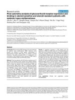
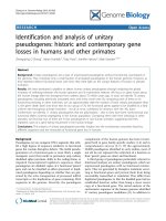
Figure 1 Characteristics of Y. lipolytica introns. (a) Size distribution of the 1,083 introns from strain E150 located within the coding regions of genes.
Introns are separated into three size classes: multiples of 3 nucleotides (blue line), multiples of 3 plus 1 nucleotides (orange line), and multiples of 3
plus 2 nucleotides (green line). For each class, the number of introns is reported as a function of size, with a window of 20 nucleotides from 41 nucle-
otides to more than 1,000 nucleotides. (b) Position of introns within the CDS. Introns are separated according to their order in the gene model, from
start to stop: first introns of genes (red boxes), second introns of genes (orange boxes) and other introns (green boxes). Data for all introns considered
together are shown in black. The proportion of introns in each group is plotted as a function of their relative position within the CDS, with a window
of 10% of the CDS length.
0%
10%
20%
30%
40%
50%
60%
70%
<10% 10-20% 20-30% 30-40% 40-50% 50-60% 60-70% 70-80% 80-90% >90%
All introns (1083)
First intron of genes (951) Second intron of genes (109)
Other introns (23)
0
10
20
30
40
50
60
70
80
90
<41
61-80
101-120
141-160
181-200
221-240
261-280
301-320
341-360
381-400
421-440
461-480
501-520
541-560
581-600
621-640
661-680
701-720
741-760
781-800
821-840
861-880
901-920
941-960
>1000
3n+2
3n+1
3n
Intron number
intron size (bp)
1083 introns (41 – 3478 bp)
Percentage of introns
position within the CDS, from start to stop
(a)
(b)
Mekouar et al. Genome Biology 2010, 11:R65
/>Page 6 of 17
longer than 62 codons (Figure S4 in Additional file 2). We
thus compared the distribution of PTCs in introns
shorter than 186 nucleotides with the expected probabil-
ity. The proportion of stop-containing introns was higher
than would be expected by chance alone (Figure S4 in
Additional file 2). Thus, stop-free introns are scarce (88
stop-free introns). Their distribution as a function of
length and insertion frame was highly heterogeneous,
with an overrepresentation of stop-free 3n + 1 introns
inserted in phase 0 and of 3n + 2 introns in phase 1 (Fig-
ure 2d).
We hypothesized that the unusual intron boundaries in
Y. lipolytica might account for the high frequency of
PTCs in short introns. The 5'ss motif GTGAGT generates
an in-frame stop codon in introns inserted in phase 2,
whatever their size, and this situation applied to 209
introns (19.3% of the 1,083 introns; Figure 3a). Similarly,
GTAAGT, the second most frequent motif, was responsi-
ble for 1% (11 introns) of stop-containing introns in phase
2. The conserved part of the BP motif, CTAAC, also gen-
erated stop codons. Assuming that the distance between
the BP and 3'ss motifs (S2 distance) is a mean of one base
(Figure S2 in Additional file 2), three categories of introns
(phase 0 size 3n + 2, phase 1 size 3n + 1 and phase 2 size
n) are most likely to contain in-frame stop codons in BP.
Indeed, 125, 114 and 60 introns, respectively, fell into
these categories (27.6% of all introns; Figure 3b). The
involvement of the BP motif is clearly underestimated, as
the S2 distance may be different from one base (possibly
shorter or longer than one base), making it possible for
introns inserted in other phases to contribute to the pres-
ence of an in-frame stop codon in the BP motif. Finally,
the 3'ss TAG is also responsible for the generation of 4%
of stop codons (Figure 3c). These consensus sequences
together account for at least 50% of stop codons. Thus,
the constraints exerted on donor, acceptor and BP motifs
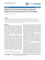
Figure 2 Distribution of introns as a function of their length and insertion frame. (a) Introns are represented according to the three possible
frames of the CDS. Phase 0 indicates that the intron is located between two codons, phase 1 indicates that it is located after the first nucleotide of a
codon and phase 2 indicates that it is located after the second nucleotide of a codon. 'All introns' corresponds to the 1,083 introns, 'first introns' to the
first intron of the 951 intron-containing genes and 'other introns' to the 131 second, third, fourth and fifth introns of genes. Differences between in-
sertion phases were statistically significant for all introns (c
2
= 64.68, P = 8.98e-15) or for the first introns (c
2
= 60.68, P = 6.63e-14) but not for introns
other than the first intron (c
2
= 5.50, P = 0.063), probably due to their limited number. (b) The proportions of each of the four bases are represented
for each base of the codons of the 6,449 protein-coding genes. Differences in nucleotide distribution were statistically significant for each position
within the codon (c
2
test, P << e-100). Stop codons were not considered. (c) Introns shown according to length categories, corresponding to a mul-
tiple of 3 (3n) or a multiple of 3 plus 1 nucleotides (3n + 1) or plus 2 nucleotides (3n + 2). There were 204 introns ≤60 nucleotides in length. The un-
derrepresentation of 3n introns was statistically significant for all introns (c
2
= 7.35, P = 0.025), first introns (c
2
= 10.90, P = 0.004) and for introns no
longer than 60 nucleotides (c
2
= 6.70, P = 0.034). (d) Stop-free introns are represented according to their insertion frame and length category.
0%
5%
10 %
15 %
20%
25%
30%
35%
40%
45%
All introns First
introns
introns
<61bp
3n
3n+1
3n+2
0
5
10
15
20
25
30
35
P hase 0 P hase 1 P hase 2
3n
3n+1
3n+2
0%
10 %
20%
30%
40%
50%
All intron First
introns
Other
introns
Phase 0
Phase 1
Phase 2
0%
5%
10%
15%
20%
25%
30%
35%
40%
AC GT
First
Second
Third
(a)
(c)
Insertion of introns within the CDS
Repartition of intron length
(d)
Distribution of stop-free introns
Number of introns
Proportion of introns Proportion of introns
(b)
Proportion of bases
Repartition of bases within the codons
Bases of
codons
Phase 0
Phase 1
Phase 2
Mekouar et al. Genome Biology 2010, 11:R65
/>Page 7 of 17
are not only necessary for splicing (intron definition
mechanism) but, together with constraints on intron size
and phasing within the codons, also contribute to intron
modeling.
Y. lipolytica uses all modes of alternative splicing
AS events were sought by two different experimental
approaches. First, transcripts of genes with multiple
introns or with large introns (>900 bp) were investigated
by RT-PCR. Subsequently, sequences obtained from
cDNA libraries were screened for splicing variants.
Multi-intronic genes
RT-PCR was carried out on 93 genes of Y. lipolytica for
which in silico predictions for more than one intron had
been made at the beginning of this study (Additional file
5). For 68 of these genes, the predicted spliced transcript
was confirmed and a single mRNA was detected. Two
other gene models (YALI0F03817g and YALI0F31427g)
were poorly predicted and, in both cases, the second
intron was not spliced in any of the three RNA prepara-
tions. It was thus considered to be part of an exon, result-
ing in a monointronic gene model. In nine RT-PCRs, no
result was obtained, due to an absence of PCR product or
non-specific amplification. For two other predicted gene
models (YALI0C07150g and YALI0D04554g), only partial
data were obtained and we were able to confirm only the
splicing of intron 2.
The last 12 RT-PCRs revealed the presence of multiple
transcripts, corresponding to different splicing variants.
For nine of these genes, we observed both transcripts
with retained introns, and transcripts efficiently spliced.
For seven of these transcripts, only the first intron of the
gene was retained, whereas, in one case (YALI0F16753g),
either intron 1 or 2 was retained and, in the last case
(YALI0C15323g), only the second intron was retained.
The last three cases involved both intron retention and
exon skipping events. For YALI0C23496g, we observed
either intron 1 retention, introducing a PTC after 11
codons, or exon 2 skipping, changing the phase of exons 3
and 4 and generating different putative proteins (Figure
4a). For YALI0F26873g, two mRNA variants were
detected in addition to the predicted fully spliced tran-
script responsible for generating the putative 505 amino
acid protein (Figure 4b). In both alternative transcripts,
exon 3 was skipped either totally (splicing between 5'ss of
intron 2 and 3'ss of intron 3) or partially (alternative 3'ss
of intron 2, leaving 45 nucleotides of exon 3). Both vari-
ants retained the stop-free intron 1, which changed the
predicted phase and generated a PTC within exon 2,
thereby resulting in a truncated 259 amino acid protein.
This gene belongs to the large septin family, which has
seven members in Y. lipolytica, as in most hemiascomyce-
tous yeasts. Surprisingly, all but one of the genes in this
family contain at least one intron, the splicing of which
was validated by cDNA clones. YALI0F26873g is the only
gene of this family with three introns and the only mem-
ber of the family with alternative transcripts. Mitrovich et
al. [16] observed that three of the seven septins of C. albi-
cans contained introns and suggested that AS might play
an important role in their regulation, consistent with our
findings.
Genes bearing long introns (>900 bp)
Long introns are rare in S. cerevisiae, with all but five of
the introns in this species being less than 700 nucleotides
long and the largest intron being 1,002 bp long. In Y.
lipolytica, gene model predictions indicate that there are
61 introns of more than 700 nucleotides in length, with a
maximal intron size of 3,478 bp (see detailed analysis
below). We focused on the genes with the largest introns,
with a view to confirming these predictions. For this pur-
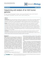
Figure 3 Presence of premature termination codons in spli-
ceosomal introns, as a function of intron size (3n, 3n + 1, 3n + 2)
and insertion frame (frame 0, 1 and 2) within the coding se-
quence. (a) A PTC is generated for all retained introns inserted in frame
2 and containing GTGAGT or GTAAGT as the 5'ss sequence, whatever
their length; 209 introns are concerned, that is, 19.3% of all intron-con-
taining genes. (b) PTCs (TAA) are also detected in the BP of 3n + 2 in-
trons in frame 0, 3n + 1 introns in frame 1 or 3n introns in frame 2 if the
S2 distance is indeed 1 bp. (c) The main 3'ss is CAG, but, in about 10.5%
of the introns, TAG is also used. This sequence generates a PTC for 3n
introns inserted in frame 0, 3n + 2 introns in frame 1 and 3n + 1 introns
in frame 2. Overall, conserved intron motifs are present in about 50%
of the PTC-containing introns.
GTGAGTTACTAAC.CAG
.GTGAGTTACTAAC.CAG
GTGAGT
Phase 0
Phase 1
Phase 2
GTAAGT
19.3 %
1.0 %
3n
3n+2
3n+1
.TAG
1.7%
(a)
(b)
(c)
Phase 0
Phase 1
Phase 2
Phase 0
Phase 1
Phase 2
3n
3n+1
3n+2
GTGAGTTACTAAC.CAG
GTGAGTTACTAAC.CAG
GTGAGTTACTAAC.CAG
3n
3n+1
3n+2
.GTGAGTTACTAAC.CAG
.GTGAGTTACTAAC.CAG
.GTGAGTTACTAAC.CAG
3n
3n+1
3n+2
GTGAGTTACTAAC.CAG
GTGAGTTACTAAC.CAG
GTGAGTTACTAAC.CAG
GTGAGTTACTAAC.CAG
.TAG
.GTGAGTTACTAAC.CAG
.TAG
GTGAGTTACTAAC.CAG
11.6%
1.5%
0.8%
5.5%
10.5%
Mekouar et al. Genome Biology 2010, 11:R65
/>Page 8 of 17
Figure 4 Schematic representation of alternative transcripts from multi-intronic genes. Gene models include exons, represented by gray rect-
angles and introns, symbolized by thin black articulated lines. Vertical bars on each of the three phases (0, +1 and +2) represent an in-frame stop
codon. The resulting mRNA variants are depicted as a concatenation of exons and the thick black vertical line represents the first in-frame codon of
the transcript. The size of the putative proteins derived from each splicing variant is indicated on the right. All three genes generate at least three
different splicing variants. (a) YALI0C23496g mRNAs are subject to intron retention (intron 1) or exon skipping (exon 2). The retention of intron 1 gen-
erates a PTC and a putative peptide of 11 amino acids. Exon 2 skipping generates a frameshift in exon 3 and in exon 4, which is slightly shortened
(exon 4'), and generates a putative protein of 65 amino acids. (b) YALI0F26873g splicing variants display retained intron 1, alternative 3'ss (intron 2)
usage or the skipping of exon 3. Both variants with a retained intron 1 generate a PTC in exon 2 and a putative truncated protein of 259 amino acids.
(c) In YALI0F32043g mRNAs, the retention of intron 5 and the use of an alternative 3'ss do not generate a PTC or a frame shift in that intron 5 is a mul-
tiple of three (60 nucleotides) nucleotides long and the difference between E4 and E4' is also a multiple of three (15 nucleotides). Both variants gen-
erate a putative protein of about the same size as that generated by the fully spliced transcript. Considering the large size of exon 6, it is shown
truncated with horizontal dashed lines.
(a)
(c)
(b)
+2
+1
0
E1
E2
E3
i1
i2
E4
i3
11 aa
65 aa
150 aa
mRNA
E1
E1
E2
E2
E3
E3
E3’
A
n
A
n
A
n
i1 E4
E4
E4’
putative
proteins
or peptides
gene models
+2
+1
0
gene models
E2
E3
i2
i3
E4
mRNA
putative
proteins
505 aa
E1
259 aa
A
n
E1 E2 E3 E4
E1 E1’ E4
E1 E1’ E3’ E4’
i1
i1
A
n
A
n
259 aa
E1
E2
5E3E
E6
i1 i2
i3 i4 i5
gene models
+2
+1
0
E4
mRNA
putative
proteins
E1
E2
E3
E5
E6
E4’
A
n
E1 E3
E6
E4’
A
n
i5
E1 E3
E6
E4
A
n
i5
1845 aa
1865 aa
1870 aa
E2
E3
E3’
E3’
E4’
i1
E1 E1’
E4’
Mekouar et al. Genome Biology 2010, 11:R65
/>Page 9 of 17
pose, 17 introns exceeding 900 bp in length (from 901 to
1,551 bp) were reverse-transcribed and amplified with
specific primers and mRNA extracted from cells grown
under the three different sets of conditions. Thirteen of
these introns were spliced as expected, one was not
amplified (cDNA clones revealed a different gene model
with no introns), two were found to have been poorly pre-
dicted (intron size larger than expected) and the last
intron, in YALI0F32043g, was found to be a mosaic of five
introns and exons (Additional file 6). Transcripts of this
last gene displayed AS due to alternative 3'ss selection
(extending exon 4 by 15 bases) and retention of the 60
nucleotides, stop-free intron 5 (Figure 4c; Additional file
5). The observed AS events did not generate in-frame
stop codons and did not modify the translation phase.
They may result in the generation of different, putatively
functional proteins.
Nine additional long introns were detected during the
cDNA analysis. The most interesting of these introns was
found in YALI0D18403g. Two transcription start sites
were found, one located 179 bases upstream of the meth-
ionine initiation codon and enabling the transcription of
a single exon (Figure 5a), and the other located about 3 kb
upstream and giving rise to a transcript with a 3,478-base
intron (Figure 5b). Surprisingly, a CDS of 1,062 bases (353
amino acids) of unknown function was predicted within
this intron and shown to be highly conserved in the
genomes of closely related species (data not shown).
All these results demonstrate the efficient splicing of
long introns not necessarily predicted in silico.
cDNA libraries
The three cDNA libraries were screened for the presence
of alternative transcripts and, more specifically, for the
presence/absence of the 1,083 introns. Eighty-six introns
matched cDNA sequences entirely or partially. For nine
of these introns, mRNAs were found in an antisense ori-
entation. Sixty-one of the remaining 75 intron sequences
corresponded to the retention of the first (58 cases) or
second (3 cases) intron of the gene. Matches for the last
14 intron sequences revealed more complex situations,
involving alternative transcription start sites, alternative
5' and 3'ss usage, exon skipping, internal exon and intron
retention or combinations of these mechanisms (Addi-
tional file 7). For example, in YALI0B15598g, which is
highly expressed (24, 9 and 28 cDNA in expo, stat and
oleic conditions, respectively), exon 2 was mostly skipped
(46 cDNAs versus 2 in which introns 1 and 2 were both
efficiently spliced). Exon 2 skipping is facilitated by the
presence of suboptimal sequences for intron 1 BP
(TGCTCAC) and intron 2 5'ss (GTCAGC). As exon 2 is
39 bp long, both variants encode putative proteins (Fig-
ure 6a) homologous to GND1 and GDN2 from S. cerevi-
siae, two 6-phosphogluconate dehydrogenases catalyzing
an NADPH-regenerating reaction in the pentose phos-
phate pathway. These proteins are highly conserved in
fungi, with the exception of the amino-terminal domain
(Figure 6b). Comparisons of gene models showed the
Figure 5 Schematic diagram of alternative variants of YALI0D18403g. The two different transcription start sites (TSS1 and TSS2) are indicated by
arrows. (a) TSS2 is located 179 bases upstream of the methionine initiation codon of YALI0D18403g1 (position 2309045 on chromosome D) down-
stream of YALI0D18436g and allows the transcription of a single exon. Translation of this mRNA generates a putative protein of 1,322 amino acids. (b)
TSS1 is located about 3 kb upstream of TSS2 and initiates a transcript with a 3,478-nucleotide intron. Surprisingly, this intron overlaps YALI0D18436g,
a CDS of 1,062 bases the translation of which generates a putative 353 amino acid protein of unknown function. Translation of the YALI0D18403g2
mRNAs generates a putative protein of 1,424 amino acids.
+2
+1
0
YALI0D18436g
YALI0D18403g1
Putative protein of 1424 aa
(a)
(b)
+2
+1
0
YALI0D18436g
Putative protein of 1322 aa
YALI0D18403g2
TSS2
TSS1
Mekouar et al. Genome Biology 2010, 11:R65
/>Page 10 of 17
presence of a large number of introns at different sites in
the various fungal phyla (Figure 6c). Only intron 4 of
YALI0B15598g was found to be conserved in all the
basidiomycetes, archiascomycetes and filamentous asco-
mycetes studied (Figure 6c). Intron 1 of S. pombe and
Ustilago maydis is located at the same position, which
differs by few nucleotides from that of Y. lipolytica intron
2 or of the single intron retained in some other hemiasco-
mycetous species, such as Arxula adeninivorans, Lachan-
cea kluyveri and Debaryomyces hansenii. Thus,
YALI0B15598g may represent an interesting example of
intron acquisition or intron slippage.
The different strategies used to detect alternative tran-
scripts in Y. lipolytica revealed that such variants were
generated from at least 88 genes (Additional files 7 and 8).
All known modes of AS were observed: alternative 5'ss (3
Figure 6 Alternative splicing in YALI0B15598g and conservation of gene models in Dikarya species. (a) Gene models for YALI0B15598g. Exons
are represented by gray or black (skipped exon) rectangles and introns by thin black lines. The size of the putative protein is 502 amino acids when
intron 1 and intron 2 are efficiently spliced, or 489 amino acids when exon 2 is skipped. (b) Amino acid alignment of the amino-terminal domain of
fungal and yeast proteins, homologs of YALI0B15598g. The size of this domain is given in amino acids, on the right, for each protein (from 20 to 41).
The black rectangle groups together hemiascomycetous yeasts or ascomycetous filamentous fungi. Archiascomycetes are represented by S. pombe
and basidiomycetes by Ustilago maydis. The numbers of spliced introns (column on the right) are colored identically when intron positions are con-
served within genes: blue for most hemiascomycetous yeasts, red for Y. lipolytica, green for all ascomycetous filamentous fungi, yellow for S. pombe
and black for U. maydis. (c) Intron localization. Triangles indicate the position of the introns for the different groups of genes (same colors as in (b)).
Only intron 4 of Y. lipolytica is conserved in all genes.
* 20 * 40
YHR183w MS ADFGLIGLAVMGQNLILN : 20
YGR256w MSKAVGDLGLVGLAVMGQNLILN : 23
CAGL0M13343g MS ADFGLIGLAVMGQNLILN : 20
ZYRO0D07876g MS ADFGLVGLAVMGQNLILN : 20
KLTH0B08668g MAQPKGDMGLIGLAVMGQNLILN : 23
SAKL0H01848g MSQPTGDIGLIGLAVMGQNLILN : 23
KLLA0A09339g MSEPAGDIGLIGLAVMGQNLILN : 23
DEHA2D06160g MSAPTGDIGLIGLAVMGQNLILN : 23
P.pastoris MVEATGDIGLIGLAVMGQNLILN : 23
ARAD0D06006g MVTPTGDIGLIGLAVMGQNLILN : 23
YALI0B15598g_sk. MTDTSNIK PVADIALIGLAVMGQNLILN : 28
YALI0B15598g_sp. MTDTSNIKLRLNQVMSQVKVKPVADIALIGLAVMGQNLILN : 41
A.fumigatus MSTQAVARLAGINVGAPARPLPSADFGLIGLAVMGQNLILN : 41
A.clavatus MSDQAVARLAGINVGAPARHLPSADFGLIGLAVMGQNLILN : 41
T.stipitatus MADQAVARLAGINVGAPARPVPSGDFGLIGLAVMGQNLILN : 41
P.chrysogenum MADQAVARLAGINVGAPAHLAPSADFGLIGLAVMGQNLILN : 41
P.marneffei MADQAVARLAGINVGAPARPEPSGDFGLIGLAVMGQNLILN : 41
A.dermatitidis MADKAVARLAGIDAGSSASSAPSGDFGLIGLAVMGQNLILN : 41
S.pombe MSQKEVADFGLIGLAVMGQNLILN : 24
U.maydis MSSQAVADIGLIGLAVMGQNLILN : 24
(a)
(b)
+2
+1
0
E1
E2
E3
E4
E5
Hemiascomycetous
yeasts
Ascomycetous
fungi
(c)
conserved intron
3
4
0
0
0
0
0
1
0
1
0
3
4
4
4
4
4
4
4
1
Spliced
introns
Mekouar et al. Genome Biology 2010, 11:R65
/>Page 11 of 17
genes) and 3'ss usage (6 genes), exon skipping (4 genes)
and intron retention (76 genes). Alternative transcription
start sites within or downstream of introns were detected
in seven genes. Alternative transcripts were observed for
9.2% of the intron-containing genes, but for only 1.8% of
these genes if intron retention was excluded. Most of the
variants observed resulted from intron retention and, if
only multi-intronic genes were considered, the intron
retained was mostly the first intron (15 of 21 genes). In
almost all cases, intron-containing transcripts revealed
by our experimental approaches, carried a PTC. This type
of mRNA is generally detected by the NMD pathway, a
quality control mechanism that recognizes and degrades
PTC-containing transcripts, preventing their translation.
The NMD machinery exists in Y. lipolytica, but some
effectors are lacking
The presence and efficiency of NMD was investigated in
Y. lipolytica. Homologs of UPF1 (YlUPF1,
YALI0D23881g) and UPF2 (YlUPF2, YALI0E24629g)
were detected in the Y. lipolytica genome by searches for
similarity to known genes. UPF3, which is less well con-
served in eukaryotes than UPF1 or UPF2, was not
detected in the chromosomes or in any non-assembled
reads, suggesting that this NMD effector is lacking or
highly divergent in Y. lipolytica. We also looked for
SMG1, SMG5, SMG6 and SMG7 (EBS1 in S. cerevisiae),
but failed to detect any homologs in Y. lipolytica.
The YALI0D23881g and YALI0E24629g genes, encod-
ing YlUPF1 and YlUPF2, were entirely deleted from the
laboratory strain PO1d. None of the single-deletion
mutants for these genes displayed a growth defect under
the conditions tested, and no defect was observed for the
double-mutant (Figure S5 in Additional file 2). This result
is consistent with the absence of a growth defect in S. cer-
evisiae strains lacking the UPF1 or UPF2 gene [68,69],
suggesting that NMD is not an essential biological mech-
anism in yeasts.
The efficiency of NMD in Y. lipolytica was assessed by
comparing the levels of PTC-containing transcripts in
wild-type and mutant strains. RT-PCR was performed on
four populations of mRNAs (YALI0B011154g,
YALI0C23496g, YALI0D05041g and YALI0F16752g) dis-
playing intron retention and resulting in the generation of
a PTC, and four populations of efficiently spliced mRNAs
(YALI0B15598g, YALI0E20031g, YALI0F03179g and
YALI0F09669g). In the efficiently spliced mRNAs, we
found no difference in the ratio of efficiently spliced to
unspliced transcripts between the wild-type and the
mutant strains (Figure S6 in Additional file 2). Among the
second set of genes, YALI0D05041g and YALI0F16752g
showed a very low level of intron retention, which did not
increase in NMD mutants (Figure S6 in Additional file 2).
This observation suggests that both genes are probably
not subjected to the NMD pathway. In contrast, for
YALI0C23496g and YALI0B11154g the ratios of spliced
and unspliced transcripts clearly differed between wild-
type and mutant strains. The intensity of the RT-PCR
product for the unspliced transcript was clearly higher in
NMD mutants (the unspliced/spliced (R/S) ratio
increased from 0.09 to 0.35 for YALI0B11154g and from
0.07 to 1.82 for YALI0C23496g; Figure 7a). Thus, despite
the lack of a conserved UPF3 homolog, NMD is func-
tional in Y. lipolytica and unspliced transcripts of
YALI0C23496g and YALI0B11154g are targeted by this
degradation pathway.
We also focused on the homolog of YDR381W (YRA1),
which is known to encode an mRNA not targeted by the
NMD pathway in S. cerevisiae [41,42]. Homologs of the
YRA1 gene are conserved in all hemiascomycetous yeasts
Figure 7 Gene expression in the NMD
-
context. (a) Variations in the
level of expression of YALI0C23496g splicing variants as a function of
NMD context. RT-PCR products from spliced (S) and unspliced tran-
scripts (intron 1 retained, R) from wild-type strains (WT) and NMD mu-
tants (NMD-). Wild-type strains are E150 (lane 1) and PO1d (lane 2).
NMD
-
strains are two independent knockouts of UPF1 (lane 3,
upf1::LEU2 clone 7; lane 4, upf1::LEU2 clone C) and one UPF2 knockout
(lane 5: upf2::LEU2 clone 7). The intensity of the unspliced transcripts is
much stronger in the mutant strains. (b) Expression of the different
transcripts of the Y. lipolytica YRA1 gene. Northern blot of total RNA of
wild-type (WT) strain PO1d (lane 1) and NMD
-
mutant strains upf1::LEU2
clone 7 (lane 2), upf2::LEU2 clone 7 (lane 3), upf1::URA3 upf2::LEU2 (lane
4), xrn1::LEU2 (lane 5). The exon probe binding to exons 1 and 3 reveals
the spliced transcript (S) in all strains and an additional splicing variant
in NMD
-
mutants only. This variant corresponds to the retention of in-
tron 1 (R). Hybridization with intron 1 confirmed that this intron is re-
tained only in NMD
-
mutants, whereas it is efficiently spliced out in
PO1d and xrn1
-
mutants.
1 2 3 4 5
R (473 bp)
S (419 bp)
506/517
396
344
298
NMD-WT
ratio R/S
0.22 0.07 0.95 1.82 0.53
1 2 3 4 5 1 2 3 4 5
NMD- NMD-
TWTW
R
S
(a)
(b)
exons intron 1
Mekouar et al. Genome Biology 2010, 11:R65
/>Page 12 of 17
and present a long intron, the BP motif of which diverges
from the canonical sequence in almost all species [41].
The Y. lipolytica gene model for the YRA1 homolog
(YALI0A20867g) follows this rule: the first intron is 850
bp long and its BP sequence, located three bases
upstream of the 3'ss motif, is TGCTGAC. RT-PCR vali-
dated the splicing of the intron in wild-type and mutant
strains but identified no transcripts in which intron 1 was
retained. However, as the difference in the lengths of the
spliced and unspliced forms may bias the PCR in favor of
the spliced variant, northern blots were performed with
probes binding to exons or the first intron (Figure 7b).
Given that YRA1 mRNA degradation requires Xrn1p in S.
cerevisiae [42], we also included a YlXRN1 mutant in our
analysis. In hybridization studies, we observed a higher
intensity of the bands corresponding to intron-retained
transcripts in NMD
-
mutants only. No such increase in
intensity was observed for the YlXRN1 mutant. This
observation suggests that, as in S. cerevisiae, the YlYRA1
transcript is not efficiently spliced in Y. lipolytica. We also
found that, by contrast to what has been reported for S.
cerevisiae, unspliced transcripts were targeted by the
NMD pathway, and their degradation seemed to be inde-
pendent of the YlXrn1 protein. These results suggest that
the S. cerevisiae YRA1 autoregulation mechanism based
on the nuclear export and cytoplasmic Edc3p-mediated
decay of the unspliced transcript [42], is probably not
conserved in Y. lipolytica.
Discussion
Hemiascomycetous yeasts are considered to have intron-
poor genomes. We show here that despite this intron
paucity, Y. lipolytica has four times as many introns as S.
cerevisiae and is the hemiascomycetous genome with the
largest number of intron-containing genes sequenced to
date. The combination of approaches used made it possi-
ble to correct many predicted gene models, to identify
new genes, such as SOA genes [51], to confirm the splic-
ing of many introns, including both large introns and
introns from weakly expressed genes, and to detect
introns in 5' UTRs. From a structural point of view, the
genome annotation of Y. lipolytica is now largely vali-
dated by experimental data and provides a reliable
genome model complementary to that of S. cerevisiae.
We show here that Y. lipolytica produces alternative
transcripts through several different mechanisms: intron
retention, exon skipping, 3' and 5' alternative splice site
usage and the use of alternative promoters. The fre-
quency of AS in Y. lipolytica is not very high, particularly
if intron retention is excluded from the analysis (1.8% of
intron-containing genes), but remains higher than that
reported for S. cerevisiae or other hemiascomycetous
yeasts, in which few naturally occurring cases
[16,26,27,29,31,38,39] or experimentally induced exam-
ples [70,71] have been described. Additional cases have
been detected in yeasts, thanks to the recent develop-
ment of genome-wide technologies providing informa-
tion about transcript polymorphism, such as tiling or
RNA-seq approaches, but these cases mostly involve
intron retention [32-34]. We report here a few interesting
examples of exon skipping, alternative 3'ss usage or pres-
ence of an intronic gene, the expression of which depends
on an alternative promoter. The situation is quite differ-
ent in basidiomycetous yeasts, such as Cryptococcus neo-
formans, which has an intron-rich genome (mean of 5.3
introns per gene) and a high frequency of AS, with high
levels of intron retention [72] but 4.2% of the transcripts
nonetheless resulting from exon skipping and alternative
3'ss or 5'ss usage [73].
In Y. lipolytica, intron retention is the main model by
which mRNA variants are generated, consistent with pre-
vious findings for ascomycetous fungi [74,75]. However,
the particular involvement of the first intron in intron
retention has not been reported before, probably because
AS was investigated mostly in hemiascomycetous yeasts
with very few multi-intronic genes [14]. It would be inter-
esting to perform a similar analysis in other phyla of asco-
mycetous fungi or in basidiomycetes. If this phenomenon
reflects an ancestral trait, then the bias should be more
marked in filamentous fungi known to possess intron-
rich genomes.
One of the key questions emerging from our study
relates to whether intron retention in Y. lipolytica plays a
physiological role, as observed for YRA1 or meiotic genes
in S. cerevisiae, or reflects an underlying background of
splicing failure. We addressed this question by investigat-
ing whether the retained introns were different from
other introns, including, specifically, whether their 5'ss,
3'ss or BP were degenerate or whether the introns were
particularly long, potentially accounting for the low splic-
ing efficiency (Additional file 8). However, no bias was
detected in primary structure, except that the first intron
of YRA1 had a degenerate BP, as it does in S. cerevisiae
[41]. More recently, YRA1 splicing inhibition has been
reported to be regulated by YRA1 exon 1, in a size-depen-
dent but sequence-independent manner [42]. We thus
investigated the size of exon 1 (coding exon plus 5' UTR)
in inefficiently spliced transcripts but, again, no bias was
detected. Another possibility, requiring further investiga-
tion for Y. lipolytica introns, stems from the reported cor-
relation between splicing efficiency and the spatial
distance between 5'ss and BP [76,77]. It has been sug-
gested that a zipper stem in the secondary structure of
three large introns of S. cerevisiae shortens the S1 dis-
tance and facilitates spliceosome assembly [77].
However, as the first intron is more often retained than
downstream introns, it is tempting to speculate that, in
most cases, intron retention probably results from a
Mekouar et al. Genome Biology 2010, 11:R65
/>Page 13 of 17
defect in the kinetics of spliceosome recruitment by the
polymerase or in spliceosome assembly. Indeed, in S. cer-
evisiae [78], as in other eukaryotes, splicing is mostly
cotranscriptional [79]. It has also been shown that the
efficiency of splicing factor recruitment during transcrip-
tion may influence splicing efficiency [80] and that the
carboxy-terminal domain (CTD) of the large subunit of
polymerase II is involved in this mechanism. We can thus
speculate that retention of the first intron of transcripts
may result from a defect in the recruitment of the spli-
ceosome by polymerase II during transcription. We are
currently investigating this hypothesis in Y. lipolytica and
determining whether there is a correlation between
intron retention and the binding kinetics of introns, splic-
ing factors and CTD.
Almost all the observed unspliced transcripts included
a premature termination codon. During the first round of
translation (before degradation by NMD), the ribosome
is thus likely to be rapidly stopped by the PTC because
the introns concerned were mostly located at the 5' end of
the CDS, close to the start codon. A statistical analysis of
the structural characteristics of Y. lipolytica introns
(intron size, frame of integration within the coding
sequence, PTC) revealed that up to 93% of introns gener-
ated a PTC, whereas only about 30% of introns in Para-
mecium generate PTCs [50]. This high percentage is due
to both intron size and, in half the cases, the sequence of
intron boundaries. The presence of stop codons in 5'ss
motifs is unusual for yeast introns, as the most frequent
motif in the hemiascomycete genomes sequenced to date
is GTATGT [14,61], but the 5'ss motif in Y. lipolytica is
GTGAGT. This observation highlights a specific evolu-
tion of intron features in Y. lipolytica, as proposed for var-
ious intron-poor lineages with strong 5'ss [60]. Introns of
size 3n were also found to be underrepresented, and stop-
free 3n introns were particularly strongly underrepre-
sented, as previously reported for other eukaryotes [50].
If retained, 3n stop-free introns do not change the trans-
lation frame and are thus considered as coding sequences
that may affect the structure and activity of the resulting
protein, with possible deleterious consequences for the
cell. In Y. lipolytica, the small number of 3n introns was
particularly pronounced for short introns, probably
reflecting an ancestral situation in which introns were
numerous and short [20]. Intron size has increased dur-
ing the course of evolution in yeasts, including Y. lipolyt-
ica in particular, to a much greater extent than in
filamentous ascomycetes. This size increase has
increased the likelihood of introns containing a PTC,
potentially limiting the need for specific constraints on
intron size (3n, 3n + 1, 3n + 2).
In eukaryotes, mRNAs with PTCs are subject to NMD,
a quality-control mechanism directing PTC-containing
transcripts for degradation to prevent their translation
(for reviews, see [43-45]). As most Y. lipolytica transcripts
containing retained introns also contain PTCs, this would
suggest that such transcripts are mostly targeted by
NMD. This may account for their lack of detection in
assays with wild-type strains, in which they were proba-
bly degraded by the NMD pathway too rapidly for detec-
tion. We therefore investigated whether NMD was active
in Y. lipolytica. Genes for only two of the core NMD fac-
tors were detected in the sequenced strain, YlUPF1 and
YlUPF2. This situation is exceptional among eukaryotes,
as all other organisms in which NMD has been studied
possess at least three major effectors (UPF1/SMG2,
UPF2/SMG3 and UPF3/SMG4). Deleting YlUPF1 or
YlUPF2 resulted in a significant increase in the propor-
tion of unspliced transcripts for some, but not all intron-
containing genes. This result confirms the existence of a
functional NMD pathway in Y. lipolytica. However, the
absence of significant growth defects in YlUPF1 and
YlUPF2 mutants suggests that NMD is not an essential
mechanism, as in S. cerevisiae and Caenorhabditis ele-
gans, whereas it has been shown to be essential in plants
and metazoans (for a review, see [43]). We now aim to
determine, at the whole-genome scale, which genes are
targets of NMD and how this pathway is regulated in this
yeast.
Conclusions
We present here an extensive survey of the transcriptome
of a yeast chosen for this study on the basis of its phyloge-
netic position, far removed from all other hemiascomyce-
tous yeasts sequenced to date. This in-depth analysis of
the transcriptome made it possible to improve the struc-
tural annotation of the Y. lipolytica genome and identified
complex cases of alternative transcripts. With a genome
slightly more complex than that of S. cerevisiae in terms
of gene structure, together with its genetic and biochemi-
cal tractability, Y. lipolytica may be a valuable organism
for studies of the regulation of AS and its impact on the
evolution of gene structure. Although considered an
intron-poor species, Y. lipolytica nonetheless displays sig-
nificant biases in its intron structure, generating PTCs in
cases of intron retention. However, further comparative
studies at a larger phylogenetic scale are clearly required
to determine whether the modeling of intron-containing
genes corresponds to an ancestral characteristic or to an
evolutionary phenomenon acquired in this particular lin-
eage.
Materials and methods
Strains and media
Y. lipolytica strains E150 (CLIB122, MATB his-1 leu2-270
ura3-302 xpr2-322) and PO1d (CLIB139, MatA leu2-270
ura3-302 xpr2-322) were routinely grown at 28°C on YPD
(yeast extract, peptone and glucose, 10 g/l each) or YNB
Mekouar et al. Genome Biology 2010, 11:R65
/>Page 14 of 17
(1.7 g/l Yeast Nitrogen Base (Difco, Detroit, MI, USA), 10
g/l glucose) supplemented for auxotrophy if necessary.
Oleic acid medium was prepared as follows: 1.7 g/l Yeast
Nitrogen Base (Difco), 50 g/l NH
4
Cl, 50 mM PO
4
NaK pH
6.8, a 100 ml/l emulsion of oleic acid (oleic acid 20% (v/v),
0.625% (v/v) Tween 40), 0.8 g/l yeast extract, 10 g/l glu-
cose. Growth phenotypes were investigated for mutant
strains of PO1d, on YPD and YNB medium, at 28°C and
18°C.
RNA extraction and RT-PCR
The RNeasy Midi Kit (Qiagen, Courtaboeuf, France) was
used to extract total RNA from cells grown in three dif-
ferent conditions: exponential growth phase in YPD
(called 'expo'), stationary phase in YPD ('stat') and expo-
nential growth phase in oleic acid medium ('oleic'). DNA
contamination was eliminated with the Turbo DNA-free
kit (Applied Biosystems/Ambion, Austin, Texas, USA).
RT-PCR was performed with Ready-To-Go™ RT-PCR
Beads (GE Healthcare Life Sciences, Orsay, France) and
PCR control with PuReTaq Ready-To-Go™ PCR Beads
(GE Healthcare Life Sciences). Primers for RT-PCR were
designed so as to obtain a 200-bp amplicon after splicing.
The resulting amplicons were subsequently inserted into
a Bluescript plasmid and sequenced to identify the differ-
ent splicing variants. The relative intensities of RT-PCR
products were estimated from ethidium bromide-stained
gels, with the ImageJ software developed at the National
Institutes of Health [81].
Northern blotting
About 20 μg of RNA was separated by electrophoresis in
a 1.2% agarose gel in 1× FA buffer (20 mM morpho-
linepropanesulfonic acid, 5 mM sodium acetate, 1 mM
EDTA, pH 7) supplemented with 1.8% formaldehyde.
After electrophoresis, the RNAs were transferred onto
GeneScreen nylon membranes (Perkin-Elmer Life Sci-
ences, Courtaboeuf, France), as previously described [82].
DNA probes were amplified by PCR from the genomic
DNA of strain E150. PCR products were purified by elec-
trophoresis in a 1% low-melting point agarose gel. DNA
probes were labeled with [α-
32
P]dCTP, with the Amer-
sham Megaprime™ DNA labeling kit (GE Healthcare Life
Sciences, Orsay, France), and hybridizations were per-
formed in Denhardt's solution-containing buffer at 65°C
[83]. Final washes were performed at 65°C, in 0.2 × SSC (1
× SSC is 0.15 M NaCl plus 0.015 M sodium citrate)-0.1%
sodium dodecyl sulfate.
cDNA library construction and sequencing
Total RNA was extracted from cells grown in three differ-
ent sets of culture conditions (expo, stat and oleic; see
above). We isolated mRNA from the total RNA prepara-
tion with the Oligotex mRNA kit (Qiagen) and the three
libraries were constructed with the CloneMiner™ cDNA
Library Construction Kit (Invitrogen, Cergy Pontoise,
France), based on Gateway
®
technology. The resulting
libraries were highly enriched in full-length, oriented
clones. We sequenced 28,434 clones (9,409, 9,620 and
9,405 clones for the expo, stat and oleic libraries, respec-
tively) by the Sanger method, first from the 5' end of the
cloning cDNAs and then from the 3' end for 1,414 chosen
clones. For 1,004 of these 1,414 selected clones, 5' and 3'
sequences have been assembled, whereas for the remain-
ing 410 clones, the 5' and 3' sequences were deposited
individually in the EMBL database. The accession numbers
for the resulting 28,844 sequences are [EMBL:FP671140
-
EMBL:FP680548
], [EMBL:FP680607-EMBL:FP690338]
and [EMBL:FP690350
-EMBL:FP700052] for the expo,
stat and oleic libraries, respectively.
Gene deletion
The complete deletion of Y. lipolytica genes (YlUPF1,
YALI0D23881g; YlUPF2, YALI0E24629g; YlXRN1,
YALI0C23144g) was performed as previously described
[84]. Primers for the PCR amplification of promoter (P)
and terminator (T) regions are listed in Additional file 9.
The ML and/or MU cassettes [84] were introduced into
the PT cassette. PO1d cells were transformed by the lith-
ium acetate method [85], with about 400 ng of purified
DNA from the disruption cassettes. Transformants were
selected on YNB medium supplemented with NH
4
Cl (5
g/l), sodium potassium phosphate buffer, pH 6.8 (50
mM), agar (2%) and uracyl (100 mg/ml) or leucine (100
mg/ml). Gene deletion was checked by PCR, with prim-
ers external to the disruption cassette, upstream from P
and downstream from T.
Auxotrophic mutants were complemented with the
URA3 or LEU2 cassettes, for comparison of their growth
rates with that of the wild-type strain, W29.
Genome sequence and sequence analysis
At the beginning of this study, the genome annotation of
Y. lipolytica strain E150 included 6,703 CDSs (genes and
pseudogenes [1]) and 742 introns (First annotation ver-
sion 3 July 2004). The genomic sequence and the different
versions of the genome annotation of Y. lipolytica strain
E150, including the version updated with our data, are
available from the Génolevures database [52].
The sequences of the cDNA clones were compared
with sequences in a nucleotide sequence database of Y.
lipolytica CDS using BLAST [86]. Only the first hit was
considered if the expected value was lower than 1.e-100
or between 1.e-50 and 1.e-100 with an identity score
exceeding 95%.
DNA logos were created with WEBLOGO version 2.8.1
[87,88].
Additional material
Additional file 1 Supplementary Table S1.
Additional file 2 Supplementary figures.
Additional file 3 Supplementary Table S2.
Mekouar et al. Genome Biology 2010, 11:R65
/>Page 15 of 17
Abbreviations
3'ss: 3' splice site; 5'ss: 5' splice site; AS: alternative splicing; bp: base pair; BP:
branch point; CDS: coding sequence; CTD: carboxy-terminal domain; NMD:
nonsense-mediated mRNA decay; PTC: premature termination codon; UTR:
untranslated region.
Authors' contributions
CN conceived and designed the experiments; MM, IBL, CO and CN performed
the experiments. CC, CDS and PW performed the cDNA sequencing; CN and
MM analyzed the data; CN and CG wrote the paper. All authors read and
approved the final manuscript.
Acknowledgements
We thank Stefan Kerscher for providing gene models experimentally validated
for genes of the complex I, and Emmanuelle Beyne, Marek Elias and Dominique
Swennen for gene model detection or modification. We also thank Stéphanie
Kervestin, Olivier Jaillon and our colleagues from the Génolevures Consortium
for helpful discussions, and Donald White of the ABIES doctoral school and
Julie Sappa of Alex Edelman and Associates for their help in correcting the
English version of the manuscript. This work was funded by the GDR CNRS
2354 'Génolevures-3' and the ANR 'Genarise' (ANR-05-BLAN-0331) programs.
Author Details
1
INRA UMR1319 Micalis - AgroParisTech, Biologie intégrative du métabolisme
lipidique microbien, Bât. CBAI, 78850 Thiverval-Grignon, France and
2
Genoscope (CEA) - Centre National de Séquençage, 2 rue Gaston Crémieux,
91057 Evry cedex, France
References
1. Dujon B, Sherman D, Fischer G, Durrens P, Casaregola S, Lafontaine I, De
Montigny J, Marck C, Neuvéglise C, Talla E, Goffard N, Frangeul L, Aigle M,
Anthouard V, Babour A, Barbe V, Barnay S, Blanchin S, Beckerich JM, Beyne
E, Bleykasten C, Boisramé A, Boyer J, Cattolico L, Confanioleri F, De Daruvar
A, Despons L, Fabre E, Fairhead C, Ferry-Dumazet H, et al.: Genome
evolution in yeasts. Nature 2004, 430:35-44.
2. Souciet JL, Dujon B, Gaillardin C, Johnston M, Baret PV, Cliften P, Sherman
DJ, Weissenbach J, Westhof E, Wincker P, Jubin C, Poulain J, Barbe V,
Ségurens B, Artiguenave F, Anthouard V, Vacherie B, Val ME, Fulton RS,
Minx P, Wilson R, Durrens P, Jean G, Marck C, Martin T, Nikolski M, Rolland
T, Seret ML, Casarégola S, Despons L, et al.: Comparative genomics of
protoploid Saccharomycetaceae. Genome Res 2009, 19:1696-1709.
3. Dietrich FS, Voegeli S, Brachat S, Lerch A, Gates K, Steiner S, Mohr C,
Pöhlmann R, Luedi P, Choi S, Wing RA, Flavier A, Gaffney TD, Philippsen P:
The Ashbya gossypii genome as a tool for mapping the ancient
Saccharomyces cerevisiae genome. Science 2004, 304:304-307.
4. Vernis L, Poljak L, Chasles M, Uchida K, Casarégola S, Käs E, Matsuoka M,
Gaillardin C, Fournier P: Only centromeres can supply the partition
system required for ARS function in the yeast Yarrowia lipolytica. J Mol
Biol 2001, 305:203-217.
5. Marck C, Kachouri-Lafond R, Lafontaine I, Westhof E, Dujon B, Grosjean H:
The RNA polymerase III-dependent family of genes in
hemiascomycetes: comparative RNomics, decoding strategies,
transcription and evolutionary implications. Nucleic Acids Res 2006,
34:1816-1835.
6. Bergeron J, Drouin G: The evolution of 5S ribosomal RNA genes linked
to the rDNA units of fungal species. Curr Genet 2008, 54:123-131.
7. Acker J, Ozanne C, Kachouri-Lafond R, Gaillardin C, Neuvéglise C, Marck C:
Dicistronic tRNA-5S rRNA genes in Yarrowia lipolytica: an alternative
TFIIIA-independent way for expression of 5S rRNA genes. Nucleic Acids
Res 2008, 36:5832-5844.
8. Fournier P, Gaillardin C, Persuy MA, Klootwijk J, van Heerikhuizen H:
Heterogeneity in the ribosomal family of the yeast Yarrowia lipolytica:
genomic organization and segregation studies. Gene 1986, 42:273-282.
9. De Schutter K, Lin Y, Tiels P, Van Hecke A, Glinka S, Weber-Lehmann J,
Rouzé P, Van de Peer Y, Callewaert N: Genome sequence of the
recombinant protein production host Pichia pastoris. Nat Biotechnol
2009, 27:561-566.
10. Casaregola S, Neuvéglise C, Bon E, Gaillardin C: Ylli, a non-LTR
retrotransposon L1 family in the dimorphic yeast Yarrowia lipolytica.
Mol Biol Evol 2002, 19:664-677.
11. Kovalchuk A, Senam S, Mauersberger S, Barth G: Tyl6, a novel Ty3/gypsy-
like retrotransposon in the genome of the dimorphic fungus Ya r r ow i a
lipolytica. Yeast 2005, 22:979-991.
12. Neuvéglise C, Feldmann H, Bon E, Gaillardin C, Casaregola S: Genomic
evolution of the long terminal repeat retrotransposons in
hemiascomycetous yeasts. Genome Res 2002, 12:930-943.
13. Neuvéglise C, Chalvet F, Wincker P, Gaillardin C, Casaregola S: Mutator-
like element in the yeast Yarrowia lipolytica displays multiple
alternative splicings. Eukaryot Cell 2005, 4:615-624.
14. Bon E, Casaregola S, Blandin G, Llorente B, Neuvéglise C, Munsterkotter M,
Guldener U, Mewes HW, Van Helden J, Dujon B, Gaillardin C: Molecular
evolution of eukaryotic genomes: hemiascomycetous yeast
spliceosomal introns. Nucleic Acids Res 2003, 31:1121-1135.
15. Juneau K, Palm C, Miranda M, Davis RW: High-density yeast-tiling array
reveals previously undiscovered introns and extensive regulation of
meiotic splicing. Proc Natl Acad Sci USA 2007, 104:1522-1527.
16. Mitrovich QM, Tuch BB, Guthrie C, Johnson AD: Computational and
experimental approaches double the number of known introns in the
pathogenic yeast Candida albicans. Genome Res 2007, 17:492-502.
17. Jeffares DC, Mourier T, Penny D: The biology of intron gain and loss.
Trends Genet 2006, 22:16-22.
18. Vanácová S, Yan W, Carlton JM, Johnson PJ: Spliceosomal introns in the
deep-branching eukaryote Trichomonas vaginalis. Proc Natl Acad Sci
USA 2005, 102:4430-4435.
19. Collins JE, Wright CL, Edwards CA, Davis MP, Grinham JA, Cole CG, Goward
ME, Aguado B, Mallya M, Mokrab Y, Huckle EJ, Beare DM, Dunham I: A
genome annotation-driven approach to cloning the human ORFeome.
Genome Biol 2004, 5:R84.
20. Stajich JE, Dietrich FS, Roy SW: Comparative genomic analysis of fungal
genomes reveals intron-rich ancestors. Genome Biol 2007, 8:R223.
21. Johnson JM, Castle J, Garrett-Engele P, Kan Z, Loerch PM, Armour CD,
Santos R, Schadt EE, Stoughton R, Shoemaker DD: Genome-wide survey
of human alternative pre-mRNA splicing with exon junction
microarrays. Science 2003, 302:2141-2144.
22. Kim E, Magen A, Ast G: Different levels of alternative splicing among
eukaryotes. Nucleic Acids Res 2007, 35:125-131.
23. Lander ES, Linton LM, Birren B, Nusbaum C, Zody MC, Baldwin J, Devon K,
Dewar K, Doyle M, FitzHugh W, Funke R, Gage D, Harris K, Heaford A,
Howland J, Kann L, Lehoczky J, LeVine R, McEwan P, McKernan K, Meldrim
J, Mesirov JP, Miranda C, Morris W, Naylor J, Raymond C, Rosetti M, Santos
R, Sheridan A, Sougnez C, et al.: Initial sequencing and analysis of the
human genome. Nature 2001, 409:860-921.
24. Srebrow A, Kornblihtt AR: The connection between splicing and cancer.
J Cell Sci 2006, 119:2635-2641.
25. Venables JP: Unbalanced alternative splicing and its significance in
cancer. Bioessays 2006, 28:378-386.
26. Habara Y, Urushiyama S, Tani T, Ohshima Y: The fission yeast prp10(+)
gene involved in pre-mRNA splicing encodes a homologue of highly
conserved splicing factor, SAP155. Nucleic Acids Res 1998, 26:5662-5669.
27. Preker PJ, Kim KS, Guthrie C: Expression of the essential mRNA export
factor Yra1p is autoregulated by a splicing-dependent mechanism.
RNA 2002, 8:969-980.
28. Juneau K, Nislow C, Davis RW: Alternative splicing of PTC7 in
Saccharomyces cerevisiae determines protein localization. Genetics
2009, 183:185-194.
29. Davis CA, Grate L, Spingola M, Ares MJ: Test of intron predictions reveals
novel splice sites, alternatively spliced mRNAs and new introns in
meiotically regulated genes of yeast. Nucleic Acids Res 2000,
28:1700-1706.
Additional file 4 Supplementary Table S3.
Additional file 5 Supplementary Table S4.
Additional file 6 Supplementary Table S5.
Additional file 7 Supplementary Table S6.
Additional file 8 Supplementary Table S7.
Additional file 9 Supplementary Table S8.
Received: 11 March 2010 Revised: 15 June 2010
Accepted: 23 June 2010 Published: 23 June 2010
This article is available from: 2010 Mekouar et al.; licensee BioMed Central Ltd. This is an open access article distributed under the terms of the Creative Commons Attribution License ( which permits unrestricted use, distribution, and reproduction in any medium, provided the original work is properly cited.Genome Biolog y 2010, 11:R65
Mekouar et al. Genome Biology 2010, 11:R65
/>Page 16 of 17
30. Rodríguez-Navarro S, Igual JC, Pérez-Ortín JE: SRC1: an intron-containing
yeast gene involved in sister chromatid segregation. Yeast 2002,
19:43-54.
31. Miura F, Kawaguchi N, Sese J, Toyoda A, Hattori M, Morishita S, Ito T: A
large-scale full-length cDNA analysis to explore the budding yeast
transcriptome. Proc Natl Acad Sci USA 2006, 103:17846-17851.
32. David L, Huber W, Granovskaia M, Toedling J, Palm CJ, Bofkin L, Jones T,
Davis RW, Steinmetz LM: A high-resolution map of transcription in the
yeast genome. Proc Natl Acad Sci USA 2006, 103:5320-5325.
33. Nagalakshmi U, Wang Z, Waern K, Shou C, Raha D, Gerstein M, Snyder M:
The transcriptional landscape of the yeast genome defined by RNA
sequencing. Science 2008, 320:1344-1349.
34. Wilhelm BT, Marguerat S, Watt S, Schubert F, Wood V, Goodhead I, Penkett
CJ, Rogers J, Bähler J: Dynamic repertoire of a eukaryotic transcriptome
surveyed at single-nucleotide resolution. Nature 2008, 453:1239-1243.
35. Roy SW, Irimia M: Intron mis-splicing: no alternative? Genome Biol 2008,
9:208.
36. Sayani S, Janis M, Lee CY, Toesca I, Chanfreau GF: Widespread impact of
nonsense-mediated mRNA decay on the yeast intronome. Mol Cell
2008, 31:360-370.
37. Pleiss JA, Whitworth GB, Bergkessel M, Guthrie C: Rapid, transcript-
specific changes in splicing in response to environmental stress. Mol
Cell 2007, 27:928-937.
38. Engebrecht JA, Voelkel-Meiman K, Roeder GS: Meiosis-specific RNA
splicing in yeast. Cell 1991, 66:1257-1268.
39. Nakagawa T, Ogawa H: The Saccharomyces cerevisiae MER3 gene,
encoding a novel helicase-like protein, is required for crossover control
in meiosis. EMBO J 1999, 18:5714-5723.
40. Vilardell J, Chartrand P, Singer RH, Warner JR: The odyssey of a regulated
transcript. RNA 2000, 6:1773-1780.
41. Preker PJ, Guthrie C: Autoregulation of the mRNA export factor Yra1p
requires inefficient splicing of its pre-mRNA. RNA 2006, 12:994-1006.
42. Dong S, Li C, Zenklusen D, Singer RH, Jacobson A, He F: YRA1
autoregulation requires nuclear export and cytoplasmic Edc3p-
mediated degradation of its pre-mRNA. Mol Cell 2007, 25:559-573.
43. Behm-Ansmant I, Kashima I, Rehwinkel J, Saulière J, Wittkopp N, Izaurralde
E: mRNA quality control: an ancient machinery recognizes and
degrades mRNAs with nonsense codons. FEBS Lett 2007, 581:2845-2853.
44. Rehwinkel J, Raes J, Izaurralde E: Nonsense-mediated mRNA decay:
Target genes and functional diversification of effectors. Trends Biochem
Sci 2006, 31:639-646.
45. Stalder L, Mühlemann O: The meaning of nonsense. Trends Cell Biol 2008,
18:315-321.
46. Kertész S, Kerényi Z, Mérai Z, Bartos I, Pálfy T, Barta E, Silhavy D: Both
introns and long 3'-UTRs operate as cis-acting elements to trigger
nonsense-mediated decay in plants. Nucleic Acids Res 2006,
34:6147-6157.
47. Kerényi Z, Mérai Z, Hiripi L, Benkovics A, Gyula P, Lacomme C, Barta E,
Nagy F, Silhavy D: Inter-kingdom conservation of mechanism of
nonsense-mediated mRNA decay. EMBO J 2008, 27:1585-1595.
48. Amrani N, Ganesan R, Kervestin S, Mangus DA, Ghosh S, Jacobson A: A
faux 3'-UTR promotes aberrant termination and triggers nonsense-
mediated mRNA decay. Nature 2004, 432:112-118.
49. Mühlemann O, Eberle AB, Stalder L, Zamudio Orozco R: Recognition and
elimination of nonsense mRNA. Biochim Biophys Acta 2008,
1779:538-549.
50. Jaillon O, Bouhouche K, Gout JF, Aury JM, Noel B, Saudemont B, Nowacki
M, Serrano V, Porcel BM, Ségurens B, Le Mouël A, Lepère G, Schächter V,
Bétermier M, Cohen J, Wincker P, Sperling L, Duret L, Meyer E:
Translational control of intron splicing in eukaryotes. Nature 2008,
451:359-362.
51. Desfougères T, Haddouche R, Fudalej F, Neuvéglise C, Nicaud J: SOA
genes encode proteins controlling lipase expression in response to
triacylglycerol utilization in the yeast Yarrowia lipolytica. FEMS Yeast Res
2009, 10:93-103.
52. Génolevures Database [ />53. Kupfer DM, Drabenstot SD, Buchanan KL, Lai H, Zhu H, Dyer DW, Roe BA,
Murphy JW: Introns and splicing elements of five diverse fungi.
Eukaryot Cell 2004, 3:1088-1100.
54. Spingola M, Grate L, Haussler D, Ares MJ: Genome-wide bioinformatic
and molecular analysis of introns in Saccharomyces cerevisiae. RNA
1999, 5:221-234.
55. Lin K, Zhang D: The excess of 5' introns in eukaryotic genomes. Nucleic
Acids Res 2005, 33:6522-6527.
56. Mourier T, Jeffares DC: Eukaryotic intron loss. Science 2003, 300:1393.
57. Hong X, Scofield DG, Lynch M: Intron size, abundance, and distribution
within untranslated regions of genes. Mol Biol Evol 2006, 23:2392-2404.
58. Schwartz SH, Silva J, Burstein D, Pupko T, Eyras E, Ast G: Large-scale
comparative analysis of splicing signals and their corresponding
splicing factors in eukaryotes. Genome Res 2008, 18:88-103.
59. Irimia M, Roy SW: Evolutionary convergence on highly-conserved 3'
intron structures in intron-poor eukaryotes and insights into the
ancestral eukaryotic genome. PLoS Genet 2008, 4:e1000148.
60. Irimia M, Penny D, Roy SW: Coevolution of genomic intron number and
splice sites. Trends Genet 2007, 23:321-325.
61. Lopez PJ, Séraphin B: Genomic-scale quantitative analysis of yeast pre-
mRNA splicing: implications for splice-site recognition. RNA 1999,
5:1135-1137.
62. Génosplicing [ />63. Fedorov A, Suboch G, Bujakov M, Fedorova L: Analysis of nonuniformity
in intron phase distribution. Nucleic Acids Res 1992, 20:2553-2557.
64. Ruvinsky A, Eskesen ST, Eskesen FN, Hurst LD: Can codon usage bias
explain intron phase distributions and exon symmetry? J Mol Evol 2005,
60:99-104.
65. Long M, de Souza SJ, Rosenberg C, Gilbert W: Relationship between
"proto-splice sites" and intron phases: evidence from dicodon analysis.
Proc Natl Acad Sci USA 1998, 95:219-223.
66. Whamond GS, Thornton JM: An analysis of intron positions in relation to
nucleotides, amino acids, and protein secondary structure. J Mol Biol
2006, 359:238-247.
67. Long M, de Souza SJ, Gilbert W: The yeast splice site revisited: new exon
consensus from genomic analysis. Cell 1997, 91:739-740.
68. Leeds P, Wood JM, Lee BS, Culbertson MR: Gene products that promote
mRNA turnover in Saccharomyces cerevisiae. Mol Cell Biol 1992,
12:2165-2177.
69. Cui Y, Hagan KW, Zhang S, Peltz SW: Identification and characterization
of genes that are required for the accelerated degradation of mRNAs
containing a premature translational termination codon. Genes Dev
1995, 9:423-436.
70. Howe KJ, Kane CM, Ares MJ: Perturbation of transcription elongation
influences the fidelity of internal exon inclusion in Saccharomyces
cerevisiae. RNA 2003, 9:993-1006.
71. Romfo CM, Alvarez CJ, van Heeckeren WJ, Webb CJ, Wise JA: Evidence for
splice site pairing via intron definition in Schizosaccharomyces pombe.
Mol Cell Biol 2000, 20:7955-7970.
72. McGuire AM, Pearson MD, Neafsey DE, Galagan JE: Cross-kingdom
patterns of alternative splicing and splice recognition. Genome Biol
2008, 9:R50.
73. Loftus BJ, Fung E, Roncaglia P, Rowley D, Amedeo P, Bruno D, Vamathevan
J, Miranda M, Anderson IJ, Fraser JA, Allen JE, Bosdet IE, Brent MR, Chiu R,
Doering TL, Donlin MJ, D'Souza CA, Fox DS, Grinberg V, Fu J, Fukushima M,
Haas BJ, Huang JC, Janbon G, Jones SJ, Koo HL, Krzywinski MI, Kwon-
Chung JK, Lengeler KB, Maiti R, et al.: The genome of the
basidiomycetous yeast and human pathogen Cryptococcus
neoformans. Science 2005, 307:1321-1324.
74. Ebbole DJ, Jin Y, Thon M, Pan H, Bhattarai E, Thomas T, Dean R: Gene
discovery and gene expression in the rice blast fungus, Magnaporthe
grisea: analysis of expressed sequence tags. Mol Plant Microbe Interact
2004, 17:1337-1347.
75. Galagan JE, Henn MR, Ma L, Cuomo CA, Birren B: Genomics of the fungal
kingdom: insights into eukaryotic biology. Genome Res 2005,
15:1620-1631.
76. Goguel V, Rosbash M: Splice site choice and splicing efficiency are
positively influenced by pre-mRNA intramolecular base pairing in
yeast. Cell 1993, 72:893-901.
77. Rogic S, Montpetit B, Hoos HH, Mackworth AK, Ouellette BF, Hieter P:
Correlation between the secondary structure of pre-mRNA introns and
the efficiency of splicing in Saccharomyces cerevisiae. BMC Genomics
2008, 9:355.
78. Görnemann J, Kotovic KM, Hujer K, Neugebauer KM: Cotranscriptional
spliceosome assembly occurs in a stepwise fashion and requires the
cap binding complex. Mol Cell 2005, 19:53-63.
79. Kornblihtt AR, de la Mata M, Fededa JP, Munoz MJ, Nogues G: Multiple
links between transcription and splicing. RNA 2004, 10:1489-1498.
Mekouar et al. Genome Biology 2010, 11:R65
/>Page 17 of 17
80. Kornblihtt AR: Promoter usage and alternative splicing. Curr Opin Cell
Biol 2005, 17:262-268.
81. ImageJ [ />82. Zimmermann M, Fournier P: Electrophoretic karyotyping of yeasts. In
Nonconventional Yeasts in Biotechnology Wolf K edition. Berlin, Germany:
Springer-Verlag; 1996:101-116.
83. Sambrook J, Fritsch EF, Maniatis T: Molecular Cloning: a Laboratory Manual
2nd edition. Cold Spring Harbor NY: Cold Spring Harbor Laboratory Press;
1989.
84. Fickers P, Le Dall MT, Gaillardin C, Thonart P, Nicaud JM: New disruption
cassettes for rapid gene disruption and marker rescue in the yeast
Yarrowia lipolytica. J Microbiol Methods 2003, 55:727-737.
85. Le Dall MT, Nicaud JM, Gaillardin C: Multiple-copy integration in the
yeast Yarrowia lipolytica. Curr Genet 1994, 26:38-44.
86. Altschul SF, Gish W, Miller W, Myers EW, Lipman DJ: Basic local alignment
search tool. J Mol Biol 1990, 215:403-410.
87. Crooks GE, Hon G, Chandonia J, Brenner SE: WebLogo: a sequence logo
generator. Genome Res 2004, 14:1188-1190.
88. Schneider TD, Stephens RM: Sequence logos: a new way to display
consensus sequences. Nucleic Acids Res 1990, 18:6097-6100.
doi: 10.1186/gb-2010-11-6-r65
Cite this article as: Mekouar et al., Detection and analysis of alternative splic-
ing in Yarrowia lipolytica reveal structural constraints facilitating nonsense-
mediated decay of intron-retaining transcripts Genome Biology 2010, 11:R65