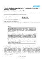
Báo cáo y học: "Long noncoding RNA genes: conservation of sequence and brain expression among diverse amniotes" doc
Bạn đang xem bản rút gọn của tài liệu. Xem và tải ngay bản đầy đủ của tài liệu tại đây (7.22 MB, 16 trang )
RESEA R C H Open Access
Long noncoding RNA genes: conservation of
sequence and brain expression among diverse
amniotes
Rebecca A Chodroff
1,2
, Leo Goodstadt
3
, Tamara M Sirey
1
, Peter L Oliver
3
, Kay E Davies
1,3
, Eric D Green
2
,
Zoltán Molnár
1*
, Chris P Ponting
1,3*
Abstract
Background: Long considered to be the building block of life, it is now apparent that protein is only one of many
functional products generated by the eukaryotic genome. Indeed, more of the human genome is transcribed into
noncoding sequence than into protein-coding sequence. Nevertheless, whilst we have developed a deep
understanding of the relationships between evolutionary constraint and function for protein-coding sequence, little
is known about these relationships for non-coding transcribed sequence. This dearth of information is partially
attributable to a lack of established non-protein-coding RNA (ncRNA) orthologs among birds and mammals within
sequence and expression databases.
Results: Here, we performed a multi-disciplinary study of four highly conserved and brain-expressed transcripts
selected from a list of mouse long intergenic noncoding RNA (lncRNA) loci that generally show pronounced
evolutionary constraint within their putative promoter regions and across exon-intron boundaries. We identify
some of the first lncRNA orthologs present in birds (chicken), marsupial (opossum), and eutherian mammals
(mouse), and investigate whether they exhibit conservation of brain expression. In contrast to conve ntional protein-
coding genes, the sequences, transcriptional start sites, exon structures, and lengths for these non-coding genes
are all highly variable.
Conclusions: The biological relevance of lncRNAs would be highly questionable if they were limite d to closely
related phyla. Instead, their preservation across diverse amniotes, their apparent conservation in exon structure, and
similarities in their pattern of brain expression during embryonic and early postnatal stages together indicate that
these are functional RNA molecules, of which some have roles in vertebrate brain development.
Background
Whilst only approximately 1.06% of the human genome
appears to encode protein [1,2] at least four times this
amount is transcribed into stable non-protein-coding
RNA (ncRNA) transcripts [3-5]. Unfortunately, the bio-
logical relevance of the vast majority of this extensive
and interleaving network of coding RNAs and ncRNAs
remains far from clear. One possibility is that many
ncRNAs result simply from transcriptional ‘noise’.Ifso,
their sequence and transcription might be expected not
to be conserved outside of restricted phyletic lineages.
Indeed, the finding that o nly 14% of the well-defined
mouse long intergenic ncRNAs (lncRNAs) identified in
the FANTOM projects [6,7] have a transcribed ortholog
in human (based on analyses of known EST and cDNA
data sets) [2] argues against their functionality. Similarly,
known human intergenic lncRNA loci are generally not
conserved in sequence at statistically significant levels in
the mouse genome [3,8,9], and there is little evidence
for conserved expression of intergenic regions (including
lncRNAs) between mouse and human [10].
On the other hand, our preconceptions of lncRNA
functionality might be g reatly prejudiced by our long-
standing knowledge of protein evolution. Just because
functional protein-coding sequence is highly con-
strained, this need not necessarily imply that largely
* Correspondence: ;
1
Department of Physiology, Anatomy, and Genetics, Le Gros Clark Building
South Parks Road, University of Oxford, Oxford OX1 3QX, UK
Chodroff et al. Genome Biology 2010, 11:R72
/>© 2010 Chodroff et al.; licensee BioMed Central Ltd. This is an open access article distributed under the terms of the Creative
Commons Attribution License ( which permits unrestricted use, distribution, and
reproduction in any medium, pro vided the origina l work is properly cited.
unconstrained n on-protein-coding sequence, free from
the need of maintaining an ORF and producing a ther-
mody namically stable protein product, is not functional.
Indeed, even well-known examples of functional mam-
malian lncRNAs, such as Gomafu [11], Evf-2 [12], XIST
[13], Air [14], and HOTAIR [9], exhibit poor sequence
conservati on across species. Moreover, there is evidence
for significant, albeit modest, evolutionary constraint
within lncRNA loci compared to neutrally evolving
DNA [15-18]. In addition, as with mRNAs, many
lncRNAs are subject to splicing, polyadenylation, and
other post-transcriptional modifications, and their loci
tend to be associated with particular chromatin marks
[15]. However, whether the observed chromatin marks
and purifying selection are most frequently directed
towards the transcribed lncRNA, the process of tran-
scription, or the underlying DNA sequence remains
unknown [19-21].
In support of functional roles for lncRNA loci, many
lncRNAs have been shown to be developmentally regu-
lated and/or expressed in specific tissues. For example, a
computational analysis of in situ hybridization data from
the Allen Brain Atlas identified 849 lncRNAs (out of
1,328 examined) showing specific expression patterns in
adult mouse brain [22]. Similarly, 945 lncRNAs were
foundtobeexpressedabovebackgroundlevelsina
microarray screen of mouse embryonic stem cells at var-
ious stages of differentiation [23]. A follow-up study
found that 5% of approximately 3,600 analyzed lncRNAs
are differentially expressed in forebrain-derived mouse
neural stem cells subjected to various developmental
paradigms [24]. Such regulated expression patterns ca n
perhaps be attributed to lncRNA loci tending to cluster
near brain-expressed protein-coding genes and tran-
scription factor-encoding genes associated with develop-
ment [15,17,25].
Nevertheless, it is im portant to stress that the above-
mentioned studies focused on only one species, namely
the laboratory mouse. There is a clear and substantial
need to investigate the evolution and expression of spe-
cific lncRNA loci for more diverse species, for example
birds, whose lineage separated from that of mammals
approximately 310 million years ago [26]. However, few,
if any, studies have identified orthologous lncRNAs
shared between birds and mammals, let alone investi-
gated either their expression in homologous develop-
mental fields or adult anatomical structures, or their
molecular functions. Whilst one study found that
Sox2ot is both dynamically regulated and transcribed
from highly conserved elements in chicken and zebra-
fish [27], this locus overlaps with a protein-coding gene
(Sox2), a pluripotency regulator, and thus is not inter-
genic. A more comprehensive study of full-length
chicken cDNA sequences identified 30 transcripts that
could be aligned with RIKEN-identified mouse
lncRNAs, although their expression in developing chick
embryos was undetectable [28]. Even Xist,whichis
involved in chromosome-wide × inactivation in euther-
ians, is not conserved as a lncRNA in birds, as its avian
ortholog is protein-coding [29].
In this study, we used a multi-disciplinary approach to
investigate a select group of highly conserved lncRNAs
that are expressed within the embryonic and early post-
natal mouse brain. We report the characterization of
four such lncRNAs, demonstrating that they are
expressed at experimentally detectable levels, are ti ssue-
specific and developmentally regulated, and are con-
served in transcript structure and expression pattern
across diverse a mniotes during brain development. To
our knowledge, this is the first description and investiga-
tion of lncRNA loci with orthologs present in eutheria,
metatheria (marsupials), and birds. As these lncRNAs
do not differ substantially from protein-coding genes in
their sequence or expression properties, we propose that
they are novel RNA genes that are likely to confer
important functions among these diverse amniotes. Our
observations provide the first indications that investiga-
tion of lncRNA orthologs in amniote model organisms
will be informative about their co ntributions to human
biology.
Results
lncRNA selection
We started with a set of 3,122 well-characterized inter-
genic lncRNAs derived from FA NTOM 2 and 3 consor-
tia collections of full-length noncoding transcripts in the
mouse [6,7,18]. While transcripts wit h evidence of p ro-
tein-coding capacity had already been discarded, we
removed additional lncRNAs that overlap either with
more-recently annotated mouse protein-coding genes or
with alignable protein-coding g enes from other species.
We also discarded lncRNAs transcribed in close proxi-
mity (<5 kb) of annotated protein-codin g genes in order
to reduce the chances of inadvertently considering
untranslated regions or alternative transcripts of these
genes. Of the remaining set of 2,055 lncRNA transcripts,
1,209 (59%) harbor strongly constrained sequence, ba sed
on overlap with phastCons-predicted conserved ele-
ments (Figure 1b) [30], consistent with a recent report
[16]. On average, 10.6% and 10.9% of the lncRNA
sequences (including and excluding introns, respectively)
overlap phastCons-predicted conserved elements.
To compare the evolution of lncRNA loci with pro-
tein-coding gene evolution, we next constructed a gen-
eric locus from 877 multi-exon lncRNA loci, and
annotated it according to the presence of conserved
sequence elements (Figure 1a). A similar portrait of evo-
lutionary conservation for protein-coding genes was
Chodroff et al. Genome Biology 2010, 11:R72
/>Page 2 of 16
presentedbytheMouseGenomeSequencingConsor-
tium (Figure 25a in [31]). As seen for protein-coding
genes, sequence conservation is not uniformly distribu-
ted across various features (exons, introns, and upstream
and downstream regions) of a generic multi-exon
lncRNA locus (Figure 1a). The putative core promoter
region (here defined as 200 bp upstream of each
lncRNA transcription start site (TSS)) is generally under
greater evolutionary constraint than lncRNA exonic
sequence, in agreement with pre vious reports [6,16,18].
Constra int peaks at 0.19 (range between 0 and 1), 43 bp
upstream of the normalized TSS, as previously observed
for human and mouse promoter sequence [32]. Just as
for protein-coding genes [31], the generic lncRNA locus’
first, middle and last exons tend to be unde r greater
evolutionary constraint than its introns, with average
phastCons scores peaking in close proximity to splice
sites.
To establish whether lncRNAs are conserved in
expression as well as in sequence, we sought to select a
small number of mouse lncRNAs and investigate their
putative orthologs in other amniotes, namely the marsu-
pial opossum (Monodelphis domestica) and the chicken
(Gallus gallus). We chose lncRN As that are h ighly con-
served, developmentally regulated, and brain-expressed.
Thesecriteriawereusedbecauseourpreviousstudy
[17] found that constrained lncRNAs with significantly
suppressed human-mouse nucleotide substitution rates
tended to be expressed in the mouse brain and, when
developmentally expressed, to be transcribed near pro-
tein-coding genes involved in transcriptional regulation.
Accordingly, we selected three lncRNAs, each having
extensive overlap with phastCons-predicted conserved
elements (Figure 1b) and each expressed in embryonic
or neonatal brain based on the origin of the cDNA
library from which they were identified. Here, we refer
to these three lncRNAs and their genomic loci accord-
ing to their database accession numbers: AK082072,
AK082467, and AK043754.
Structure of selected lncRNA loci
The three selected lncRNA loci harbor elements that
are more usually associated with protein-coding genes.
These include GT-AG donor-acceptor splice sites,
polyadenylation signals, and chromatin marks in their
putative promoter regions (Figures 2b,c, 3b,c and 4b,c;
Figure S1 in Additional file 1). Aceview annotations
[33] indicate an unspliced (single exon) transcript and
single promoter for the AK043754 locus (spanning
1.75 kb on mouse chromosome 6qG1), a single canoni-
cal GT-AG intron and promoter for the AK082072
locus (39.7 kb on mouse chromosome 13qC3), and
Figure 1 Sequence conservation among lncRNAs. (a)
Conservation across a generic lncRNA locus, based on 877 mouse
multi-exon lncRNAs. We sampled 200 evenly spaced bases across
each region listed, with regions containing fewer than 200 bases
sampled entirely. The graph shows the average vertebrate
phastCons score at each genomic position across all multi-exon
lncRNA loci. Note phastCons score peaks within the putative
promoter region (200 bp upstream) and near donor and acceptor
splice sites (analysis inspired by Figure 25a in [31]). (b) Overlap
between vertebrate phastCons-predicted conserved elements and
mouse lncRNA exons. Of 2,055 lncRNAs with signatures of purifying
selection initially identified in mouse [18], 1,095 contain exons that
overlap phastCons-predicted vertebrate conserved elements (log-
odds score range 1 to 1,000) [30]. Depicted is a histogram showing
the percentage of each lncRNA transcript that overlaps a
phastCons-predicted vertebrate conserved element. The relative
positions of three selected lncRNAs (AK082072, AK043754, and
AK082467 with overlaps of 36.7, 44.8, and 51.7%, respectively) are
shown.
Chodroff et al. Genome Biology 2010, 11:R72
/>Page 3 of 16
31 different GT-AG introns in at least 16 different
mRNA splice variants and 6 probable alternative pro-
moters for the AK082467 locus (94 kb on mouse chro-
mosome 10qC2). Each lncRNA sequence is supported
by several GenBank cDNA records, representing
cDNAs derived primarily from mouse embryonic or
neonatal central nervous system tissues, including
hypothalamus, diencephalon, cortex, cerebellum, and
spinal cord. Many of the supporting GenBank records
additionally support poly(A) and 5′ cap structures,
indicating that each lncRNA is most likely transcribed
by RNA polymerase II. Chromatin marks from either
mouse embryonic stem cells or adult mouse whole
brain [34] are present at each putative lncRNA promo-
ter (Figures 2b, 3b and 4b).
In contrast to most protein-coding genes, the
lncRNA loci each harbor at least one Evofold-predicted
RNA secondary structure (Figures 2b, 3b and 4b) [35].
This reflects the general tendency of conserved brain-
expressed lncRNA loci to contain such structures [17].
The three ln cRNA transcripts each la ck long (>100
amino acids) ORFs. While it remains possible that the
lncRNAs encode short peptides, there is no evidence
for constraint on their protein-coding capacity, as the
frequenci es of synonymous and non-synonymous sub-
stitutions across eutherians are roughly equal (that is,
Figure 2 Evolutionary constraint of AK043754. (a) The genomic region of mouse chromosome 6 (chr6) e ncompassing the lncRNA locus
AK043754 (1.7 kb) is depicted. Note the locations of flanking protein-coding genes: Grin2B (glutamate receptor, ionotropic, NMDA2B (N-methyl-D-
aspartic acid)) and Emp1 (epithelial membrane protein 1). Also shown are the positions of mouse-chicken ECRs (evolutionarily conserved regions
at least 100 bp in size with 70% sequence identity between the mouse and chicken genomes); ECRs within protein-coding regions are shown in
blue. (b) A more detailed representation of AK043754 (single exon highlighted in orange) and its immediate flanking regions, including the 3’
end of Grin2B. Below the gene structures are the positions of H3K4me1 chromatin marks (green) detected in mouse embryonic stem cells
(obtained from UCSC Genome Browser), EvoFold predictions of RNA secondary structures (grey), a SinicView conservation plot [68] based on a
21-vertebrate multispecies sequence alignment (using Threaded Blockset Aligner) generated with mouse as the reference sequence, and Gmaj
[66] views of alignments between mouse and the indicated species’ sequences (note the detected homology with the orthologous lizard and
chicken, but not frog, sequences). (c) Conservation and relative sizes of AK043754 orthologs in various species. The TSSs (arrows) and transcript
lengths are depicted in each case. Note the conserved position of a polyA signal (red) and increased sequence conservation (relative to the
mouse sequence) towards the 3’ end. ECR, evolutionarily conserved region.
Chodroff et al. Genome Biology 2010, 11:R72
/>Page 4 of 16
dN/dS ≈ 1 ± 0.16) for the longest predicted ORF of
each lncRNA [36].
These findings imply that the three selected tran-
scripts might be functional noncoding RNA genes.
AK082467 is an alternative splice variant that contains
the first three exons and retains the second intron of a
previously described long noncoding RNA, Rmst (rhab-
domyosarcoma 2 associated transcript, also known as
NCRMS); the human RMST ortholog was initially iden-
tified as a differentially expressed transcript in alveolar
versus embryonic rhabdomyosarcoma (a malignant soft
tumor tissue), but its function remains undocumented
[37]. To our knowledge, AK043754 and AK082072
have not been experimentally investigated. To examine
their potential functions, we first studied the expres-
sion patterns of the three lncRNAs during mouse
development.
Expression of selected lncRNAs in mouse
Analysis of the three selected lnc RNAs by in situ hybri-
dization of mouse tissues at different developmental
time points revealed that each exhibits a specific expres-
sion pattern that , in g eneral, is restricted to the brain.
Our findings further suggest their expression is tightly
regulated, as opposed to stochastic background
transcription.
Figure 3 Evolutionary constraint of AK082072. (a) The genomic region of mouse chromosome 13 (chr13) encom passing lncRNA AK082072
(523 bp) is depicted. Note the locations of the flanking protein-coding genes: Tmem161b (transmembrane protein 161b) and Mef2C (myocyte
enhancer factor 2C). (b) A more detailed representation of AK082072 (exons highlighted in orange) and its immediate flanking regions. Below
the gene structures are the positions of H3K4me3 chromatin marks (green) detected in mouse brain, VISTA conserved non-coding midbrain
enhancer element 268 (obtained from the UCSC Genome Browser), and a BLAT alignment of the chicken AK082072 ortholog, as well as similar
tracks as those in Figure 2b. Note the detected homology with orthologous frog sequence in exon 1. (c) Conservation and relative sizes of
AK082072 orthologs in various species. Note the sequence conservation (relative to the mouse sequence) at both the 5’ and 3’ ends and the
conserved position of splice sites (green). Unlike the other vertebrate genomes considered, the zebra finch genome did not align to the
proximal promoter or first exon of mouse AK082072. This apparent lack of sequence identity might reflect either an unannotated gap in its
genome assembly or rapidly evolving sequence within its orthologous genomic region. Other details are provided in the legend to Figure 2.
ECR, evolutionarily conserved region.
Chodroff et al. Genome Biology 2010, 11:R72
/>Page 5 of 16
Figure 4 Evolutionary constraint of AK082467 and Rmst. (a) The genomic region of mouse chromosome 10 (chr 10) encompassing lncRNAs
AK082467 (2.7 kb) and Rmst (2.7 kb) is depicted. Note the presence of the protein-coding gene Nedd1 (neural precursor expressed
developmentally down-regulated protein 1) upstream of AK082467 and Rmst. (b) A more detailed representation of AK082467 and Rmst (exons
highlighted in yellow and orange, respectively), microRNAs mir-1251 and mir-135a-2, and their immediate flanking regions. Below the gene
structures are the positions of H3K4me3 (green) and H3K27me3 (red) chromatin marks detected in mouse brain (obtained from the UCSC
Genome Browser) as well as similar tracks as those in Figure 2b. Note the detected homology with orthologous frog sequence in Rmst exons 1,
2, 4, and 11. (c) Conservation and relative sizes of AK082467 and Rmst orthologs in various species. Note the conserved splice sites (green bars)
in mouse Rmst exons 1, 4, and 11 as well as the sequence conservation (relative to mouse sequence) in exons 1 and 11, but differences in total
exon number among species. The 3’ ends of opossum and chicken orthologs have not been experimentally verified. Other details are provided
in the legend to Figure 2. ECR, evolutionarily conserved region.
Chodroff et al. Genome Biology 2010, 11:R72
/>Page 6 of 16
AK043754 is initially expressed in the primo rdial
plexiform layer or preplate. This is the first of the devel-
opmental cell layers to appear during mammalian
embryogenesis and is, most likely, homologous to the
simpler amphibian and avian cortical structures (Figure
5a(i,ii,iv,v)) [38]. At embryonic day 17 (E17), AK043754
is expressed prominently within the marginal zone along
the pial surface in a pattern similar to that of reelin-
expressing Cajal-Retzius cells. Of note, the expressed
transcript is also present within the ventricular zone of
the ganglionic eminence, a source of GABAergic migra-
tory neurons (including some Cajal-Retzius cells) that
ultimately colonize the marginal zone, intermediate
zone, and subplate; this suggests that AK043754-expres-
sing cells might originate in the ganglionic eminence
and then migrate to the preplate and marginal zone
[39]. Reinforcing this transcript’s potential association
with inhibitory GABAergic neurons, hybridization is
also seen in the latero-caudal migratory path of inter-
neurons from the basal telencephalon to the striatum.
This is best illustrated at stage E17 and within the inter-
nal granule cell layers of the olfactory b ulb at postnatal
day 3 (P3; Figure 5a(vii)).
Cells expressing AK082072 at stage E13 primarily
populate the roof of the midbrain and the cortical hem
(the most caudomedial edge of the telencephalic neuroe-
pithelium), one of the major patterning centers of the
developing telencephalon and, as recently shown by
Monuki and Tole and colleagues, a hippocampal precur-
sor (Figure 5b(i,iv)) [40,41]. By stage E17, expression
continues to be apparent within the roof of the mid-
brain, and, as illustrated at higher magnification, is
strongest in the soma and outward projections of cells
lining the midbrain ventricle (Figure 5b(v)). Also visible
in the E17 image is the expression of AK082072 along
the caudal ganglionic eminence, a major source of
GABAergic neurons that preferentially migrate caudally
to the caudal cortex and hippocampus [42]. At postnatal
stages, AK082072 expression is restricted to the hippo-
campus (mostly wit hin CA1), the rostral migratory
stream, and the internal plexiform and granule cell layer
of the olfactory bulb. Reinforcing our observations, a
previous independent study that utilized a probe
designed from another region of the AK082072 tran-
script yielded similar results [43].
AK082467 is expressed early in mouse brain develop-
ment, with its transcription mostly attenuated after
birth. The antisense riboprobe designed to an intron-
spanning region of this lncRNA transcript partially over-
laps the 5’ region of Rmst, such that all observations
coul d reflect the ex pression pattern(s) of one or both of
these transcripts. Consistent with the expression pattern
of Rmst described by Bouchard et al. [44], our riboprobe
hybridized to the mid-hindbrain organizer region in
developing mouse embryos, most clearly illustrated in
Figure 5c(ii). We also found expression in two additional
Pax2-expressing regions, including the optic stalk at
stage E9 and within the accessory olfactory bulb postna-
tally (Figure 5c(i,iv)).
lncRNA orthologs in other vertebrates
AK082072, AK082467, Rmst,andAK043754 are each
transcribed from regions of the m ouse genome whose
sequence aligns to vertebrate genome sequences from
species at least as distantly related as chicken, with
greater than 80% nucleotide identity within some inter-
vals. We sought to determine whether conservation in
lncRNA sequence also extends to conservation in the
expression of these lncRNAs among diverse vertebrate
species. In order to identify orthologs in other verte-
brates, we aligned g enomic sequences orthologous to
each lncRNA locus from species ranging from frog to
human, and including birds and marsupials (see Materi-
als and methods; Figures 2b, 3b and 4b).
Each lncRNA locus and its closest flanking protein-
coding genes show conserved synteny across amniotic
species from mouse to chicken, and a portion of each
mouse lncRNA locus aligns to all the genomic
sequences we analyzed (Figures 2a, 3a and 4a). The pat-
terns of nucleotide conservation for these lncRNA loci
exemplify the more general trends we observed for all
such loci, including greater conservation near exon
boundaries (Figure 1a). In these respects, these lncRNA
loci differ markedly from protein-coding genes, which
typically c ontain more uniformly distributed and strong
conservation within exons [31].
AK043754
Blo cks of aligned sequence with at least 70% nucl eotide
identity across all the examined amniote species are
restricted to the 3’ end (approximately 500 bp) of
AK043754 (Figure 2). We could find no evidence of
AK043754-aligning sequence within non-amniote verte-
brate genomes, suggesting that this locus has either
evolved extremely rapidly or originated within the
amniote lineage after divergence from other vertebrates.
The sequence of the putative proximal promoter, pre-
sumed to reside within the 400 bp upstream of the TSS,
aligns to orthologous sequences in metatheria and
eutheria; such orthologous sequence could not be iden-
tified in monotremata (platypus) and non-mammalian
vertebrates. Finally, a polyaden ylation signal (ATAAA)
located 30 bp upstream of the 3’ end of AK043754 in
mouse is present in all examined amniote sequences.
Guided by the multi-species sequence alignments, we
cloned the AK043754 orthologs from opossum and
chicken poly(A)-selected re verse-tran scribed cDNA. As
illustrated in Figure 2c, the orthologous opossum and
chicken sequences (as well as the orthologous zebra
Chodroff et al. Genome Biology 2010, 11:R72
/>Page 7 of 16
Figure 5 lncRNAs are specifically expressed and developmentally regulated in the mouse brain. (a-c) Digoxigenin-labeled riboprobes
complementary to AK043754 (a), AK082072 (b), and AK082467 (c) were hybridized to sagittal sections of C57BL/6J mouse brains at different
development stages (E9, E13, E17, and P3). (a) The AK043754 probe hybridized to the first generated cell layer of the preplate or primordial
plexiform zone (red arrowheads) at E13 (i, iv) and E17 (ii, v), the ventricular zone of the medial and lateral ganglionic eminences (black
arrowhead) at E13, the latero-caudal migratory path from the basal telencephalon to the striatum (green arrowhead) at E17 (ii, v), and the
hippocampus (iii, vi) and the olfactory bulb (iii, vii) at P3. Scale bar (shown in (i)) is 500 μm in (i), 543 μ m in (ii), 322 μm in (iii), 292 μm in (iv), 300
μm in (v), 167 μm in (vi), and 214 μm in (vii). (b) The AK082072 probe hybridized to the hem of the embryonic cerebral cortex (blue arrowheads)
and the roof of the midbrain (black arrowheads) at E13 (i, iv) and E17 (ii, v), and to the hippocampus (iii, vi), rostral migratory stream (iii, vi), and
internal plexiform and granule cell layer of the olfactory bulb (iii, vi) at P3. Scale bar (shown in (i)) is 500 μm in (i), 595 μm in (ii), 422 μm in (iii),
357 μm in (iv), 386 μm in (v), and 311 μm in (vi). (c) The AK082467 probe hybridized to the optic stalk (black arrowheads) at E9 (i, v), the cortical
hem (blue arrowheads) at E13 (ii, vi) and E17 (ii, vii), and the accessory olfactory bulb (iii, viii) at P3. Scale bar (shown in i)) is 500 μm in (i), 637
μm in (ii), 684 μm in (iii), 522 μm in (iv), 182 μm in (v), 177 μm in (vi), 176 μm in (vii), and 110 μm in (viii).
Chodroff et al. Genome Biology 2010, 11:R72
/>Page 8 of 16
finch sequence [GenBank: DQ213170]) align to the
mouse AK043754 sequence. Based on BlastN local align-
ments, the opossum (1,307 bp), chicken (1,912 bp), and
zebra finch (938 bp) transcripts share approximately
38%, 29%, and 29% nucleotide sequence identity with
the mouse transcript, respectively. Consistent with the
multi-species genome sequence alignment, each tran-
script has a unique (non-aligning) TSS (indicated by
grey arrows), but harbors a conserved poly(A) signal
(red band) and 3’ end. As with mouse AK043754,the
examined orthologs lack long or conserved ORFs, indi-
cating that this locus is unlikely to have possessed pro-
tein-coding capacity over the span of amniote evolution.
AK082072
Orthologous sequences in each of the 16 vertebrate gen-
omes we examined (with one exception - see below)
aligned to the proximal promoter and first exon of mouse
AK082072 with sequence identities exceeding 85% (Figure
3b). Notably, a 5’ consensus splice-site sequence (MAG|
GTRAG) for U2 introns in pr e-mRNA is constrained.
However, sequence conservation of the second exon,
including an adjacent 3’ AG acceptor site and poly(A) sig-
nal, is detectable only in mammals, suggesting that this
region might have arisen within the mammalian lineage
after divergence from other amniotes.
AK082072 orthologs were identified in frog (754 bp),
chicken (759 bp), and human (553 bp) ( [GenBank:
CX847574.1, CR35248.1, DA317999.1], respectively) from
a BLASTn query of the NCBI (nr/nt) database. In addition,
we cloned and sequenced the full-length (725 bp) opos-
sum ortholog from poly(A)-selected reverse-transcribed
cDNA. Based on the resulting BLASTn alignments, we
found that the frog, chicken, opossum, and human
sequences share approximately 11%, 21%, 53%, and 67%
sequence identity, respectively, with their mouse ortholog
(Figure 3c). Consi stent with the multi- species genome
sequence alignment, all transcripts utilize a co nserved 5’
donor site. By contrast, only the mammalian transcripts
use the predicted 3’ acceptor site and terminate immedi -
ately after the predicted poly(A) signal (depicted as blue
and red bands, respectively, in Figure 3c).
While the relative structure of the first and last exons
is conserved across therian mammals, the opossum and
human orthologs contain an additional and non-homo-
logous central exon, in each case buttressed by non-con-
served AG/GT accep tor/donor sites and residing within
poorly constrained genomic sequence. In fact, the opos-
sum middle exon lies within a genomic region contain-
ing a MAR1 element (a tRNA-derived SINE (short
interspersed element) specific to M. domestica [45]).
The terminal mammalian AK082072 exons lack
demonstrable homology with those in the chicken and
frog orthologs (Figure 3b). The second exon in chicken
AK082072 is transcribed f rom an e volutionarily
conserved region that shares >70% sequence identity
with the orthologous mouse sequence (highlighted in
grey) across 200 bp and harbors a poly(A) signal with
100% sequence conservation in all examined vertebrates
except zebra finch. While suggestive of a highly con-
served exon, we were unable to clone similar splice var-
iants from either mouse or opossum cDNA. In contrast,
the sec ond exon of frog AK082072 appears to be speci-
fic to amphibians and, like opossum AK082072, includes
a repeat element, in this case a X. tropicalis DNA trans-
poson hAT.
AK082467/Rmst
AK082467 and Rmst orthologs from human to frog also
exhibit >70% sequenc e identity over their proximal pro-
moters, first exons, and 5’ splice donor sites (Figure 4b).
In all examined eutherians, we identified putative two-
exon AK082467 orthologs that share a TSS, splice site,
and exonic structure. While genomic regions containing
the second exon of AK082467 share at l east 60%
sequence identity among the examined vertebrates, the
non-eutherian vertebrates lack an upstream 3’ acceptor
site; hence, we expected either unspliced or differentially
spliced orthologs in these species. Indeed, we cloned
unspliced and differentially spliced AK082467 orthologs
from chicken (30% sequence identity) and opossum
(26% sequence identity) cDNA, respectively, each s har-
ing similar 5’ and 3’ ends with mouse AK082467 (Figure
4c). The opossum AK082467 3’ acceptor site is not con-
served, as it aligns approximately 10 bp upstream of
that in mouse, although this may reflect inaccuracies in
the sequence alignment. Chicken AK082467 contains an
additional approximately 200-bp stretch that spans the
mouse intronic region . Importantly, the identified mam-
malian intron in AK082467 (approximately 320 bp),
which is almost entirely composed of simple repeats, is
not alignable to chicken or to other non-mammalian
vertebrategenomes.Also,wewereunabletoidentifya
poly(A) signal within the AK082467 orthologs despite
the fact that the t ranscripts were derived from poly(A)-
selected cDNA, suggesting that the isolated transcripts
were either unpolyadenylated contaminants within our
cDNA samples or that the transcripts are recapped deri-
vatives of larger RNA molecules.
Our multi-species sequence alignment (Figure 4b)
revealed that only exons 1, 4, and 11 of mouse Rmst
share the same exonic structure (including alignable
donor and acceptor splice sites) across the examined
vertebrates. At least one >50-bp stretch of >60%
sequence identity resides within each of these exons.
Sequences of the remaining mouse exons align to
regions of varying sequence conse rvation among mam-
mals, suggesting relaxed evolutionary constraint on their
structures. Accordingly, we predicted vertebrate Rmst
orthologs containing at least three conserved exons and
Chodroff et al. Genome Biology 2010, 11:R72
/>Page 9 of 16
a variable number of total exons. Of note, we also iden-
tified a eutherian-specific poly(A) signal residing
approximately 25 bp upstream of the termination site
within the mouse transcript, suggesting that other
eutherians also share the same transcription stop site.
We cloned and sequenced the chicken and opossum
Rmst orthologs, which contain four and seven exons,
respectively. While we only identified one splice variant
for each species, alternative transcripts could exist.
Alignment of the identified orthologs along with the
mouse and human [GenBank: NR_024037] Rmst
sequences revealed striking conservation of the struc-
tures of exons 1, 4, a nd 11 and of the sequences of
exons 1 and 11 (Figure 4c). In contrast, the mouse,
opossum, and chicken Rmst exon 4 o rthologs share
<50% sequence identity. Furthermore, the overall
sequence identity, calculated by BLASTn, between
mouse Rmst and the chicken, opossum, and human
orthologs is only 4%, 7%, and 22%, respectively.
Expression of selected lncRNA orthologs in the
developing brain
Given the evidence that lncRNA orthologs are tran-
scribed in diverse species, we next sought to dete rmine
whether the tissue pattern of transcription is similarly
conserved. Indeed, w e identified numerous homologous
ESTs and c DNAs from nervous system tissue isolated
from diverse species (human to zebra finch; Table 1).
To observe lncRNA expression at a finer resolution,
we performed in situ hybridization of mouse, opossum,
and chicken brains harvested at early and late embryo-
nic stages, using probes specific to approximately 300-
bp portions of phastCons conserved elements within
AK043754, AK082072,andAK082467 exons. While the
expression patterns of thelncRNAorthologsarenot
identical among these species, we encountered evidence
of spatio-temporal regulation f or each locus, with tran-
scription typically regionally restricted within embryonic
and neonatal b rain tissue. Many of these regions have
been implicated in the evolution of the mammalian cer-
ebral cortex [46,47].
Probes specific to chicken, opossum, and mouse
AK043754 orthologs hybridize to the germinal zone of
the telencephalic cortex in coronal and sagittal sections
of early developmental brain in all three species (red
arrowheads in Figure 6a). While the neuroanatomical
homology relationships between mammalian and avian
brains remain controversial (see [46] for a review), most
Table 1 AK043754, AK082072, and AK082467 orthologs among vertebrates
lncRNA Species (common name) GenBank accession Tissue type Dev. stage
AK043754 M. musculus (mouse) [Genbank:AK043754]* Cortex Neonate
R. norvegicus (rat) [Genbank:BF565173] Brain Adult
C. jacchus (marmoset) [Genbank:EH380404] Hippocampus Adult
H. sapiens (human) [Genbank:DB326634] Brain Fetal
B. taurus (cow) [Genbank:CO886535] Brain Adult
S. scrofa (pig) [Genbank:EW186118] Cerebellum Fetal
T. guttata (zebra finch) [Genbank:DV959637] Brain Pooled
AK082072 M. musculus (mouse) [Genbank:AK082072]* Cerebellum Neonate
R. norvegicus (rat) [Genbank:CB798977] Hypothalamus Unknown
M. fascicularis (macaque) [Genbank:CJ466564] Parietal lobe Adult
H. sapiens (human) [Genbank:DA317999] Hippocampus Unknown
C. lupus familiaris (dog) [Genbank:CO685831] Kidney Adult
B. taurus (cattle) [Genbank:DV836210] Hypothalamus Adult
S. scrofa (pig) [Genbank:EV900652] Cerebellum Unknown
G. gallus (chicken) [Genbank:BU232759] Head Embryo
AK082467/Rmst M. musculus (mouse) [Genbank:AK082467]* Cerebellum Neonate
M. musculus (mouse) [Genbank:AK086758]* Head Embryo
R. norvegicus (rat) [Genbank:BF397583] Whole embryo Embryo
H. sapiens (human) [Genbank:DA347802] Substantia nigra Unknown
C. lupus familiaris (dog) [Genbank:CO586030] Brain Adult
B. taurus (cow) [Genbank:CB447323] Pooled Unknown
S. scrofa (pig) [Genbank:BI405055] Anterior pituitary Adult
*Sequences used as queries in BLASTN searches against the NCBI nr database to identify orthologous ESTs. The cut-off for significance was set at E-value < 1
×10
-10
.
Chodroff et al. Genome Biology 2010, 11:R72
/>Page 10 of 16
researchers agree that the telencephalic germinal zone is
a source of neural progenitors in bo th mammals and
birds [48]. We found that AK043754-expressing cells
appear to migrate radially away from the v entricular
germinal zone to the pial sur face as development pro-
gresses in all three species. At later developmental stages
(E12, P20, and P0 in chicken, opossum, and mouse,
respectively), AK043754 is expressed within the piriform
(olfactory) cortex (black arrowheads in Figure 6a). This
conserved expression pattern - from the telencephalic
germinal zone to a specific cortical substructure -
implies negative selection acting on as yet unidentified
AK043754 regulatory elements.
Early in development, chicken, opossum, and mouse
prominently express AK082072 within the stria termina-
lis, a fiber bundle connecting the amygdala to the
hypothal amus and other basal telencephalic regions, and
the telencephalic ventricular zone (red and green arrow-
heads in Figure 6b). This expression is reduced at later
developmental stages in all three species, suggesting that
the locus has retained temporal in addition to spatial
regulatory elements during amniote evolution.
The clearest example of a conserved expression pat-
tern among chicken, opossum, and m ouse is seen for
AK082467, which hybridizes specifically to the ventricu-
lar zone of the hippocampal formation (green arrow-
heads in sagittal brain sections in Figure 6c), an area
rich in Wnt signaling among vertebrates [49]. We also
found modest conservation in expression within the pre-
optic area o f the hypothalamus among birds and mam-
mals and within the thalamus among mammals.
Discussion
The application of new DNA sequencing technologies
over the past decade has revealed that the vertebrate
transcriptome is extensive, complex, and developmen-
tally dynamic [5]. Most components of this interleaved
network of transcripts appear to have little protein-cod-
ing capacity, and their general contribution to pheno-
type has often been questioned. In light of the evolving
definition of a ‘gene’ [50,51], we argue that the lncRNA
transcriptional products we characterized here exhibit
signatures of evolutionary constraint on sequence and
transcriptional regulation that are similar to, although
less pronounced than, those for protein-coding genes.
These lncRNA l oci thus are biologically relevant, and
should be considered genes.
Conservation of lncRNA sequence
Reinforcing previous observations [6,16,18], our analyses
of vertebrate phastCons scores across lncRNA transcrip-
tional units revealed substantial evidence for more strin-
gent purifying selectio n within proximal promoter
sequences than within the transcripts themselves.
Figure 6 Conservation of lncRNA expression in developing
avian and mammalian brains. (a-c) Digoxigenin-labeled
riboprobes complementary to lncRNAs AK043754 (a), AK082072 (b),
and AK082467 (c) were hybridized to chicken (E4, 6, 8,12), opossum
(P12, 20), and mouse (E13, 15, 17, 18 and P0) brain sections. (a)
AK043754: strong hybridization seen in the germinal zone of the
telencephalic cortex at early developmental time points (red
arrowheads) and then concentrated within the piriform (olfactory)
cortex at later stages (black arrowheads). (b) AK082072: hybridization
signals seen in the stria terminalis (red arrowheads) and the
telencephalic ventricular zone (green arrowheads). Signal was
undetectable at later developmental stages. (c) AK082467:
hybridization signals seen in the ventricular zone of the
hippocampal formation (green arrowheads), the preoptic area of the
hypothalamus (red arrowheads), and the epithalamus (black
arrowheads). Signal was undetectable at later developmental stages.
Scale bars = 200 μm.
Chodroff et al. Genome Biology 2010, 11:R72
/>Page 11 of 16
Exemplifying this trend, the inferred promoter regions
of AK082072 and AK082467 are highly conserved across
vertebrates, with only punctuated conservation across
the primary transcript sequences. Nevertheless, and in
contrast to coding sequence, exonic conservation was
observed to b e <30% and was as low as 4% (for Rmst)
between confirmed chicken and mouse orthologs.
Multi-exonic lncRNA loci were found to exhibit
greater evolutionary constraint within exons than
within introns (Figure 1a). This observation is consis-
tent with the functionality of RNA molecules tran-
scribed from such loci rather than, for example,
functionality being imparted by the act of transcrip-
tional elongation and chromat in remodeling. It is nota-
ble that constraint tends to be lowest on bases furthest
from exon boundaries (Figure 1a). This tendency has
previously been noted for protein-coding exons, where
it has been associated with reduced rates of nucleotide
substitution within int ron-proximal exonic s plicing
enhancers [52]. However, lower constraint within the
central portions of exons may also reflect the insertion
of large transposable element sequences, which are
generally free of selective constraint [53] within
lncRNA exons in early eutherian evolution. In this
model, large insertions into exons result in functional
sequence becoming closer (in terms of fractional exo-
nic size) to intron-exon boundaries.
Mammalian and bird AK082072, Rmst,andAK082467
orthologs share some, but not a ll, splice sites, exons,
and introns (Figures 3c and 4c). Multi-species genomic
sequence alignments of these loci revealed 100%
sequence conservation across all examined vertebrates
within a subset of donor and acceptor splice sites. Con-
sensus splice-site motifs adjacent to exon boundaries
were found to be under particularly strong constraint, as
we found previously [18]. This indicates that rather than
the opportunistic use of incidental splice sites by the
splicing machinery, the presence and location of splice
sites are evolutionarily conserved and likely to be rele-
vant to the function(s) of these lncRNA loci.
Conservation of spl ice-site location may al so demar-
cate an intron containing functional modules with sec-
ondary structures (such as primary miRNAs (pri-
miRNAs)). As previously reported [17], lncRNA loci are
enriched in Evofold-predicted RNA secondary struc-
tures. Two miRNAs (eutherian-conserved MIR1251 and
vertebrate-conserved MIR135A2) are embedded in
introns of Rmst alternative splice variants, indicating
that this lncRNA might function as a miRNA host tran-
script. Similarly, numerous Evofold-predicted RNA sec-
ondary structures, which could represent as yet
undiscovered miRNAs, lie within the single AK082072
intron.
Conservation of lncRNA transcription
The identification of transcribed AK082072, Rmst,
AK082467,andAK043754 orthologs in birds and mam-
mals provides strong evidence for their functionality
over the 310 million years since these lineages last
shared a common ancestor. Over this time span, how-
ever, it appears likely that considerable evolution of
each lncRNA locus has occurred. TSSs, exon structures,
and poly-adenylation signals are not always well-con-
served (Figures 2c, 3c, and 4c). The structure of the
AK043754 locus, for example, appears to have been
altered considerably because its proximal promoter
sequence in mouse is not conserved with that in chicken
(Figure 2b).
We also observed similar spatio-temporal expression
patterns of each lncRNA locus among distantly related
vertebrates. Far from being the result of spurious tran-
scription, the expression of these lncRNAs might instead
be tightly regulated by conserved transcription fac tors.
Indeed, Rmst transcript levels are significantly reduced
in Pax2-deficient tissues [44] and AK043754 has
recently been reported as a direct target of the homeo-
box transcription factor Nanog, which is critical for
embryonic stem cell pluripotency [54]. Furthermore, a
described mid-hindbrain enhancer element [55] lies
within an intron of AK082072 (Figure 3b), although
whether this elemen t facilitates expression of AK082072
or a neighboring protein-coding gene remains unknown.
lncRNA functions
The observed conservation in the sequence, transcrip-
tion, and expression of these lncRNA loci over hundreds
of millions of years of evolution indicates that these
genes must confer important functions across diverse
vertebrates. Because the transcription of each of these
lncRNAsislargelylimitedtothedevelopingnervous
system in distantly related vertebrates (Table 1), the
transcripts could play critical roles in neuroge nesis and
neuronal differentiation in specific sectors of the devel-
oping telencephalon. The underlying molecular mechan-
isms could, as discussed above, involve the generation of
precursor short RNAs, including pri-miRNAs.
Sequence-conserv ed and brain-expressed lncRNA loci
tend to be located adjacent to protein-coding genes that
are also brain-expressed and are involved in transcrip-
tional regulation or in nervous system development [17].
Many such lncRNA loci may thus be involved in the cis-
regulation of neighboring protein-coding transcription
factor genes [17,21]. Consequentl y, establishing whether
expression of AK082072 transcriptionally regulates
Mef2C (Figure 3a), a gene implicated in autism and
intellectual disability phenotypes [56, 57], warrant s
detailed investigation.
Chodroff et al. Genome Biology 2010, 11:R72
/>Page 12 of 16
The study of lncRNAs in cortical development and
evolution reflects relatively uncharted territory. Several
transcription factors are expressed at specific times and
regions during telencephalic development and cerebral
cortex formation [58,59]. We hypothesize that slight dif-
ferences in vertebrate developmental programs estab-
lished during evolution are responsible for the radial
expansion, which contributed to increas ed lamination of
the mammalian cortex and, later, to the tangential
expansion of cortical surface area that ultimately pro-
duced the human cerebral cortex [46,60,61]. The differ-
ential expression of lncRNA genes in a specific
spatiotemporal pattern may promote neuronal diversity
[62]. It is an exciting challenge to determine whether
thelncRNAsevolvedtodifferentiallymodulatethe
expression of relevant transcription factors or to act
independently during telencephalic development and
evolution. Our study represents an important first step
by demonstrating that lncRNAs are conserved with
respect to transcription, exon structure, and brain tis-
sue-specific developmental expression during embryonic
and early postnatal stages.
Conclusions
Initially selected for their extensive overlap with phast-
Cons-predicted conserved elements and mouse bra in-
specific expression, the three murine lncRNA loci we
examined in this stud y exhibit several indicators of tran-
script functionality. Despite a lack of extensive primary
sequence conservation across amniotes, we successfully
identified AK043754, AK082072, AK082467,andRmst
lncRNA orthologs with modest evolutionary constraint
of exon-structure and spatio-temporal transcriptional
regulation in distantly related amniotes spanning at least
310 million years of evolutionary divergence. T he regu-
latory control of transcription and splicing patterns, evo-
lutionary conservation of exon structure, stability of
mature transcripts, and presence of predicted secondary
structures suggest that the transcriptional products from
each locus are functional, and should therefore b e con-
sidered genes. Furthermore, similarities of spatiotem-
poral expression patterns for these transcripts in therian
and avian developing nervous systems suggest that these
lncRNA loci might contribute to neurogenesis and/or
neuronal differentiation programs. Experimental inquiry
of these lncRNAs will hopefully elucidate thei r roles in
vertebrate brain development and evolution.
Materials and methods
Multi-species sequence alignments
Regions orthologous to AK043754, AK082467, Rmst, and
AK082072 (including 100 kb on either side) of the fol-
lowing whole-genome assemblies [63] were used in this
study: frog (Xenopus tropicalis; xenTro2), chicken
(Gallus gallus;galGal3),songbird(Taeniopygia guttata;
taeGut1), lizard (Anolis carolinensis; anoCar1), platypus
(Ornithorhyncus anatinus; ornAna1), opossum (Mono-
delphis domestica; monDom 4), mouse (Mus musculus;
Mm9) rat (Rattus norvegicus;Rn4),guineapig(Cavia
porcellus; cavPor3), marmoset (Callithrix jacchus;cal-
Jac1), macaque (Macaca mulatta; rheMac2), orang utan
(Pongo abelli;ponAbe2),human(Homo sapiens; Hg18),
chimpanzee (Pan troglodytes;panTro2),horse(Equus
caballus;equCab1),dog(Canis familiaris; canFam2),
and cattle (Bos taurus;bosTau3)(Figures2,3and4;
coordinates provided in Table S1 in Addi tional file 2).
We additionally used deep sequence from a chicken
BAC [GenBank: AC192716] to fill a gap in the chicken
whol e-geno me assembly. The liftOver program [64] was
used to iden tify orthologous regions in all non-mouse
species listed. We used TBA (Threaded Blockset
Aligner) to generate multisequence a lignments as
described previously [65], and then visualized each
alignment with the program Gmaj (Generalized Multiple
Alignme nts with Java) [66]. We used evolutionarily con-
served regions (ECRs; defined as genomic segments at
least 100 bp in size with at least 70 % sequence identity
between mouse and chicken)withinandbetweenthe
flanking protein-coding genes as anchors to facilitate the
generation of multi-species sequence alignments [67].
Finally, percent sequence identity plots across all species
considered in each alignment were graphed with the
program SinicView (Sequence-alignin g INnovative an d
Interactive Comparison VIEWer) [68].
cDNA preparation, RACE and sequencing of lncRNA
orthologs
Total RNA was extracted from whole brains removed
from mouse (E17), chicken (E8), and opossum (P12)
using RNAeasy miniprep kit (Qiagen, Hilden, Germany)
and then treated with DNAse (Roche, Basel, Switzer-
land). Poly-A selected RACE-ready first-strand cDNA
was then g enerated from each RNA sample (1 μg) with
the GeneRacer kit, according to the manufacturer’ s
instructions (Invitrogen, Carlsbad, CA, USA). To obtain
full-length 5’ and 3’ ends of opossum and chicken
lncRNA orthologs, RLM-RACE (RNA ligase-mediated
rapid amplification of cDNA ends) was performed with
the opossum or chicken cD NA as template, and GeneR-
acer (Invitrogen) and gene-specific primers designed
near the predicted 5’ and 3’ ortholog ends. Nested PCR
of the RACE products was performed if needed. The
resulting RACE products were cloned into the PCR4-
TOPO vector (Invitrogen) and the inserts were
sequenced. Using sequence information obtained from
5’ and 3’ RACE, PCR amplification and sequencing were
performed with primers spanning the remaining portion
of each ortholog. All primer sequences can be found in
Chodroff et al. Genome Biology 2010, 11:R72
/>Page 13 of 16
Table S2 in Additional file 2. Finally, the overlapping
sequence fragments were merged into the predicted full-
length cDNA with the program SeqMan (DNAStar,
Madison, WI, USA). Identif ied lncRNA ortholog cDNA
sequences were deposited into GenBank as follows:
AK043754 chicken ortholog [GenBank:GU951674],
AK043754 opossum ortholog [GenBank:GU951677],
AK082072 opossum ortholog [GenBank:GU951678],
AK082467 chicken ortholog [GenBank:GU951675],
AK082467 opossum ortholog [GenBank:GU951679],
Rmst chicken ortholog [GenBank:GU951676], and Rmst
opossum ortholog [GenBank:GU951680].
Tissue preparation
All animal procedures were a pproved by the local Ethi-
cal Review Committee and performed under license
from the UK Home Office (Scientific Procedures Act,
1986). Embryonic (E11, E13, E15, and E17) and postna-
tal (P0, P 3, and adult) mice (M. musculus); embryonic
(E4, E6, E8, and E12) chicken (G. gallus), and postnatal
(P4, P12, and P20) opossum (M. domestica)werealso
used. Mouse embryos were obtained by caesarean sec-
tion of time-mated pregna nt dams sacrificed by cervical
dislocation. Chicken embryos were anesthetized on ice
and then extracted from their shells. Postnatal animals
were anesthetized either on ice or by pentobarbital
intraperitoneal injection (45 mg/kg). Followin g anesthe-
sia, animals were decapitated, and the heads or brains
were immediately embedded in Tissue-Tek embedding
compound (Ted Pella, Redding, CA, USA), f rozen on
dry ice, and then s tored at -80°C. For in situ hybridiza-
tion studies, frozen sections (10 to 15 mm) were cut
with a cryostat (Leica, Wetzlar, Germany) and mounted
onto Superfrost Plus slides (Thermo Fisher Scientific
Inc., Waltham, MA, USA).
In situ hybridization
For generation of in situ hybridization probes, universal
degenerate oligonucleotide primers were designed from
the most evolutionarily conserved regions of the selected
mouse lncRNA loci and then PCR was performed using
chicken, opossum, or mouse cDNA as template (pri mer
sequences listed in Table S 2 in Additional file 2). PCR
products were cloned into the PCR4-TOPO vector
(Invitrogen) and then sequenced to confirm authenticity.
Sense and antisense probes were generated from
selected PCR4-TOPO clones using T7 and T3 RNA
polymerases and labeled with digoxigenin (DIG; Roche).
Tissue frozen sections were postfixed with 4% parafor-
maldehyde in phosphate-buffered saline, deproteinized
with 0.1N HCl for 5 minutes, acetylated with acetic
anhydride (0.25% in 0.1 M triethanolmine hydrochlor-
ide), and prehybridized at room temper ature for at least
1 hour in a solution containing 50% formamide, 10 mM
Tris (pH 7.6), 200 μg/ml Escherichia coli tRNA, 1× Den-
hardt’ s solution, 10% dextran sulfate, 600 mM NaCl,
0.25% SDS, and 1 mM EDTA. Sections were then hybri-
dized in the same buffer containing the DIG-labeled
probe overnight at 65°C. After hybridization, sections
were washed to a final stringency of 30 mM NaCl/3
mM sodium citrate at 65°C and detected using anti-
DIG-alkaline phosphatase (Roche), essentially as
described previously [69]. Sense probe hybridizations
(Additional File 1) were used as background controls
when analyzing corresponding antisense probe
hybridizations.
Additional material
Additional file 1: Figure S1: splice-site and poly(A)-signal
conservation among AK043754, AK082072,andAK082467 orthologs.
Figure S2: sense probe controls for in situ hybridization.
Additional file 2: Table S1: genome coordinates used in multi-
species sequence alignments. Table S2: PCR primers used for
amplification of in situ hybridization probes and 3’ and 5’ lncRNA
ortholog RACE.
Abbreviations
BP: base pair; DIG: digoxigenin; E: embryonic day; ECR: evolutionarily
conserved region; EST: expressed sequence tag; LNCRNA: long noncoding
RNA; MIRNA: microRNA; NCRNA: noncoding RNA; ORF: open reading frame;
P: postnatal day; PRI-MIRNA: primary microRNA; RACE: rapid amplification of
cDNA ends; RMST: rhabdomyosarcoma 2 associated transcript; TBA: Threaded
Blockset Aligner; TSS: transcription start site.
Acknowledgements
Leah Krubitzer and Sarah Karlen (UC Davies), and Helen Stolp, Carl Joakim Ek
and Norman Saunders (University of Melbourne) for M. domestica tissue; Jo
Begbie (University of Oxford) for G. gallus tissue; Lisa Bluy (University of
Oxford) for histological assistance; Juan Montiel (Pontificia Universidad
Católica de Chile) for comments on G. gallus expression patterns, Darryl Leja
and Julia Fekecs (NHGRI) for assistance with figures; Shih-Queen Lee-Lin
(NHGRI) for technical assistance; and Shurjo Kumar Sen and Belen Hurle
(NHGRI) for critical reading of the manuscript. RAC was supported by an
NIH-Oxford Graduate Studentship in the laboratories of EDG and ZM. The
project was supported from a BBSRC Project Grant BB/F003285/1 to ZM in
collaboration with EDG, KED and CPP, and a BBSRC Research Grant BB/
F007590/1 to CPP. This work was also supported in part by the Intramural
Research Program of the National Human Genome Research Institute of the
National Institutes of Health, the UK Medical Research Council, and the
European Research Council (DARCGENs).
Author details
1
Department of Physiology, Anatomy, and Genetics, Le Gros Clark Building
South Parks Road, University of Oxford, Oxford OX1 3QX, UK.
2
Genome
Technology Branch, National Human Genome Research Institute, National
Institutes of Health, 50 South Drive, Building 50, Room 5222, Bethesda, MD
20892, USA.
3
MRC Functional Genomics Unit, Le Gros Clark Building, South
Parks Road, University of Oxford, Oxford OX1 3QX, UK.
Authors’ contributions
RAC and LG performed the bioinformatic analyses and multi-species
sequence alignments; RAC, TS, and PLO contributed to the in situ
hybridizations; RAC carried out the RACE experiments and prepared the
manuscript with assistance from KED, EDG, ZM, and CPP. ZM, CPP, EDG and
RAC designed and coordinated the study. All authors read and approved
the final manuscript.
Chodroff et al. Genome Biology 2010, 11:R72
/>Page 14 of 16
Received: 4 March 2010 Revised: 17 May 2010 Accepted: 12 July 2010
Published: 12 July 2010
References
1. Birney E, Stamatoyannopoulos JA, Dutta A, Guigo R, Gingeras TR,
Margulies EH, Weng Z, Snyder M, Dermitzakis ET, Thurman RE, Kuehn MS,
Taylor CM, Neph S, Koch CM, Asthana S, Malhotra A, Adzhubei I,
Greenbaum JA, Andrews RM, Flicek P, Boyle PJ, Cao H, Carter NP,
Clelland GK, Davis S, Day N, Dhami P, Dillon SC, Dorschner MO, Fiegler H,
et al: Identification and analysis of functional elements in 1% of the
human genome by the ENCODE pilot project. Nature 2007, 447:799-816.
2. Church DM, Goodstadt L, Hillier LW, Zody MC, Goldstein S, She X, Bult CJ,
Agarwala R, Cherry JL, DiCuccio M, Hlavina W, Kapustin Y, Meric P,
Maglott D, Birtle Z, Marques AC, Graves T, Zhou S, Teague B, Potamousis K,
Churas C, Place M, Herschleb J, Runnheim R, Forrest D, Amos-Landgraf J,
Schwartz DC, Cheng Z, Lindblad-Toh K, Eichler EE, et al: Lineage-specific
biology revealed by a finished genome assembly of the mouse. PLoS Biol
2009, 7:e1000112.
3. Bertone P, Gerstein M, Snyder M: Applications of DNA tiling arrays to
experimental genome annotation and regulatory pathway discovery.
Chromosome Res 2005, 13:259-274.
4. Cheng J, Kapranov P, Drenkow J, Dike S, Brubaker S, Patel S, Long J,
Stern D, Tammana H, Helt G, Sementchenko V, Piccolboni A, Bekiranov S,
Baily DK, Ganesh M, Ghosh S, Bell I, Gerhard DS, Gingeras TR:
Transcriptional maps of 10 human chromosomes at 5-nucleotide
resolution. Science 2005, 308:1149-1154.
5. Kapranov P, Willingham AT, Gingeras TR: Genome-wide transcription and
the implications for genomic organization. Nat Rev Genet 2007, 8:413-423.
6. Carninci P: Constructing the landscape of the mammalian transcriptome.
Journal of Experimental Biology 2007, 210:1497-1506.
7. Okazaki Y, Furuno M, Kasukawa T, Adachi J, Bono H, Kondo S, Nikaido I,
Osato N, Saito R, Suzuki H, Yamanaka I, Kiyosawa H, Yagi K, Tomaru Y,
Hasegawa Y, Nogami A, Schonbach C, Gojobori T, Baldarelli R, Hill DP,
Bult C, Hume DA, Quackenbush J, Schriml LM, Kanapin A, Matsuda H,
Batalov S, Beisel KW, Blake JA, Bradt D, Brusic V, et al: Analysis of the
mouse transcriptome based on functional annotation of 60,770 full-
length cDNAs. Nature 2002, 420:563-573.
8. Kampa D, Cheng J, Kapranov P, Yamanaka M, Brubaker S, Cawley S,
Drenkow J, Piccolboni A, Bekiranov S, Helt G, Tammana H, Gingeras TR:
Novel RNAs identified from an in-depth analysis of the transcriptome of
human chromosomes 21 and 22. Genome Res 2004, 14:331-342.
9. Rinn JL, Kertesz M, Wang JK, Squazzo SL, Xu X, Brugmann SA,
Goodnough LH, Helms JA, Farnham PJ, Segal E, Chang HY: Functional
demarcation of active and silent chromatin domains in human HOX loci
by noncoding RNAs.[see comment]. Cell 2007, 129:1311-1323.
10. Babak T, Blencowe BJ, Hughes TR: A systematic search for new
mammalian noncoding RNAs indicates little conserved intergenic
transcription. BMC Genomics 2005, 6:104.
11. Sone M, Hayashi T, Tarui H, Agata K, Takeichi M, Nakagawa S: The mRNA-
like noncoding RNA Gomafu constitutes a novel nuclear domain in a
subset of neurons. J Cell Sci 2007, 120:2498-2506.
12. Feng J, Bi C, Clark BS, Mady R, Shah P, Kohtz JD: The Evf-2 noncoding RNA
is transcribed from the Dlx-5/6 ultraconserved region and functions as a
Dlx-2 transcriptional coactivator. Genes Dev 2006, 20:1470-1484.
13. Okamoto I, Arnaud D, Le Baccon P, Otte AP, Disteche CM, Avner P, Heard E:
Evidence for de novo imprinted X-chromosome inactivation
independent of meiotic inactivation in mice.[see comment]. Nature 2005,
438:369-373.
14. Sleutels F, Zwart R, Barlow DP: The non-coding Air RNA is required for
silencing autosomal imprinted genes. Nature 2002, 415:810-813.
15. Guttman M, Amit I, Garber M, French C, Lin MF, Feldser D, Huarte M, Zuk O,
Carey BW, Cassady JP, Cabili MN, Jaenisch R, Mikkelsen TS, Jacks T,
Hacochen N, Bernstein BE, Kellis M, Regey A, Rinn JL, Lander ES: Chromatin
signature reveals over a thousand highly conserved large non-coding
RNAs in mammals. Nature 2009, 458:223-227.
16. Marques AC, Ponting CP: Catalogues of mammalian long noncoding
RNAs: modest conservation and incompleteness. Genome Biol 2009, 10:
R124.
17. Ponjavic J, Oliver PL, Lunter G, Ponting CP: Genomic and transcriptional
co-localization of protein-coding and long non-coding RNA pairs in the
developing brain. PLoS Genet 2009, 5:e1000617.
18. Ponjavic J, Ponting CP, Lunter G: Functionality or transcriptional noise?
Evidence for selection within long noncoding RNAs. Genome Res 2007,
17:556-565.
19. Amaral PP, Dinger ME, Mercer TR, Mattick JS: The eukaryotic genome as
an RNA machine. Science 2008, 319:1787-1789.
20. Ponting CP, Oliver PL, Reik W: Evolution and functions of long noncoding
RNAs. Cell 2009, 136:629-641.
21. Valadkhan S, Nilsen TW: Reprogramming of the non-coding transcriptome
during brain development. J Biol 2010, 9:5.
22. Mercer TR, Dinger ME, Sunkin SM, Mehler MF, Mattick JS: Specific
expression of long noncoding RNAs in the mouse brain. Proc Natl Acad
Sci USA 2008, 105:716-721.
23. Dinger ME, Amaral PP, Mercer TR, Pang KC, Bruce SJ, Gardiner BB, Askarian-
Amiri ME, Ru K, Solda G, Simons C, Sunkin C, Crowe ML, Grimmond SM:
Long noncoding RNAs in mouse embryonic stem cell pluripotency and
differentiation. Genome Res 2008, 18:1433-1445.
24. Mercer TR, Qureshi IA, Gokhan S, Dinger ME, Li G, Mattick JS, Mehler MF:
Long noncoding RNAs in neuronal-glial fate specification and
oligodendrocyte lineage maturation. BMC Neurosci 2010, 11:14.
25. Engstrom PG, Suzuki H, Ninomiya N, Akalin A, Sessa L, Lavorgna G, Brozzi A,
Luzi L, Tan SL, Yang L, Kunarso G, Ng EL, Batalov S, Wahlestedt C, Kai C,
Kawai J, Carninci P, Hayashizaki Y, Wells C, Bajic VB, Orlando V, Reid JF,
Lenhard B, Lipovich L: Complex Loci in human and mouse genomes.
PLoS Genet 2006, 2:e47.
26. Hedges SB, Parker PH, Sibley CG, Kumar S: Continental breakup and the
ordinal diversification of birds and mammals. Nature 1996, 381:226-229.
27. Amaral PP, Neyt C, Wilkins SJ, Askarian-Amiri ME, Sunkin SM, Perkins AC,
Mattick JS: Complex architecture and regulated expression of the Sox2ot
locus during vertebrate development. Rna 2009,
15:2013-2027.
28. Hubbard SJ, Grafham DV, Beattie KJ, Overton IM, McLaren SR, Croning MD,
Boardman PE, Bonfield JK, Burnside J, Davies RM, Farrell ER, Francis MD,
Griffiths-Jones S, Humphray SJ, Hyland C, Scott CE, Tang H, Taylor RG,
Tickle C, Brown WR, Birney E, Rogers J, Wilson SA: Transcriptome analysis
for the chicken based on 19,626 finished cDNA sequences and 485,337
expressed sequence tags. Genome Res 2005, 15:174-183.
29. Duret L, Chureau C, Samain S, Weissenbach J, Avner P: The Xist RNA gene
evolved in eutherians by pseudogenization of a protein-coding gene.
Science 2006, 312:1653-1655.
30. Siepel A, Bejerano G, Pedersen JS, Hinrichs AS, Hou M, Rosenbloom K,
Clawson H, Spieth J, Hillier LW, Richards S, Weinstock GM, Wilson RK,
Gibbs RA, Kent WJ, Miller W, Haussler D: Evolutionarily conserved
elements in vertebrate, insect, worm, and yeast genomes. Genome Res
2005, 15:1034-1050.
31. Waterston RH, Lindblad-Toh K, Birney E, Rogers J, Abril JF, Agarwal P,
Agarwala R, Ainscough R, Alexandersson M, An P, Antonarakis SE,
Attwood J, Baertsch R, Bailey J, Barlow K, Beck S, Berry E, Birren B, Bloom T,
Bork P, Botcherby M, Bray N, Brent MR, Brown DG, Brown SD, Bult C,
Burton J, Butler J, Campbell RD, Carninci P, Cawley S, et al: Initial
sequencing and comparative analysis of the mouse genome. Nature
2002, 420:520-562.
32. Taylor MS, Kai C, Kawai J, Carninci P, Hayashizaki Y, Semple CA: Heterotachy
in mammalian promoter evolution. PLoS Genet 2006, 2:e30.
33. Thierry-Mieg D, Thierry-Mieg J: AceView: a comprehensive cDNA-
supported gene and transcripts annotation. Genome Biol 2006, 7(Suppl
1):S12, 11-14.
34. Mikkelsen TS, Ku M, Jaffe DB, Issac B, Lieberman E, Giannoukos G, Alvarez P,
Brockman W, Kim TK, Koche RP, Lee W, Mendenhall E, O’Donovan A,
Presser A, Russ C, Xie X, Meissner A, Wernig M, Jaenisch R, Nusbaum C,
Lander ES, Bernstein BE: Genome-wide maps of chromatin state in
pluripotent and lineage-committed cells. Nature 2007, 448:553-560.
35. Pedersen JS, Bejerano G, Siepel A, Rosenbloom K, Lindblad-Toh K,
Lander ES, Kent J, Miller W, Haussler D: Identification and classification of
conserved RNA secondary structures in the human genome. PLoS
Comput Biol 2006, 2:e33.
36. Yang Z: PAML 4: phylogenetic analysis by maximum likelihood. Mol Biol
Evol 2007, 24:1586-1591.
37. Chan AS, Thorner PS, Squire JA, Zielenska M: Identification of a novel gene
NCRMS on chromosome 12q21 with differential expression between
rhabdomyosarcoma subtypes. Oncogene 2002, 21:3029-3037.
Chodroff et al. Genome Biology 2010, 11:R72
/>Page 15 of 16
38. Goffinet AM, Daumerie C, Langerwerf B, Pieau C: Neurogenesis in reptilian
cortical structures: 3H-thymidine autoradiographic analysis. J Comp
Neurol 1986, 243:106-116.
39. Lavdas AA, Grigoriou M, Pachnis V, Parnavelas JG: The medial ganglionic
eminence gives rise to a population of early neurons in the developing
cerebral cortex. J Neurosci 1999, 19:7881-7888.
40. Bulchand S, Grove EA, Porter FD, Tole S: LIM-homeodomain gene Lhx2
regulates the formation of the cortical hem. Mech Dev 2001, 100:165-175.
41. Mangale VS, Hirokawa KE, Satyaki PR, Gokulchandran N, Chikbire S,
Subramanian L, Shetty AS, Martynoga B, Paul J, Mai MV, Li Y, Flanagan LA,
Tole S, Monuki ES: Lhx2 selector activity specifies cortical identity and
suppresses hippocampal organizer fate. Science 2008, 319:304-309.
42. Yozu M, Tabata H, Nakajima K: Birth-date dependent alignment of
GABAergic neurons occurs in a different pattern from that of non-
GABAergic neurons in the developing mouse visual cortex. Neurosci Res
2004, 49:395-403.
43. Magdaleno S, Jensen P, Brumwell CL, Seal A, Lehman K, Asbury A,
Cheung T, Cornelius T, Batten DM, Eden C, Norland SM, Rice DS,
Dosooye N, Shakya S, Mehta P, Curran T: BGEM: an in situ hybridization
database of gene expression in the embryonic and adult mouse
nervous system. PLoS Biol 2006, 4:e86.
44. Bouchard M, Grote D, Craven SE, Sun Q, Steinlein P, Busslinger M:
Identification of Pax2-regulated genes by expression profiling of the
mid-hindbrain organizer region. Development 2005, 132:2633-2643.
45. Gentles A, Jurka J: MAR1_MD, a tRNA-derived SINE element from
Monodelphis domestica. Repbase Reports 2005, 5:391.
46. Molnar Z, Metin C, Stoykova A, Tarabykin V, Price DJ, Francis F, Meyer G,
Dehay C, Kennedy H: Comparative aspects of cerebral cortical
development. Eur J Neurosci 2006, 23:921-934.
47. Cheung AF, Kondo S, Abdel-Mannan O, Chodroff RA, Sirey TM, Bluy LE,
Webber N, DeProto J, Karlen SJ, Krubitzer L, Stolp HB, Saunders NR,
Molnar Z: The subventricular zone is the developmental milestone of a
6-layered neocortex: comparisons in metatherian and eutherian
mammals. Cereb Cortex 2010, 20:1071-1081.
48. Puelles L, Kuwana E, Puelles E, Bulfone A, Shimamura K, Keleher J, Smiga S,
Rubenstein JL: Pallial and subpallial derivatives in the embryonic chick
and mouse telencephalon, traced by the expression of the genes Dlx-2,
Emx-1, Nkx-2.1, Pax-6, and Tbr-1. J Comp Neurol 2000, 424:409-438.
49. Salinas PC, Zou Y: Wnt signaling in neural circuit assembly. Annu Rev
Neurosci 2008, 31:339-358.
50. Gerstein MB, Bruce C, Rozowsky JS, Zheng D, Du J, Korbel JO,
Emanuelsson O, Zhang ZD, Weissman S, Snyder M: What is a gene, post-
ENCODE? History and updated definition. Genome Res 2007, 17:669-681.
51. Gingeras TR: Origin of phenotypes: genes and transcripts. Genome Res
2007, 17:682-690.
52. Parmley JL, Urrutia AO, Potrzebowski L, Kaessmann H, Hurst LD: Splicing
and the evolution of proteins in mammals. PLoS Biol 2007, 5:e14.
53. Lunter G, Ponting CP, Hein J: Genome-wide identification of human
functional DNA using a neutral indel model. PLoS Comput Biol 2006, 2:e5.
54. Sheik Mohamed J, Gaughwin PM, Lim B, Robson P, Lipovich L: Conserved
long noncoding RNAs transcriptionally regulated by Oct4 and Nanog
modulate pluripotency in mouse embryonic stem cells. Rna 16:324-337.
55. Pennacchio LA, Ahituv N, Moses AM, Prabhakar S, Nobrega MA, Shoukry M,
Minovitsky S, Dubchak I, Holt A, Lewis KD, Plajzer-Frick I, Akiyama J, De
Val S, Afzal V, Black BL, Couronne O, Eisen MB, Visel A, Rubin EM: In vivo
enhancer analysis of human conserved non-coding sequences. Nature
2006, 444:499-502.
56. Le Meur N, Holder-Espinasse M, Jaillard S, Goldenberg A, Joriot S, Amati-
Bonneau P, Guichet A, Barth M, Charollais A, Journel H, Auvin S, Boucher C,
Kerckaert JP, David V, Manouvrier-Hanu S, Saugier-Veber P, Frebourg T,
Dubourg C, Andrieux J, Bonneau D: MEF2C haploinsufficiency caused by
either microdeletion of the 5q14.3 region or mutation is responsible for
severe mental retardation with stereotypic movements, epilepsy and/or
cerebral malformations. J Med Genet 47:22-29.
57. Li H, Radford JC, Ragusa MJ, Shea KL, McKercher SR, Zaremba JD,
Soussou W, Nie Z, Kang YJ, Nakanishi N, Okamoto S, Roberts AJ, Schwarz JJ,
Lipton SA: Transcription factor MEF2C influences neural stem/progenitor
cell differentiation and maturation in vivo. Proc Natl Acad Sci USA 2008,
105:9397-9402.
58. Hevner RF, Hodge RD, Daza RA, Englund C: Transcription factors in
glutamatergic neurogenesis: conserved programs in neocortex,
cerebellum, and adult hippocampus. Neurosci Res 2006, 55:223-233.
59. Guillemot F, Molnar Z, Tarabykin V, Stoykova A: Molecular mechanisms of
cortical differentiation. Eur J Neurosci 2006, 23:857-868.
60. Kriegstein A, Noctor S, Martinez-Cerdeno V: Patterns of neural stem and
progenitor cell division may underlie evolutionary cortical expansion.
Nat Rev Neurosci 2006, 7:883-890.
61. Rakic P: Evolution of the neocortex: a perspective from developmental
biology. Nat Rev Neurosci 2009, 10:724-735.
62. Molyneaux BJ, Arlotta P, Menezes JR, Macklis JD: Neuronal subtype
specification in the cerebral cortex. Nat Rev Neurosci 2007, 8:427-437.
63. available from UCSC Genome Browser. [].
64. Kent WJ, Baertsch R, Hinrichs A, Miller W, Haussler D: Evolution’s cauldron:
duplication, deletion, and rearrangement in the mouse and human
genomes. Proc Natl Acad Sci USA 2003, 100:11484-11489.
65. Margulies EH, Cooper GM, Asimenos G, Thomas DJ, Dewey CN, Siepel A,
Birney E, Keefe D, Schwartz AS, Hou M, Taylor J, Nikolaev S, Montoya-
Burgos JI, Loytynoia A, Whelan S, Pardi F, Massingham T, Brown JB, Bickel P,
Holmes I, Mullikin JC, Ureta-Vidal A, Paten B, Stone EA, Rosenbloom KR,
Kent WJ, Bouffard GG, Guan X, Hansen NF, Idol JR: Analyses of deep
mammalian sequence alignments and constraint predictions for 1% of
the human genome. Genome Res 2007, 17:760-774.
66. Blanchette M, Kent WJ, Riemer C, Elnitski L, Smit AF, Roskin KM, Baertsch R,
Rosenbloom K, Clawson H, Green ED, Haussler D, Miller W: Aligning
multiple genomic sequences with the threaded blockset aligner. Genome
Res 2004, 14:708-715.
67. Loots G, Ovcharenko I: ECRbase: database of evolutionary conserved
regions, promoters, and transcription factor binding sites in vertebrate
genomes. Bioinformatics 2007, 23:122-124.
68. Shih AC, Lee DT, Lin L, Peng CL, Chen SH, Wu YW, Wong CY, Chou MY,
Shiao TC, Hsieh MF: SinicView: a visualization environment for
comparisons of multiple nucleotide sequence alignment tools. BMC
Bioinformatics 2006, 7:103.
69. Wilkinson DG: In situ hybridization: a practical approach Oxford; New York:
IRL Press at Oxford University Press 1992.
doi:10.1186/gb-2010-11-7-r72
Cite this article as: Chodroff et al.: Long noncoding RNA genes:
conservation of sequence and brain expression among diverse
amniotes. Genome Biology 2010 11:R72.
Submit your next manuscript to BioMed Central
and take full advantage of:
• Convenient online submission
• Thorough peer review
• No space constraints or color figure charges
• Immediate publication on acceptance
• Inclusion in PubMed, CAS, Scopus and Google Scholar
• Research which is freely available for redistribution
Submit your manuscript at
www.biomedcentral.com/submit
Chodroff et al. Genome Biology 2010, 11:R72
/>Page 16 of 16