Differential Diagnosis in Neurology and Neurosurgery - part 4 ppt
Bạn đang xem bản rút gọn của tài liệu. Xem và tải ngay bản đầy đủ của tài liệu tại đây (1.03 MB, 35 trang )
92
Argyll Robertson pupils
Neurosyphilis Very rarely, may cause unilateral miosis
Advanced age
MAO: monoamine oxidase.
Diplopia
Monocular Diplopia
This condition may be psychogenic, or may be due to a refractive distur-
bance in the eye.
Astigmatism or opacity of the cornea or lens
Corneal dystrophy
Iridodialysis
Foreign body (e.g., air bubbles, glass, parasites)
Large retinal tear
Retinal macular cyst
Occipital lobe lesions
Tonic conjugate gaze deviation
Lack of correspondence between the frontal eye fields and occipital associative
areas
Palinopsia
Binocular Diplopia
If double vision is relieved by occlusion of either eye, it is due to
malalignment of the visual axes.
Extraocular muscle
disorders
Myasthenia gravis
Thyroid orbitopathy
Orbital apex trauma
with connective tissue
and muscle entrapment
Orbital myositis
Neuro-Ophthalmology
Tsementzis, Differential Diagnosis in Neurology and Neurosurgery © 2000 Thieme
All rights reserved. Usage subject to terms and conditions of license.
93
Tumors E.g., pituitary adenoma and growth hormone –secret-
ing adenoma. The tumors cause enlargement of the
extraocular muscles
Oculomotor nerve dis-
orders
Severe head trauma E.g., sphenoid fractures (orbital apex) affect the oculo-
motor nerves, temporal bone fractures affect cranial
nerves VI and VII
Microvascular ischemia Associated with diabetes mellitus
Compression
– Tumor Meningioma, pituitary adenoma with apoplexy,
metastases (particularly from nasopharyngeal carci-
noma)
– Giant intracranial
aneurysm
Increased intracranial
pressure
E.g., uncal and tonsillar herniation affecting cranial
nerves III and VI
Meningeal infection,
basal inflammation and
carcinomatosis
Central pathway dis-
orders
Internuclear ophthal-
moplegia
A lesion of the medial longitudinal fasciculus (MLF) be-
tween cranial nerves III and VI produces disconjugate
eye movements and diplopia on lateral gaze
Skew deviation This is thought to represent damaged otolithic inputs.
It occurs frequently with unilateral MLF lesions, but
may also occur in many brain stem lesions. Usually,
the higher eye is on the side of the lesion
Divergence insuffi-
ciency
E.g., bilateral sixth cranial nerve palsies, increased in-
tracranial pressure
Convergence insuffi-
ciency
E.g., convergence spasm suggested by associated
miosis due to the near response
Decompensated stra-
bismus
Usually of no pathological importance
Optical system disorders
Nuclear lens sclerosis
Uncorrected refractory
error
Corneal disease
– Keratoconus E.g., Gorlin–Goltz syndrome or focal dermal hypo-
plasia, Crouzon’s disease
– Megalocornea E.g., Marfan’s syndrome, Pierre Robin’s syndrome
– Microcornea E.g., Bardet–Biedl syndrome
Diplopia
Tsementzis, Differential Diagnosis in Neurology and Neurosurgery © 2000 Thieme
All rights reserved. Usage subject to terms and conditions of license.
94
Peripheral iridectomy
Disorders of the lens
– Dislocated lens E.g., Alpor t’s syndrome, Marfan’s disease
– Spherophakia E.g., hyperlysinemia, sulfite oxidase deficiency
Unclear or combined
disorders
Chronic progressive ex-
ternal ophthalmoplegia
Toxic ophthalmoplegia E.g., botulism and diphtheria
Miller–Fisher syndrome,
Guillain–Barré syn-
drome
E.g., postviral neuropathy
Metabolic E.g., Wernicke’s encephalopathy
Eaton–Lambert myas-
thenic syndrome
Myotonic dystrophy
MLF: medial longitudinal fasciculus.
Vertical Binocular Diplopia
Blowout fracture of orbital floor with entrapment of the inferior rectus muscle
Thyroid orbitopathy with tight inferior rectus muscle
Ocular myasthenia
Cranial nerve III (oculomotor) palsy
Cranial nerve IV (trochlear) palsy
Skew deviation
Horizontal Binocular Diplopia
Blowout fracture of medial orbital wall and entrapment of the medial rectus
muscle
Thyroid orbitopathy with tight medial rectus muscle
Ocular myasthenia
Internuclear ophthalmoplegia
Convergence insufficiency
Decompensated strabismus
Cranial nerve III (oculomotor) palsy
Cranial nerve VI (abducens) palsy
Neuro-Ophthalmology
Tsementzis, Differential Diagnosis in Neurology and Neurosurgery © 2000 Thieme
All rights reserved. Usage subject to terms and conditions of license.
95
Ptosis
Congenital
Isolated Drooping is unilateral in 70% of congenital ptosis
cases
Familial Very rare, bilateral
Sympathetic denerva-
tion
Congenital Horner’s syndrome
Anomalous synkinesis
between cranial nerves
III and V
Marcus Gunn phenomenon, jaw winking
Blepharophemosis syn-
dromes
Neonatal myasthenia
Neurogenic E.g., due to third nerve lesions
Nuclear lesions Severe bilateral ptosis, medial rectus weakness, up-
ward gaze paresis and pupillary dilation if the lesion is
complete
Peripheral lesions Unilateral ptosis, mydriasis, and ophthalmoplegia
Myopathy
Myasthenia gravis
Oculopharyngeal
muscular dystrophy
Chronic progressive ex-
ternal ophthalmoplegia
Polymyositis
Chronic use of topical
steroid eye drops/oint-
ment
Orbit
Inflammatory disease
– Thyroid orbitopathy
– Idiopathic orbital in-
flammatory disease
Orbital pseudotumor
– Tolosa–Hunt syn-
drome
– Orbital ape x syn-
drome
Painful ophthalmoplegia
Tumors Infantile rhabdomyosarcoma, dermoid cyst, heman-
gioma, metastatic neuroblastoma, optic glioma
Ptosis
Tsementzis, Differential Diagnosis in Neurology and Neurosurgery © 2000 Thieme
All rights reserved. Usage subject to terms and conditions of license.
96
Trauma Iatrogenic, especially after surgery for strabismus, reti-
nal detachment, and cataract
Pseudoptosis
Secondary to ocular irri-
tations, foreign body
(e.g., protective)
Blepharospasm
Enophthalmos
Pathological contra-
lateral lid retraction
Contralateral exoph-
thalmos
Huntington’s chorea
(lid-opening apraxia)
Hysterical
Acute Ophthalmoplegia
Unilateral
Aneurysm or
anomalous vessels
The nerve palsy is considered to be due to hemor-
rhage, either within the aneurysmal sac to which the
nerve is adherent, or directly into the nerve
– Oculomotor nerve
palsy
Aneurysms at the junction of the posterior communi-
cating and internal carotid arteries
– Abducens nerve
palsy
Aneurysm of the anterior inferior cerebellar artery and
basilar artery
Small brain stem
hemorrhages
E.g., emboli, leukemia, blood coagulopathies
Ophthalmoplegic
migraine
Transitory palsy affecting the oculomotor nerve in
85% of cases, and the abducens and trochlear nerves
in only 15%
Cavernous sinus throm-
bosis
Originating almost exclusively from spread of infection
from the mouth, nose, or face
Inferior petrosal sinus
thrombosis (Gradenigo
syndrome)
Originating from infections of the middle ear and af-
fecting the abducens nerve, facial nerve, and trigemi-
nal ganglion
Cavernous sinus fistula Traumatic in origin
Neuro-Ophthalmology
Tsementzis, Differential Diagnosis in Neurology and Neurosurgery © 2000 Thieme
All rights reserved. Usage subject to terms and conditions of license.
97
Brain tumors Brain stem glioma, craniopharyngioma, pituitary ade-
noma, nasopharyngeal carcinoma, lymphoma, pineal
region tumors
Idiopathic cranial ner ve
palsy
Transitory nerve palsy, attributed to a viral infection
and affecting the abducens nerve more often than the
oculomotor or trochlear nerves
Myasthenia gravis And other pharmacological or toxic causes of neuro-
muscular blockade
Orbital
– Tumors Dermoid cyst, hemangioma, metastatic neuroblas-
toma, optic glioma, rhabdomyosarcoma
– Inflammatory dis-
ease
Tolosa–Hunt syndrome, orbital pseudotumor, sarcoid
Trauma E.g., blowout fracture of the orbit with entrapment
myopathy
Increased intracranial
pressure
E.g., uncal herniation, pseudotumor cerebri
Demyelination E.g., fascicular, affecting all three nerves
Bilateral Most of the conditions causing unilateral acute oph-
thalmoplegia may also produce bilateral ophthal-
moplegia
Botulism
Intoxication Ocular motility may be impaired by drugs such as anti-
convulsants, tricyclic antidepressants, and other psy-
chotropic medications at toxic serum concentrations
Encephalitis of the brain
stem
Caused by echovirus, coxsackievirus, and adenovirus
Diphtheria
Cavernous sinus throm-
bosis
Caroticocavernous
fistula
Myasthenia gravis,
thyrotoxicosis
Acute Ophthalmoplegia
Tsementzis, Differential Diagnosis in Neurology and Neurosurgery © 2000 Thieme
All rights reserved. Usage subject to terms and conditions of license.
98
Internuclear Ophthalmoplegia
This is a disorder of horizontal eye movements due to a lesion of the me-
dial longitudinal fasciculus (MLF) in the mid-pons, between the third
and sixth cranial nerves. The MLF lesion produces disconjugate eye
movements and diplopia on lateral gaze, since impulses to the lateral
rectus travel abnormally, whereas those to the medial rectus are intact.
Brain stem infarction Most common in the older population; the syndrome
is unilateral, and is caused by occlusion of the basilar
artery or its paramedian branches
Multiple sclerosis Most common in the young adults, especially when
the syndrome is bilateral
Intrinsic and extra-axial
brain stem and fourth
ventricular tumors
E.g., glioma, metastasis
Brain stem encephalitis E.g., viral or other forms of infection
Drug intoxication E.g., tricyclic antidepressants, phenothiazines, barbitu-
rates, phenytoin
Metabolic en-
cephalopathy
E.g., hepatic encephalopathy, maple syrup urine dis-
ease
Lupus erythematosus
Head trauma
Degenerative condi-
tions
E.g., progressive supranuclear palsy
Syphilis
Chiari types II and III
malformation and as-
sociated syringobulbia
Pseudointernuclear
ophthalmoplegia
As a feature of myasthenia gravis, Wernicke’s en-
cephalopathy, Guillain–Barré syndrome, exotropia,
Fisher’s syndrome
Neuro-Ophthalmology
Tsementzis, Differential Diagnosis in Neurology and Neurosurgery © 2000 Thieme
All rights reserved. Usage subject to terms and conditions of license.
99
Vertical Gaze Palsy
Tumors
– Pineal area
– Midbrain
– Third ventricle
Aqueduct stenosis and hydrocephalus
Infarction or hemorrhage of the dorsal midbrain
Head trauma
Multiple sclerosis
Miller–Fisher syndrome
Vitamin B
12
or B
1
deficiency
Neurovisceral lipid storage diseases
– Gaucher’s disease
– Niemann–Pick disease, type C
Congenital vertical oculomotor apraxia
The syndrome can be mimicked by:
– Progressive supranuclear palsy
– Thyroid ophthalmopathy
– Myasthenia gravis
– Guillain–Barré syndrome
– Congenital upward gaze limitation
Unilateral Sudden V isual Loss
Vascular disturbances
Ischemic optic atrophy
due to arteriosclerosis
Pallor of the optic nerve head, pale retinas, pseudo-
papilledema and incomplete blindness are the promi-
nent diagnostic features
Transient monocular
blindness or amaurosis
fugax
Stenosis of the internal carotid artery or cardiogenic
emboli are mainly responsible
Temporal arteritis Affects elderly individuals, and frequently leads to
complete blindness; patients complain of headaches,
and the ESR is usually raised
Unilateral Sudden Visual Loss
Tsementzis, Differential Diagnosis in Neurology and Neurosurgery © 2000 Thieme
All rights reserved. Usage subject to terms and conditions of license.
100
Bilateral Sudden Visual Loss
Cortical blindness Loss of vision with preservation of the pupillary light
reflex and normal ophthalmoscopic examination
Transient blindness Mild head trauma, migraine, hypoglycemia, hypoten-
sion
Acute retrobulbar neuritis
Acute inflammatory re-
action of the optic
nerve in response to:
– Multiple sclerosis Up to 50% of cases have other manifestations of mul-
tiple sclerosis
– Metabolic and toxic
insults
– Birth control pill
Patients complain of
impairment of central
vision (e.g., “puff of
smoke,” “fluffy ball”).
The examination re-
veals impaired visual
acuity (20/200), a cen-
tral scotoma, and occa-
sionally papilledema
(when the inflamma-
tion is just behind the
nerve head)
Differential diagnosis – Papilledema (due to the severe visual loss, since vi-
sion remains normal in papilledema unless there is
hemorrhage or exudate into the macula retinal
area, which leads into rapid central visual loss
– Optic chiasmal compression (central vision is
served by the papillomacular bundle, which is more
sensitive to external compression than the rest of
the optic nerve fibers. The presence of optic atro-
phy and bitemporal field defects are the clues to
the diagnosis
– Trauma (fracture of the anterior cranial fossa ex-
tending into the optic foramen)
– Amblyopia with papilledema (transient attacks as-
sociated with raised intracranial pressure, e.g.,
benign intracranial hypertension)
ESR: erythrocyte sedimentation rate.
Neuro-Ophthalmology
Tsementzis, Differential Diagnosis in Neurology and Neurosurgery © 2000 Thieme
All rights reserved. Usage subject to terms and conditions of license.
101
Permanent blindness
– Anoxia
Infarction ț Sudden and marked impairment of the basilar
artery flow, usually in elderly individuals
ț Posttraumatic intracranial hypertension, leading to
tentorial herniation and causing compression of
the posterior cerebral arteries
Hemorrhage E.g., traumatic, or rarely spontaneous
– Multifocal metastatic
tumors in the occipi-
tal lobes
– Multifocal primary
tumors
E.g., malignant gliomas
– Multifocal abscess in
the occipital lobes
Optic neuropathy
Ischemic neuropathy E.g., infarction of the anterior portion of the optic
nerve due to systemic vascular disease or hypotension
Traumatic neuropathy E.g., severe head trauma with indirect optic neu-
ropathy from nerve swelling, tear, or hemorrhage
Toxic nutritional neuro-
pathy
– Drugs E.g., barbiturates, streptomycin, chloramphenicol,
isoniazid, sulfonamides
– Alcohol E.g., methyl alcohol: overnight visual loss; tobacco and
ethyl alcohol: progressive visual loss
– Vitamin B
1
, B
12
, folic
acid deficiencies
Progressive visual loss over weeks
Demyelinating neu-
ropathy
Binocular visual loss in more than 50% of children,
whereas in adults it is usually monocular
Retinal disease
Retinal ischemia E.g., central retinal artery occlusion
– Hemodynamic Usually with aortic arch syndrome, after a sudden
change from the recumbent to the upright position in
elderly individuals
– Retinal migraine In one-third of cases in children and young adults
– Coagulopathies E.g., increased platelet activity, and increased factor
VIII
– Miscellaneous risk
factors
E.g., congenital heart disease, sickle-cell disease,
vasculitis, and pregnancy
Blind trauma E.g., retinal contusion, tear, or detachment
Trauma to carotid or
vertebral arteries
Symptoms develop over several hours, or sometimes
days
Pituitary apoplexy E.g., hemorrhagic infarction of the pituitary gland oc-
curring usually in preexisting pituitary tumor
Bilateral Sudden Visual Loss
Tsementzis, Differential Diagnosis in Neurology and Neurosurgery © 2000 Thieme
All rights reserved. Usage subject to terms and conditions of license.
102
Psychogenic blindness The pupillary reaction to light is normal, and fundus-
copy is unremarkable; the patient is not alarmed by
the sudden blindness, and has not suffered any of the
known causes of blindness
Slowly Progressing V isual Loss
Compressive optic
nerve atrophy
Mostly unilateral
– Aneurysm of the
carotid artery
– Tumors Pituitary adenoma, meningioma, optic nerve and hy-
pothalamic glioma in children, craniopharyngioma,
dermoid
Hereditary optic atrophy
– Macular degeneration
– Leber’s familial optic
atrophy
– Wolfram’s syndrome Juvenile diabetes mellitus, optic atrophy, and bilateral
hearing loss
– Infantile Refsum dis-
ease
Blindness, deafness, dementia, ataxia
Prolonged elevation of
the intracranial pressure
– Pseudotumor cerebri
– Obstructive hydro-
cephalus
Intraocular tumors E.g., retinoblastoma
Toxic agents E.g., industrial solvents
Tapetoretinal degenera-
tion
– Aminoacidopathy
– Abnormal lipid me-
tabolism
– Abnormal carbohy-
drate metabolism
– Cockayne syndrome Primary pigmentary degeneration of the retina,
ataxia, spasticity, deafness, peripheral neuropathy
Neuro-Ophthalmology
Tsementzis, Differential Diagnosis in Neurology and Neurosurgery © 2000 Thieme
All rights reserved. Usage subject to terms and conditions of license.
103
Transient Monocular Blindness
Embolic 3–5 minutes in duration; quadrantic, altitudinal, or
total visual loss, corresponding in distribution of reti-
nal arterioles; associated with contralateral hemiplegia
with or without hemihypoesthesia. The most common
type of embolus is cholesterol embolus, manifesting
as a glistening, shiny, slightly irregular object with the
narrowed retinal vessel, corresponding to a field de-
fect, and in other retinal areas, since the cholesterol
emboli are often multiple. Fibrin platelet emboli mani-
fest as creamy white molding on the arterial tree, re-
sembling an amorphous plug; they may coexist with
cholesterol emboli. Calcific emboli are the rarest, and
appear as jagged, bright white spots within the ves-
sels, originating exclusively from the heart valves
Carotid bifurcation
thromboembolism
The most frequent source
Cardiogenic emboli Valve, mural thrombus, intracardial tumor
Great vessel or distal
internal carotid
atheroembolism
Drug abuse-related
intravascular emboli
Hemodynamic Binocular attacks of visual loss, predominantly in the
elderly, lasting a few seconds to minutes, and de-
scribed as a graying-out or dimming-out of vision.
They are related to posture and/or cardiac arrhyth-
mias. They may be associated with occasional tinnitus,
diplopia, vertigo, and perioral paresthesias
Extensive atheromatous
occlusive disease
Inflammatory arteritis Takayasu’s disease
Hypoperfusion E.g., cardiac failure, acute hypovolemia, coagulopathy,
blood viscosity
Ocular
Anterior ischemic optic
neuropathy
Central or branch reti-
nal artery occlusion
(often embolic)
Central retinal vein oc-
clusion
Nonvascular causes E.g., hemorrhage, pressure, tumor, congenital
Transient Monocular Blindness
Tsementzis, Differential Diagnosis in Neurology and Neurosurgery © 2000 Thieme
All rights reserved. Usage subject to terms and conditions of license.
104
Neurological Extremely brief and secondary episodes of visual dim-
ming affecting both eyes simultaneously, or either eye
alternately; these episodes occur in association with
papilledema
Brain stem, vestibular,
or oculomotor
Optic neuritis Compression of optic nerve or chiasm
Papilledema
Multiple sclerosis
Migraine
Psychogenic
Idiopathic
Adapted from: Amaurosis Fugax Study Group. Current management of amaurosis fugax.
Stroke 1990; 21: 201– 8.
Transient Visual Loss
Embolic Usually monocular, lasting 3– 10 minutes. Most
frequently, the source is an ulcerated plaque at the
carotid bifurcation, but it can also be cardiac valves,
mural thrombi, and atrial myxomas. Clinically, there is
a quadrantic, altitudinal, or total pattern of visual loss,
corresponding to the distribution of the retinal arteri-
oles. In the case of a central TIA, the condition is as-
sociated with contralateral hemiplegia, with or
without hemihypoesthesia
Cholesterol embolus or
carotid bifurcation
thromboembolism
50%. The most common type is cholesterol embolus,
most often from the ipsilateral carotid bifurcation and
less frequently from the distal internal carotid and the
great vessels. At funduscopy, it is seen as a glistening,
shiny, slightly irregular object within the vessel and
sometimes at a bifurcation
Fibrin platelet emboli or
cardiogenic emboli
4%. These emboli may come from thrombotic changes
in ulcerated plaques, mural thrombi in the heart, ab-
normalities of the valves, or drug abuse– related intra-
vascular emboli and intracranial tumor. At funduscopy,
they have a soft and creamy appearance, and mold
themselves to the arterial tree like an amorphous plug;
they may coexist with cholesterol emboli (79%)
Calcific emboli 9%. Very rare, appearing as bright white spots within
the vascular tree, and originating almost exclusively
from heart valves
Neuro-Ophthalmology
Tsementzis, Differential Diagnosis in Neurology and Neurosurgery © 2000 Thieme
All rights reserved. Usage subject to terms and conditions of license.
105
Other Rarer emboli include cardiac myxomas, fat (Purt-
scher’s retinopathy and pancreatitis), air, amniotic
fluid, and particles injected by intravenous drug
abusers
Hemodynamic Uniocular or binocular attacks of blindness, usually de-
scribed as a total and rarely as an altitudinal graying-
out or dimming-out of vision. The elderly patients
who are predominantly affected may describe a flick-
ering of the field like “snow” on a television screen, or
may have attacks without complaining. The attacks
last from a few seconds to minutes, and are occa-
sionally associated with tinnitus, diplopia, vertigo, and
rarely perioral paresthesias
The attacks of blindness
are related to:
– Hypoperfusion E.g., cardiac failure, cardiac arrhythmia, compression
of the vertebral artery, postural hypotension, acute
hypovolemia, coagulopathy, blood viscosity
– Extensive vascular
occlusive disease
E.g., of the orbit or carotid distribution, making the
orbital circulation susceptible to slight decreases in per-
fusion that would not normally affect visual function
– Inflammatory ar-
teritis
Takayasu’s disease (“pulseless disease”)
Ocular
Anterior ischemic optic
neuropathy (AION)
Presents with a sudden uniocular decrease in visual
acuity and color vision on awakening, with swelling of
the optic head cup, an afferent pupillary defect, and
microhemorrhages within the nerve fibers. AION oc-
curs with increased incidence in those with systemic
diseases (e.g., diabetes mellitus, atherosclerosis, hy-
pertension, hypotension, hypoxia, migraine, carotid
occlusive disease), vasculitides (e.g., temporal arter-
itis, SLE, postviral vasculitis, radiation necrosis, postim-
munization), hematological conditions (e.g., poly-
cythemia vera, hyperviscosity, increased antiphos-
pholipid antibodies, protein C deficiency, sickle-cell
disease), and infectious and inflammatory diseases
(e.g., sarcoidosis, syphilis, Lyme disease, cytomegalo-
virus, herpes)
Central or branch reti-
nal artery occlusion
(often embolic)
About 20% of central artery occlusions are due to em-
boli; most others are arteriosclerotic and inflam-
matory in nature. Contributing processes include hy-
pertension, diabetes mellitus, sarcoidosis, fungi, tem-
poral arteritis, hypercoagulable states. Clinically, there
is a sudden severe visual loss, and funduscopy would
show an opaque posterior retina and cherry-red mac-
ula, whereas the fovea and peripheral retina maintain
a normal color.
Transient Visual Loss
Tsementzis, Differential Diagnosis in Neurology and Neurosurgery © 2000 Thieme
All rights reserved. Usage subject to terms and conditions of license.
106
Central retinal vein oc-
clusion
After a few hours or days of fluctuating visual acuity,
this finally leads to very poor vision (20/200) and pho-
topsias, with funduscopy showing a massive retinal
hemorrhage, tortuous and dark distended veins, and
papilledema. Spontaneous recovery of visual acuity
often occurs 6– 12 months later (up to 20/50 in half of
the cases). Important factors in the pathogenesis of
venous occlusions are: atherosclerosis and hyperten-
sion (75%), glaucoma (15%), diabetes, and hypervis-
cosity states
Nonvascular causes E.g., hemorrhage, pressure, tumor, congenital
Neurological
“Classic” migraine By far the most frequent cause of transient visual loss
is “classic” migraine, manifesting in a bilateral homo-
nymous visual field loss, often followed by a scotoma.
This is considered to be due to vascular spasm or arte-
riovenous shunting, which rarely leads to infarction,
usually clears within 10 – 20 minutes, and is almost in-
variably followed by headache, which lasts for hours to
more than a day and may be associated with nausea
and photophobia
Optic neuritis, multiple
sclerosis
Optic neuritis is the most frequent cause of neuro-
genic blindness in patients under the age of 50. Optic
neuritis is often a manifestation of demyelination
(e.g., idiopathic multiple sclerosis, Schilder’s disease,
or other leukodystrophy), and it is the first symptom
in 20– 75% of MS patients. Demyelination is the most
frequent cause of optic neuritis, and MS is the most
frequent cause of demyelination
Brain stem, vestibular,
or oculomotor
Papilledema The only symptom with true papilledema may be ob-
scurations or momentary episodes of visual blurring—
usually unilateral at each occurrence, but either eye
can be affected. True papilledema with equivocal disk
swelling from generalized increased ICP is not as-
sociated with visual loss until the disk swelling has be-
come chronic, and atrophy begins. Visual loss can
occur in association with papilledema secondary to
compression of the optic nerve or chiasma by intra-
cranial tumors (e.g., craniopharyngioma, pituitary
adenoma)
Psychogenic
AION: anterior ischemic optic neuropathy; ICP: intracranial pressure; MS: multiple sclerosis;
SLE: systemic lupus erythematosus; TIA: transient ischemic attack.
Neuro-Ophthalmology
Tsementzis, Differential Diagnosis in Neurology and Neurosurgery © 2000 Thieme
All rights reserved. Usage subject to terms and conditions of license.
107
Swollen Optic Disks (Papilledema)
The term “papilledema” is usually reserved for bilateral swelling of the
optic disk, associated with increased intracranial pressure. All other
types should be described as a “swollen disk” or “disk swelling” and the
majority are unilateral. True papilledema with raised intracranial pres-
sure is not associated with visual loss unless the disk swelling becomes
chronic and atrophy sets in.
Pseudopapilledema
Congenital disk eleva-
tion
A false impression of papilledema, usually caused by
hyaline bodies (drusen) within the nerve head. Found
in 4% of adults; children below the age of 10 years do
not have optic nerve head drusen
“Small full disk” Slightly indistinct disk margins, late-branching central
vessels, and no central cup; a true normal variant
True papilledema Almost always bilateral
Increased intracranial
pressure
– Intracranial mass
lesion
E.g., tumor, abscess, hematoma
– Diffuse brain swell-
ing
E.g., posttraumatic, infectious
– Acute obstructive
hydrocephalus
– Pseudotumor cerebri
Perineuritis, neuritis,
neuroretinitis
Syphilitic; sarcoid; viral meningoencephalitis; Lyme
disease
Unilateral disk swelling
Without visual loss
– The large blind spot
syndrome
Possibly a viral form of optical meningitis
– Juvenile diabetes
With visual loss
– Papillitis E.g., papilledema, central scotoma, profound decrease
in color vision, afferent pupillary defect, pain on
movement
– Anterior ischemic
optic neuropathy
E.g., sudden decrease in visual acuity, optic nerve
head swelling, afferent pupillary reflex, decrease in
color vision, altitudinal field defect
– Foster–Kennedy syn-
drome
Optic atrophy in one eye and a swollen disk in the
other, associated with anosmia
Swollen Optic Disks (Papilledema)
Tsementzis, Differential Diagnosis in Neurology and Neurosurgery © 2000 Thieme
All rights reserved. Usage subject to terms and conditions of license.
108
– Pseudo-Foster–Ken-
nedy syndrome
More common: a swollen disk due to acute anterior
ischemic optic neuropathy (AION) and atrophy of the
other eye from a previous AION. May be due to co-
caine abuse or orbital groove meningioma
– Other ischemic optic
neuropathies
ț Infectious and inflammatory diseases (e.g., sar-
coidosis, syphilis, Lyme disease, cytomegalovirus,
Epstein–Barr virus, and herpes virus infections can
give rise to an ischemic appearance)
ț Systemic arteritis (e.g., lupus erythematosus)
ț Tumor invasion of the optic nerve head: primary
(e.g., hemangioma, hemangioblastoma, melanocy-
tomas); metastatic (e.g., leukemia, reticulum cell
sarcoma, meningeal carcinomatosis, breast cancer,
lung cancer)
ț Tumors compressing the optic nerve in the orbit
AION: anterior ischemic optic neuropathy
Optic Nerve Enlargement
MRI scanning is able to differentiate between most of the vascular le-
sions and can help to reduce the large numbers of confusing lesions
within the orbit.
Tumors
Optic nerve gliomas
– Astrocytic tumors of
the anterior visual
pathway
These occur predominantly in prepubertal children,
and one-third of the tumors are associated with neu-
rofibromatosis. Clinically, they present with unilateral
visual loss, proptosis, disk pallor and/or swelling, and
strabismus. Half of childhood gliomas have a stable
clinical course, particularly those associated with neu
rofibromatosis; the other half of these tumors undergo
continuing progressive enlargement.
Neuroimaging work-up with CT and MRI demonstrates
a characteristic fusiform shape of the glioma, optic
canal enlargement if the tumor extends out if the
orbit, and associated abnormalities of the sphenoid
ridge
– Malignant glioma or
glioblastoma
Rare, affecting adults; may present as optic neuritis
with unilateral visual loss. The contralateral optic
nerve becomes involved rapidly, and the disease prog-
resses within a few months to total blindness and fi-
nally to death within a year
Neuro-Ophthalmology
Tsementzis, Differential Diagnosis in Neurology and Neurosurgery © 2000 Thieme
All rights reserved. Usage subject to terms and conditions of license.
109
Meningiomas
– Primary menin-
giomas of the optic
nerve sheaths
Classically in middle-aged women, with insidious and
minor visual loss and with time proptosis. Neuroimag-
ing usually shows a “railroad-track” enlargement of
the optic nerve shadow, sometimes associated with
calcification on both CT and MRI
– Meningiomas origi-
nating intracranially
These may involve the optic nerve, either by invasion
along its sheaths or by compression. Intracranial
meningiomas arise from the sphenoid ridge, the
planum sphenoidale and areas of the tuberculum
sella. En plaque meningiomas originate from the outer
third of the sphenoid wing, form a thin layer of tumor,
spread medially, and infiltrate the optic nerve. They
produce massive hyperostosis, significant proptosis
with chemosis, vascular engorgement, and enlarge-
ment of the extraocular muscles
Other tumors of neuro-
genic origin
E.g., plexiform neuroma causing massive enlargement
of nerves within the orbit, often coexistent with en-
largement of nerves within the cavernous sinus and
associated with neurofibromatosis
Metastases The most common are as follows.
– In children ț Neuroblastoma
ț Ewing’s sarcoma
– In women ț Breast cancer
– In men ț Lung cancer
ț Prostate cancer
Leukemic infiltration
Idiopathic inflam-
matory pseudotumor
An inflammation that acts like a tumor and resembles
one histologically, with orbital lymphomas
Central retinal vein oc-
clusion
Optic neuritis Inflammation of the optic nerve, causing an acute or
subacute decrease in central vision, which ranges from
20/15 to no light perception over hours to days, with
contrast sensitivity in 98% and photopsia in 30%,
diminution of color vision, pain on eye movement,
and an afferent pupillary defect. There is an excellent
prognosis for visual recovery over a period of months
Idiopathic
Demyelination This is the most common cause of optic neuritis
– Multiple sclerosis The most frequent cause of demyelination, and the
first symptom in 20– 75% of MS patients
– Devic’s disease
– Adrenoleukodystro-
phy
Schilder’s disease
Optic Nerve Enlargement
Tsementzis, Differential Diagnosis in Neurology and Neurosurgery © 2000 Thieme
All rights reserved. Usage subject to terms and conditions of license.
110
Viral Measles, mumps, rubella, polio, coxsackie, viral en-
cephalitis, herpes zoster, infectious mononucleosis
Special infections Toxoplasmosis, cryptococcus, histoplasmosis, Lyme
disease, syphilis, tuberculosis
Inflammatory E.g., sarcoidosis may involve chiasmal, sellar and para-
sellar structures, and is usually associated with
meningeal thickening on contrast-enhanced CT or MRI
Associated with sys-
temic disease
Crohn’s disease, ulcerative colitis, Whipple’s disease,
Reiter’s syndrome, autoimmune disorders
CT: computed tomography; MRI: magnetic resonance imaging; MS: multiple sclerosis
Neuro-Ophthalmology
Tsementzis, Differential Diagnosis in Neurology and Neurosurgery © 2000 Thieme
All rights reserved. Usage subject to terms and conditions of license.
111
Intracranial Tumors
Cerebral Hemispheres
Adults
Astrocytoma
– Anaplastic astrocytoma (10– 30% of gliomas)
– Glioblastoma multiforme (45– 50% of gliomas)
Meningioma
Metastases
Pituitary adenoma
Oligodendroglioma
Primary CNS lymphoma
Ependymoma
Ganglioganglioma
Sarcoma
Young adults and children
Glioblastoma
Ganglioglioma
Gangliosarcoma
Malignant astrocytoma
Meningioma
Meningiosarcoma
Oligodendroglioma
Juvenile pilocytic astrocytoma
Solitary metastasis
Pleomorphic xanthoastrocytoma
Fibrous histiocytoma
Fibrous xanthomas
Infants
Primitive neuroectodermal tumor (PNET)
Supratentorial ependymomas
Astrocytoma
Desmoplastic infantile gangliogliomas
Dysembryoplastic neuroepithelial tumors
CNS: central nervous system.
Tsementzis, Differential Diagnosis in Neurology and Neurosurgery © 2000 Thieme
All rights reserved. Usage subject to terms and conditions of license.
112
Intraventricular
Lateral ventricles Favored sites
Astrocytoma Anaplastic, glioblastoma
Subependymal giant
cell astrocytoma
Foramen of Monro
Ependymoma Fourth ventricle
Subependymoma Fourth ventricle
Oligodenroglioma (neu-
rocytoma)
Septum pellucidum, lateral ventricle
Choroid plexus cysts,
xanthogranulomas
Atrium of lateral ventricle
Meningioma Atrium of lateral ventricle
Metastases All sites
Choroid plexus
papilloma, carcinoma
Atrium of lateral ventricle
Epidermoid, dermoid
Primary cerebral neuro-
blastoma
Hamartomas Ependyma of lateral ventricle
Cerebral hemangiomas All sites
Spongioblastomas
Neurinomas
Cysticercosis All sites
Ependymal cyst
Choroidal xanthoma Foramen of Monro
Third ventricle
Colloid cyst
Pilocytic astrocytoma,
astrocytoma
Oligodendroglioma
Ependymoma
Metastases
Lymphoma
Intracranial Tumors
Tsementzis, Differential Diagnosis in Neurology and Neurosurgery © 2000 Thieme
All rights reserved. Usage subject to terms and conditions of license.
113
Sarcoid
Cysts Glioependymal, choroid, or inflammatory
Extrinsic mass
– Pituitary adenoma
– Vein of Galen AVM
– Astrocytoma Or other neoplasm arising from the hypothalamus,
quadrigeminal body
– Pinealoma, teratoma
Fourth ventricle,
aqueduct
Adults
Metastases
Hemangioblastoma
Brain stem glioma
Choroid plexus papillo-
ma
Subependymoma
Dermoid, epidermoid
Nonneoplastic masses Inflammatory cysts, vascular malformations, cysticer-
cosis
Children
Medulloblastoma
Astrocytoma
Ependymoma
Choroid plexus papilloma
Brain stem glioma
Dermoid cyst
Meningioma
AVM: arteriovenous malformation.
Intraventricular
Tsementzis, Differential Diagnosis in Neurology and Neurosurgery © 2000 Thieme
All rights reserved. Usage subject to terms and conditions of license.
114
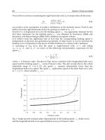
Fig. 6 Pineal lesions
1. Germinoma. Sagittal T1 WI with a large, solid space-occupying lesion originat-
ing from the pineal gland and a high postcontrast signal intensity causing com-
pression of the brain stem and cerebellum with distortion of the 4th ven-
tricle.There is also descent of the cerebellar tonsils.
2. Astrocytoma and suprasellar metastasis. Sagittal T1 WI shows a postcontrast
enhancing mass in the pineal region producing compression of the quad-
rigeminal plate. A second suprasellar mass compresses the pituitary stalk. The
patient presented clinical signs of diabetes insipidus.
3. Medulloblastoma. Sagittal T1 WI with a solid, multilobular space-occupying le-
sion, which presents an intermediate, heterogenous postcontrast enhance-
ment and is housed in the upper region of the cerebellum and 4th ventricle.
4. Basilar aneurysm. Sagittal T1 WI demontrates a partially thrombosed giant
aneurysm of the basilar artery, which acts as a space-occupying mass and thus
compresses the pons, the cerebral peduncles, and the 3nd ventricle, extend-
ing retrochiasmatically into the suprasellar cisterns.
Intracranial Tumors
Tsementzis, Differential Diagnosis in Neurology and Neurosurgery © 2000 Thieme
All rights reserved. Usage subject to terms and conditions of license.
115
Pineal Gland
(Fig. 6)
Germ-cell tumors
– Pure germinoma The most common variant of germ-cell neoplasm in
this area, accounting for 50% of pineal neoplasms
– Embryonal cell carci-
noma
– Choriocarcinoma
– Teratoma
– Mixed germ-cell
tumor
– Yolk sac tumor Endodermal sinus
Pineal parenchymal (cell
origin) tumors
– Pineoblastoma
– Pineocytoma
Tumors of supportive
tissues and adjacent
structures
– Astrocytoma
– Ependymoma
– Meningioma
– Hemangiopericytoma
– Ganglioneuroma
– Ganglioglioma
– Chemodectoma
– Craniopharyngioma
– Lipoma (quadrige-
minal cistern)
Metastatic tumors of
the pineal gland
Extremely rare; 75 reported cases in total
– Lung
– Breast
– Stomach
– Kidney
Nonneoplastic tumor-
like conditions
– Pineal cysts Degenerative cysts lined with fibrillary astrocytes
– Arachnoid cysts
– Cysticercus cysts
– Vascular lesions Aneurysmal dilation of the vein of Galen, vertebro-
basilar dolichoectasia, basilar tip aneur ysm
Pineal Gland
Tsementzis, Differential Diagnosis in Neurology and Neurosurgery © 2000 Thieme
All rights reserved. Usage subject to terms and conditions of license.
116
Cerebellopontine Angle
(Figs. 7 and 8)
Acoustic schwannoma Most common mass, up to 75% of cases
Meningioma Second most common lesion, up to 10% of cases
Ectodermal inclusion tu-
mors
– Epidermoid Also known as “congenital cholesteatoma” or “pearly
tumor”; 5 – 7%
– Dermoid
Metastases
Paraganglioma Also known as “glomus jugulare tumor”; a chemodec-
toma arising from the jugular foramen and extending
into the CPA; 2–10%
Other schwannomas 2– 5%. The trigeminal and facial nerves are probably
the most common sites of nonacoustic schwannomas.
Other cranial nerves involved are: VI, IX, X, XI, and
rarely XII
Vascular 2–5%
– Dolichobasilar ec-
tasia
3– 5%
– Aneurysm 1– 2%
– Vascular malforma-
tion
1%
Choroid plexus papil-
loma
1%; primary in the CPA or extension via the lateral
foramina of Luschka
Ependymoma 1%; extension from the fourth ventricle
Rare lesions Incidence Ͻ 1%
– Arachnoid cyst
– Lipoma
– Exophytic brain stem
or cerebellar astrocytoma
– Chordoma
– Osteocartilaginous tu-
mors
– Cysticercosis
CPA: cerebellopontine angle.
Intracranial Tumors
Tsementzis, Differential Diagnosis in Neurology and Neurosurgery © 2000 Thieme
All rights reserved. Usage subject to terms and conditions of license.