Differential Diagnosis in Neurology and Neurosurgery - part 6 docx
Bạn đang xem bản rút gọn của tài liệu. Xem và tải ngay bản đầy đủ của tài liệu tại đây (850.65 KB, 35 trang )
162
Blood constituents
Erythrocyte disorders Polycythemia vera, sickle-cell disease
Platelet dysfunction Thrombocytosis
Protein abnormalities Anticardiolipin/antiphospholipid antibodies, protein C
and S deficiency, lupus anticoagulant
Emboli Cardiogenic sources, infective endocarditis, atrial
myxoma, mitral valve prolapse, lupus, paradoxical
emboli, etc.
CNS: central nervous system.
Cervical Bruit
Internal carotid artery stenosis
External carotid artery stenosis
Internal carotid artery dissection
Internal carotid artery kink
Fibromuscular dysplasia
Subclavian or Innominate artery stenosis
Radiated cardiac murmur
High flow state
– Intracranial arteriovenous malformations
– Caroticocavernous fistula
– Hyperthyroidism
Venous hum
Cerebral Arteritis
Conditions associated with arteritis probably account for some portion
of the approximately 25% of strokes that are of undetermined etiology.
Infection
Syphilis
AIDS
Lyme disease (borreliosis)
Tuberculous meningitis
Mycoplasma angiitis
Sarcoid
Cerebrovascular Disease
Tsementzis, Differential Diagnosis in Neurology and Neurosurgery © 2000 Thieme
All rights reserved. Usage subject to terms and conditions of license.
163
Stroke
Determining whether a stroke is hemorrhagic or ischemic has important
implications for the patient’s prognosis and for decisions concerning
surgery or anticoagulant treatment. The suddenness of onset and the
focal neurological signs give these syndromes the popular term “stroke,”
and help to distinguish cerebrovascular disease from other neurological
disorders. Hypertension, atherosclerosis, or other evidence of vascular
disease are commonly present. The disappearance of symptoms within
minutes or hours allows transient ischemic attacks (TIAs) to be distin-
guished from stroke.
Drug abuse
Amphetamines
Heroin
LSD
Cocaine
Phenylephrine (Neo-Synephrine)
Diseases of altered immunity (including hypersensitive states)
Hodgkin’s disease with CNS vasculitis
Non-Hodgkin’s lymphoma with CNS vasculitis
Serum sickness
Systemic necrotizing vasculitides
Giant-cell ar teritis
Polyarteritis nodosa
Takayasu’s arteritis
Wegener’s granulomatosis
Henoch–Schönlein purpura
Connective-tissue diseases
Sjögren’s syndrome
Progressive systemic sclerosis
Polymyositis, dermatomyositis
Systemic lupus erythematosus
Rheumatoid disease
Behçet’s syndrome
Cryoglobulinemia
AIDS: acquired immune deficiency syndrome; CNS: central nervous system; LSD: lysergic acid
diethylamide.
Stroke
Tsementzis, Differential Diagnosis in Neurology and Neurosurgery © 2000 Thieme
All rights reserved. Usage subject to terms and conditions of license.
164
Cerebral embolism This is suggested by a sudden onset and a focal neuro-
logical deficit attributable to brain surface ischemia,
e.g., pure aphasia, pure hemianopia
Cerebral thrombosis A more complex and extensive neurological deficit
suggests a thrombosis, particularly when the stroke
has been preceded by transient ischemic attacks.
When the deficits are of sudden onset, thrombus is
clinically indistinguishable from embolus. The two
mechanisms of thrombosis are difficult to distinguish
on clinical grounds
Cerebral hemorrhage The neurological symptoms have a characteristically
smooth onset and evolution. However, if the syn-
drome advances within minutes, or is halted at an
early stage with only minor signs, the clinical picture
may then become indistinguishable from that of in-
farction
Trauma Sudden onset also characterizes trauma, subsequent
to which epidural and subdural hematomas may
occur, possibly mimicking stroke. Although the
trauma itself is sudden, the accumulation of the he-
matoma takes time: minutes or hours for epidural
hemorrhage, and as long as week for subdural hemor-
rhage
Seizures Seizures may be a sign of lobar hemorrhage. The im-
mediate postictal deficit mimics that caused by major
stroke. A small percentage of seizures develop months
or years after a stroke. A proper history may help rule
out a new stroke
Migraine This represents a major source of difficulty in the diag-
nosis of TIA. Migraine affects young people and in-
volves repeated attacks, with the patients experienc-
ing classic visual migraine auras at other times. Symp-
toms include a pounding headache contralateral to
the sensory or motor symptoms hours after the attack
Cerebral neoplasia The focal cerebral disturbance evolves gradually over
days or weeks, which is a longer period than stroke.
CT in tumors demonstrates an enhancing mass, but in
ischemic stroke, by contrast, the CT is often negative
Cerebral abscess Clinical and CT findings similar to those of a brain
tumor
Cerebrovascular Disease
Tsementzis, Differential Diagnosis in Neurology and Neurosurgery © 2000 Thieme
All rights reserved. Usage subject to terms and conditions of license.
165
Metabolic disturbances In comatose patients, consideration should be given
to other conditions causing focal neurological signs,
which often remit when the cause is removed
– Metabolic glucose dis-
turbances
– Renal failure
– Severe disturbances
of electrolyte balance
– Alcohol intoxication
– Barbiturate intoxica-
tion
CT: computed tomography; TIA: transient ischemic attack.
Clinical Grading Scales in Subarachnoid
Hemorrhage
Botterell scale Grade
Conscious, with or without signs of bleeding in the subarachnoid space I
Drowsy, without significant neurological deficit II
Drowsy, with significant neurological deficit III
Major neurological deficit, deteriorating, or older with preexisting
cerebrovascular disease IV
Moribund or near-moribund, failing vital centers, extensor rigidity V
Hunt–Hess scale Grade
Asymptomatic or mild headache I
Moderate to severe headache, nuchal rigidity, may have oculomotor
palsy II
Confusion, drowsiness, or mild focal signs III
Stupor or hemiparesis IV
Coma, moribund appearance, and/or extensor posture V
Clinical Grading Scales in Subarachnoid Hemorrhage
Tsementzis, Differential Diagnosis in Neurology and Neurosurgery © 2000 Thieme
All rights reserved. Usage subject to terms and conditions of license.
166
World Federation of Neurologic Surgeons (WFNS) scale Grade
Glasgow Coma Scale score 15:
– no headache or focal signs I
Glasgow Coma Scale score 15:
– headache, nuchal rigidity, no focal signs II
Glasgow Coma Scale score 13–14:
– may have headache, nuchal rigidity, no focal signs III
Glasgow Coma Scale score 13 – 14:
– may have headache, nuchal rigidity, or focal signs IVa
Glasgow Coma Scale score 9–12:
– may have headache, nuchal rigidity, or focal signs IVb
Glasgow Coma Scale score 8 or less:
– may have headaches, nuchal rigidity, or focal signs V
Cooperative Aneurysm Study scale Grade
Symptom-free I
Mildly ill, alert and responsive, headache present II
Moderately ill III
– Lethargic, headache, no focal signs
– Alert, focal signs present
Severely ill
– Stuporous, no focal signs
– Drowsy, major focal signs present
IV
Cerebral Salt-Losing Syndrome and Syndrome of
Inappropriate Secretion of Antidiuretic Hormone
after Subarachnoid Hemorrhage
Clinical parameter SIADH Cerebral salt-losing
syndrome
Blood pressure Normal Low or postural
hypotension
Heart rate Slow or normal Resting or postural
tachycardia
Cerebrovascular Disease
Tsementzis, Differential Diagnosis in Neurology and Neurosurgery © 2000 Thieme
All rights reserved. Usage subject to terms and conditions of license.
167
Clinical parameter SIADH Cerebral salt-losing
syndrome
Blood volume Normal or increased Decreased
Hematocrit Normal or low Elevated
Hydration Well hydrated Dehydrated
Body weight Normal or increased Decreased
Glomerular filtration rate Increased Decreased
Blood urea nitrogen/creatinine Normal or low Normal or high
Urine volume Normal or low Normal or low
Urine concentration High High
Hyponatremia Dilutional (false) True
Hypo-osmolality Dilutional (false) True
Mean day of appearance 8 (range 3–15) 4 – 5 (range 2 – 10)
Treatment Fluid restriction Sodium and volume
expansion
SIADH: syndrome of inappropriate secretion of antidiuretic hormone.
Syndrome of Inappropriate Secretion of
Antidiuretic Hormone and Diabetes Insipidus
The syndrome of inappropriate secretion of antidiuretic hormone
(SIADH) involves the release of antidiuretic hormone (ADH) at levels in-
appropriate for a low serum osmolality. Due to continued water inges-
tion, the elevated ADH results in water retention, hyponatremia, and
hypo-osmolality. SIADH results from partial damage to the supraoptic
and paraventricular nuclei or neighboring areas, or from production of
ADH by tumor or inflammatory tissue outside the hypothalamus.
The laboratory criteria for the diagnosis of SIADH are as follows.
– Low serum sodium (Ͻ 135 mEq/L)
– Low serum osmolality (Ͻ 280 mOsm/Kg)
– Elevated urinary sodium level (25 mEq/L)
– Urine osmolality that is inappropriately high compared to the serum
osmolality
– Absence of clinical evidence of volume depletion or diuretic use and normal
thyroid, renal, and adrenal function. Symptoms of hyponatremia include
confusion, muscle weakness, seizures, anorexia, nausea and vomiting, and
stupor, when the serum sodium falls below 110 mEq/L
Syndrome of Inappropriate Secretion
Tsementzis, Differential Diagnosis in Neurology and Neurosurgery © 2000 Thieme
All rights reserved. Usage subject to terms and conditions of license.
168
Diabetes insipidus involves a lack of free water due to a partial or
complete deficiency in ADH. The clinical symptoms include polyuria
(urine output greater than 300 mL/h or 500 mL/2 h), thirst, dehydration,
hypovolemia, and polydipsia. Diabetes insipidus results from the de-
struction of at least 90% of the large neurons in the supraoptic and para-
ventricular nuclei. The lesion of ten involves the supraoptic and hy-
pophysial tract rather than the neuronal bodies themselves.
The laboratory criteria for the diagnosis of diabetes insipidus are as
follows.
– Urine specific gravity of less than 1.005
– Urine osmolality between 50 and 150 mOsm/Kg
– Serum sodium greater than 150 mEq/L, unaccompanied by a corresponding
fluid deficiency. Sodium levels reaching 170 mEq/L are accompanied by
muscle cramping, tenderness and weakness, fever, anorexia, paranoia, and
lethargy
Syndromes of Cerebral Ischemia
Occluded artery Signs and symptoms
Common carotid artery – May be asymptomatic
– Ipsilateral blindness
Middle cerebral artery – Contralateral hemiplegia (face and arm greater
than leg)
– Contralateral hemisensory deficit (face and arm
greater than leg)
– Homonymous hemianopsia
– Horizontal gaze palsy
– Language and cognitive deficits in the left hemi-
sphere: aphasia (motor, sensory, global); apraxia
(ideomotor and ideational); Gerstmann syndrome
(agraphia, acalculia, left – right confusion, and fin-
ger agnosia)
– Language and cognitive deficits in the right hemi-
sphere: constructional/spatial defects (con-
structional apraxia, or apractognosia, dressing
apraxia); agnosias (atopognosia, prosopagnosia,
anosognosia, asomatognosia); left-sided unilateral
neglect; amusia
Cerebrovascular Disease
Tsementzis, Differential Diagnosis in Neurology and Neurosurgery © 2000 Thieme
All rights reserved. Usage subject to terms and conditions of license.
169
Occluded artery Signs and symptoms
Anterior cerebral artery – Contralateral hemiparesis (distal leg more than arm)
– Contralateral sensory loss (distal leg more than arm)
– Urinary incontinence
– Left-sided ideomotor apraxia or tactile anomia
– Severe behavior disturbance (apathy or “abulia,”
motor inertia, akinetic mutism, suck and grasp re-
flexes, and diffuse rigidity—“gegenhalten”)
– Eye deviation toward side of infarction
– Reduction in spontaneous speech, perseveration
Posterior cerebral
artery
– Contralateral homonymous hemianopia or quad-
rantanopia
– Memory disturbance with bilateral inferior tem-
poral lobe involvement
– Optokinetic nystagmus, visual perseveration
(palinopsia), hallucinations in the blind field
– There may be alexia (without aphasia or agraphia),
and anomia for colors, in dominant hemisphere in-
volvement
– Cortical blindness, with patient not recognizing or
admitting the loss of vision (Anton’s syndrome),
with or without macular sparing, poor eye–hand
coordination, metamorphopsia, and visual agnosia
when cortical infarction is bilateral
– Pure sensory stroke: may leave anesthesia dolorosa
with “spontaneous pain,” in cortical and thalamic
ischemia
– Contralateral hemiballism and choreoathetosis in
subthalamic nucleus involvement
– Oculomotor palsy, internuclear ophthalmoplegia,
loss of vertical gaze, convergence spasm, lid retrac-
tion (Collier’s sign), corectopia (eccentrically posi-
tioned pupils), and some times lethargy and coma
with midbrain involvement
Anterior choroidal
artery
May cause varying combinations of:
– Contralateral hemiplegia
– Sensory loss
– Homonymous hemianopia (sometimes with a strik-
ing sparing of a beak-like zone horizontally)
Syndromes of Cerebral Ischemia
Tsementzis, Differential Diagnosis in Neurology and Neurosurgery © 2000 Thieme
All rights reserved. Usage subject to terms and conditions of license.
170
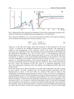
Brain Stem Vascular Syndromes
Midbrain (Fig. 15a)
Syndrome Structures involved Manifestations
Weber’s
syndrome
ț Ventral midbrain
ț CN III corticospinal
track
ț Ipsilateral CN III palsy, including
parasympathetic paresis (i.e.,
dilated pupil)
ț Contralateral hemiplegia
Benedikt’s
syndrome
ț Midbrain tegmen-
tum
ț Red nucleus
ț CN III brachium con-
junctivum
ț Ipsilateral CN III palsy, usually
with a dilated pupil
ț Contralateral involuntary
movements (intention tremor,
hemichorea, or hemiathetosis)
Claude’s
syndrome
ț Dorsal mesence-
phalic tegmentum
ț Dorsal red nucleus
ț Brachium conjunc-
tivum
ț CN III
ț Ipsilateral CN III palsy, usually
with a dilated pupil
ț Prominent cerebellar signs
ț Contralateral involuntary
movements (nucleus ruber
tremor, hemiataxia, and no
hemiballismus)
Parinaud’s
syndrome
ț Dorsal rostral mid-
brain
ț Pretectal area
ț Posterior commis-
sure
ț Paralysis of conjugate upward
(and occasionally downward)
gaze
ț Pupillary abnormalities (disso-
ciation of pupil response close
to light)
ț Convergence–retraction nys-
tagmus on upward gaze
ț Pathological lid retraction
(Collier’s sign)
ț Lid lag
ț Pseudo-abducens palsy
CN: cranial nerve.
Cerebrovascular Disease
Tsementzis, Differential Diagnosis in Neurology and Neurosurgery © 2000 Thieme
All rights reserved. Usage subject to terms and conditions of license.
171
superior colliculus
CN III n. nucleus
(Edinger-Westphal)
mesencephalic n. CN V
ventral + lateral
spinothalamic tracts
MLF
medial lemniscus
mesencephalic
reticular
formation
red nucleus
CN III
pyramidal tract
(corticospinal)
cortico-
pontine
tracts
Substantia nigra
medial geniculate
body
a
Parinaud syndrome
Benedict
syndrome
Weber
syndrome
Claude syndrome
a
Fig. 15a
Fig. 15 Brain stem vascular syndromes:
a Midbrain (superior colliculus): Weber syndromes: a) corticospinal and corti-
copontine tracts (contralateral hemiplegia including the face); b) parasympa-
thetic root fibres of CN III (ipsilateral oculomotor nerve paresis with fixed and di-
lated pupil); c) substantia nigra (Parkinsonian akinesia). Benedict syndrome: a)
red nucleus (contralateral involuntary movements, including intention tremor,
hemichorea, and hemiathetosis; b) brachium conjuctivum (ipsilateral ataxia); c)
parasympathetic root fibres of CN III (ipsilateral oculomotor paresis with fixed and
dilated pupil). Claude syndrome: a) dorsal red nucleus (contralateral involuntary
movements, including intention tremor, hemichorea, and hemiathetosis; b)
Brain Stem Vascular Syndromes
Tsementzis, Differential Diagnosis in Neurology and Neurosurgery © 2000 Thieme
All rights reserved. Usage subject to terms and conditions of license.
172
b
b
superior cerebellar peduncle
MLFparamedian raphe
mesencephalic
motor nuclei
lateral
system
lateral lemniscus
medial lemniscus
pontine nuclei
pontine reticular
formation
pyramidal tract
middle cerebellar
penduncle
pontine tracts
pontine reticular
formation
pyramidal tract
CN V
lateral pontine syndrome
(Marie-Foix
syndrome)
Raymond-Cestan syndrome
midpontine base
syndrome
Fig. 15b
brachium conjuctivum (prominent cerebellar signs and no hemiballismus); c) dor-
sal midbrain tegmentum. Parinaud sydrome: a) superior colliculi (conjugated
gaze paralysis upward); b) medial longitudinal fasciculus (nystagmus and internal
ophthalmoplegia); c) eventual paresis of the CNs III and IV; d) cerebral aqueduct
stenosis/obstruction (hydrocephalus). Involvement of the inferior colliculi pro-
duces hearing loss.
b Pons (rostral): Raymond–Cestan syndrome: a) superior cerebellar peduncle
(cerebellar ataxia with a coarse “rubral” tremor); b) medial lemniscus and
Cerebrovascular Disease
Tsementzis, Differential Diagnosis in Neurology and Neurosurgery © 2000 Thieme
All rights reserved. Usage subject to terms and conditions of license.
173
Fig. 15c
c
c
medial lemniscus
inferior cerebellar penduncle MLF CN V nucleus and tract
ventral and lateral
spinothalamic tracts
pontine tracts
pontine reticular
formation
pyramidal tract
CN VIII
CN VII
CN VI
locked-in syndrome
ventral pontine
(Millard-Gubler)
syndrome
dorsal pontine (Foville) syndrome
spinothalamic tract (contralateral decrease in all sensory modalities, involving
face and extremities). Ventral extension of the lesion involves additionally; c) cor-
ticospinal tract (contralateral hemiparesis), d) paramedian pontine reticular for-
mation (paralysis of the conjugate gaze towards the side of the lesion). Marie–
Foix syndrome: a) superior and middle cerebellar peduncles (ispilateral cerebel-
lar ataxia); b) corticospinal tract (contralateral hemiparesis); c) spinothalamic
tract (variable contralateral hemihypesthesia for pain and temperature). Midpon-
Brain Stem Vascular Syndromes
Tsementzis, Differential Diagnosis in Neurology and Neurosurgery © 2000 Thieme
All rights reserved. Usage subject to terms and conditions of license.
174
d
inferior
cerebellar
peduncle
solitary
nucleus
reticular
formation
CN VII nuclei
MLF
medial lemniscus
pyramidal tract
ambiguus nucleus
CN XII
inferior olivary
nucleus
CN XI
CN X
ventral + lateral
spinothalamic tract
d
lateral medullary
(Wallenberg) syndrome
Fig. 15d
tine base syndrome: a) middle cerebellar peduncle (ipsilateral ataxia and asyn-
ergy); b) corticospinal tract (contralateral hemiparesis); c) corticopontine fibres
(ipsilateral dystaxia); d) root fibres of CN V (ipsilateral hemianesthesia of all mo-
dalities and flaccid paralysis of chewing muscles).
c Pons (caudal): Foville syndrome: a) nucleus and fascicles of CN VII (ipsilateral
peripheral type facial palsy), b)nucleus of CN VI (gaze is “away from” the lesion), c)
corticospinal tract (contralateral hemiplegia with sparing of the face), d) parame-
dian pontine reticular formation. Millard–Gubler syndrome: a) pyramidal tract
(contralateral hemiplegia sparing the face); b) CN VI (diplopia accentuated when
the patient “looks towards” the lesion); c) CN VII (ipsilateral peripheral facial nerve
paresis). Locked-in syndrome: a) bilateral corticospinal tracts in the basis pontis
(tetraplegia); b) corticobulbar fibres of the lower CNs (aphonia); c) occasionally
bilateral fascicles of the CN VI (impairment of horizontal eye movements).
Cerebrovascular Disease
Tsementzis, Differential Diagnosis in Neurology and Neurosurgery © 2000 Thieme
All rights reserved. Usage subject to terms and conditions of license.
175
e
vestibulospinal tract
CN V nerve
nucleus and tract
solitary nucleus gracile nucleus
and fasciculus
cuneate nucleus
and fasciculus
accessory olivary
nucleus
ventral and lateral
spinothalamic tract
medial lemniscus
medullary reticular
formation
Medial medullary
syndrome
(Dejerine's anterior
bulbar syndrome)
e
CN XII
Fig. 15e
d Medulla (rostral): Lateral medullary (Wallenberg) syndrome: a) nucleus
and tract of CN V (ipsilateral facial pain and hypalgesia and thermoanesthesia); b)
spinothalamic tract (contralateral trunk and extremity hypalgesia and thermoan-
esthesia); c) nucleous ambiguus (ipsilateral palatal, pharyngeal, and vocal cord
paralysis with dysphagia and dysarthria); d) vestibular nuclei (vertigo, nausea, and
vomiting); e) descending sympathetic fibers (ipsilateral Horner’s syndrome); f) in-
ferior cerebellar peduncle and cerebellum (ipsilateral cerebellar signs and symp-
toms); g) medullary respiratory centers (hiccups); h) lower pons (diplopia).
e Medulla (caudal): Medial medullary (Dejerine) syndrome: a) CN XII (ipsi-
lateral paresis atrophy, and fibrillation of the tongue; b) pyramidal tract (con-
tralateral hemiplegia with sparing of the face); c) medial lemniscus (contralateral
loss of position sense and vibration occasionally); d) medial longitudinal nystag-
mus (upbeat nystagmus).
Brain Stem Vascular Syndromes
Tsementzis, Differential Diagnosis in Neurology and Neurosurgery © 2000 Thieme
All rights reserved. Usage subject to terms and conditions of license.
176
Pons (Figs. 15b and 15c)
Syndrome Structures involved Manifestations
Millard–Gubler syn-
drome
ț Ventral paramedian
pons
ț CN VI and VII
fascicles
ț Cor ticospinal tract
ț Contralateral hemiplegia (spar-
ing the face)
ț Ipsilateral lateral rectus palsy
with diplopia
ț Ipsilateral peripheral facial
paresis
Dysarthria–clumsy
hand syndrome
ț Basis pontis (lacunar
infarction) at junc-
tion of upper one-
third and lower two-
thirds of pons
ț CN VII
ț Clumsiness and paresis of the
hand, ipsilateral hyperreflexia,
and Babinski sign
ț Facial weakness
ț Severe dysarthria and
dysphagia
Differential diagnosis: this syndrome has also been described with lesions in a)
the genu of the internal capsule or b) with small deep cerebellar hemorrhages.
Pure motor hemi-
paresis
ț Lacunar infarction in-
volving the cortico-
spinal tracts in the
basis pontis
ț Pure motor hemiplegia
ț With or without facial involve-
ment
Ataxic hemiparesis ț Lacunar infarction in-
volving the basis
pontis at the junc-
tion of the upper
third and lower two-
thirds of the pons
ț Hemiparesis more severe in the
lower extremity
ț Ipsilateral hemiataxia
ț Occasional dysar thria, nystag-
mus, and paresthesias
Differential diagnosis: this syndrome has also been described with lesions in a)
the contralateral thalamocapsular area, b) the contralateral posterior limb of
the internal capsule, and c) the contralateral red nucleus
Locked-in syn-
drome
(deefferentation)
ț Bilateral ventral pon-
tine lesions (infarc-
tion, tumor, hemor-
rhage, trauma, cen-
tral pontine my-
elinolysis)
ț Tetraplegia due to bilateral cor-
ticospinal tract involvement
ț Aphonia due to involvement of
the corticobulbar fibers
destined for the lower cranial
nerves
ț Occasionally, impairment of
horizontal eye movements due
to bilateral involvement of the
fascicles of CN VI
Cerebrovascular Disease
Tsementzis, Differential Diagnosis in Neurology and Neurosurgery © 2000 Thieme
All rights reserved. Usage subject to terms and conditions of license.
177
Syndrome Structures involved Manifestations
Primary pontine
hemorrhage syn-
dromes
ț Classic type (60%):
severe pontine de-
struction
ț Tetraparesis, coma, and death
ț Hemipontine type
(20%)
ț Hemiparesis, skew deviation,
dysarthria, unilateral absent
corneal reflex, CN VII palsy,
ipsilateral facial sensory
changes, survival with func-
tional recovery
ț Dorsolateral
tegmental type
(20%)
ț Gaze paresis and/or ipsilateral
CN VI palsy, unilateral CN VII
palsy, contralateral extremity
and ipsilateral facial sensory
loss, dysarthria, preserved con-
sciousness, motor sparing, oc-
casional gait or limb ataxia
Foville’s syndrome ț Dorsal pontine teg-
mentum in the
caudal third of the
pons, PPRF
ț Contralateral hemiplegia (with
facial sparing)
ț Ipsilateral peripheral-type facial
palsy (involvement of CN VII
fascicles)
ț Gaze palsy to side of lesion
Raymond–Cestan
syndrome
ț Rostral lesions of the
dorsal pons
ț Cerebellar signs (ataxia)
ț Contralateral reduction of all
sensory modalities (face and
extremities)
ț Contralateral hemiparesis
ț Paralysis of conjugate gaze in
PPRF involvement
Marie–Foix syn-
drome
ț Lateral pontine le-
sions
(especially brachium
pontis)
ț Ipsilateral cerebellar ataxia
ț Contralateral hemiparesis
ț Variable contralateral hemihy-
pesthesia for pain and
temperature
CN: cranial nerve; PPRF: paramedian pontine reticular formation.
Brain Stem Vascular Syndromes
Tsementzis, Differential Diagnosis in Neurology and Neurosurgery © 2000 Thieme
All rights reserved. Usage subject to terms and conditions of license.
178
Medulla (Figs. 15 d and 15e)
Syndrome Structures involved Manifestations
Dejerine anterior
bulbar syndrome
ț Medial medulla ob-
longata (corti-
cospinal tract,
medial lemniscus,
CN XII)
ț Ipsilateral paresis, atrophy
(tongue deviates toward the le-
sion)
ț Contralateral hemiplegia with
sparing of the face
ț Contralateral loss of position and
vibratory sensation. Pain and
temperature sensation are
spared
Wallenberg’s syn-
drome
ț Lateral medulla
ț Inferior cerebellum
(inferior cerebellar
peduncle, de-
scending sympa-
thetic tract,
spinothalamic
tract, CN V nu-
cleus)
ț Ipsilateral facial hypalgesia and
thermoanesthesia
ț Contralateral trunk and extrem-
ity hypalgesia and thermoan-
esthesia
ț Ipsilateral palatal, pharyngeal,
and vocal cord paralysis with
dysphagia and dysarthria
ț Ipsilateral Horner’s syndrome
ț Vertigo, nausea, and vomiting
ț Ipsilateral cerebellar signs and
symptoms
ț Occasionally, hiccups and di-
plopia
Lateral ponto-
medullary syn-
drome
ț Lateral medulla
ț Inferior cerebellum
ț Lower pons (to the
region of exit of
CNs VII and VIII)
ț All clinical findings seen in the
lateral medullary syndrome
ț Ipsilateral facial weakness
ț Ipsilateral tinnitus and occa-
sionally hearing disturbance
CN: cranial nerve.
Cerebrovascular Disease
Tsementzis, Differential Diagnosis in Neurology and Neurosurgery © 2000 Thieme
All rights reserved. Usage subject to terms and conditions of license.
179
Differentiation of the Various Types of Cerebral Ischemic Vascular Lesion
Ischemic vascu-
lar lesions
Clinical and radiological characteristics
Risk factors Onset/cause Anatomical
characteristics
Associated signs Imaging characteris-
tics
Systemic
hypoperfusion
Heart disease,
trauma, GI bleeding,
hypotension
Systemic disease
present (cardiac ar-
rest, bleeding
Border zone regions
between AC A, MCA,
PCA and SCAs, PICA,
AICA
Pallor, sweating,
hypotension
Located in watershed
CT: low density (dark)
MRI: hypointensity
(dark) in T1-weighted
images and hyper-
intensity (white) in
T2-weighted images
Embolism Heart/coronary dis-
ease, peripheral
vascular disease in
white men, smoking
hyperlipidemia
Sudden onset in 80%
of cases during first
24 h; progressive in
20%
Middle cerebral
artery region most
frequently, followed
by PCA or PICA dis-
tribution
Headache during and
after the onset of
cerebral embolism is
prominent in 25% of
cases
Superficial or deep
wedge-shaped areas
CT: low density (dark)
MRI: hypointensity
(dark) in T1-weighted
images and hyper-
intensity (white) in
T2-weighted images
Cont. ̈
Differentiation of the Various Types of Cerebral Ischemic Vascular Lesion
Tsementzis, Differential Diagnosis in Neurology and Neurosurgery © 2000 Thieme
All rights reserved. Usage subject to terms and conditions of license.
180
Ischemic vascu-
lar lesions
Clinical and radiological characteristics
Risk factors Onset/cause Anatomical
characteristics
Associated signs Imaging characteris-
tics
Large artery
thrombosis
Heart/coronary dis-
ease, peripheral
vascular disease in
white men, smoking
hyperlipidemia
Fluctuating, progres-
sive and remitting,
manifested by a TIA
in appprox. 40% of
cases
Middle cerebral
artery region most
frequently, followed
by PCA or PICA dis-
tribution
Headache during and
after the onset of
cerebral embolism is
prominent in 25% of
patients
Located in watershed
areas or center of
arterial supply
CT: low density (dark)
MRI: hypointensity
(dark) in T1-weighted
images and hyper-
intensity (white) in
T2-weighted images
Small artery
thrombosis
Systemic hyperten-
sion, diabetes, poly-
cythemia
Fluctuating, progres-
sive and remitting,
manifested by a TIA
in approx. 25% of
patients
Small perforating ar-
teries of deep brain
structures, basal gan-
glia, thalamus, pons,
cerebellum, cerebral
white matter
Usually none Small, deep lesions
(lacunar infarcts)
CT: low density (dark)
MRI: hypointensity
(dark) in T1-weighted
images and hyper-
intensity (white) in
T2-weighted images
ACA: anterior cerebral artery; AICA: anterior inferior cerebellar artery; CT: computed tomography; GI: gastrointestinal; MCA: middle cerebral arter y; MRI:
magnetic resonance imaging; PCA: posterior cerebral artery; PICA: posterior inferior cerebellar artery; SCA: superior cerebellar artery; TIA: transient
ischemic attack.
Cerebrovascular Disease
Tsementzis, Differential Diagnosis in Neurology and Neurosurgery © 2000 Thieme
All rights reserved. Usage subject to terms and conditions of license.
181
Predisposing Factors and Associated Disorders of
Cerebral Veins and Sinuses Thrombosis
Primary idiopathic thrombosis
Secondary thrombosis
Pregnancy
Postpartum
Head injury
Tumors
– Meningioma
– Metastatic neoplasia
Malnutrition and dehydration (marasmus in infancy)
Infection involving sinuses, mastoids, and leptomeninges
Hypercoagulable states and coagulopathies
– Polycythemia
– Sickle-cell anemia
– Leukemia
– Disseminated intravascular coagulation
– Oral contraceptives
– Inflammatory bowel disease
– Nephrotic syndrome
– Protein S and protein C deficiencies
– Antithrombin III deficiency
Paraneoplastic syndromes
– Cerebellar degeneration
– Encephalomyelitis
– Subacute necrotizing myelopathy
– Peripheral polyneuropathy
– Cerebrovascular disease
– Neuromuscular junction
Chemotherapeutic agents (L-asparaginase)
Cyanotic congenital heart disease
Predisposing Factors and Associated Disorders
Tsementzis, Differential Diagnosis in Neurology and Neurosurgery © 2000 Thieme
All rights reserved. Usage subject to terms and conditions of license.
182
Venous Thrombosis
Vessel involved Structures involved Clinical findings
Superior sagit-
tal sinus
ț Venous drainage
from the hemi-
spheres and medial
cerebral cortex
ț New-onset headaches (simple or
severe headaches that can be posi-
tionally aggravated)
ț Increased intracranial pressure
Extension of clot into the larger cere-
bral veins (as is common in septic
thrombosis and in a high percentage
in the nonseptic type) may cause the
following:
ț Convulsive seizures
ț Hemiplegia
ț Aphasia
ț Hemianopia
ț Lethargy or coma
Lateral sinus ț Venous drainage
from the posterior
fossa
ț Drainage from the
confluence of
sinuses (secondary
to otitis media and
mastoiditis)
ț Pain, especially behind the ear
(coinciding with acute or chronic
otitis or mastoiditis)
ț Increased intracranial pressure
Extension of infection into the veins
draining the lateral surface of the
hemisphere may cause the following:
ț Jacksonian seizures
ț Hemiplegia
ț Gradenigo’s syndrome
ț CNs IX, X, XI (jugular foramen disten-
sion)
ț Drowsiness and coma
Differential diagnosis: cerebral abscess
Cavernous
sinus
ț CNs IV, V, and/or VI
ț Internal carotid
artery, possibly
ophthalmic artery
(originates in
suppurative
processes of the
orbit, nasal sinuses,
upper half of face)
ț Retro-orbital pain
ț Proptosis
ț Orbital congestion with edema and
chemosis of the conjunctivae and
eyelids
ț Ptosis
ț Facial sensory loss
ț Signs of carotid artery occlusion
ț Visual loss
ț Disks are swollen, with small hemor-
rhages
Differential diagnosis: a) orbital tumors in the region of the sphenoid; b) malig-
nant exophthalmos; c) arteriovenous aneurysms
CN: cranial nerve.
Cerebrovascular Disease
Tsementzis, Differential Diagnosis in Neurology and Neurosurgery © 2000 Thieme
All rights reserved. Usage subject to terms and conditions of license.
183
Spontaneous Intracerebral Hemorrhage
Spontaneous intracerebral hemorrhage (ICH) accounts for approxi-
mately 10% of cases of stroke. Arterial hypertension is by far the most
common cause of ICH; other causes are the intracranial aneurysms,
vascular malformation, bleeding diathesis, cerebral amyloidosis, brain
tumors, vasculitis, or drug abuse.
The clinical features of ICH depend on the location, size, direction of
spread, and rate of development of the hematoma. The clinical presenta-
tion of lobar hemorrhages is often misinterpreted as a thromboembolic
cerebral infarction. Posterior fossa spontaneous hemorrhages occur in
10% of patients with spontaneous hemorrhage, and may affect either the
cerebellum or the pons. Differentiation of cerebellar or pontine hemor-
rhages often is not possible on clinical grounds, since they share the sud-
den presenting symptoms and often signs. An accurate diagnosis is
achieved quickly by computed tomography and magnetic resonance im-
aging.
Structure involved Clinical manifestations
Lobar hemorrhage
Frontal lobe – Abulia
– Contralateral hemiparesis
– Bifrontal headache (maximum ipsilateral)
– Occasionally, mild gaze preference away from the
hemiparesis
Parietal lobe – Contralateral hemisensory loss
– Neglect of the contralateral visual field
– Headache (usually anterior temporal location)
– Mild hemiparesis
– Occasionally, hemianopia or anosognosia
Temporal lobe – Wernicke’s aphasia (dominant temporal lobe)
– Conduction or global aphasia (dominant temporal-
parietal lobe)
– Variable degrees of visual field deficit
– Headache around or anterior to ipsilateral ear
– Occasionally, agitated delirium
Occipital lobe – Ipsilateral orbital pain
– Contralateral homonymous hemianopia
Spontaneous Intracerebral Hemorrhage
Tsementzis, Differential Diagnosis in Neurology and Neurosurgery © 2000 Thieme
All rights reserved. Usage subject to terms and conditions of license.
184
Structure involved Clinical manifestations
Putaminal hemor-
rhage
The putamen is the most common site of hyperten-
sive ICH
– Hemiparesis or hemiplegia and, to a lesser degree,
hemisensory deficit
– Transient global aphasia with dominant
hemispheric lesions
– Agnosia or unilateral neglect with nondominant
hemispheric lesions
– Homonymous hemianopia
– Contralateral gaze palsy: the patient looks toward
the hematoma and away from the hemiplegia
Alloesthesia: a noxious stimulus on the side of the
hemisensory disturbance is perceived at the corre-
sponding area of the other (normal) side
Thalamic hemorrhage
Findings – Hemisensory deficit and, to a lesser degree, hemi-
paresis
– Anomic aphasia with impaired comprehension,
with lesions of the dominant thalamus
– Convergence–retraction nystagmoid movements,
impairment of vertical gaze, and pupillary near-
light dissociation
– Downward–inward deviation of the eyes
– Unilateral or bilateral pseudo-sixth ner ve paresis
– Skew deviation
– Conjugate gaze palsy to the side of the lesion
(wrong side) or conjugate horizontal gaze deviation
Cerebellar hemor-
rhage
Most common in the area of the dentate nucleus
Symptoms – Sudden occipital headache
– Nausea and repeated vomiting
– Dizziness, vertigo
– Inability to stand
Findings – Variable degrees of alertness
– Small reactive pupils
– Skew deviation
– Ipsilateral gaze palsy
– Ocular bobbing and nystagmus toward the gaze;
paresis
– Ipsilateral peripheral facial weakness
– Ipsilateral absence or decrease of corneal reflex
– Slurred speech
– Gait or truncal ataxia
– Bilateral hyperreflexia and Babinski signs
Cerebrovascular Disease
Tsementzis, Differential Diagnosis in Neurology and Neurosurgery © 2000 Thieme
All rights reserved. Usage subject to terms and conditions of license.
185
Structure involved Clinical manifestations
Pontine hemorrhage
Symptoms – Headache, vomiting, vertigo, dysarthria
– Sudden loss of consciousness, often progressing
into deep coma
Findings – Sudden-onset coma
– Quadriparesis, quadriplegia
– Respiratory abnormalities
– Hyper thermia
– Pinpoint reactive pupils
– Eyes fixed in a central position
– Loss of brain stem reflexes, including the oculo-
cephalic (doll’s head) and the ocuovestibular re-
flexes
– Ocular bobbing
ICH: intracerebral hemorrhage.
Spontaneous Intracerebral Hemorrhage
Tsementzis, Differential Diagnosis in Neurology and Neurosurgery © 2000 Thieme
All rights reserved. Usage subject to terms and conditions of license.
186
Spinal Disorders
Failed Back Syndrome
The syndrome involves recurrent or residual low back pain after lumbar
disk surgery; the incidence ranges from 5% to 40%.
Incorrect original diag-
nosis
Permanent nerve root
injury from the original
disk herniation
Deafferentation pain, which is usually constant and
burning
Residual or recurrent
disk
Postoperative compli-
cations
– Immediate ț Permanent injury to the nerve roots from surgery
(deafferentation pain, which is usually constant and
burning, and is responsible for 6–16% of persistent
symptoms in postoperative patients)
ț Epidural hematoma
ț Infection
ț Postoperative swelling
– Late ț Pseudomeningocele, from a dural tear at the time
of surgery. Differential diagnosis includes: a) post-
operative serous fluid collections, b) infected col-
lections
ț Epidural fibrosis (scar or granulation tissue forma-
tion, causing compression and mechanical distor-
tion of the nerve root)
ț Arachnoiditis. Once very common after contrast
myelography, particularly with the combination of
hemorrhage from myelography/surgery and re-
tained contrast material. Differential diagnosis in-
cludes: a) Intradural mass, b) CSF tumor spread,
and c) spinal stenosis)
ț Diskitis. Incidence after lumbar diskectomy 0. 2%;
intractable back pain 1–4 weeks postoperatively
after a period of symptomatic relief. Differential di-
agnosis includes: a) neoplasm, b) degenerative dis-
ease, and c) osteomyelitis
Tsementzis, Differential Diagnosis in Neurology and Neurosurgery © 2000 Thieme
All rights reserved. Usage subject to terms and conditions of license.