Neurology Seminars in Clinical Neurology - part 2 ppt
Bạn đang xem bản rút gọn của tài liệu. Xem và tải ngay bản đầy đủ của tài liệu tại đây (396.31 KB, 11 trang )
Diagnosis, Classification, and Pathophysiology of Dystonia
3
C
lassification by Etiology
TABLE 1.3
Primary Dystonia
• Dystonia is the only sign without associated neurologi-
cal findings.
• Evaluation does not reveal any other cause for dystonia.
G
enetic
• DYT1: Onset typically in childhood with spread to
become generalized dystonia. Gene isolated. Clinical
testing available.
• DYT2, 4, 7, 11, 13: No clinical testing available.
Sporadic
• No family history.
• Most adult-onset dystonia. Some may have genetic
basis.
Secondary Dystonia
Associated with hereditary neurologic syndromes.
1. Dystonia Plus
Dopa-responsive dystonia
• GCHI mutations (DRD or DYT5)
• Tyrosine hydroxylase mutations
• Other biopterin deficient states
• Dopamine agonist responsive dystonia due to decar-
boxylase deficiency
• Myoclonus—Dystonia
2. Other inherited (degenerative) disorders
• Autosomal-dominant
• Rapid-onset dystonia-parkinsonism
•
Huntington's disease
• Machado-Joseph's disease/SCA3 disease
• Other SCA subtypes
• DRPLA
• Familial basal ganglia calcifications
•
Autosomal-r
ecessive
• Wilson's
•
Gangliosidoses
• Metachromatic leukodystrophy
• Homocystinuria
• Hartnup disease
•
Glutaric acidemia
• Methylmalonic aciduria
• Hallervorden-Spatz disease
•
Dystonic lipidosis
• Ceroid-lipofuscinosis
• Ataxia-telangiectasia
• Neuroacanthocytosis
• Intraneuronal inclusion disease
• Juvenile Parkinsonism (Parkin)
•
X-linked recessive
• Lubag (X-linked dystonia-parkinsonism or DYT3)
• Lesch-Nyhan syndrome
• Deafness/Dystonia
• Mitochondrial
• MERRF/MELAS
• Leber's disease
3. Due to acquired/exogenous causes
• Perinatal cerebral injury
• Encephalitis, infectious, and postinfectious
• Head trauma
• Pontine myelinolysis
• Primary antiphospholipid syndrome
• Stroke
• Tumor
• Multiple sclerosis
• Cervical cord injury or lesion
• Peripheral injury
• Drugs
•
T
oxins
• Psychogenic
4. Dystonia due to degenerative parkinsonian disorders
•
Parkinson Disease
• Multiple system atrophy
•
Progressive supranuclear palsy
• Cortico basal ganglionic degeneration
ics, such as perchlorpromazine or metoclopramide, or
antipsychotics, such as haloperidol or pimozide. These
usually present with forced eye deviations and involun-
t
ary trunk and neck extensions (oculogyric crisis), and
are infrequently confused with primary dystonia. Acute
drug-induced dystonic reactions are transient, resolving
with drug discontinuation, and are acutely responsive to
anticholinergic administration. However, the chronic
administration of the same class of dopamine receptor
antagonist drugs may cause tardive dystonia, which may
be either focal or generalized and often presents as
trunk and neck extension, sometimes associated with
stereotypic mouth movements. Tardive dystonia is
chronic and persists with discontinuation of the offend-
ing drug. The history of a temporal relationship of the
onset of dystonia following sustained use of these drugs
suggests this diagnosis.
The list of the genetic forms of dystonia has expand-
ed greatly over the past decade. The most frequent
genetic form of dystonia with childhood onset and sec-
ondary generalization is DYT1 dystonia. In youth-onset
primary dystonia, especially in Ashkenazi Jews, this is
the most common genetic form of dystonia. Although
inherited in an autosomal-dominant fashion, the pene-
trance of the gene is reduced, and only 30%–40% of
those carrying the gene will have symptoms of dysto-
nia. This means that, despite the absence of a family
history of dystonia in this patient, it is likely that the
patient will have a genetic form of dystonia, and may
have the DYT1 gene. This gene is located on chromo-
some 9, in the 9q32-34 region. It is a GAG deletion that
gives rise to a deletion in a glutamic acid residue in a
protein called torsin A. The function of torsin A has not
been elucidated, but it is widely distributed in the brain.
Most patients with dystonia due to DYT1 have symp
-
tom onset before the age of 26 years, with 1 or more
limbs affected. Testing for DYT1 is recommended for
patients with dystonia onset befor
e the age of 26 years,
and in those with onset over the age of 26, but with a
relative who has early-onset dystonia. This patient
would fall within the guidelines for obtaining DYT1
testing, if affordable. Genetic counseling for the patient
and family would be also recommended if available.
In summary, this patient had the typical history and
physical findings of youth-onset primary dystonia. In
the absence of any other associated neurologic abnor-
malities and no other putative cause for dystonia, a trial
of levodopa would be recommended to rule out the
possibility of dopa-responsive dystonia.
No other testing is essential. Obtaining a DYT1 gene
test would clarify whether the patient had this one
form of inherited dystonia, but would not be useful in
diagnosing the dystonic syndrome.
CASE 2
A 42-year-old woman presented with right-sided neck
pain that started 3 years previously. She initially attrib-
u
ted the pain to a stiff neck or arthritis. However, the
pain increased in intensity and she noticed that her head
tended to move to the right. She felt that movement to
the left was restricted. Over the following year, the move-
ment to the right became more pronounced, and was
obser
ved by her coworkers. When attempting to hold her
head in a forward position, she would have a side-to-side
tr
emor. If she touched her chin, or held her head in her
hand, her movements would abate. She developed an
ulnar neuropathy from resting her head on her left hand
with her elbow on the table. Over the past year, she also
reported difficulties with her handwriting. Although not
occurring during any other activity with her right hand,
when trying to write, she noticed that her second and
third fingers would bend forward and that her hand
would tend to supinate. There was no family history of
similar problems, although a maternal aunt had devel-
oped tremor in both hands when she was 60 years old.
Neurologic examination of this patient was remarkable
for head posturing to the right with an elevation of the
right shoulder, and ulnar neuropathy on the left. There
was neither tremor nor bradykinesia in the limbs. When
writing, flexion of the index and third finger occurred,
with flexion at the wrist and internal rotation of the arm.
In contrast to the first patient, this patient developed
symptoms in mid-adulthood. Her first symptom was
pain localized to an area of her neck. Involuntary, sus-
tained tur
ning of her head, tr
emor, and writing difficul-
ties followed. This patient had a history typical for
cervical dystonia (CD) with subsequent development
of writer's cramp.
CD is a focal dystonia with involvement of the neck
muscles. Pr
eviously known as spasmodic torticollis, it
is a common form of adult-onset dystonia with occur-
r
ence of symptoms in the fifth decade. CD is 1.5 to 3
times mor
e common in women than in men. It usual
-
ly r
emains localized to the neck area, though it may
spread to a contiguous body area as it did in this
patient, and become part of a segmental dystonia. As
is true with all adult-onset focal dystonias, it is rar
e for
this dystonia to become generalized.
Head postur
es associated with CD vary. Ther
e may
be a tur
ning of the head (torticollis) to one side, a lat
-
DYSTONIA
4
A
trial of levodopa is recommended in childhood-onset
dystonia, or in adults with generalized dystonia,
especially if accompanied by additional neurologic
a
bnormalities such as parkinsonism or spasticity.
eral flexion of the neck (laterocollis), a forward flexion
of the head (anterocollis), or a posterior extension of
the head (retrocollis). There may also be a shifting of
t
he head on the shoulders in a sagittal plane. In many
patients, the movement is not a single movement, but
rather a combination of the above. In addition, there
may be overlying muscle spasms, as were observed in
this patient, causing quick, repetitive jerking move-
ments that may be mistaken for essential tremor.
Although there may be an association of essential
tremor with dystonia, in this patient the directional pre-
ponderance of the movement to the right, along with
the positional quality of the tremor—only occurring
when turning to the left—suggest this to be a dystonic
tremor.
Cervical pain occurs in as many as 60% of patients
with CD, and may be the most disabling feature of this
disease. Although pain may derive directly from dysto-
nia, other causes include cervical arthritis and radicu-
lopathy. Some patients report pain in the suboccipital
region radiating unilaterally into the scalp. This sug-
gests an occipital neuralgia that may arise due to com-
pression of the greater occipital nerve as it emerges
from the base of the skull to provide sensory innerva-
tion for the top of the head.
Among the most interesting features of dystonia is
the presence of the geste antagoniste, or “sensory
trick,” that occurs in many patients with focal dystonia.
This is a gesture or touch that can transiently alleviate
the symptoms of dystonia. In CD, patients will find that
a touch to the cheek or the back of the head allows
them to bring their head forward. Electromyogram
shows reduction in dystonic muscle activity when per-
forming a sensory trick. The presence of these tricks
sometimes leads inaccurately to a misdiagnosis of a
psychogenic movement disorder. However, the pres-
ence of them is one of the hallmarks of dystonia.
CD is the most common dystonia seen in r
eferral
centers, but is relatively rare, with an estimated preva-
lence of approximately 90 to 120 per 1 million persons.
Other common types of focal dystonia with onset in
adulthood include blepharospasm, spasmodic dyspho-
nia, and writer's cramp. If this patient had initially
developed a focal dystonia in the leg, it would have
strongly suggested that the dystonia was secondary.
Adult-onset focal foot dystonia may be the first symp-
tom of young-onset Parkinson's disease or sympto-
matic of a structural lesion in the spinal cord or brain.
CD with predominant anterocollis can be seen in
patients with multiple system atrophy, but is rarely a
presenting feature of the disorder.
Primary CD is rare in infancy and childhood, usual-
ly occurring secondary to other disorders. In infancy,
the most common cause of torticollis is congenital
muscular torticollis, with shortening of a sternocleido-
mastoid muscle, causing a head tilt. Other causes of
t
orticollis developing in infancy include intrauterine
crowding, malformations of the cervical spine, and
Arnold–Chiari malformations. In childhood, torticollis
is usually caused by either cervical abnormalities or
rotational atlantoaxial subluxation. Nasopharyngeal
infections and posterior fossa and cervical cord lesions
are other local causes of torticollis. Abnormal posturing
of the head may occur to compensate for visual distur-
bances such as diplopia or congenital nystagmus.
Sandifer's syndrome arising from gastroesophageal
reflux and esophagitis should also be considered.
Although onset of torticollis in adulthood is almost
always primary, CD may arise as a tardive syndr
ome
following exposure to dopamine receptor antagonists.
Torticollis occurring at any age with sudden onset,
severe pain, restricted range of movement, and no
improvement during sleep is likely to have originated
from an underlying structural lesion.
The pathophysiology of focal dystonia is not
known. Electrophysiologic studies suggest loss of cen-
tral inhibitory mechanisms. Imaging studies suggest
abnormalities in the lenticular nucleus and dorsal stria-
tum. Modulation of CD symptoms by gesture or touch
(geste antagoniste) suggests involvement of sensory
input.
Although most cases of CD appear to be sporadic,
clinical investigations have suggested that an autoso-
mal-dominant genetic mutation with reduced pene-
trance is responsible for this disease in many patients.
The DYT1 gene has been excluded as a cause of famil-
ial CD. Both DYT6 (chromosome 8) and DYT7 (chro-
mosome 18p) have been identified as possible loci in
large families with CD. This disease is likely to be
genetically heterogeneous, as both DYT6 and DYT7
have been ruled out in several lar
ge families.
This patient also had dystonia of her hand manifest-
ed as writer's cramp. Task-specific dystonia is dystonia
that occurs only during the per
formance of specific
tasks, such as writing. The task that causes the dysto-
nia may vary in different patients. A piano player may
have dystonia only while trying to play certain
sequences of keys, a typist may have dystonia while
typing but not with writing, or a woodwind player may
develop dystonia of the mouth or jaw only while play-
ing his or her instrument (embouchure dystonia). Task-
specific dystonias are not understood, although they
have been hypothesized to arise from overuse of the
limb in question.
In summary, this patient demonstrated the typical
features of adult-onset CD with subsequent spread to
Diagnosis, Classification, and Pathophysiology of Dystonia
5
the hand as segmental dystonia. Unless unusual fea-
tures are present, additional workup is rarely neces-
sary. Treatment of focal dystonia has largely been
through chemodenervation of the overactive dystonic
muscles, using botulinum toxin. This procedure, how-
ever, is expensive and needs to be repeated at approx-
imately 3- to 4-month intervals. If botulinum toxin
treatment is not available, pharmacologic agents—
specifically, anticholinergic drugs, baclofen, clon-
azepam, and tetrabenazine—may be tried, although
the success of these treatments is often limited by the
occurrence of adverse effects. Bilateral deep-brain
stimulation surgery has been observed recently to be
effective for symptoms of dystonia. Some experts have
suggested that bilateral pallidotomy may be just as
effective, although with ablative surgery, possible com-
plications including dysarthria, cognitive change, and
spasticity are not reversible.
CASE 3
A 56-year-old woman with a history of hypertension pre-
sented with dystonic posturing of her right arm and leg.
The symptoms began suddenly approximately 1 month
e
arlier and had been stable since onset. She had difficul-
ty using her right hand, and found that she was unable
to write. She also had problems with right foot inversion
t
hat caused pain and swelling in the ankle joint. She had
had no previous problems with involuntary movements.
Her family history was negative for dystonia.
H
er neurologic examination showed inversion of the
right foot with extension of the great toe. There was an
internal rotation of the leg at the right hip. Her right
a
rm was flexed at the elbow and wrist, with the fingers
of the hand flexed at the metacarpophalangeal and
proximal interphalangeal joints. There was a mild hyper-
reflexia of the right side. Sensory examination was nor-
mal. She was able to walk only with assistance. The
diagnosis was hemidystonia. A magnetic resonance
imaging scan showed an infarct in the left putamen.
In contrast to primary dystonia, symptomatic dysto-
nia is often associated with lesions involving the basal
ganglia. In particular, pathologic processes of the puta-
men ar
e most likely to give rise to hemidystonia in the
contralateral body. Lesions in other areas have also
been associated with dystonia, including those located
in the thalamus, cortex, cerebellum, brainstem, and
spinal cord. Secondary blepharospasm has been
observed following an infarct of the upper brainstem.
The most common pathologic lesion observed is
infarction, although tumors and vascular malformations
may also be associated with this dystonia.
DYSTONIA
6
Treatment of dystonia is symptom oriented, and
i
ncludes pharmacologic agents, chemodenervation with
botulinum toxin, and surgical approaches.
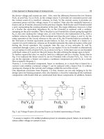
The motor circuit of the basal ganglia showing the direct and indirect pathways. Excitatory
pathways ar
e the filled ar
r
ows and inhibitor
y pathways are the dashed arrows.
FIGURE 1.1
Cortex
Striatum
Globus pallidus
externa
Globus pallidus
interna
Subthalamic
Nucleus
Thalamus
Brainstem
Spinal cord
Direct
Indirect
The description of hemidystonia secondary to basal
ganglia lesions provides an invaluable clue as to the
underlying anatomy of the dystonia. The basal ganglia
have dense fiber connections to the thalamus and the
cerebral cortex. The motor loops of the basal ganglia
include direct and indirect pathways (Figure 1.1). The
direct pathway flows from the striatum directly to the
globus pallidus internus (GPi) and inhibits it. The indi-
rect pathway flows from the striatum to the globus pal-
lidus externa to the subthalamic nucleus and has an
excitatory effect on the GPi. The primary outflow from
the basal ganglia to the thalamus is an inhibitory path-
Diagnosis, Classification, and Pathophysiology of Dystonia
7
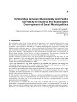
The motor circuits of the basal ganglia in Parkinson's disease with increased affected pathways.
Thin arrows show a decrease output and thick arrows show an increase in output.
FIGURE 1.2
Cortex
Striatum
Globus pallidus
externa
Globus pallidus
interna
Subthalamic
Nucleus
Thalamus
Brainstem
Spinal cord
Indirect
Direct
The motor circuits of the basal ganglia in dystonia. Thin arrows show a decrease in output and
thick ar
r
ows show an incr
ease in output. Ir
regular lines indicate irregular outputs.
FIGURE 1.3
Cortex
Striatum
Globus pallidus
externa
Globus pallidus
interna
Subthalamic
Nucleus
Thalamus
Brainstem
Spinal cord
Direct
Indirect
DYSTONIA
8
way originating from the GPi. Parkinson disease is
mediated primarily through an increase in the excitato-
ry effect of the indirect pathway, causing an increase in
G
Pi inhibition of the thalamus. In contrast, dystonia is
hypothesized to involve both direct and indirect path-
ways, causing abnormalities in discharge rates and pat-
tern of firing of the GPi neurons.
To summarize, this patient had a symptomatic
hemidystonia with an infarction in the contralateral
basal ganglia. It was through investigations of similar
patients that researchers had the first glimmer of under-
standing of the underlying pathophysiology and anato-
my of dystonia.
In patients with other forms of secondary dystonia,
a careful history and physical and neurologic examina-
tion are essential to investigate for the underlying
cause. An important secondary dystonia to consider is
Wilson's disease. To assess for this disease, a slit lamp
examination for Kayser–Fleischer rings, a serum ceru-
loplasmin, and a 24-hour urine test for copper are rec-
ommended. A patient with Wilson's disease may be
treated successfully by chelation therapy.
ADDITIONAL READING
Bressman S. Dystonia update. Clin Neuropharmacol 2000;23:
239–251.
Bressman SB, Sabatti C, Raymond D, de Leon D, Klein C, Kramer PL,
et al. The DYT1 phenotype and guidelines for diagnostic testing.
Neurology 2000;54:1746–1752.
Chan J, Brin MF
, Fahn S. Idiopathic cervical dystonia: clinical charac
-
teristics.
Mov Disord 1991;6:119–126.
Claypool DW. Epidemiology and outcome of cervical dystonia (spas-
modic torticollis) in Rochester, Minnesota.
Mov Disord
1995;10:608–614.
Eidelberg D, Moeller JR, Antonini A, Dhawan V, Spetsieris P, de Leon
D, et al. Functional brain networks in DYT1 dystonia.
Ann
Neurol
1998;44:303–312.
Epidemiologic Study of Dystonia in Europe (ESDE) Collaborative
Group. Sex-related influences on the frequency and age of onset
of primary dystonia.
Neurology 1999;53:1871–1873.
Fahn S, Bressman SB, Marsden CD. Classification of dystonia. Adv
Neurol 1998;78:1–10.
Fahn S, Marsden CD, Calne DB. Classification and investigation of
dystonia. In: Marsden CD, Fahn S, (eds.) Movement Disorders 2.
London: Butterworth and Co; 1987:332–358.
Greene P, Kang UJ, Fahn S. Spread of symptoms in idiopathic dysto-
nia. Mov Disord 1995;10:143–152.
Jankovic J, Fahn S. Dystonic disorders. In: Jankovic J, Tolosa E, (eds.)
Parkinson's Disease and Movement Disorders. 2nd ed. Baltimore:
Williams & Wilkins; 1993:337–374.
Kaji R. Basal ganglia as a sensory gating device for motor control. J
Med Invest 2001;48:142–146.
Kostic VS, Stojanovic-Svetel M, Kacar A. Symptomatic dystonias asso-
ciated with brain structural lesions: report of 16 cases. Can J
Neurol Sci
1996;23:53–56.
Kramer LP, de Leon D, Ozelius L, Risch NJ, Bressman SB, Brin MF, et al.
Dystonia gene in Ashkenazi Jewish population is located in chromo-
some 9q32-34.
Ann Neurol 1990;27:114–120.
Lowenstein DH, Aminoff MJ. The clinical course of spasmodic torti-
collis.
Neurology 1988;38:530–532.
Marsden CD, Obeso JA, Zarranz JJ, Lang AE. The anatomical basis of
symptomatic hemidystonia. Brain 1985;108:463–483.
Muller J, Wissel J, Masuhr F, Ebersbach G, Wenning GK, Poewe W.
Clinical characteristics of the geste antagoniste in cervical dysto-
nia.
J Neurol 2001;248:478–482.
Nutt JG, Muenter MD, Aronson A, Kurland LT, Melton LJ.
Epidemiology of focal and generalized dystonia in Rochester,
Minnesota.
Movement Dis 1988;3:188–194.
Nygaard TG, Trugman JM, de Yebenes JG, Fahn S. Dopa-responsive
dystonia: the spectrum of clinical manifestations in a large North
American family. Neurology 1990;40:66–69.
Ozelius L, Kramer PL, Moskowitz CB, Kwiatkowski DJ, Brin MF,
Bressman SB, et al. Human gene for torsion dystonia located on
chromosome 9q32-34.
Neuron 1989;2:1427–1434.
Suchowersky O, Calne DB. Non-dystonic causes of torticollis. Adv
Neurol 1988;50:501–508.
Vitek JL. Pathophysiology of dystonia: a neuronal model. Mov Disord
2002;17(suppl 3):S49–S62.
Vitek JL, Chockkan V, Zhang JY, Kaneoke Y, Evatt M, DeLong MR, et
al. Neuronal activity in the basal ganglia in patients with general-
ized dystonia and hemiballism. Ann Neurol 1999;46:22–35.
In progressive dystonia associated with cognitive or
psychiatric featur
es, testing for Wilson's disease is
necessary.
9
C
HAPTER 2
THE GENETICS OF DYSTONIA
M
. Tagliati, MD, M. Pourfar, MD, and Susan B. Bressman, MD
INTRODUCTION
Dystonia comprises a heterogeneous group of disor-
ders characterized by sustained and involuntary muscle
contractions generally resulting in an abnormal twist-
ing posture. These disorders have been divided into
primary (or idiopathic) and secondary (or sympto-
matic) subsets. Since Ozelius and colleagues first
described a mutation in the DYT1 gene in 1989, the
genetic underpinnings of many of the dystonias have
become evident. There are currently more than a
dozen genetic loci associated with the clinical expres-
sion of dystonia, and the number of other genes asso-
ciated with dystonic disorders continues to grow
steadily. Despite this growing body of information, the
majority of genes that cause primary dystonias have yet
to be identified. This overview will focus on the pres-
ent delineation of genetically associated primary dysto-
nias along with some of the “dystonia-plus” syndromes
in which other features may coexist with the dystonia.
T
able 2.1 outlines the major genetic loci associated
with dystonia. The discussion here will focus mainly
on the more common and better-described types,
namely DYT1, DYT6, DYT7, and DYT13 in the “pure”
dystonia group; DYT5, DYT11, and DYT12 in the “dys-
tonia-plus” group; and PKD and PKND in the paroxys-
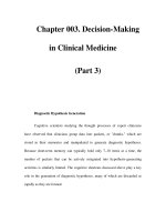
mal dystonia group. Figure 2.1 illustrates the chromo-
somal locations of the most common genetic defects
associated with dystonia. Several extensive reviews in
the “Additional Reading” section provide more cover-
age of the broad range of genetic dystonia.
Classification of Genetic Loci Associated with Dystonia
TABLE 2.1
Gene Locus Location Inheritance Phenotype Gene Product
DYT1 9q34 AD Early limb–onset PTD TorsinA
DYT2 Not mapped AR Early onset
DYT3
Xq13.1
XR Lubag dystonia/parkinsonism Multiple transcript system
DYT4 Not mapped AD Whispering dysphonia
DYT5 14q22.1 AD DRD/parkinsonism GCH1
DYT6 8p21-p22 AD “mixed” cranial/cervical/limb onset Not identified
DYT7 18p AD Adult cervical Not identified
DYT8
2q33-25
AD
PDC/PNKD
Myofibrillogenesis
r
egulator 1
DYT9 1p21 AD Episodic choreoathetosis/ataxia Not identified
with spasticity
DYT10
16
AD
PKC/PKD (EKD1 and 2)
Not identified
DYT11 7q21 AD Myoclonus dystonia
⑀-sarcoglycan
DYT12 19q AD Rapid-onset dystonia parkinsonism Na+/K+ ATPase ␣3
DYT13
1p36
AD
Cervical/cranial/brachial Not identified
DYT14 14q13 AD DRD Not identified
AD=Autosomal dominant; DRD=dopa-resistant dystonia; EKD=Endokinin D; PDC=Paroxysmal dystonic choreathetosis; PKC=Paroxysmal
kinesigenic choreoathetosis; PKD=paroxysmal kinesigenic dystonia/dyskinesia; PNKD=paroxysmal nonkinesigenic dystonia/dyskinesia;
PTD=Primar
y torsion dystonia; XR= X-linked r
ecessive.
In addition to the general subdivi-
sion into primary and secondary
forms, dystonia can be also classified
b
y age of onset (early vs adult) and
by the extent of muscle involvement
and disability (generalized, focal,
and mixed types). When viewed
from a genetic perspective, it can be
appreciated that the same mutation
can cause varying phenotypes in dif-
ferent individuals both in terms of
age of onset and localization. When
studied on pathologic examination,
primary dystonias are generally char-
acterized by a lack of consistent neu-
r
odegenerative or neurochemical
changes. They are also unified (with
the notable exception of dopa-
responsive dystonia [DRD]) by a lack
of consistently efficacious pharmaco-
logic treatment. However, recent
experience supports pallidal deep
brain stimulation (DBS) as a safe and
efficacious treatment, in particular
for patients with primary dystonia.
PRIMARY DYSTONIAS
Dystonic muscle contractions are the
only neurologic abnormality in pri-
mary dystonias, and evaluation does
not reveal an identifiable exogenous
cause or other inherited or degener-
ative disease. Primary dystonias can
be further classified (Table 2.2)
according to their prevalent age of
onset as:
1. Childhood and adolescent onset
(DYT1 and other genes to be identified), character-
ized by early limb onset and frequent spread to
other muscles.
2. Adult onset (DYT7 and other genes to be identified),
characterized by onset in cervical, cranial, or
brachial muscles and limited spread.
3. Mixed phenotype (DYT6, DYT13, and other genes
to be identified).
DYT1
The gene responsible for the most common of the
genetically identifiable dystonias was described by
Ozelius and colleagues in 1989 and named DYT1 (or
TOR1A). The defect leading to dystonia is a deletion of
an inframe GAG trinucleotide localized to chromosome
9q32-34. The DYT1 gene encodes torsinA, a protein
expressed throughout the central nervous system that
belongs to the family of AAA+ proteins (ATPases asso-
ciated with a variety of activities).
These proteins often serve as chaperones and are
involved in a variety of functions, including protein fold-
ing and degradation, cytoskeletal dynamics, membrane
traf
ficking and vesicle fusion, and response to stress. The
function of torsinA remains elusive and the mechanism(s)
by which mutant torsinA may compromise neuronal
function are unknown, but may include an altered
response to stress-induced changes in protein structure.
Neuronal degeneration has not been identified in the
brains of patients with DYT1 dystonia. Although brain-
stem neuronal inclusino have recently been described.
DYSTONIA
10
Chromosomal locations of genetic dystonias.
FIGURE 2.1
The same GAG deletion is responsible for dystonia in
families and patients from diverse ethnic groups (Table
2.3). In the Ashkenazi population, dystonia due to DYT1
h
as an estimated prevalence between 1/3000 and
1/9000 with a carrier frequency of 1/1000 to 1/3000.
This represents as much as a 10-fold increased preva-
lence as that found in the non-Ashkenazi population.
The increased frequency in Ashkenazi Jews is thought to
be the result of a founder mutation that was introduced
into the population approximately 350 years ago, origi-
nating in the area of Lithuania or Byelorussia. The pat-
tern of inheritance is autosomal dominant, with 30%
penetrance. Thus, first-degree relatives of affected indi-
viduals have a 15% risk and second-degree relatives
have about a 7%–8% risk of developing the disorder. In
this population, the TOR1A GAG deletion accounts for
an estimated 80%–90% of early limb–onset cases. Unlike
that observed in the Ashkenazi population, the DYT1
mutation is a less common cause of early limb–onset
primary dystonia in the non-Ashkenazi population, con-
stituting about 30%–50% of the cases. There is no
known founder effect and clearly other genes, yet to be
identified, are important in non-Jewish populations.
Clinical expression of the DYT1 GAG deletion is
generally similar across ethnic groups. While there is
marked clinical variability, the disorder characteristical-
ly first affects an arm or leg beginning in mid to late
childhood. Ultimately, more than 95% of patients expe-
rience involvement of the arm, while less than 15%
develop cranial or cervical involvement. Patients with
leg onset tend to be younger at onset and are more
likely to progress to generalized dystonia compared
with those with initial involvement of the arm.
Progressive spread of dystonia to involve multiple
muscle gr
oups as generalized or multifocal dystonia is
The Genetics of Dystonia
11
E
tiologic Classification of Dystonia
TABLE 2.2
Primary
Dystonia is the only neurologic sign. Evaluation does
not reveal an identifiable exogenous cause or other
inherited or degenerative disease.
Childhood and adolescent onset
• DYT1: Autosomal dominant with reduced
penetrance (~30%), early limb onset with
predominant family phenotype
• Other genes to be identified
Adult onset
• DYT7: Autosomal dominant, cervical onset in
adult life
• Other genes to be identified
Mixed phenotype
• DYT6, DYT 13: Autosomal dominant, early and
late onset with possible cranial, cervical, and
sometimes limb onset and variable spread
• Other genes to be identified
Secondary
Variety of lesions, mostly involving the basal ganglia
and/or dopamine synthesis.
Inherited nondegenerative (dystonia plus)
• Dopa-responsive dystonia: due to DYT5 and
other genetic defects
• Myoclonus dystonia: due to DYT11 and
possibly other genetic defects
• Rapid-onset dystonia parkinsonism: due to
DYT12
Inherited degenerative
• Autosomal dominant, autosomal recessive,
X-linked (DYT3), mitochondrial
Degenerative disorders of unknown etiology
•
Parkinson disease
• Progressive supranuclear palsy
• Corticobasal ganglionic degeneration
Acquired
• Drugs (dopamine-receptor blockers), other
toxins
• Head trauma
• Stroke, hypoxia
• Encephalitis, infectious and postinfectious
•
T
umors
•
Peripheral injuries
Other movement disorders with dystonic
phenomenology
•
T
ics, par
oxysmal dyskinesias (DYT8, DYT9,
DYT10)
Psychogenic Dystonia
DYT1 Features in Ashkenazi and
Non-Jewish Populations
TABLE 2.3
Ashkenazi Non-Jewish
Mode of inheritance
100% AD
85% AD
Penetrance 30% 40% (in AD)
9q haplotype
Y
es
No
GAG TOR1A deletion
90%
40%–65%
% new mutation Rare 14%
Incidence
1/6000–1/2000 1/160,000
Age of onset Uncommon 10%–15%
>40 years
AD=autosomal dominant.
observed in about 65% of patients; about 25% remain
focal and 10% are segmental.
C
ASE 1
KW had normal psychomotor development until age 7,
w
hen she initially showed turning in of her feet and pos-
turing of the legs with prolonged walking. She subse-
quently developed difficulty writing and marked loss of
t
runk control, with difficulty maintaining erect sitting
position, inability to transfer from sitting to standing
position, and inability to control the left arm due to con-
stant shoulder movements. Fixed equinovarus deformity
of the left foot and varus posture of the right foot
ensued over a period of 2 or 3 years. She demonstrated
little response to a variety of medications, including lev-
odopa, anticholinergics, baclofen, and benzodiazepines.
Neur
ologic examination revealed cervical dystonia with
head turning to the left, bilateral arm dystonia at rest
with internal rotation, spasmodic back arching of the
trunk, and dystonic flexion of the right leg at the knee
and of the left foot. Brain magnetic resonance imaging
(MRI) was normal. Genetic testing revealed that she was
a carrier of the DYT1 mutation.
With the identification of the DYT1 gene, it is now
possible to diagnose one of the most frequent causes of
generalized dystonia. The DYT1 GAG deletion accounts
for a significant proportion of early-onset (<26 years of
age) primary dystonia. As all cases of DYT1 dystonia are
due to the same GAG deletion, screening is relatively
easy and commercially available. The test should be con-
sidered for all patients with primary dystonia with onset
by age 26 and for individuals with later-onset dystonia
who have an early-onset blood relative. DYT1 testing
(when positive) will obviate other expensive diagnostic
tests, including MRI, unless ther
e ar
e other findings on
exam to suggest an independent central nervous system
(CNS) or spinal cord lesion. We recommend preliminary
genetic counseling when DYT1 diagnostic and car
rier
testing are employed.
After 6 years of disease, KW was wheelchair bound.
After the failur
e of all available medications for dysto-
nia, she underwent bilateral implant of pallidal DBS elec-
trodes. Progressive and sustained improvement of dysto-
nia was noted over the following months. The patient
was able to walk and run 18 months after DBS surgery.
She was practically dystonia free when stimulated.
Mor
eover, she was able to completely discontinue her
medications. We as well as other researchers have
reported that in select cases of intractable primary dysto-
nia, including DYT1-positive cases, DBS may be a safe
and effective alternative over current best medical man-
agement.
D
YT6
This type of primary dystonia is referred to as a mixed
type because of the varying body distribution and age
at onset of the dystonia within affected families.
Described in 2 Mennonite families, it has been mapped
t
o chromosome 8 (8p21-8q22). It is autosomal domi-
nant with decreased penetrance, and appears to be the
result of a founder mutation. About 1/2 of affected
family members had onset of symptoms in childhood,
with the rest exhibiting symptoms during the third and
fourth decades. There was a wide range of body
regions first affected (arm, cranial muscles, neck, and
leg), and almost all had some degree of spread—or
progression of dystonia—to other body regions, but
again this varied widely. Most had cervical and cranial
involvement, and for the majority, the greatest disabil-
ity stemmed from dystonia of the neck and cranial
muscles, including speech involvement.
DYT7
Leube and colleagues first described this primary focal
dystonia locus in a large German family in 1996. The
gene was localized to the short arm of chromosome 18.
Focal in nature, it manifests primarily as cervical dysto-
nia (familial torticollis). The age of onset varies from
the second to seventh decade, with an average age of
43 years.
DYT13
This relatively indolent, typically segmental dystonia
has been identified in 1 Italian family and has been
mapped to the short arm of chromosome 1. It is an
autosomal-dominant disorder with r
educed penetrance
and begins between ages 5 and 40 years. This dystonia
is often limited to the cranial, neck, and/or upper limbs
muscles, but can occasionally generalize.
SECONDARY DYSTONIAS
This group is comprised of disorders in which dystonia
is often accompanied by other neurologic manifesta-
tions such as parkinsonism and myoclonus. They can
be inherited, acquired, psychogenic, or of unknown
etiology (Table 2.2). The inherited forms that are rele-
vant for this chapter can be further classified as:
1. Inherited nondegenerative or “dystonia plus,”
including DRD due to DYT5 and other genetic
defects; myoclonus dystonia due to DYT11 and pos-
sibly other genetic defects; and rapid-onset dystonia
parkinsonism (RPD) due to DYT12.
2. Inherited degenerative, which can have an autoso-
mal-dominant, autosomal-recessive, X-linked, or
mitochondrial patter
n of inheritance.
DYSTONIA
12
The Genetics of Dystonia
13
3. Paroxysmal dyskinesias (DYT8, DYT9, DYT10),
which are frequently categorized separately from
dystonia but which have been assigned DYT loci.
DRD (DYT5)
DRD is a form of dystonia whose hallmark feature is a
remarkable response to low dosages of levodopa. The
most common cause of DRD is mutation in the gene
encoding guanosine triphosphate cyclohydrolase 1
(GCH1) on chromosome 14 (see Figure 2.2). DRD due
to GCH1 mutations is autosomal dominant (mutations
are heterozygous), and penetrance appears to be influ-
enced by gender, being higher in females. A less com-
mon autosomal-recessive variant of DRD involves the
tyr
osine hydroxylase gene on chromosome 11.
T
ypically, DRD due to GCH1 mutations (DYT5)
begins in early childhood and presents with limb or
truncal dystonia, a dystonic-spastic–appearing gait,
and mild parkinsonism (bradykinesia and postural
instability). Onset in infancy mimicking cerebral palsy
may also occur. Hyperreflexia and diurnal fluctuation
of symptoms, with progressive deterioration during
the day, are common. Affected individuals are all char-
acterized by a dramatic and sustained response to lev-
odopa, and an excellent response to cholinergic med-
ications has also been described. The diagnosis of
DRD depends on both the clinical findings and a dra-
matic response to low-dose levodopa therapy. Total
daily dosages of as little as 50 to 200 mg of levodopa
usually result in complete or near-complete reversal of
s
ymptoms and signs, which is maintained without
fluctuations.
CASE 2
AS was born by normal, spontaneous, vaginal delivery,
w
ith the first four months of gestation complicated by
maternal vaginal bleeding. The patient had normal cog-
nitive development. When she began walking at the age
of 9 1/2 months, her parents noticed that she had a clum-
sy gait, and that her toes turned inward. By age 10, she
had had bilateral achilles tendon releases because of
dystonic posturing of her feet. At age 11, she was diag-
nosed with DRD after responding well to a trial with lev-
odopa. She continued to do well thr
oughout puberty
and was able to compete in running races. At age 13, she
first experienced subtle extra movements after taking
the medications. Her Sinemet (carbidopa-levodopa) dose
was reduced from 350 mg/day to 200 mg/day with reso-
lution of her abnormal movements. By age 20, she was
taking only 1 Sinemet 25/100 per day. At age 25, she con-
tinued to do very well. On examination, she had minimal
clumsiness when performing rapid successive move-
ments of the left foot. She was maintained on 1 Sinemet
25/100 per day and continued to complain of left toe
curling and cramping under physical exertion.
Although both Parkinson’s disease
and DRD respond symptomatically to
levodopa, the 2 differ both patho-
physiologically and in their response
to Sinemet. In contrast to Parkinson’s
disease, DRD is a nondegenerative
condition and DRD patients do not
usually experience clinically signifi-
cant fluctuations, dyskinesias, or
decreasing dosage efficacy after long-
term treatment with levodopa.
Familial Myoclonus Dystonia
(DYT11)
Although very rapid dystonic jerks can
be part of the clinical manifestations
of DYT1 and other primary dystonias,
myoclonus dystonia is a distinct
genetic disorder in which dystonia,
usually mild and not always present,
is associated with marked myoclonus.
There are no other neurologic signs.
Myoclonus dystonia is autosomal
dominant with reduced penetrance,
Diagram of GCH1 defect pathway
.
FIGURE 2.2