EMERGENCY NEURORADIOLOGY - PART 10 doc
Bạn đang xem bản rút gọn của tài liệu. Xem và tải ngay bản đầy đủ của tài liệu tại đây (1.43 MB, 40 trang )
lae. In such clinically suspected but MRI-nega-
tive cases, myelography and spinal angiography
may be indicated, and when positive will defin-
itively reveal the site of the dural fistula, the
feeding arteries and the dilated draining veins.
In any case, angiography is a prerequisite for
therapeutic dural fistula embolization, the
treatment of choice in these patents.
Cavernous angiomas are usually indolent
vascular malformations that are nevertheless
prone to haemorrhage and intrinsic thrombo-
sis. T1- and T2-weighted MRI typically shows
a central hyperintense core with a peripheral
margin or margins of hyper- and hypointensity
due to the presence of mixed subacute and
chronic haemoglobin metabolites (Fig. 5.46).
Some cases demonstrate central enhancement
after IV contrast medium administration, rep-
resenting the residual patent vascular compo-
nent of the angioma. Cavernous angiomas can
present acutely with signs and symptoms relat-
ed to intramedullary haemorrhage (Fig. 5.47).
Acute spinal cord syndromes can also be
caused by viral or granulomatous infections. In
364 V. SPINAL EMERGENCIES
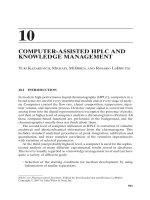
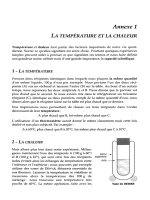
Fig. 5.47 - Acute haemorrhage within intramedullary cavernous
angioma. The spinal T1-weighted spinal MRI images demon-
strate an extensive acute-subacute (deoxyhaemoglobin and
methaemoglobin) thoracic intramedullary haemorrhage associ-
ated with an intramedullary cavernous angioma showing intrin-
sic hypointensity on T2-weighted acquisitions consistent chron-
ic peripheral microhaemorrhages (haemosiderin). The MRI of
the brain showed several cavernous angiomas demonstrating the
multicentric potential of this pathologic process. [a) sagittal T2-
weighted spinal MRI b) sagittal T1-weighted weighted spinal
MRI; c) sagittal T2*-weighted spinal MRI; d) axial T1-weighted
spinal MRI; e) axial T2*-weighted cranial MRI].
a
b
Fig. 5.46 - Chronic haemorrhage within thoracic intramedullary
cavernous angioma. T2*-weighted sagittal MRI reveals hy-
pointensity within the upper thoracic spinal cord as a conse-
quence of deposition of haemosiderin associated with thrombo-
sis-haemorrhage within an intramedullary cavernous angioma.
cases of infectious myelitis, MRI demonstrates
multisegmental non-specific intramedullary
hyperintensity on T2-weighted sequences, with
a variable enhancement after contrast medium
administration.
Spinal cord abscess formation is rare and is
usually caused by the direct extension of in-
fections from adjacent perispinal tissues or
from penetrating trauma. T2-weighted MRI
demonstrates an intramedullary mass that is
hyperintense on T2-weighted sequences and
reveals rim enhancement after IV gadolinium
administration. Distinguishing this pattern
from other spinal cord lesions such as neopla-
sia is not always possible on the basis of the
images alone.
Acute transverse myelitis is an acute in-
flammatory process with a poor prognosis.
The aetiology is unknown, however it is prob-
ably autoimmune in nature. Acute transverse
myelitis can be associated with various con-
ditions such as multiple sclerosis, paraneo-
plastic syndromes, prior vaccinations, vas-
culitis or known autoimmune disorders. Clin-
ically there is an acute onset of a profound
spinal cord neurological deficit in the absence
of other findings. It is for this reason that
transverse myelitis is always a diagnosis of ex-
clusion. On MRI, acute transverse myelitis
demonstrates areas of hyperintensity on T2-
weighted imaging associated with spinal cord
swelling and irregular contrast enhancement
following gadolinium administration due to
an associated breakdown in the blood-cord
barrier. In the chronic phase, the spinal cord
5.4 EMERGENCY IMAGING OF THE SPINE IN THE NON-TRAUMA PATIENT 365
Fig. 5.47 (cont.).
e
c
d
can appear atrophic, and areas of high signal
on T2-weighted images may persist due to
gliosis.
Acute disseminated encephalomyelitis is a
monophasic autoimmune disease that follows
within days or weeks of an antiviral vaccination
or viral infection. As the name indicates, it in-
volves the brain but also concomitantly affects
the spinal cord. Pathologically the lesions are
similar to those of MS. The prognosis is typi-
cally good and the majority of patients respond
rapidly to steroid treatment. MRI shows hyper-
intense areas on T2-weighted sequences within
the parenchyma of the brain and spinal cord
that enhance after IV gadolinium; the spinal
cord may be swollen.
The development of radiation myelopathy
in part depends on the dose of radiation and
the time period over which it was adminis-
tered. In the acute milder forms, typically pre-
senting approximately three months after ra-
diation is applied to the spinal cord, the pa-
tient experiences sensations similar to electric
shocks in the lower limbs; MRI may show no
abnormality. However in severe cases the pa-
tient reveals a severe, rapidly progressive
myelopathy; in such cases, the spinal cord is
swollen, hyperintense on T2-weighted images
and enhances following IV contrast injection.
Evolution of spinal cord atrophy will occur
over time (Fig. 5.48).
366 V. SPINAL EMERGENCIES
c
Fig. 5.48 - Radiation induced thoracic myelitis/spondylitis
three years following radiotherapy. T2-weighted MRI with fat
suppression shows hyperintense MRI signal within the thoracic
spinal cord and within the bone marrow of several contiguous
vertebral bodies, both of which are caused in this case by the
preceding radiation therapy. Axial T2*-weighted images
demonstrate again the intramedullary location of the patholog-
ic process. No contrast enhancement of the intramedullary
process can be identified. Note the postsurgical alterations. [a)
sagittal T2-weighted MRI; b) sagittal T1-weighted MRI follow-
ing IV Gd; c) axial T2*-weighted MRI].
a
b
REFERENCES
1. Baleriaux DL: Spinal cord tumours. Eur Radiol 9 (7):1252-
1258, 1999.
2. Chen CJ, Chen CM, Lin TK: Enhanced cervical MRI in
identifying intracranial dural arteriovenous fistulae with
spinal perimedullary venous drainage. Neuroradiology 40:
393-407, 1998.
3. Fortuna A, Ferrante L, Acqui M et al: Spinal cord ischemia
diagnosed by MRI. J Neuroradiol 22:115-122, 1995.
4. Karampekios S: Inflammatory, vascular and demyelinating
diseases of the spine and spinal cord. Eur Radiol (S1)10:36,
2000.
5. Liou RJ, Chen CY, Chou TY et al: Hypoxic - ischaemic
injury of the spinal cord in systemic shock: MRI. Neurora-
diology 38:S 181-183, 1996
6. Lyclama à Nijeholt GJ, Uitdehaag BMJ et al: Spinal cord
magnetic resonance imaging in suspected multiple sclero-
sis. Eur Radiol 10:368-376, 2000.
7. Obenberg J, Seidi Z, Plas J: Osteoblastoma in lumbar ver-
tebral body. Neuroradiology 41:279-282, 1999.
8. Rimmelin A, Clouet PL, Salatino S et al: Imaging of thora-
cic and lumbar spinal extradural arachnoid cysts: report of
two cases. Neuroradiology 39:203-206, 1997.
9. Rocca MA, Mastronardo G, Horsfield MA et al: Comparison
of three MR sequences for detection of cervical cord lesions in
patients with multiple sclerosis. AJNR 20:1710-1716, 1999.
10. Silbergleit R Brunberg JA, Patel SC et al: Imaging of spinal
intradural arachnoid cysts: MRI, myelography and CT.
Neuroradiology 40:664-668, 1998.
11. Suzuki K, Meguro K, Wada M et al: Anterior spinal ar-
tery syndrome associated with severe stenosis of the ver-
tebral artery. AJNR 19:1353-1355, 1998.
12. Wilmink JT: MR imaging of the spine: trauma and degene-
rative disease. Eur Radiol 9 (7):1259-1266, 1999.
13. Yamada K, Shrier DA, Tanaka H et al: A case of subacute
combined degeneration: MRI finding. Neuroradiology 40:
398-400, 1998.
5.4 EMERGENCY IMAGING OF THE SPINE IN THE NON-TRAUMA PATIENT 367
VI
NEUROPAEDIATRIC EMERGENCIES
371
INTRODUCTION
This chapter covers the most common emer-
gency situations encountered in neuropaedi-
atrics, including cerebrovascular disease, head
injuries, infections of the central nervous sys-
tem (CNS) and intracranial hypertension.
CEREBROVASCULAR DISEASE
Cerebrovascular disease (15, 32) is rare in in-
fants and newborns and when encountered does
not have the same aetiological factors as in adults:
the most common causes in the young age group
are congenital vascular abnormalities and those
secondary to systemic illnesses. Various areas of
the brain show significant differences in their sus-
ceptibility to cerebral vasculopathy. In addition,
there are also important physiological differences
in the blood vessels of different areas of the brain.
The pathological conditions of cerebrovascular
disease are haemorrhage and ischaemia.
H
AEMORRHAGE
NEWBORNS AT TERM AND YOUNG INFANTS
In newborns at term, a large number of possi-
ble pathological events may result in intracranial
haemorrhage: a) trauma: subdural haematoma,
epidural haematoma. subarachnoid haemor-
rhage, intracerebral haemorrhage, intracerebellar
haemorrhage, b) clotting disorders: clotting de-
fects, thrombocytopenia, c) vascular disorders:
aneurysms, arteriovenous malformations, d)
metabolic disorders, and e) idiopathic intra-
parenchymal haemorrhage.
The clinical signs of an intracranial haemor-
rhage lesion in a newborn are often modest and
non-specific: apathy or irritability/hyperex-
citability without focal neurological signs,
seizure, tremors, breathing disorders. Fre-
quently acidosis, hypoglycaemia and hypoten-
sion are associated with such haemorrhages.
a) Labour trauma is the most frequent cause
of bleeding in newborns.
Subdural haematomas and subarachnoid
haemorrhage are the most common types of
haemorrhagic lesion. The most frequent site of
subdural bleeding is over the cerebral convexi-
ty and within the temporal fossa, however
haemorrhages can also be encountered adja-
cent to the falx cerebri, tentorium cerebelli and
in the posterior cranial fossa.
Intraparenchymal haemorrhages are less fre-
quent and can be associated with subarachnoid
and subdural haemorrhage, should the bleed-
ing extend into the ventricles. The prognosis of
6
NEUROPAEDIATRIC EMERGENCIES
N. Zamponi, B. Rossi, G. Polonara, U. Salvolini
small lesions is good, however serious sequelae
are typically observed following larger haemor-
rhages.
Diagnostic imaging should first include CT,
which demonstrates the presence, site and ex-
tent of the cerebral bleed at an early stage; on
the other hand, haemorrhages associated with
cerebral infarcts will only become evident some
days after the ischaemic event.
Ultrasound may not show epidural or sub-
dural haemorrhages localized to the cranial
convexity or in the posterior fossa, whereas
larger haemorrhages are clearly visible on ultra-
sound images as hyperechoic areas, and later
hypo- anechoic regions.
Both ultrasound and CT are capable of doc-
umenting and sequentially monitoring the most
important sequelae: porencephalic cysts and
hydrocephalus.
Porencephalic cysts usually form off of the
bodies of the lateral ventricles. They typically
develop from a haemorrhage that ruptures into
the lateral ventricle or the subarachnoid space.
Associated posthaemorrhagic hydrocephalus
develops in 10-15% of patients with intraven-
tricular haemorrhage. The hydrocephalus halts
or improves in most cases; more rarely it pro-
gresses and can require surgical ventricular-
peritoneal shunt placement.
b) Various clotting and platelet disorders can
result in intracranial haemorrhage in newborns.
The most frequent causes of thrombocytopenia
are the use of medicines during pregnancy, ma-
ternal infections, immunological disorders and
disseminated intravascular coagulation.
c) Vascular malformations and intracranial
aneurysms may present with intracranial haem-
orrhages in newborns in rare occasions.
Ultrasound may prove useful in diagnosis,
especially in infants with aneurysmal dilatation
of the vein of Galen. This is a rare congenital
disorder wherein abnormal arteriovenous for-
mations drain into the deep, dilated vessels of
the galenic system. These direct connections
with the vein of Galen can be by large fistulae
or by multiple smaller arteriovenous connec-
tions. The pathogenesis would seem to involve
intrauterine vascular thrombosis or absence of
formation of the superior sagittal venous sinus.
In 90% of cases, signs and symptoms of vas-
culopathy arise in early infancy: intracranial
haemorrhage (intraparenchymal or subarach-
noid) and rapidly progressive hydrocephalus
are the most frequent presentations. In new-
borns these complications are frequently asso-
ciated with cardiac insufficiency, the final
pathophysiological result of a preexistent con-
genital haemodynamic anomaly (e.g., increase
in blood flow across an arteriovenous fistula,
increase in blood return to the right atrium,
right-to-left blood flow through cardiac de-
fects). In addition, the large blood flow through
the fistula can create a secondary state of cere-
bral ischaemia.
In newborns, the aneurysmal dilatation of
the vein of Galen can be simply diagnosed us-
ing ultrasound. On colour Doppler images,
turbulent flow can usually be seen.
On CT without IV contrast medium, the
vein of Galen appears as a rounded mass in the
region of the tentorial incisura and straight ve-
nous sinus; aqueduct compression may cause
obstructive hydrocephalus. After IV contrast
medium administration, intense enhancement
is typically seen within the aneurysmally dilated
vessel which is smooth and well defined. In the
presence of thrombosis of this structure, vari-
able degrees of non-enhancement will be ob-
served.
On MRI the vascular malformation appears
hypointense on both T1- and T2-weighted se-
quences due to the rapid blood flow within the
abnormal vessels. The arteries that supply the
malformation can be reasonably well shown
with MRA. Conventional selective angiography
will still better define the arterial feeding ves-
sels and the draining venous structures and
may assist in presurgical planning (27, 32, 37).
P
REMATURE NEWBORNS
Subependymal and intraventricular haemor-
rhages are more frequently encountered in pre-
mature newborns than in those born at term (3,
5, 11). Babies with a gestational age of less than
35 weeks or a birth weight of less than 1.5 kg
have a higher risk of such haemorrhages, which
372 VI. NEUROPAEDIATRIC EMERGENCIES
commonly present during the second or third
day of life. The haemorrhage originates from
the germinal matrix that surrounds the lateral
cerebral ventricles. Small haemorrhages remain
confined to the subependymal regions, howev-
er, when the bleeding is larger it can extensive-
ly involve the cerebral parenchyma or rupture
into the ventricular system. Certain factors
make haemorrhage in this area more likely. The
vessels of the germinal matrix are fragile and
contain little connective tissue. This germinal
matrix begins to involute at approximately the
35
th
week of gestation. Until that time it has a
high arterial perfusion with consonantly elevat-
ed venous and capillary pressure.
Two clinical syndromes have been described
in association with subependymal and intraven-
tricular haemorrhage. The catastrophic syn-
drome has an acute onset and a rapid evolution
towards coma; the mortality rate is high. The
salt losing syndrome is a disorder of conscious-
ness, accompanied with a reduction in sponta-
neous movements, hypotonia and oculomotor
abnormalities; these signs evolve slowly and are
often followed by a period of stabilization fol-
lowed by a second phase of deterioration. The
mortality rate is lower for this syndrome than
for the catastrophic syndrome.
From the standpoint of medical imaging,
germinal matrix haemorrhages can be broken
down into 4 stages: stage I is characterized by a
small germinal matrix haemorrhages together
with a small intraventricular haemorrhage;
stage II is characterized by germinal matrix
haemorrhage accompanied by a large intraven-
tricular haemorrhage; stage III is characterized
by a subependymal haemorrhage, intraventric-
ular haemorrhage, and hydrocephalus; and,
stage IV indicates the spread of the parenchy-
mal haemorrhage into one or both cerebral
hemispheres.
The use of ultrasound, which can be per-
formed safely at the bedside, has lead to an in-
crease in the identification and characterization
of neonatal subependymal and intraventricular
haemorrhages. On ultrasound, stage I germinal
matrix subependymal haemorrhage appears as
a hyperechoic mass lesion, which is either uni-
or bilateral and is primarily located in the head
of the caudate nucleus. Generally speaking, in
order to be visualized, it must measure 4-5 mm
in diameter. A Stage II haemorrhage appears as
hyperechoic material within the lateral ventri-
cle(s). A stage III haemorrhage is represented
by a dilatation of the ventricular system and the
presence of intraventricular hyperechoic blood.
The intraparenchymal component of a stage IV
haemorrhage appears on ultrasound as an in-
tensely hyperechoic lesion located in the deep
white matter of the centrum semiovale.
Subsequent ultrasound scans will show pro-
gressive stages of resolution of the subependy-
mal-intraventricular haemorrhage. An exten-
sive haemorrhage can evolve over 2-3 months
towards the formation of porencephalic cysts
or the development of cystic encephalomalacia.
On CT, acute germinal matrix haemorrhages
appear as hyperdense foci, usually adjacent to
the lateral ventricle near the head of the cau-
date nucleus. MRI is also fairly sensitive and
specific in demonstrating acute germinal ma-
trix haemorrhage.
In premature neonates with the hypoxic-is-
chaemic syndrome white matter alterations are
also frequently detected: periventricular leuko-
malacia appears on ultrasound as widespread,
poorly defined hyperechoic periventricular re-
gions. These are especially prominent in the
ventricular trigone regions and adjacent to the
foramina of Monroe. The hyperecho findings
are due to oedema and petechial haemorrhage.
The abnormality is generally bilateral, but is of-
ten asymmetric. After 2-3 weeks, small cysts
form within the hyperechoic region that coa-
lesce to form a multicystic lesion, before col-
lapsing, fusing and being replaced by glial
scars. In this late phase of glial scarring the ul-
trasound findings are usually unremarkable.
During the acute phase of ischemia, CT can
be normal or can show a minor attenuation in the
parenchyma of the periventricular regions; dur-
ing the subacute phase it is only possible to iden-
tify medium-sized cysts, whereas chronic glial
scarring is not usually visible on CT (Fig. 6.1).
MRI is rarely used in the acute phase, how-
ever it is the best technique for highlighting
chronic periventricular leukomalacia. On T2-
weighted scans the residual glial scars localized
NEUROPAEDIATRIC EMERGENCIES 373
to the periventricular areas appear hyperin-
tense. These areas generally border the ventri-
cles and typically spread into the adjacent white
matter in a flame-shaped configuration. A thin-
ning of the posterior body and splenium of the
corpus callosum are seen in the chronic phase
as a result of degeneration of the transcallosal
fibres, ventricular dilatation and atrophy of the
hemispheric white matter (Fig. 6.2).
I
NFANTS
Vascular malformations are the most com-
mon cause of haemorrhage in infants (15, 32)
and can be broken down into four main
types: arteriovenous malformations, venous
angiomas, capillary telangiectasias and cav-
ernous angiomas. The most common clinical
manifestations are headache and seizures
rather than haemorrhage; however, if the lat-
ter do occur, they may be subarachnoid, in-
traparenchymal or combined.
Arteriovenous malformations (AVM’s) con-
sist of an aggregate of abnormal vessels with
thin walls (i.e., nidus) in which there is direct
continuity between dilated arteries and veins
without the interposition of capillaries. Ap-
proximately 90% are superficial and are locat-
ed within the cerebral hemispheres. AVM’s are
responsible for up to 40% of spontaneous in-
tracranial haemorrhages in infants. The mortal-
ity rate associated with the rupture of an AVM
is approximately 10%.
On unenhanced CT, a typical AVM appears
as a heterogeneous area with slightly increased
density compared to the normal surrounding
parenchyma. After IV contrast medium admin-
istration, intense enhancement of the malfor-
mation and its afferent and efferent vessels is
observed.
On MRI the fast blood flow within AVM’s
creates flow voids on spin echo sequences. The
nidus appears as a tangle of tubular shaped
black vessels. However, in order to obtain an
accurate anatomical map of the vascular mal-
formation, an angiographic examination is re-
quired. Typically a tangle of small, irregular
blood vessels supplied by dilated and twisted
arteries and drained by dilated veins that fill
rapidly.
In the case of haemorrhage, unenhanced CT
details the haemorrhagic spread into the sub-
374 VI. NEUROPAEDIATRIC EMERGENCIES
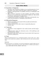
Fig. 6.1 - Hypoxic-ischaemic injury in newborn. a, b) Unenhan-
ced CT shows widespread hypodensity of the subcortical and
periventricular white matter and enlargement of the cerebral
ventricular system.
a
b
arachnoid space, cerebral parenchyma and
cerebral ventricles. In severe cases haemor-
rhage can obscure the underlying vascular mal-
formation. In the acute phase the haematoma
appears hyperdense and relatively homoge-
neous (Fig. 6.3); in the chronic phase, en-
cephalomalacia, rarely accompanied by calcifi-
cations, may result.
Cavernous vascular malformations consist of
a tangle of dilated vessels that do not possess
the characteristics of normal arteries or of
veins. With the exception that thrombi can be
present, the draining veins and arteries usually
have a normal calibre. The malformation may
contain small intrinsic areas of neural tissue.
Most of these lesions are located in the cerebral
parenchyma, and although they are usually iso-
lated they can also be multiple and have a fa-
milial pattern of expression. The clinical pres-
entation is typically seizures, but more rarely it
can be cerebral haemorrhage. In fact, subclini-
cal haemorrhages often occur. The diagnosis is
currently based on MRI due to the characteris-
tic imaging findings, including a mixed signal
core surrounded by a hypodense haemosiderin
ring on T2-weighted sequences.
Venous malformations are often incidentally
detected on MR or CT scans. The risk of
bleeds is generally low. MRI, which is more
sensitive than CT, shows a branching network
of small draining veins that unite to form a sin-
gle, large terminal vein. In the venous phase,
conventional angiography shows a collection
of abnormal veins (i.e., “Medusa head”) that
drain into a single large collecting vein before
emptying into a superficial cortical vein or dur-
al venous sinus.
Aneurysms are rare in children under the age
of ten years; males are more frequently affected
than are females. The clinical presentation is
typically a subarachnoid haemorrhage, howev-
er, some patients present with seizures.
Aneurysms in children under 2 years usually
originate from the anterior cerebral or the in-
ternal carotid arteries. Such aneurysms are usu-
ally larger than 1 cm in diameter.
CT at presentation shows an acute sub-
arachnoid haemorrhage. If the aneurysm is suf-
ficiently large it will demonstrate intense en-
NEUROPAEDIATRIC EMERGENCIES 375
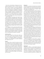
Fig.6.2 - Periventricular leukomalacia. A, b) Axial FLAIR MRI
shows an increase in the subependymal and periventricular whi-
te matter MR signal with sickle shape of the cerebral ventricles.
a
b
hancement with a smooth, round or oval con-
figuration after IV contrast medium adminis-
tration. Internal thrombosis may be observed.
While MR and angio-MR better define the
aneurysm, conventional angiography defini-
tively visualizes the lumen and neck of the
aneurysm, and its relationship to the vessel of
origin.
ISCHAEMIA
Cerebrovascular occlusions may occur in ar-
teries, veins or capillaries, as a single acute
event, a recurrence or a progressive phenome-
non. They can be associated with a number of
pathological conditions including inflamma-
tion, infection, cardiac disease, neoplasia, trau-
ma, primary arterial dysplasia, vascular malfor-
mations and metabolic disease (26, 32).
The clinical symptoms vary according to the
age of the infant and the vascular territory in-
volved. The most common sign of internal
carotid occlusion is acute hemiplegia. A vascu-
lar occlusion in the vertebrobasilar circulation
can result in pyramidal and cerebellar signs,
hemiparesis, paralysis of the cranial nerves, lat-
eral conjugate deviation of the eyes, dizziness,
nausea and vomiting.
Acute phase CT is typically normal. After
24-48 hours the infarction appears as a hypo-
dense area with poorly defined margins and is
accompanied by varying mass effect. After the
second to third week, enhancement is usually
observed after IV contrast medium administra-
tion. In the later stages encephalomalacia,
porencephalic cyst formation and focal atrophy
are typical terminal sequelae.
MRI is more sensitive in detecting acute/sub-
acute postinfarction oedema, which appears
hyperintense relative to the normal cerebral
parenchyma on T2-weighted sequences. MRA
(Fig. 6.4) may reveal vascular thrombosis of the
cranial circulation (Fig. 6.5).
Despite the fact that it is the most sensitive
technique for examining the cranial vascular sys-
tem, in infants angiography is reserved for se-
lected cases where it will clearly influence future
therapy or when the diagnosis is in doubt (12).
376 VI. NEUROPAEDIATRIC EMERGENCIES
Fig.6.3 - Intraparenchymal haematoma caused by AVM hae-
morrhage. a, b) Unenhanced CT demonstrates inhomogeneous
right frontoparietal intraparenchymal haemorrhage associated
with compression of the right lateral ventricle and contralateral
shift of the midline structures.
a
b
THE MOYA-MOYA PHENOMENON
The moya-moya phenomenon (6, 32) is a
disorder that characteristically affects children
and adolescents. Approximately 70% of cases
are diagnosed within the first 20 years of life.
The disorder consists of an idiopathic pro-
gressive stenosis of the supraclinoid internal
carotid arteries. A prominent collateral circu-
lation is formed by small branches of the
rubrothalamic arteries and the lenticulostriate
arteries, resulting in an MRA and convention-
al angiographic appearance similar to that of a
puff or cloud of smoke (moya-moya means
foggy in Japanese).
The aetiology and pathogenesis are unknown,
although there are many pathological condi-
tions that are sometimes associated with moya-
moya type angiographic patterns (e.g., neurofi-
bromatosis, tuberculosis, Down’s syndrome,
tuberous sclerosis, prior radiation therapy,
etc.). Nevertheless, in certain cases the disease
presents as an isolated phenomenon. In infants,
moya-moya phenomenon clinically reveals
transient-relapsing ischaemic episodes, with the
appearance of neurological deficits and convul-
sions. In adolescents, headaches and cerebral
haemorrhages are the most common clinical
presentations.
Unenhanced CT typically reveals the pres-
ence of multiple cerebral infarcts in different
stages of evolution. In certain cases, enhanced
CT may demonstrate absence of visualization
of the proximal internal carotid vessels and the
vessels of the circle of Willis. These findings are
more clearly visible on MRI and MRA. Suffi-
ciently large collateral vessels are seen at the
base of the brain in the region of the basal gan-
glia (Fig. 6.6). In time regional cerebral en-
cephalomalacia and intracranial calcifications
may develop.
The definitive diagnosis is angiographic: the
supraclinoid sections of the internal carotid ar-
teries are stenotic or completely occluded as
may be the proximal segments of the anterior
and middle cerebral arteries. Distal to the oc-
clusion the collateral vessels appear as a tangle
of dilated, twisted vessels. The marked, dense
blood flow within these small collateral vessels
NEUROPAEDIATRIC EMERGENCIES 377
Fig.6.4 - Ischaemia of the basal ganglia. a) Unenhanced CT
shows hypodensity in the head of the caudate nucleus and the
frontal aspect of the putamen on the right side, with involve-
ment of the anterior limb of the internal capsule. b) Corre-
sponding MRI.
a
b
378 VI. NEUROPAEDIATRIC EMERGENCIES
Fig.6.5 - Left cerebellar ischaemia. a) Unenhanced CT scan shows minor cortical-subcortical hypodensity. b, c) Unenhanced T2-wei-
ghted MRI demonstrates areas of increased signal in the left cerebellar hemisphere and midbrain-pontine junction. d) Enhanced T1-
weighted MRI reveals breakdown in the blood-brain barrier following IV gadolinium administration.
b
a
c
d
can produce the typical cloud of smoke ap-
pearance.
FIBROMUSCULAR DYSPLASIA
This is a rare progressive idiopathic condi-
tion typically encountered in children and ado-
lescents and adult women. The most common
NEUROPAEDIATRIC EMERGENCIES 379
Fig. 6.5 (cont.).
Fig.6.6 - Moya-Moya syndrome. a) coronal MR angiogram shows that the flow signal of the internal carotid arteries is not visible in
an intracranial vessels above the supraclinoid segments of the internal carotid artery siphons. b) MR angiogram reveals absence of
flow signal in the arterial vessels of the circle of Willis, which have been replaced by a number of small, irregular vessels at the base
of the brain. c) T2-weighted MRI demonstrates poor visualisation of the anterior and middle cerebral arteries and the presence of nu-
merous irregular vascular structures.
e
a
b
c
form consists of concentric rings of mural fi-
brous proliferation and smooth muscle hyper-
plasia resulting in thickening of the media asso-
ciated with destruction of the elastic lamina. In
addition to the cervical and intracranial arter-
ies, this disease process can also affect the renal
arteries. The findings are frequently bilateral al-
though asymmetric. The diagnosis is an angio-
graphic one, having the appearance of a typical
string of beads pattern as a result of a series of
multiple constrictions alternating with dilata-
tions along the course of the artery involved.
Signs and symptoms may result from either dis-
section of the diseased vessel and/or thrombo-
sis as well as spontaneous intracranial haemor-
rhage. Intracranial aneurysms also may be asso-
ciated with this condition, and may themselves
lead to haemorrhage upon rupture.
CEREBROVASCULAR OCCLUSIONS SECONDARY
TO SYSTEMIC ILLNESSES
Venous thromboses, venous sinus throm-
boses and arterial embolic disease are not in-
frequent consequences of cyanogenic congen-
ital heart malformations, in particular tetralo-
gy of Fallot and the transposition of the great
vessels. The initial signs/symptoms are typi-
fied by the sudden onset of focal neurological
deficits and/or intracranial hypertension. Ve-
nous thrombosis is the most commonly en-
countered complication, often related to the
polycythaemia typically present in such pa-
tients.
Arterial embolism usually occurs as a conse-
quence of right-to-left vascular/cardiac shunts
or the presence of septic endocarditis. Falci-
form cell anaemia results in stroke in up to 8%
of cases, especially in the 5-10 year age group.
A condition of homozygous protein C deficit,
one of the components of the antithrombotic
system, may present in newborns with a purpu-
ra fulminans and cerebral venous/venous sinus
thrombosis.
The MELAS syndrome (i.e., mitochondrial
encephalomyopathy, lactic acidosis and stroke-
like episodes) presents with repeated migraine-
like events, with vomiting at onset and repeat-
ed stroke-like episodes later in the evolution of
the disease. Children may be short in stature,
with multisystem involvement. MR shows mul-
tifocal hyperintense areas on T2-weighted se-
quences that involve the cerebral cortex and
the subcortical white matter. MR spectroscopy
may demonstrate characteristic patterns with a
certain degree of specificity (25, 43, 45).
HEAD INJURIES
Head injuries (9, 18, 30, 33) are a not un-
common cause of disability and death in the in-
fant population. Approximately one in ten chil-
dren experiences posttraumatic loss of con-
sciousness during childhood. However, most
traumatic incidents are minor and do not re-
quire hospitalization or any specific treatment.
Falls are the most common cause of head in-
juries in children under ten and road traffic ac-
cidents are the most frequent cause in adoles-
cents.
Head injuries can be classified in various
ways: according to the general type of trauma
(e.g., closed or open), the location and extent of
the traumatic lesion (e.g., skull fracture, focal
intracranial lesion, widespread intracranial le-
sion) and the severity of the traumatic lesion
(e.g., minor, moderate, severe). The severity of
head injuries is clinically defined using the
Glasgow Coma Scale (GCS) score, modified to
suit children with the Paediatric Coma Scale.
Severe head injuries are associated with a GCS
score lower than or equal to 8, moderate head
injuries with a GCS between 9 and 13 and mi-
nor head injuries with a GCS score between 13
and 15.
M
INOR/MODERATE DEGREE HEAD INJURIES
Patients with minor or no external signs of
trauma, who are awake and cooperative with a
normal orthopaedic and neurological examina-
tion, and who have no symptoms with the ex-
ception of slight headache and/or nausea-vom-
iting, do not necessarily require emergency
medical imaging examinations (e.g., skull x-ray,
380 VI. NEUROPAEDIATRIC EMERGENCIES
CT), but they should be hospitalized for an ob-
servation period of 24 hours. Should they re-
quire general anaesthesia in order to operate on
trauma to other body parts, cranial CT should
be performed prior to the surgery.
Brief immediate posttraumatic loss of con-
sciousness and/or confusion and disorientation
are not necessarily indicators of cerebral struc-
tural damage, however, CT should be consid-
ered in such cases.
In general, the indications for CT in cases of
minor/moderate head trauma include: the on-
set/progression of neurological signs; progres-
sive reduction of the level of consciousness; and
patients whose mental status is difficult to eval-
uate.
Currently, certain practitioners recommend
abandonment of the use of standing-order skull
radiography in favour of CT. However, certain
cases of minor/moderate head injury, and spe-
cific situations such as x-ray evidence of frac-
tures (e.g., depressed fracture, skull base frac-
ture, etc.), can mandate the need for hospital-
ization for clinical observation as well as for
emergency CT.
S
EVERE DEGREE HEAD INJURIES
Children with severe head injuries have a
GCS of less than 9 and are incapable of fulfill-
ing simple commands due to their impaired
state of consciousness. The disability and mor-
tality associated to this type of trauma can be
dramatically reduced by the rapid initiation of
specific treatment (e.g., stabilization of the vital
functions, respiratory control, reduction of in-
tracranial hypertension) in order to curb the
consequences of the primary traumatic lesion
as well as the sequelae resulting from hypoten-
sion, hypoxia, hypercapnia, ischaemia and
oedema.
The initial imaging examination of choice in
cases of severe head trauma is unenhanced CT,
using 5 mm thick slices for the base of the skull
and posterior fossa, and 10 mm thick slices for
the remainder of the brain. Cervical spine CT
should also be included in the protocol. Dia-
gram 6.1 illustrates a recommended diagnostic
pathway (21).
Extracranial traumatic lesions
In newborns, the presence of a cephalo-
haematoma (i.e., subperiosteal scalp haematoma)
is most frequently a consequence of the applica-
tion of the forceps during delivery, but can
also occur in 1% of unassisted vaginal births.
Cephalohaematomas appear on ultrasound, CT
and MRI as a crescent-shaped extracranial soft
tissue mass directly adjacent to the outer table of
NEUROPAEDIATRIC EMERGENCIES 381
HEAD INJURY
slight moderate severe
no x-ray in the absence of CT: skull x-ray + observation
no worsening worsening
observation hospitalisation
at home for brief period CT + Neurosurgery
Diagram 6.1
the skull, limited by the cranial sutures (Fig. 6.7).
On CT in the acute phase, cephalohaematomas
are hyperdense, becoming progressively hypo-
dense in the chronic phase. Calcification may oc-
cur late in the evolutionary process.
Another common post-partum posttraumat-
ic lesion is so-called caput succedaneum charac-
terized by haemorrhagic oedema of the scalp
secondary to a trauma occurring in the vagina
at the time of labour and delivery. They can be
382 VI. NEUROPAEDIATRIC EMERGENCIES
Fig.6.7 - Cerebral haematoma. a) T1-, b) T2-, c) T2*-, d) T1-weighted MRI shows a large extracranial haemorrhage in the right pa-
rietal region.
a
b
c
d
distinguished from cephalohaematomas by
their superficial site and the fact that they tra-
verse the cranial suture lines.
The third type of extracranial posttraumatic
lesion encountered in newborns is the subgaleal
haematoma, which consists of a haematoma de-
lineated externally by the calvarial aponeurosis
that covers the scalp beneath the frontal and
occipital scalp muscles.
Traumatic bony lesions
Fractures of the vertex and the base of the
skull can be linear or stellate, depressed or non-
depressed. Subgaleal haematomas are often as-
sociated with skull fractures of the calvaria.
Therefore the detection of such haematomas in
a child would indicate the performance of a
skull x-ray.
A “ping-pong ball” type depressed fracture
in newborns may be a consequence of the use
of the forceps during labour or due to falls
from a height. In most cases, cerebral pulsation
re-establishes the normal bone contour within
weeks of the initial injury.
The so-called “growing fracture” or lepto-
meningeal cyst on the other hand occurs when
the leptomeninges are entrapped between the
edges of a skull fracture. Vascular and CSF pul-
sations result in the progressive enlargement of
the fracture site.
NEUROPAEDIATRIC EMERGENCIES 383
Fig.6.8 - Acute extradural haematoma. CT shows a biconvex
extradural haemorrhage in the right frontoparietal region.
Marked mass effect upon the underlying cerebral structures is
present.
a
c
b
Depressed skull fractures are a consequence
of major traumatic impact and are often associ-
ated with serious underlying cerebral injury.
Therefore, CT must always be performed even
when there are no clinical neurological signs or
symptoms. This being said, most depressed
fractures smaller than 1 cm in diameter do not
require surgical elevation. Fractures of the skull
base must be suspected in cases of periorbital
or retroauricular ecchymoses. They can some-
times be associated with oto- or rhinorrhea and
mandate the performance of a CT scan.
384 VI. NEUROPAEDIATRIC EMERGENCIES
Fig. 6.9 - Posterior fossa extradural haematoma. a) CT shows a fracture of the base of the skull involving the occipital bone on the
left with anterior extension into the left petrous bone. b, c and d) CT reveals a posterior fossa extradural haemorrhage with signs of
compression on the left cerebellar hemisphere and the 4
th
ventricle.
a
b
c
d
Meningeal lesions
Epi- or extradural haematomas usually occur
following arterial laceration in the space be-
tween the inner and outer layers of the cranial
dura mater. The haematoma can potentially ex-
tend to the margins of the dura mater, only be-
ing delimited by the cranial sutures. These
haematomas appear as a sickle-shaped or bi-
convex/lens-shaped lesion in cross section on
CT (Figs 6.8, 6.9). Approximately 75% of
these haematomas are associated with overly-
ing skull fractures. If there are no other cranial
lesions, the patient may remain conscious and
devoid of neurological deficits until the in-
tracranial structures are significantly compro-
mised (i.e., the clinically lucid interval); if the
haematoma continues to enlarge, this early
phase if followed by a rapid deterioration in
consciousness and the onset of focal neurolog-
ical symptoms. At this point, surgical drainage
becomes imperative, and if performed swiftly
typically results in a good outcome. In infants,
the lucid interval may be longer and the clini-
cal signs less rapidly progressive. This is due, at
least in part, to the open state of the sutures in
infancy which allows for a limited expansion of
the cranial case in the presence of a growing
haematoma. About half of infants do not lose
consciousness. Another complication, anaemia
and hypovolemic shock, may be observed in
smaller babies.
Subdural haematomas are usually a conse-
quence of the rupture of the bridging veins be-
tween the brain and the dura mater. On CT
acute subdural haematomas appear as a cres-
cent-shaped extraaxial hyperdense collection
(Fig. 6.10). There may be associated lesions of
the underlying cerebral parenchyma. In pa-
tients under one year of age, peripheral sub-
dural haematomas, parenchymal haematomas
and/or parafalcian haematomas are typical of
the battered child syndrome. These findings
are in turn frequently associated with extracra-
nial soft tissue ecchymoses and cranial fractures
which are often star-shaped or depressed. Oth-
er bony structures may also be fractured in bat-
tered children, especially the ribs and limbs
(Fig. 6.11).
Shaking trauma is a common cause of in-
tracranial traumatic lesions. When babies are
grasped by the chest and shaken violently the
head is subject to intense whiplash and rotation
forces due in part to the relative weakness of
NEUROPAEDIATRIC EMERGENCIES 385
a
b
Fig.6.10 - Acute subdural haematoma. Unenhanced CT shows
a) a left occipital skull fracture and b) an acute right frontal
subdural haematoma.
the muscles of the neck and to the dispropor-
tionate size of the head as compared to the
body at this age. This type of movement can
cause the rupture of the bridging veins. Inter-
hemispheric or convexity haematomas can
evolve into the chronic stage. In such cases CT
reveals low density blood collections, whereas
MRI shows low signal on T1-weighted scans
and high signal on T2-weighted sequences.
Neuroradiological investigations may reveal
the simultaneous presence of acute and chron-
ic haematomas, the consequences of repeated
traumatic abuse. Subarachnoid haemorrhage
and extradural haematomas are less frequently
encountered, being more typically an expres-
sion of direct, nonshaking trauma. Physically
abused children may also show a variety of
parenchymal lesions, including: oedema, non-
haemorrhagic contusions, and intraparenchy-
mal haematomas.
In subjects who have suffered severe rota-
tional forces, there may be a sudden rapid com-
pression of the cervical spinal cord with a con-
sequent contusion with or without haemor-
rhage, typically observed first at the grey-white
matter junction (10, 16, 24, 29).
Subarachnoid haemorrhage. In the more se-
vere cranial injuries, there may be a widespread
haemorrhage into the subarachnoid space,
which exposes the patient to the risk of subse-
quent development hydrocephalus due to ab-
normalities in CSF reabsorption. CT reveals
hyperdensity within the basal subarachnoid cis-
terns, the parietal subarachnoid spaces or the
interhemispheric fissure.
Posttraumatic parenchymal lesions
On CT oedema appears as a focal hypodense
lesion that is sometimes on the opposite side of
that of the traumatic impact. Alternatively, wide-
spread swelling associated with small ventricles
and flattening of the cerebral sulci against the
overlying inner table of the skull. Oedema can
also typically be detected on the periphery of
acute/subacute haemorrhagic lesions after the
first few hours following the event.
The malignant cerebral oedema syndrome is
encountered exclusively in children, with an av-
erage age at presentation of 6 years. The patho-
genesis is not clear, however it is probably re-
lated in part to a loss of cerebrovascular au-
toregulation with resultant uncontrolled hyper-
aemia. CT shows collapsed cerebral ventricles
and an obliteration of the cranial subarachnoid
spaces. Frank oedema is more clearly visible in
the peripheral regions of the cerebral hemi-
spheres; it is usual to observe relative sparing of
the basal ganglia, thalami and structures of the
posterior cranial fossa.
Cerebral contusion. Parenchymal contusion
is the manifestation of direct trauma to the
brain tissue. Its macroscopic appearance de-
rives from oedema, haemorrhage and necrosis.
On CT, the appearance of brain contusion de-
pends upon the components of the haemor-
rhage: in most cases haemorrhage appears as a
heterogeneous hyper- hypodense, mixed signal
lesion with indistinct margins. Multiple contu-
sions are the sign of widespread cerebral dam-
age. These extensive injuries are particularly
well shown on MRI.
386 VI. NEUROPAEDIATRIC EMERGENCIES
Fig.6.11 - Chronic subdural haematoma resulting from shaking
injury in child. Unenhanced CT demonstrates a chronic left he-
misphere subdural haematoma with a fluid-fluid level due to
the sedimentation of the haemorrhagic content. Also note the
signs of compression of the left lateral ventricle and shift of the
midline cerebral structures.
Diffuse axonal injury (DAI) is caused by ac-
celeration-deceleration phenomena that affect
different areas of the brain as the diffusion of
the forces are applied on impact. The appear-
ance of DAI on CT can vary from minor de-
grees having a reduction in the differentiation
between white and grey matter, small cerebral
ventricles and small quantities of intraventricu-
lar blood, to more severe situations with multi-
ple brain contusions, diffuse brain swelling,
disappearance of the basilar subarachnoid cis-
terns and involvement of the brainstem. The
initial CT appearance may lead to an underesti-
mation of the severity of the lesion, however the
patient’s clinical condition rapidly worsens.
DAI may progress over the first 48-76 hours of
the traumatic incident, making serial CT re-
evaluations necessary. A system for classifying
the severity of DAI has recently been proposed,
based on the obliteration of the basal subarach-
noid cisterns and on the degree of midline shift
when present (Tab. 6.1) (28, 42).
MRI is presently assuming a more important
role in the evaluation of paediatric patients
with acute head injuries. The advantages of the
technique include safety, multiplanar imaging
capability, excellent anatomical definition, ma-
jor blood vessel identification without using
contrast agents and critical posterior fossa
analysis absent of artefacts typically present on
CT. The technique’s main disadvantages are the
relatively long acquisition times and the unsuit-
ability of the method in critical patients requir-
ing continuous extracorporeal monitoring and
life support devices.
Therefore, with the exception of certain sit-
uations (e.g., very small extraaxial haematomas
over the cranial convexity or the posterior cra-
nial fossa, small contusions, DAI), the tech-
nique of choice in the acute phase of cranial
trauma remains CT. MRI is of greater diagnos-
tic utility in the subacute phase (e.g., suba-
cute/chronic haematomas that may be isodense
on CT, posttraumatic white matter lesions such
as DAI) and in the evaluation of late sequelae
(e.g., hydrocephalus, atrophy, monofocal/mul-
tifocal encephalomalacia) (46).
CNS INFECTIONS
CNS infections are a rather varied group of
disorders. Acute meningitis is an inflammation
of the meninges and the CSF spaces. En-
cephalitis is an inflammation of the cerebral
parenchyma. Encephalitis may result as an ex-
tension of a meningitic process or as a de novo
isolated event.
The associated clinical symptoms vary in
part with age: in children the onset is abrupt,
with fever, headache, vomiting, neck stiffness
and gait abnormality that may or may not be as-
sociated with disorders of consciousness. In
newborns and infants the clinical signs and
symptoms are dominated by alterations of be-
haviour and the sleep-wake pattern, digestive
disorders, hypotonia and tension/protrusion of
the cranial fontanels.
Meningitis
In the absence of complications, neuroradio-
logical investigations may not be useful in viral
meningitis (4, 35, 40). If performed, CT and
MRI may be entirely normal or may show evi-
NEUROPAEDIATRIC EMERGENCIES 387
DEGREE OF CT APPEARANCE DEATH RATE
DIFFUSE LESION
I Normal 9.6%
II Cisterns present/Shift < 5mm 13.5%
III Cisterns compressed/Shift < 5mm 34%
IV Shift > 5mm 56.2%
Tab. 6.1.
dence of minor extraaxial fluid accumulation
surrounding the cerebral hemispheres (2). In
bacterial meningitis CT and MRI can be normal,
show an increase in the density of the CSF or
variable, diffuse enhancement of the meninges
after IV contrast medium administration.
During the course of the illness, the persist-
ence of fever and the appearance of seizures,
focal neurological signs and/or signs of in-
tracranial hypertension suggest the presence of
complications and indicate the need for the ex-
ecution of diagnostic imaging examinations. In
young infants with purulent meningitis (esp.
Haemophilus influenzae) it is possible to ob-
serve the formation of extraaxial subdural fluid
collections that may be either uni- or bilateral.
The pathophysiologic mechanism that leads to
the formation of such collections has not been
clarified. The fluid is serous-haematic, rich in
polymorphonucleocytes, and is usually sterile.
On CT, such collections show increased densi-
ty as compared to the normal CSF and follow-
ing IV contrast medium injection, enhance-
ment of the overlying meninges is typically ob-
served (Fig. 6.12). In most cases spontaneous
resolution of the extraaxial collections occurs;
more rarely, these collections become chronic,
may progressively increase and can undergo
fibrinous organization.
Approximately 90% of newborns with bac-
terial meningitis present with concomitant ven-
triculitis, an occurrence that is more rarely en-
countered in older children. On CT or MRI di-
latation of the cerebral ventricles is observed,
often associated with intense contrast enhance-
ment of the ventricular ependyma. Cerebral
parenchymal alterations can also be seen in cas-
es of purulent meningitis. Typically these alter-
ations are small superficial infarctions due to
septic microemboli. On CT these infarcts ap-
pear as hypodense, oedematous areas near the
grey-white matter junction. On occasion there
may be enhancement after IV contrast medium
administration. More widespread cerebral in-
farctions caused by septic thrombosis or spasm
of the major cerebral arteries are rare.
Thromboses of the cranial venous sinuses,
especially the sagittal sinus, are only rarely en-
countered. Early in the process, CT demon-
strates hyperdensity of the venous sinus in-
volved. Subsequently, the thrombus becomes
isodense in relation to the cerebral parenchy-
ma; at this point the IV administration of con-
trast media shows an absence of enhancement
388 VI. NEUROPAEDIATRIC EMERGENCIES
Fig. 6.12 - Subdural hygroma associated with haemophilus me-
ningitis. CT shows a left hemispheric subdural hygroma with si-
gns of mass effect upon the midline structures.
a
b
of the intraluminal thrombus (i.e., delta sign)
and marked enhancement of the collateral dur-
al venous draining structures. On MRI, throm-
bosis of a venous sinus appears as a hyperin-
tense signal on T1-weighted scans associated
with partial/complete loss of the normal flow
void within the sinus itself.
A possible complication of bacterial menin-
gitis and septic embolization is the formation of
a cerebral abscess. A parenchymal abscess ap-
pears as a heterogeneous mass resulting in the
displacement of adjacent structures. On CT
following IV contrast medium administration
there is typically peripheral enhancement in the
form of a thin, regular thickness ring surround-
ing a hypodense round-oval center. In more ad-
vanced stages, the abscess may be multilocular,
and adjacent secondary satellite abscesses may
be identified. The abscesses are typically locat-
ed at the grey-white matter junction.
On MRI, during the initial phases of abscess
formation, the wall of the lesion appears hyper-
intense on both T1- and T2-weighted sequences,
whereas the centre appears hypointense on T1-
and hyperintense on T2-weighted images. In
mature abscesses, the wall is isointense on T1-
weighted scans and markedly hypointense in T2-
eighted acquisitions; the necrotic central portion
is slightly hypointense on T1-weighted and hy-
perintense on T2-weighted scans. The enhance-
ment pattern after IV contrast medium injection
is the same as that described for CT.
Tuberculous (TB) meningitis is today a rela-
tively rarely encountered disease process in in-
dustrialized countries, although with the AIDS
epidemic, the infection is once again on the rise
in incidence. TB meningitis has a bimodal dis-
tribution, affecting young infants and the elder-
ly. The involvement of the central nervous sys-
tem results from haematogenous dissemination
of the bacilli, usually from a site in the lung; TB
meningitis in infants almost always coincides
with primary pulmonary TB. The clinical pres-
entation in this young age group often differs
from that of classic bacterial meningitis (e.g.,
inconsistent fever, stiff neck, headache, non-spe-
cific prodromal signs, greater incidence of focal
neurological deficit, involvement of the cranial
nerves, disorders of consciousness including
coma). The involvement of the meninges can be
secondary to the rupture of a small tuberculo-
ma of the adjacent cerebral cortex or spinal
cord, or to direct haematogenous dissemination
to the meninges. A gelatinous exudate fills the
basal subarachnoid cisterns; vasculitis and
thrombosis of the lenticulostriate and thalam-
operforating arteries frequently result from this
exudate and communicating hydrocephalus is
common (i.e., 50-75% of cases).
The typical CT finding in TB meningitis is
homogeneous hypodensity within the basal sub-
arachnoid cisterns, with marked enhancement
after IV contrast medium administration. MRI
shows a hyperintensity of the basal cisterns on
T1-weighted sequences. Septic emboli leading
to the formation of tuberculomas localized at the
grey-white matter junction may be isolated or
multiple and supra- and/or infratentorial. On
CT tuberculomas appear hypodense with indis-
tinct margins with mass effect and intense en-
hancement after IV contrast medium injection.
The differential diagnosis in such cases in-
cludes other forms of granulomatous/neoplas-
tic meningitis, such as cryptococcosis, coccid-
ioidomycosis, sarcoidosis and diffuse carcino-
matosis.
Encephalitis
Viral encephalitis (23, 34, 36, 44) caused by
HSV1 (herpes virus 1) is one of the most com-
mon forms of cytotoxic encephalitis encoun-
tered in infancy and childhood. The clinical syn-
drome in infants is characterized by the pres-
ence of fever, seizures that are often unilateral,
disorders of consciousness including coma and
focal neurological signs. In newborns, patients
may be asymptomatic, but the ultimate mortali-
ty is higher than in the older age groups. The di-
agnosis is based on CSF data (e.g., the presence
of specific IgM antibodies, the demonstration of
viral replication through PCR), EEG recordings
(e.g., temporal, periodic, slow wave abnormali-
ties) and medical imaging findings.
CT is the first diagnostic imaging examina-
tion performed in emergency situations in these
cases. The initial findings may be negative, in
NEUROPAEDIATRIC EMERGENCIES 389
part due to the relatively poor resolution of the
temporal fossae as a result of bone-related arte-
facts. A negative CT examination must not ex-
clude immediate, specific antiviral treatment.
Some days later, CT may show hypo- hyper-
density of one or both temporal lobes, oede-
ma/mass effect and sometimes contrast en-
hancement.
390 VI. NEUROPAEDIATRIC EMERGENCIES
Fig. 6.13 - Herpes encephalitis. a, b) CT shows cortical and subcortical temporal-insular hypodensity with signs of associated cere-
bral swelling. Corresponding c) coronal T2-weighted and d) sagittal T2-weighted MRI in the subacute phase.
c
d
a
b