Handbook of EEG interpretation - part 10 pdf
Bạn đang xem bản rút gọn của tài liệu. Xem và tải ngay bản đầy đủ của tài liệu tại đây (572.13 KB, 28 trang )
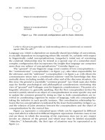
FIGURE 7.19. Intraoperative MEP monitoring during posterior spinal
fusion for scoliosis showing stable responses in both upper extremities and the
right lower extremity, but a transient loss of the MEP response in the left lower
extremity.
U
nlike BAEP and SEP, there is disagreement as to what is a signif-
icant MEP change. Some investigators suggest that a significant
change occurs when the stimulus intensity has to be increased during
the case to elicit the same response. Others suggest a significant
change occurs only when the response is completely lost, regardless of
the stimulation intensity. In the author’s experience, a significant
response is one in which the response disappears completely or by at
least 90%. In the example above, at the start of the case MEP
responses are noted in both upper (
first two columns of each graph;
thin arrows
) and lower (last column in each graph; thick arrows)
extremities. With distraction, there was loss of the left lower extrem-
ity MEP (
dashed arrow). The surgeon was notified and the distraction
was relaxed with return of the MEP (
dotted arrow).
Neurophysiologic Intraoperative Monitoring
249
FIGURE 7.20. Intraoperative MEP monitoring in a patient undergoing
spinal cord tumor biopsy showing the initial absence of the MEP responses in
the lower extremities due to administration of neuromuscular blocking agents
during induction and the subsequent return with drug cessation.
M
EPs are very sensitive to inhalational anesthetics and neuromus-
cular-blocking agents. When these drugs are given in boluses
(i.e., during induction), the affect on MEPs is striking. Maintaining
low doses of both agents may be compatible with MEP monitoring.
In the above example, initially the upper extremity MEP (
first col-
umn
) were seen (thin arrows), but lower extremity responses were
absent (
thick arrows) during induction with neuromuscular- blocking
agents. When further boluses of neuromuscular-blocking agents were
not administered, after a few minutes robust MEPs were seen for both
upper and lower extremities (
dashed arrows).
CHAPTER 7
250
When nerve roots are at risk during surgery, monitoring spontaneous and stimu-
lated EMG provides useful information to help preserve the nerve roots. Various
abnormalities can be detected with this type of NIOM and can help reduce neuro-
logic morbidity.
FIGURE 7.21. Intraoperative free-running EMG monitoring showing a neu-
rotonic discharge primarily arising from the right anterior tibialis muscle (L4
to L5 root) during tethered cord release. One second is displayed.
M
onitoring of the peripheral nervous system can be performed
with the use of free-running EMG, stimulated EMG, or nerve
action potentials. To record free-running (or stimulated) EMG, needle
or wire electrodes are placed in muscles innervated by nerves that are
at risk. Significant injury to nerves during dissection produces high-
frequency discharges called neurotonic discharges. Short bursts of
neurotonic discharges signify transient nerve injury; if persistent, the
injury may be irreversible. In the figure above, the channels monitored
are left vastus lateralis, left anterior tibialis, left medial gastrocnemius,
left semitendinosis, right vastus lateralis, right anterior tibialis, right
medial gastrocnemius, right semitendinosis, and anal sphincter mus-
cles using needle electrodes. There is a high-frequency run of dis-
charges consistent with a neurotonic discharge arising from the right
Neurophysiologic Intraoperative Monitoring
251
ELECTROMYOGRAPHY
anterior tibialis muscle (thin arrow) and to a lesser extent from the
right hamstring muscle (
thick arrow). Upon hearing the discharge, the
surgeon stopped dissecting, irrigated the surgical field, and the neuro-
tonic discharge resolved.
CHAPTER 7
252
FIGURE 7.22. Intraoperative free-running EMG monitoring data showing
occasional spontaneous muscle activity arising from the left anterior tibialis
and medial gastrocnemius muscles. The left vastus lateralis, left anterior tib-
ialis, left medial gastrocnemius, left semitendinosis, anal sphincter, right vastus
lateralis, right anterior tibialis, right medial gastrocnemius, and right semi-
tendinosis muscles are being monitored.
M
inor irritation of a nerve often causes spontaneous firing of
motor units supplied by that nerve. While monitoring free-run-
ning EMG, this is manifested as low-frequency, short discharges.
These discharges are not associated with postoperative morbidity. The
example above displays 50 msec of data from a patient undergoing
tethered cord release surgery. During irrigation low-frequency dis-
charges are noted in the left anterior tibialis (
thin arrow) and medial
gastrocnemius (
thick arrow) muscles that disappeared after a few sec-
onds.
Neurophysiologic Intraoperative Monitoring
253
FIGURE 7.23. Intraoperative stimulated EMG monitoring data showing a
response in the left anterior tibialis and medial gastrocnemius muscles. This is
a 100 msec sample. The montage is left vastus lateralis, left anterior tibialis,
left medial gastrocnemius, left semitendinosis, anal sphincter, right vastus lat-
eralis, right anterior tibialis, right medial gastrocnemius, and right semitendi-
nosis muscles.
S
timulated EMG can be used to identify neural structures during
surgery. For example, if a tumor is surrounding neural tissue,
focal stimulation in various areas of the tumor can be helpful in deter-
mining where neural elements are present. Alternatively, often when
anatomy is not clear, structures in the surgical field can be stimulated,
and according to the pattern of response seen, they can be correctly
identified. In the figure above, stimulation of a nerve root produced a
triggered response in the left anterior tibialis (
thin arrow) and the
medial gastrocnemius (
thick arrow) muscles. The root stimulated is
most likely the left L5 root.
CHAPTER 7
254
FIGURE 7.24. Intraoperative EMG monitoring data showing an artifact
that resembles a neurotonic discharge. One second is displayed. The montage
is left anterior tibialis, left medial gastrocnemius, left semitendinosis, anal
sphincter, right anterior tibialis, right medial gastrocnemius, and right semi-
tendinosis muscles.
A
s with other types of monitoring, artifacts are common in EMG
monitoring as well. Differentiating artifacts from neurotonic dis-
charges is critical to avoid unnecessary surgical intervention. In the
figure above, the patient is undergoing tethered cord release surgery.
Although runs of high-frequency discharges are seen, they are not
neurotonic discharges. Their widespread, rhythmic, and similar mor-
phology in all channels (arrows) provides proper identification as arti-
fact.
Neurophysiologic Intraoperative Monitoring
255
The EEG may demonstrate changes as a reflection of cerebral blood flow.Therefore,
EEG is commonly used in the operating suite during surgeries that may impair blood
flow to the brain. The EEG may also be useful to directly record epileptiform,
nonepileptiform, or evoked potentials during surgical resections that require identi-
fication of eloquent cortical function.
FIGURE 7.25. Intraoperative EEG during right carotid endarterectomy
demonstrating bilateral symmetrical cerebral activity after clamping of the right
carotid artery. The montage is a longitudinal bipolar montage (left over right;
parasagital over temporal). The Fp1 and Fp2 electrodes were not applied
because of anesthesia monitor placement in that location.
E
EG monitoring is often used when the vascular supply to the
brain may be interrupted. Carotid endarterectomy (CEA) is a
common indication for such monitoring. During CEA, if slowing is
noted ipsilateral to the side of clamping of the carotid artery, bypass
CHAPTER 7
256
ELECTROENCEPHALOGRAPHY
(shunting) procedures are considered. If slowing or voltage reduction
is seen over the ipsilateral hemisphere, it usually occurs within a
minute after clamping. No changes in the EEG implies adequate col-
lateral perfusion. The preceeding example (Figure 7.25) is a 10-sec
sample taken several minutes after clamping the carotid artery. The
EEG continued to look bilaterally symmetrical, implying adequate
collateral circulation.
Neurophysiologic Intraoperative Monitoring
257
FIGURE 7.26. Intraoperative EEG taken from the same patient as in the last
figure. A 60-sec page is displayed. Note the bilaterally symmetric activity.
W
hen monitoring EEG during CEA, often a slower (60 sec) dis-
play is useful to accentuate asymmetrical slowing and/or loss
of faster frequencies.
CHAPTER 7
258
FIGURE 7.27. Intraoperative EEG showing loss of faster frequencies over
the right hemisphere after clamping of the right carotid artery.
A
s noted previously, slower visual displays (paper speed) can be
helpful in identifying slowing and loss of faster frequencies.
When slowing occurs during clamping of the carotid artery, shunt
placement to bypass the iatrogenicly induced ischemia is considered.
The example shown above was taken from a patient that was under-
going a right CEA, and approximately 1 min after clamping the right
carotid artery, there was a loss of faster frequencies in that hemisphere
(
arrows). The clamp was removed, the EEG returned to baseline, and
no postoperative deficit was incurred.
Neurophysiologic Intraoperative Monitoring
259
ADDITIONAL RESOURCES
James ML, Husain AM. Brainstem auditory evoked potential monitoring:
when is change in wave V significant? Neurology 2005;65(10):1551-1555.
Legatt AD. Mechanisms of intraoperative brainstem auditory evoked potential
changes. J Clin Neurophysiol 2002;19(5):396–408.
MacDonald DB. Safety of intraoperative transcranial electrical stimulation
motor evoked potential monitoring. J Clin Neurophysiol 2002;19(5):
416–429.
Nuwer MR, Dawson EG, Carlson LG, et al. Somatosensory evoked potential
spinal cord monitoring reduces neurologic deficits after scoliosis surgery:
results of a large multicenter survey. Electroencephalogr Clin Neurophysiol
1995;96(1):6–11.
Radtke RA, Erwin CW, Wilkins RH. Intraoperative brainstem auditory evoked
potentials: significant decrease in postoperative morbidity. Neurology
1989;39(2 Pt 1):187–191.
Robertson SC, Traynelis VC, Yamada TT. Identification of the sensorimotor
cortex with SSEP phase reversal. In: Loftus CM, and Traynelis VC, eds.
Intraoperative Monitoring Techniques in Neurosurgery. McGraw-Hill,
New York, 1994:107–111.
Seyal M, Mull B. Mechanisms of signal change during intraoperative
somatosensory evoked potential monitoring of the spinal cord. J Clin
Neurophysiol 2002;19(5):409–415.
CHAPTER 7
260
261
Index
Abnormal nonepileptiform EEG,
51–69
diffuse slowing as, 53–60
encephalopathies and, 51–52
focal abnormalities in, 61–68
lateralization/localization of
abnormalities using, 51
Absence seizure, 99–101
Activation procedures, 39–40
hyperventilation and, 39–40
pharmacologic methods as, 39
photic stimulation and, 39, 41
sleep deprivation as, 39
Age, 1, 55
Airflow, polysomnography,
151–152
Alpha frequency/alpha rhythm,
28–29
alpha asymmetries as, 61
Bancaud’s phenomenon and, 29
coma and, 139, 140
diffuse slowing and, 55, 60
multiple sleep latency test
(MSLT) in, 216
occipital lobe seizures, 116
paradoxical alpha and, 29
polysomnography and, with delta
pattern in, 207–208, 209
sleep and, 35, 153–156
Alpha asymmetries, 61
American Clinical Neurophysiology
Society (ACNS), 141
American Sleep Disorders
Association (ASDA), 183
Amplifiers, artifacts related to, 22
Apneas, 167–168
arousal during, 174, 175
bradyarrhythmia in, 196–198
cardiac arrhythmias and, 196
central, 176, 177
Cheyne-Stokes respirations in,
178
continuous positive airway
pressure (CPAP) and, 170,
177
mixed, 179, 180
obstructive sleep (OSA), 170
obstructive, 167, 169, 180
oxygen desaturation in, 171–172,
173
periodic limb movements (PLMs)
in, 190–195
respiratory event related arousal
(RERA) in, 175
sinus pause in, 197, 198
snoring associated with, 181
supraventricular tachycardia in,
201
ventricular tachycardia in, 200
Arousal abnormalities on PSM,
183–189
K complexes and, 187, 188
REM sleep and, 185, 186
scoring rules for, 183–184
stage II sleep and, 36, 187, 188
wakening in, 189
Arousal during apneic period, 174,
175
Artifacts, extracerebral, 10–27
60-cycle, 22
amplifier-related, 22
ballistocardiographic, 213
bruxism in, 211
chewing, 18, 212
combined/background, 19
electrocardiogram (EKG) activity
in, 134
electromyograph (EMG) and, 17
electroretinogram (ERG) and, 14
equipment/machinery as cause
of, 27
eye movement, 11–14, 16, 19, 28
eye squeak, 28
leg lead artifact on PSM, 215
mechanical, 26
mu frequency/mu rhythm in, 30
muscle (myogenic), 17–18
phone-ring, 27
photomyoclonic response as, 15
pulse artifact as, 10
REM and, 16
single-electrode, 20
sphenoidal, 24
subclinical seizures and, 117
vagus nerve stimulation (VNS), 25
Artifacts, polysomnographic, 207
Asymmetry, alpha, 61
Atrioventricular block, 196
Auras, 106
Background artifacts, 19
Background diffuse slowing, 55, 60
Ballistocardiographic artifact, 213
Bancaud’s phenomenon, 29
Barbiturates, beta frequency/beta
rhythm, 31
Bell’s phenomenon, 11
Benign childhood epilepsy with
centrotemporal spikes
(BCECTS), 71, 80, 81, 83
Benign epileptiform transients of
sleep (BETS), 46
Benign variants of uncertain
significance, 42–49
14- and 6-Hz positive bursts as,
45
6-Hz spike-and-wave burst as,
44
benign epileptiform transients of
sleep (BETS) as, 46
phantom spike-and-wave as, 44
subclinical rhythmic electro-
graphic discharge in adults
(SREDA) as, 49
theta bursts as, rhythmic
temporal, 42
theta, central, 43
wicket waves as, 47
Benziodiazepines, beta
frequency/beta rhythm, 31
Beta frequency/beta rhythm, 31
breach rhythm and, 31
coma and, 139
diffuse slowing and, 60
occipital lobe seizures, 116
sleep and, 35
Bilateral PLEDs (BiPLEDs), 123,
137
Bilateral sharp-and-slow wave, 78
Bipolar montage, 7
Blinking, 11, 214.
See also eye
movement artifacts
Bradyarrhythmia, 196–198
Brain
cerebral potentials of, basic
physiology of, 2–3
electrical signals of, 1, 2
Brain death, 141
Brainstem, 2
Brainstem auditory evoked poten-
tials (BAEPs), NIOM, 223,
224–234
Index
262
Breach rhythm, 31
Bruxism, 211
Burst suppression pattern, 135
Cardiac arrhythmias, 196–201
apneas and, 196
atrioventricular block in, 196
ballistocardiographic artifact in,
213
bradyarrhythmia in, 196–198
continuous positive airway
pressure (CPAP) and, 198
sinus arrest in, 196
sinus arrhythmia in, 199
sinus bradycardia in, 196
sinus pause in, 197, 198
supraventricular tachycardia in,
201
ventricular tachycardia in, 200
Carotid endarterectomy (CEA),
256–257, 258, 259
Central apnea, 176, 177
Central IEDs, 83
Central spikes, 72
Central theta waves, 43
Cerebral palsy, 83, 86
Cerebral, potentials of, basic
physiology of, 2–3
Channels (potassium, sodium,
chloride), 2
Chewing artifact, 18, 212
Cheyne-Stokes respirations, 178
Chloral hydrate, beta
frequency/beta rhythm, 31
Chloride channels, 2
Clonic seizures, generalized tonic-
clonic (GTC) seizure, 103
Colloidion, 5
Coma and stupor, 121, 132–141
alpha coma in, 139, 140
beta coma in, 139
bilateral PLEDs (BiPLEDs) in, 137
brain death, 141
burst suppression pattern in, 135
diffuse slowing and, 60
generalized periodic epileptiform
discharges (GPEDs) in, 136,
138
periodic lateralized epileptiform
dicharges (PLEDs) in, 137
spindle coma in, 139, 140
status epilepticus (SE) and, 132,
145
theta/delta, 139
triphasic waves in, 132–133, 134
Combined/background artifacts, 19
Complex partial SE, 144
Compound muscle action potentials
(CMAPs), 246–247
Continuous generalized diffuse
slowing, 59
Continuous positive airway pres-
sure (CPAP), 170, 177
artifacts produced by, 26
cardiac arrhythmias and, 198
polysomnography and, 151–152
Continuous regional slowing, 65
Continuous spike-and-wave dis-
charges (CSWS), 146
Convulsive status epilepticus (CSE),
122
Cortex, 4
focal interictal epileptiform
dishcarges (IEDs) and,
73–74
layers of, potentials and, 3
Craniotomy, breach rhythm, 31
Creutzfeld-Jakob disease (CJD),
periodic lateralized
epileptiform discharges
(PLEDs), 129
Data display, 4
Deflection and polarity, 9
Index
263
Delta frequency/delta rhythm, 34
coma and, 139
diffuse slowing and, 54–59
focal abnormalities and, 62
focal interictal epileptiform dish-
carges (IEDs) and, 79
frontal intermittent rhythmic
delta activity (FIRDA) and,
57
lateralized polymorphic delta
slowing in, 66
localized polymorphic delta
slowing in, 66
orbital intermittent rhythid delta
activity (ORIDA) as, 58
polymorphic burst, 64
polysomnography and, with
alpha pattern in, 207–208,
209
sleep and, 37, 163
status epilepticus (SE) and, 142
temporal intermittent rhythmic
delta activity (TIRDA) as,
63
Designation of electrodes, 5
Diffuse slowing, 53–60
alpha frequencies and, 55, 60
background, 55, 60
beta frequencies in, 60
coma and, 60
continuous generalized, 59
delta frequencies and, 54–58
frontal intermittent rhythmic
delta activity (FIRDA) and,
57
generalized, 54
intermittent, 56
low-voltage EEG and, 60
orbital intermittent rhythid delta
activity (ORIDA) as, 58
theta frequencies and, 53–55
tilt-table testing and, 54
Dipole, 3
epileptiform abnormalities and,
71
Double banana montage, 7
Driving, photic, 41
Electrical signals of the brain, 1, 2
Electrical status epilepticus of slow
sleep (ESES), 146
Electrocardiogram (EKG/ECG), 6
EEG recording of activity from, 134
polysomnography and, 151–152,
196–197.
See also cardiac
arrhythmias
pulse artifacts on EEG and, 10
Electrochemical equilibrium, 2
Electrode box (jackbox), 4
Electrodes
artifacts derived from, 20
colloidion and, 5
designation of, numbers and let-
ters in, 5
extracerebral, 6
frontotemporal, 5
impedance of, 5, 22
placement of, 5, 152
polysomnography and, 152
sphenoidal, 5, 24
sphenoidal artifacts and, 24
subdermal, 5
true temporal, 5
Electroencephalogram (EEG)
neurophysiologic intraoperative
monitoring (NIOM) and,
223, 256–259
polysomnography and, 151–152
Electromyogram (EMG), 6
muscle (myogenic) artifacts on
EEG and, 17
neurophysiologic intraoperative
monitoring (NIOM) and,
223, 251–255
Index
264
photomyoclonic response as, 15
polysomnography and, 151–152,
210
Electroretinogram (ERG), 14
Encephalopathies, 51–52, 121
Epilepsia partialis continua,
142–143
Epileptiform abnormalities, 71–94
benign childhood epilepsy with
centrotemporal spikes
(BCECTS) and, 71, 80, 81
central spikes and, 72
diagnosis of epilepsy and, 72
dipoles in, 71
focal IEDs and, 72, 73–88
generalized, 88–94, 88
interictal epileptiform discharges
(IEDs) and, 71
occipital spikes and, 72
parietal spikes and, 72
photic stimulation and, 71
photoparoxysmal response and,
71
scalp EEGs and, limitations of,
73–74
spike-and-wave discharges and,
71
Equipment/machinery-related arti-
facts, 27
Excitatory postsynaptic potentials
(EPPs), 2
Extracerebral artifacts. See artifacts,
extracerebral
Extracerebral electrodes, 6
Eye movement artifacts, 11–14, 28,
122
alpha frequency/alpha rhythm
and, 28–29
Bancaud’s phenomenon and, 29
blinks in, 155, 214
combined/background, 19
electroretinogram (ERG) in, 14
lambda frequency/lambda
rhythm and, 33
mu frequency/mu rhythm in, 30
photomyoclonic response as, 15
polysomnography and, 151–152,
155
pseudogeneralized spike-and-
wave discharge in, 19
REM and, 16, 38
“squeak”, 28
Eye movement monitors, 6
Fast generalized slow wave (GSW),
90
Fencer’s posture, 114
Filters
60-cycle artifact and, 23
deflection and, 9
polarity and, 9
Focal abnormalities, 61–68
alpha asymmetries as, 61
continuous regional slowing as,
65
delta, 62
lateralized polymorphic delta
slowing in, 66
localized polymorphic delta
slowing in, 66
polymorphic burst, 64
sleep spindles as, 67
sleep-onset REM as, 68
status epilepticus (SE) and, 122
temporal intermittent rhythmic
delta activity (TIRDA) as,
63
Focal interictal epileptiform dis-
charges (IEDs), 72, 73–88
benign childhood epilepsy with
centrotemporal spikes
(BCECTS) and, 80, 81
bilateral sharp-and-slow wave in,
78
Index
265
Focal interictal epileptiform dis-
charges (IEDs) (continued)
central IEDs and, 83
centrotemporal spikes in, 80
cortex and, 73–74
delta frequencies in, 79
frontal lobe epilepsy (FLE) and,
82
idiopathic generalized epilepsy
(IGE) in, 82
mid-temporal lobe site of, 79
midline spike wave in, 84
multifocal spikes in, 86
needle spikes in, 85
occipital IEDs in, 85
polarity of, 75, 76
polyspike-and-wave discharge as,
75
polyspikes as, 75
positive spikes in, 77
rapid eye movement (REM) sleep
and, 78
rolandic sharp waves in, 81
scalp EEGs and, limitations of,
73–74
secondary bilateral synchrony
(SBS) in, 82, 87
sharp transients as, 75
sharp waves as, 75, 77, 79–81
sharp-and-slow waves in, 87
sleep and, 78
spike waves as, 75, 80
spike-and-polyspike discharge in,
82
spike-and-slow wave discharge
as, 76, 83, 85
spike-and-wave discharge as, 75,
82
temporal lobe as site of, 78
temporal lobe epilepsy (TLE)
and, 79
theta frequencies in, 79
Focal seizures, 106–119
frontal lobe epilepsy (FLE) and, 113
frontal lobe seizures, 112, 113
lateral or neocortical temporal
seizures, 108
mesial frontal lobe seizures, 114
mesial temporal lobe seizure, 107
occipital lobe seizures, 116
parietal lobe seizures, 115
partial seizures, 110
psychogenic nonepileptic seizures
(PNES), 118–119
regional onset seizures, 111
simple partial, 106
subclinical seizures, 117
supplementary motor seizures, 114
temporal lobe seizure, 109
14- and 6-Hz positive bursts, 45
Frontal intermittent rhythmic delta
activity (FIRDA), 57
Frontal lobe epilepsy (FLE), 113
focal interictal epileptiform dish-
carges (IEDs) and, 82
frontal lobe seizures, 112, 113
mesial frontal lobe seizures, 114
Frontal lobe seizures, 112, 113
Frontotemporal electrodes, 5
Ganglioglioma, focal delta, 62
Generalized epileptiform discharges,
88–94
3-Hz spike-and-slow wave dis-
charge in, 89
fast generalized slow wave
(GSW) in, 90
generalized paroxysmal fast
activity (GPFA) in, 94
generalized polyspike and wave
(PSW), 91
generalized slow wave (GSW) in,
89
hypsarrhythmia in, 92
Index
266
photoparoxysmal response, 88
slow spike-and-wave (SSW) in,
93
status epilepticus (SE) and, 122
Generalized paroxysmal fast activ-
ity (GPFA), 94, 123
Generalized periodic epileptiform
dischargers (GPEDs), 123,
127, 136, 138
Generalized seizures, 54, 97,
98–105
absence, 99–101
generalized tonic-clonic (GTC),
103
infantile spasms, 104
myoclonic, 102
polyspike-and-wave in, 98
recruiting rhythm in, 103
spike wave in, 98
tonic seizure, 105
Generalized slow wave (GSW), 89
fast, 90
Generalized tonic-clonic (GTC)
seizure, 103
Gradients, ionic, 2
Grand mal, 103.
See also general-
ized tonic-clonic (GTC)
seizure
Gray matter injury, periodic lateral-
ized epileptiform discharges
(PLEDs), 131
Grounding, 22
artifacts related to, 22
Head injury, localized polymorphic
delta slowing, 66
Herpes simplex encephalitis (HSE),
periodic lateralized epilepti-
form discharges (PLEDs),
128
Hyperventilation
activation using, 39–40
delta frequency/delta rhythm in,
34
spike-and-slow wave and, 89
theta frequency/theta rhythm in,
32
Hypnogram, 150
Hypoapneas, 167, 171–173.
See
also apneas
Hypsarrhythmia, 92
Idiopathic generalized epilepsy
(IGE), 82, 123
fast generalized slow wave
(GSW) in, 90
generalized tonic-clonic (GTC)
seizure in, 103
photoparoxysmal response and, 88
Impedance, electrode, 5, 22
Infantile spasms, 104
hypsarrhythmia in, 92
Infarctions
alpha asymmetries and, 61
continuous regional slowing in, 65
localized polymorphic delta
slowing in, 66
periodic lateralized epileptiform
discharges (PLEDs) and, 126
polymorphic delta activity in, 64
Inhibitory postsynaptic (IPP) poten-
tials, 2
Interictal epileptiform discharges
(IEDs), 1, 63, 71, 123
repetitive, continuous, 124
Intermittent diffuse slowing, 56
Ionic gradients, 2
Jackbox.
See electrode box
Juvenile myoclonic epilepsy (JME)
fast generalized slow wave
(GSW) in, 90
myoclonic seizure and, 102
polyspike-and-wave (PSW), 91
Index
267
K complexes
arousal from sleep and, 187, 188
sleep and, 36, 157, 159–161,
162, 163
Lambda frequency/lambda rhythm,
33
Landau-Kleffner syndrome (LKS),
status epilepticus (SE), 146
Laplacian montages, 7
Lateral or neocortical temporal
seizures, 108
Lateralization/localization of
abnormalities, 51
Lateralized polymorphic delta
slowing, 65–66
Leg lead artifact on PSM, 215
Lennox-Gastaut syndrome
generalized paroxysmal fast
activity (GPFA) in, 94
hypsarrhythmia in, 92
multifocal spikes in, 86
slow spike-and-wave (SSW) in,
93
tonic seizure in, 105
Localization related epilepsy (LRE),
repetitive IEDs, 124
Localized polymorphic delta
slowing, 62–64
Long-term monitoring, 122
Longitudinal bipolar (double
banana) montage, 7
Low-voltage EEG, diffuse slowing,
60
Mechanical artifact, 26
Mental retardation, multifocal
spikes, 86
Mesial frontal lobe seizures, 114
Mesial temporal lobe epilepsy
(MTLE), 107
simple partial seizures in, 106
Mesial temporal lobe seizure, 107
Midline spike wave, 84
Migraine, 83
localized polymorphic delta
slowing in, 66
Montage selectors, 4, 7
Motor evoked potentials (MEPs), in
NIOM, 223, 246–250
Motor seizures, supplementary, 114
Movements, on PSM, 166
Mu frequency/mu rhythm, 30, 83
parietal lobe seizures, 115
Multifocal spikes, 86
Multiple sleep latency test (MSLT),
216–220
sleep-onset REM periods
(SOREMP) in, 217, 220
Muscle (myogenic) artifacts, 17
Myoclonic seizures, 102, 122
generalized periodic epileptiform
discharges (GPEDs) in, 138
periodic lateralized epileptiform
discharges (PLEDs), 129
photomyoclonic (photomyo-
genic) response, 41
Myogenic artifact.
See muscle
(myogenic) artifacts
Myotonic seizures, photomyoclonic
response, 15
Narcolepsy, sleep-onset REM, 68
Needle spikes, 85
Neocortical temporal seizures,
108
Neurophysiologic intraoperative
monitoring (NIOM),
223–259
brainstem auditory evoked
potentials (BAEPs) in, 223,
224–234
carotid endarterectomy (CEA) in,
256–258
Index
268
compound muscle action poten-
tials (CMAPs) in, 246–247
electroencephalography (EEG)
and, 223, 256–259
electromyography (EMG) and,
223, 251–255
motor evoked potentials (MEPs)
in, 223, 246–250
somatosensory evoked potentials
(SEPs) in, 223, 235–245
Non rapid eye movement (NREM)
sleep, 149, 150, 207, 209
Nonconvulsive status epileptics
(NCSE), 136, 145
Nonepileptic seizures, psychogenic
nonepileptic seizures
(PNES), 118
Nonepileptiform EEG. See abnor-
mal nonepileptiform EEG,
51
Normal EEG, 28–34
activation procedures in, 39–40
alpha frequency/alpha rhythm,
28–29
Bancaud’s phenomenon and, 29
benign variants of uncertain sig-
nificance in, 42–49
beta frequency/beta rhythm in,
31
breach rhythm and, 31
cerebral potentials and, basic
physiology of, 2–3
deflection and polarity in, 9
delta frequency/delta rhythm in,
34
diagnosing illness and, 1
extracerebral artifacts and,
10–27
eye open/close shown on, 28
lambda frequency/lambda
rhythm in, 33
mu frequency/mu rhythm in, 30
sleep and, 35–38
theta frequency/theta rhythm in,
32
Occipital IEDs, 85
Occipital lobe seizures, 116
Occipital spikes, 72
Orbital intermittent rhythid delta
activity (ORIDA), 58
Oxygen saturation/desaturation,
151–152, 171–172, 173
Pacemakers, artifacts derived from,
25
Paradoxical alpha, 29
Parietal lobe seizures, 115
Parietal spikes, 72
Paroxysmal seizures
generalized paroxysmal fast
activity (GPFA) in, 94, 123
photoparoxysmal response in,
15, 71, 88
Partial-onset seizures, periodic later-
alized epileptiform dis-
charges (PLEDs), 125
Partial seizures, 110
Periodic discharges (PD), 122
Periodic epileptiform discharges,
123–131
bilateral PLEDs (BiPLEDs) as, 123
generalized paroxysmal fast
activity (GPFA) as, 123
generalized periodic epileptiform
dischargers (GPEDs) as,
123, 127
interictal epileptiform discharges
(IEDs) and, 123, 124
periodic lateralized epileptiform
discharges (PLEDs) as, 123,
125, 126, 128–131
sharp and slow waves as, 130
slow waves as, 130
Index
269
Periodic epileptiform discharges
(continued)
subacute sclerosing panencephali-
tis (SSPE) and, 130
Periodic lateralized epileptiform
discharges (PLEDs),
123–131, 137
Periodic limb movements (PLMs),
190–195
Petit mal, 99–101. See also absence
seizures
Phantom spike-and-wave discharge,
44
Pharmacologic activation methods,
39
Phone-ring artifact, 27
Photic driving, 41
Photic stimulation, 39, 41, 71
driving, photic driving and, 41
photomyoclonic (photomyo-
genic) response and, 15, 41
photoparoxysmal response and,
15, 71, 88
polyspike-and-wave (PSW) and, 91
pseudogeneralized spike-and-
wave discharge in, 19
spike-and-slow wave in, 89
Photomyoclonic (photomyogenic)
response, 15, 41
Photoparoxysmal response, 15, 71,
88
Placement of electrodes, 5
Polarity
deflection and, 9
focal interictal epileptiform dish-
carges (IEDs) and, 75, 76
Polymorphic delta activity, 64
Polysomnography, 149–222.
See
also sleep
airflow recorded in, 151–152
alpha-delta sleep pattern in,
207–208
alpha-delta sleep pattern in, 209
arousal abnormalities, 183–189
K complexes and, 187, 188
REM sleep and, 185, 186
scoring rules for, 183–184
stage II sleep and, 36, 187, 188
wakening in, 189
artifacts in, 207
artifacts in, from leg leads, 215
ballistocardiographic artifact in,
213
bruxism in, 211
cardiac arrhythmias in, 196–201
apneas and, 196
atrioventricular block in, 196
bradyarrhythmia in, 196–198
continuous positive airway
pressure (CPAP) and, 198
sinus arrest in, 196
sinus arrhythmia in, 199
sinus bradycardia in, 196
sinus pause in, 197, 198
supraventricular tachycardia
in, 201
ventricular tachycardia in, 200
chewing in, 212
continuous positive airway pres-
sure (CPAP) in, 151–152
electrocardiogram (EKG/ECG)
in, 151–152, 196–197. See
also cardiac arrhythmias
electrode placement in, 152
electroencephalogram (EEG) in,
151–152
electromyogram (EMG) in,
151–152, 210
eye blinks in, 214
eye movements recorded in,
151–152
hypnogram of, 150
miscellaneous findings in,
207–215
Index
270
movements in, 166
multiple sleep latency test
(MSLT) in, 216–220
non rapid eye movement
(NREM) sleep and, 149,
150, 207, 209
normal sleep in, 153–166
alpha activity in, 153–156
blinking eyes in, 155
delta waves in, 163
K complexes in, 157, 159–163
movement time in, 166
rapid eye movement (REM),
164, 165
sawtooth waves in, 165
sharp waves in, 157
slow waves in, 162
slow, rolling eye movements
in, 157
spindles in, 157, 159, 160,
162, 163
stage I in, 35, 156, 157, 158
stage II in, 36, 159–161
stage III in, 37, 162
stage IV in, 163
vertex potentials in, 158, 159,
162, 163
W stage in, 153–155
onset of sleep, 218
oxygen saturation recorded in,
151–152
paper speed and, 154
periodic limb movements (PLMs)
in, 190–195
polyspike waves in, 206
rapid eye movement (REM) sleep
and, 149, 150
REM sleep behavior disorder
(RBD) in, 210
respiratory abnormalities,
167–182
apnea in, 167–168
apnea, central, 176, 177
apnea, mixed, 179, 180
apnea, obstructive sleep
(OSA), 170
apnea, obstructive, 167, 169,
180
arousal during apneic period
in, 174, 175
Cheyne-Stokes respirations in,
178
continuous positive airway
pressure (CPAP) and, 170,
177
hypoapnea in, 167, 171–173
oxygen desaturation in,
171–173
periodic limb movements
(PLMs) in, 190–195
respiratory event related
arousal (RERA) in, 175
snoring associated with apnea
in, 181
snoring without apnea in, 182
respiratory effort recorded in,
151–152
routine, typical, 151–152
seizures during, 202–206
sleep stages and, 149, 150
sleep-onset REM periods
(SOREMP) in, 217, 220
spike waves in, 202–206
stage I sleep, 35, 219
terminology of, 149
Polyspike-and-wave (PSW), 75
generalized seizures and, 98
generalized, 91
myoclonic seizure and, 102
Polyspike waves, 75
periodic lateralized epileptiform
discharges (PLEDs) as, 126
polysomnography and, 206
status epilepticus (SE) and, 122
Index
271
Polyspike-and-slow wave
photoparoxysmal response, 88
Pooled synchronous activity, sleep,
4
Positive bursts, 6- and 14-hz, 45
Positive occipital sharp transients
(POSTS), 36, 371
sleep and, 35
Positive spikes, 77,
Potassium channels, 2
Potentials
ballistocardiographic, 10
brainstem auditory evoked
potentials (BAEPs) in, 223,
224–234
cerebral, basic physiology of, 2–3
channels in (potassium, sodium,
chloride), 2
compound muscle action poten-
tials (CMAPs) in, 246–247
cortex and, 4
dipole activity and, 3
electrochemical equilibrium and,
2
excitatory postsynaptic (EPP), 2
inhibitory postsynaptic (IPP), 2
layers of cortex and, 3
motor evoked potentials (MEPs)
in, 223, 246–250
myogenic, 17
pooled synchronous activity of, 4
pyramidal cells and, 3
resting (diffusion) membrane, 2
scalp recordings of, 4
sinks and source in, 2
somatosensory evoked potentials
(SEPs) in, 223, 235–245
synaptic, 2
volume conduction and, 3
Pseudo-pseudoseizures, 114
Psychogenic nonepileptic seizures
(PNES), 118–119
Psychogenic seizures, 114
Pulse artifact, 10
Pyramidal cells, 3
QRS complexes, 10
Rapid eye movement (REM), 16,
149, 150, 164, 165
arousal from, 185, 186
benign epileptiform transients of
sleep (BETS) as, 46
focal interictal epileptiform dish-
carges (IEDs) and, 78
mesial temporal lobe seizure, 107
REM sleep behavior disorder
(RBD) in, 210
in sleep, 38
sleep-onset REM periods
(SOREMP) in, 217, 220
sleep-onset, 68
Recruiting rhythms, 103
Referential montage, 7
Regional onset seizures, 111
Regional slowing, continuous, 65
REM sleep behavior disorder
(RBD), 210
Respiratory abnormalities on PSM,
167–182
apnea in, 167–168
apnea, central, 176, 177
apnea, mixed, 179, 180
apnea, obstructive sleep (OSA),
170
apnea, obstructive, 167, 169,
180
arousal during apneic period in,
174, 175
ballistocardiographic artifact in,
213
bradyarrhythmia in, 196–198
Cheyne-Stokes respirations in,
178
Index
272
continuous positive airway pres-
sure (CPAP) and, 170, 177
hypoapnea in, 167, 171–173
oxygen desaturation in, 171–173
periodic limb movements (PLMs)
in, 190–195
respiratory event related arousal
(RERA) in, 175
sinus arrhythmia in, 199
sinus pause and, 197, 198
snoring associated with apnea in,
181
snoring without apnea in, 182
supraventricular tachycardia and,
201
ventricular tachycardia and, 200
Respiratory effort, polysomnogra-
phy showing, 151–152
Respiratory event related arousal
(RERA), 175
Respiratory monitors, 6
Resting (diffusion) membrane
potential, 2
Rhythmic temporal theta bursts,
Rolandic sharp waes, 81
Sawtooth waves, in sleep, 165
Scalp electrodes, 74
Secondary bilateral synchrony
(SBS), 82, 87
Seizures, 97–120
clinical vs. occult manifestation
of, 97
focal seizures in, 106–119
generalized, 97, 98–105
ictal EEG diagnosis and, 97
polysomnography and, 202–206
Sharp-and-slow wave, 87
bilateral, 78
periodic epileptiform discharges
and, 130
Sharp transients, 75
Sharp waves, 75, 77, 79, 80
left anterior temporal, 8
periodic epileptiform discharges
and, 130,
periodic lateralized epileptiform
discharges (PLEDs) as, 126
rolandic, 81
single-electrode artifact mimicking,
21
sleep and, 157
sphenoidal artifact as, 24
status epilepticus (SE) and, 122
Simple partial seizures, 106
Single-electrode artifacts, 20
Sinks, 2
Sinus arrest, 196
Sinus arrhythmia, 199
Sinus bradycardia, 196
Sinus pause, 197, 198
6- and 14-Hz positive bursts, 45
6-Hz spike-and-wave burst, 44
60-cycle artifact, 22
Sleep, 2, 35–38, 149.
See also
polysomnography
absence seizures in, 99–101
alpha, 35
alpha-delta pattern in, 207–208,
209
apneas and. See apnea
arousal abnormalities in,
183–189
benign epileptiform transients of
sleep (BETS) as, 46
beta frequency/beta rhythm in,
31, 35
bruxism in, 211
cardiac arrhythmias in, 196–201
chewing in, 212
continuous spike-and-wave
discharges (CSWS) in, 146
delta frequency/delta rhythm in,
34, 37, 163
Index
273