Intraocular Drug DelIvery - part 6 ppsx
Bạn đang xem bản rút gọn của tài liệu. Xem và tải ngay bản đầy đủ của tài liệu tại đây (709.73 KB, 39 trang )
weight: 5000) at weight ratios of 80/20. These plugs included 25% GCV. The high-
molecular-weight PLA may play a substantial role in the framework of the device
and restrict the degradation rate of the low-molecular-weight PLA. Also the low-
molecular-weight PLA may regulate drug release by slowing pore formation during
the diff usional phase. The degradation rate of other biodegra dable devices such as
microspheres and intrascleral implants is also controlled in a similar manner.
Drug Delivery Systems
General Overview
Several different intraocular drug delivery systems using biodegradable polymers
such as microspheres (5–9), intraocular implants (10–13), scleral plugs (3,4,14–22),
and intrascleral implants (23) have been developed.
Moritera et al. (5) first reported an intravitreal drug delivery system using bio-
degradable polymer microspheres. PLA microspheres containing doxorubicin hydro-
chloride (6) and PLGA microspheres containing retinoic acid (7) have been reported
for the treatment of proliferative vitreoretinopathy (PVR). GCV-loaded PLGA
microspheres have been developed using a new oil-in-oil emulsion technique with
fluorosilicone (8). Interestingly, after intravitreal injection of PLA nanoparticles
with the mean size of 310 nm, nanoparticles transversed the retina and reached
the retinal pigment epithelium (9). Targeted drug delivery to the retina and retinal
pigment epithelium could be feasible using PLA nanoparticles.
Surodex
1
(Oculex Pharmaceuticals, Inc.) is a PLGA rod containing dexa-
methasone, which is implanted at cataract surgery for treatment of postsurgical
inflammation (10). In a multicenter, randomized, double-masked, parallel group
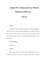
Figure 3 Cumulative release GCV from the scleral plugs of PLA-70,000 and PLA-5000
(whose content ratio was 80:20) containing 25% of GCV. The values shown are mean Æ SD.
The duration of GCV release was prolonged further compared with the plug made of PLGA
(75/25)-121,000. Abbreviations: GCV, ganciclovir; PLA, polylactic acid; PLGA, polyglycolic
acid. Source: From Ref. 4.
Biodegradable Systems 177
study, Surodex
1
safely and effectively suppressed postoperative inflammation after
uncomplicated cataract surgery (11). Posurdex
1
(Allergan, Inc.), which has a similar
design, is implanted in the vitreous cavity to deliver dexamethasone to the posterior
segment of the eye. Clinical trials for Posurdex
1
are ongoing for the treatment of
macular edema associated with diabet es and other conditions (see Chapter 19). For
the treatment of PVR, two intravitreal implants have been reported; a PLGA rod
containing 5-fluorouracil (12) and a multiple drug delivery implant consisted of three
cylindrical segments, each of which contained one of the following drugs: 5-fluoro-
uridine, triamcinolone, or human recombinant tissue plasminogen activator (13).
The scleral plug is a device that is implanted through a sclerotomy at the pars
plana; it releases the drug intravitreally (Fig. 4). Its shape is similar to that of a
metallic scleral plug, which is used temporarily during pars plana vitrectomy. Con-
trolled release of doxorubicin hydrochloride [adriamycin (ADR)] (15,16), GCV
(3,4,17,19,21), fluconazole (18), 5-fluorouracil (20), and tacrolimus (FK506) (22)
have been reported.
The intrascleral implant is a device that is implanted in the sclera; it delivers
the drug through the sclera to the intraocular tissues (Fig. 5). Transscleral delivery
may be an effective method of achieving therapeutic concentrations of drugs in the pos-
terior segment (24–27). The intrascleral implant that incorporated betamethasone phos-
phate (BP) successfully delivered the drug to the retina/choroid and vitreous (28). The
concentration of BP was maintained at a level that should suppress inflammation in
the retina–choroid for more than eight weeks, and did not produce any ocular toxicity.
Relative Advantages and Disadvantages of Different Biodegradable Systems
Microspheres can be administered into the vitreous cavity by injection as a suspen-
sion. Although this is an advantage of this system, it can be disadvantageous as
a large quantity of microspheres cannot be given by intravitreal injection and
microspheres may cause a temporary disturbance in vitreous transparency. In
Figure 4 Scleral plug made of biodegradable polymers. The plug weighs 8.5 mg and is
5.0 mm long. Source: From Ref. 14.
178 Kimura and Ogura
contrast, relatively large amounts of the drug can be loaded into scleral plugs,
intrascleral implants, and intravitreal devices without decreasing vitreous transpar-
ency. Furthermore, scleral plugs can be applied at the sclerotomy sites at the end
of pars plana vitrectomy as an adjunctive therapy. Intrascleral implants are less inva-
sive than microspheres and scleral plugs, as complications such as endophthalimitis,
vitreous hemorrhages, retinal detachment, and potential risks of intraocular systems,
are virtually eliminated. In intravitreal drug delivery systems such as microspheres and
scleral plugs, the drug is released intravitreally, reaches the surface of the retina,
and diffuses into the retina–choroid. Transvitreal permeation into the retina is
limited for relatively large molecules, such as tissue plasminogen activator
(70 kDa), because the inner limiting membrane is a barrier to penetration (28). In
contrast, large molecules such as immunoglobulin (150 kDa) have been reported
to penetrate the retina through a transscleral route (26) . Accordingly, we speculate
that intrascleral implants may be more useful for site-specific treatment in the
retina–choroid and for intraocular deliv ery of large molecular compounds, such as
bioactive protein and antibody, than intravitreal systems.
SPECTRUM OF DISEASES FOR WHICH BIODE GRADABLE
SYSTEMS MAY BE USEFUL
All intraocular disorders that require systemic administration or frequent local
administration of the drug may be appropriate for these biodegradable systems.
Uveitis is a chronic disorder that requires long-term medical therapy. Topical drug
treatment is not effective in the treatment of posterior uveitis because of limited
intraocular penetration. Systemic administration of corticosteroid or immuno-
suppressive agents may be effective but are associated with systemic side effects.
Sustained drug delivery systems may be effective in the treatment of uveitis. In
Figure 5 Intrascleral implant made of biodegradable polymers. The implant weighs 7 mg
and is 0.5 mm thick and 4 mm in diameter. Source: From Ref. 23.
Biodegradable Systems 179
addition, sp ecific inflammatory disorders such as cytomegalovirus retinitis or fungal
endophthalmitis may be treated with sustained delivery systems of antiviral agents,
antibiotics, or antifungal agents. Especially, for very chronic inflammation, repeat
administration would likely be necessary even with sustained drug delivery systems.
The exudative type of age-related macular degeneration (AMD), which is asso-
ciated with choroidal neovascularization, also may be a good target for biodegrad-
able drug delivery systems. Numerous anti-angiogenic agents have been investigated
in the treatment of AMD. In addition, AMD is a chronic disease and it can be
expected that any pharmacological therapy will likely require long-term treatment.
Therefore, sustained drug delivery may be beneficial.
Recently, macular edema associated with uveitis (29), diabetic retinopathy (30),
and central retinal vein occlusion (31,32) has been treated with intravitreal injection
of triamci nolone acetonide. Macular edema decreased after treatment but recurred
three to six months after injection. A sustained-release steroid delivery system may
be more attractive than a simple injection of triamcinolone as it could reduce or elim-
inate the need for multiple intravitreal injections.
PVR is a serious complication of retinal detachment surgery. Inhibition of
cellular proliferation and postoperative inflammation may reduce the development
of PVR. Inhibition of postoperative inflammation would eliminate one of the
components of PVR and biodegradable sustained delivery systems that contain
anti-inflammatory agents may be useful.
ANIMAL MODELS USED TO TEST BIODEGRADABLE
DRUG DELIVERY SYSTEMS
Experimental Cytomegalovirus Retinitis
Experimental cytomegalovirus retinitis was induced by intravitreal injection
of human cytomegalovirus (HCMV) solution (21). HCMV AD169 was grown on
human fetal lung fibroblast monolayers. HCMV AD169 supernatant was collected
and injected onto confluent monolayers of Hs68 cells. HCMV-infected cells were har-
vested, and their culture medium was collected. Eyes of pigme nted rabbits were inocu-
lated with 0.1 mL (5 Â 10
6
pfu/mL) HCMV supernatant. The eyes were examined by
ophthalmoscopy at one, two, three, and four weeks after HCMV inoculation. Poster-
ior segment disease was graded on a 0þ to 4þ scale of increasing severity. The retinal
and choroidal diseases were scored as follows: 0þ, no abno rmalities; 1 þ, focal white
retinal infiltrates; 2þ, focal-to-geographic retinal infiltrates and vascular engorge-
ment; 3þ, severe retinal infiltrates, vascular engorgement, and hemorrhage; and
4þ, all the foregoing, plus retinal detachment and necrosis.
Experimental Uveitis
A relatively severe, nonspecific experimental uveitis model was created according
to a modification of a previously published protocol (33,34). Pigmented rabbits were
injected subcutaneously with 10 mg of Mycobacterium tuberculosis H37RA antigen
suspended in 0.5 mL of mineral oil. One week later, a second injection of the same
amount of subcutaneous antigen was given. A microparticulate suspension of
M. tuberculosis H37RA antigen was prepared by ultrasonicating a suspension of crude
extract in sterile balanced salt solution. Fifty micrograms of antigen suspended in
0.1 mL of balanced salt solution was injected into the vitreous cavity (first challenge).
180 Kimura and Ogura
To simulate chronic inflammation with exacerbations, the eyes were challenged again
with the same amount of intravitreal antigen on day 14 (second challenge). On days
3, 7, 14, 17, 21, and 28 after the first challenge, slit-lamp biomicroscopy and indirect
ophthalmoscopy were used to evaluate the severity of inflammation. Anterior cham-
ber cells and flare were graded on a 0–4 scale based on the cell number and opa city
observed by slit-lamp examination. The vitreous opacity was also graded on a 0–4
scale based on the examination of the posterior pole by indirect ophthalmoscopy.
Aqueous protein was measured using a protein assay kit, and aqueous cell count
was measured by hemocytometer.
Experimental Proliferative Vitreoretinopathy
Experimental PVR was induced by intravitreal injection of fibroblasts in the pigmented
rabbit eye. The eyes received injections of 0.3 mL of sulfur hexafluoride (SF
6
) gas. Seven
days after the first injection, an additional 0.3 mL of SF
6
gas was injected to achieve
complete compression of the vitreous. Homologous fibroblasts from Tenon’s capsule
were cultured. Seven days after the second gas injection, 1 Â 10
5
cultured fibroblasts were
injected over the medullary wings. The animals were then placed immediately in a supine
position for one hour to allow the cells to settle on the vascularized retina. Fundus
changes were observed for four weeks by indirect ophthalmoscopy. The fundus findings
weregradedasfollows: stage 1, normalretinaorwrinkling of the medullary wing;stage2,
pucker formation; and stage 3, traction retinal detachment (5).
RESULTS OF EFFICACY STUDIES
Scleral Plugs Containing GCV for Experimental
Cytomegalovirus Retinitis
Scleral plugs were prepared by dissolving PLA with an average molecular weight of
70,000 and 5000 (PLA-70,000 and PLA-5000, respectively) whose content ratio was
80:20 and 25% of GCV in aceti c acid.
Scleral plugs were prepared by dissolving PLA and GCV in acetic acid in the
ratio 3:1. The PLA us ed was a blend of two molecular weight ranges, 80% had an
average molecular weight of 70,000 (PLA-70,000) and 20% had an average molecular
weight of 5000 (PLA-5000).
The resultant solution was lyophilized to obtain a homogeneous cake. The cake
then was compressed into a scleral plug on a hot plate. In a rabbit study the scleral
plug containing GCV was found to maintain GCV concentrations in the vitreous
in a therapeutic range adequate to treat HCMV retinitis for more than 200 days
(4). The 20 eyes of 20 pigmented rabbits that were inoculated with HCMV were
divided into two groups. One week after HCMV inoculation, the co ntrol group
(n ¼ 10) received no treatment. In the treatment group (n ¼ 10), a scleral plug contain-
ing GCV was impl anted at the pars plana (21).
In the control eyes, whitish retinal exudates developed three days after HCMV
inoculation and increased gradually until three weeks after inoculation. Thereafter
the chorioretinitis decreased until four weeks after injection. In the treated group,
scores for vitreoretinal lesions were significantly lower than those in the control
group at three weeks after HCMV inoculation (Fig. 6).
Sustained release of GCV into the vitreous cavity with biodegradable scleral
plugs was thus effective for the treatment of experimentally induced HCMV retinitis
in rabbits.
Biodegradable Systems 181
Scleral Plugs Containing Tacrolimus (FK506) for Experimental Uveitis
Scleral plugs were prepared by dissolving a bioerodible polyme r (99%) and FK 506
(1%) in 1,4-Dioxane. We used poly(
Dl-lactide-co-glycolide), with a weight-averaged
molecular weight of 63,000, whose copolymer ratio of
DL-lactide to glycolide was
50:50 (22). In in vitro tests, the scleral plug released FK506 for more than 35 days.
Figure 6 Clinical disease grading. (A) HCMV-inoculated rabbit eyes (control). (B) Treated
eyes with scleral plug containing ganciclovir in HCMV-inoculated rabbit eyes.
Ã
P < 0.01,
unpaired t-test. Abbreviation: HCMV, human cytomegalovirus. Source: From Ref. 21.
182 Kimura and Ogura
Efficacy studies were conducted in the experimental rabbit uveitis model described
above. After preimmunization with M. tuberculosis H37RA antigen, the treated eyes
(n ¼ 8) received scleral plugs containing FK506, and the control eyes received blank
plugs. One day after implantation, 50 mg of antigen was injected into the vitreous
cavity. Both the treated eyes and the control eyes were challenged again with the
same amount of intravitreal antigen on day 14.
The results of anterior chamber cell, flare, and vitreous opacity clinical grading
following the first challenge are shown in Figure 7. In the control eyes, several severe
uveitis secondary complications including corneal neovascularization, corneal opa-
city, marked posterior synechia, and cataract were observed. In contrast, such com-
plications were not seen in the treated eyes. Persis tent marked vitreous opacity was
observed for at least 28 days in untreated eyes, but was minimal throughout the
observation period in the treated eyes. Evaluated by clinical criteria, the treated eyes
had significantly less inflammation than did the control eyes. Aqueous protein con-
centration and aqueous cell count in the treated eyes were significantly lower than
those in the control eyes.
Together, the results show that biodegradable scleral plugs containing FK506
are highly effe ctive in suppressing the inflammation of experimental uveitis in the
rabbit.
Scleral Plugs Containing ADR for Experimental PVR
Scleral plugs have been tested in a rabbit model of PVR. For these experiments, scleral
plugs composed of 99% PLA (average molecular weight of 20,000) and 1% of ADR
were prepared (16). The scleral plug released ADR over five weeks in vitro (Fig. 8).
Experimental PVR was induced in 22 eyes of 22 pigmented rabbits as described
above. At the time of fibroblas t intravitreal injection, the treatment group (n ¼ 11)
received scleral plugs containing ADR and the control groups (n ¼ 11) received no
treatment. Fundus changes were observed by indirect ophthalmoscopy.
The scleral plug decreased the incidence of traction retinal detachment from
100% to 64% at 28 days after implantation (Fig. 9). The differences in traction retinal
detachment rate between control and treatment groups were significant (P ¼ 0.002,
two-way ANOVA).
PHARMACOKINETIC AND PHARMACODYNAMIC STUDIES
Scleral Plugs Containing GCV
For in vivo release studies, scleral plugs prepared from blends of PLA-70,000
and PLA-5000 at weight ratios of 80/20 and 25% of GCV were used. The scleral
plugs containing GCV were implanted in pigmented rabbits. Animals were killed
at days 1 and 3 and at weeks 1, 2, 3, 4, 6, 8, 10, 12, 14, 16, 18, 20, and 24 after implan-
tation, and the eyes were enucleated. Five rabbits were used at each time point.
The intravitreal GCV concentration was determined by high-performance liquid
chromatography (HPLC).
The scleral plugs maintained a constant vitreous GCV concentration within
the ED
50
range (0.1–2.75 mg/mL) for six months without any sudden burst
(Fig. 10) (4). However, further studies may be needed to evaluate effective GCV
concentrations clinically, as the ED
50
s are values determined in various conditions
in vitro.
Biodegradable Systems 183
Scleral Plugs Containing ADR
To evaluate scleral plug ADR vitreous pharmacokinetics, 1% ADR-loaded PLA
scleral plugs with a weight-averaged molecular weight of 20,000 were used (16).
Pigmented rabbits underwent vitrectomy, and a scleral plug was implanted at the
pars plana. Vitreous fluid (0.2 mL) was aspirated through the pars plana with a
30-gauge needle from the center of the vitreous cavity. Samples of vitreous humor
Figure 7 Clinical disease grading. (A) Anterior chamber cell grade. (B) Anterior chamber
flare grade. (C) Fundus opacity grade (mean Æ SEM, P < 0.001, a Mann–Whitney U nonpara-
metric test). Source: From Ref. 22.
184 Kimura and Ogura
were collected at days 1, 3, 7, 14, 21, and 28 after implantation. The concentrations
of ADR in the vitreous humor were determined by HPLC.
The vitreous humor ADR concentrations are shown in Figure 11. ADR
was maintained between 0.27 Æ 0.06 and 0.76 Æ 0.38 ng/mL between day 1 and day
7 and from a level 3.72 Æ 0.57 to 8.07 Æ 0.76 ng/mL between day 14 and day 21.
Intrascleral Implants Containing Betamethasone Phosphate
Intrascleral implants were prepared with 25% betamethasone phophate (BP) and
75% PLA with a weight-averaged mo lecular weight of 20,000 (23). Each intrascl-
eral implant weighed approximately 7 mg and was 0.5 mm thick and 4 mm in dia-
meter. The in vitro BP release from the implant was evaluated. The cumulative
release of BP from the intrascleral implants is plotted in Figure 12. The data show
biphasic release profiles, with an initial burst and a second stage. An initial burst
(35%) was observed during the first day, and then BP was gradually released over
50 days.
We used 20 eyes of 20 pigmented rabbits to study in vivo release of BP from the
implant and to evaluate pharmacodynamics in the ocular tissues after implantation.
A scleral pocket was made at a depth of about one-half the total scleral thickness
with a crescent knife 2 mm from the limbus. The scleral implant was inserted in
the scleral pocket. At one, two, four, and eight weeks after implantation, animals
were killed and four eyes were immediately enucleated at each time point. The
concentrations of BP in the implants and samples of ocular tissues (aqueous humor,
vitreous, and retina/cho roid) were determined by HPLC.
Figure 8 Profiles of in vitro release of adriamycin from the implant. The values are shown as
mean Æ SD. Abbreviation: ADR, adriamycin. Source: From Ref. 16.
Biodegradable Systems 185
Figure 13 shows the profile of in vivo release of BP from the implant at the
sclera. The profile was obtained by estimating the percentage of BP remaining versus
the initial content in the implant. In contrast with the in vitro release profile, no
initial burst was observed. In addition, more than 80% of BP was released at 28 days.
Figure 9 Effect of scleral plug containing adriamycin on experimental proliferative vitreo-
retinopathy. The plugs significantly reduced the incidence of traction retinal detachment
(P ¼ 0.002). Abbreviation: PVR, prolitrative vitreoretinopathy. Source: From Ref. 16.
186 Kimura and Ogura
Figure 10 GCV concentrations in the vitreous after implantation of the scleral plug
prepared from the blend of PLA-70,000 and PLA-5000 with a ratio of 80/20. The values
are shown as mean Æ SD. The shaded area indicates the ED
50
range of GCV for CMV replica-
tion. Abbreviations: CMV, cytomegalo virus; GCV, ganciclovir; PLA, ploylactic acid; PLGA,
polyglycolic acid. Source: From Ref. 4.
Figure 11 ADR concentrations in the vitreous after implantation of the scleral plug contain-
ing ADR. The values are shown as mean Æ SD. Abbreviation: ADR, adriamycin. Source: From
Ref. 16.
Biodegradable Systems 187
The BP concentrations in the vitreous and the retina–choroid after implanta-
tion are shown in Figure 14. The level of BP in the retina–choroid was significantly
higher than in the vitreous at all times. Both in the vitreous and in the retina–
choroid, maximum concentrations were obs erved at two weeks after implantation.
Thereafter, the levels of BP gradually decreased. The BP concentrations in the
vitreous and retina–choroid remained within the concentration range capable of
suppressing inflammatory responses (0.15–4.0 mg/mL) for more than eight weeks
(35–40). In the aqueous humor, BP was below the detection limit during the
observation period.
Figure 12 Profiles of in vitro release of BP from the implant. The values are shown as
mean Æ SD. Abbreviation: BP, betamethasone phosphate. Source: From Ref. 23.
Figure 13 Profiles of in vivo release of BP from the implant. The values are shown as
mean Æ SD; Abbreviation: BP, betamethasone phosphate. Source: From Ref. 23.
188 Kimura and Ogura
SUMMARY AND FUTURE HORIZONS
In this chapter, we have described a variety of biodegradable polymeric devices, and
have presented data on two systems, scleral plugs and intrascleral implants, in more
detail. The scleral plugs are illustrative of intravitreal drug delivery systems in which
the released drug diffuses in the vitreous and reaches the retina. The intrascleral
implant is a transscleral drug delivery system in which the released drug diffuses into
the retina–choroid. A drug with low molecular weight can easily diffuse into intra-
ocular tissues after intravitreal administration. The ocular penetration of macro-
molecules such as antibodies is, howeve r, generally poor. Especially in intravitreal
delivery of the drug, macromolecules cannot penetrate through the internal limiting
membrane of the retina (28). In contrast, macromolecules can reach the retina by
transscleral delivery (25,26). Therefore, drug delivery systems may be selected
according to drug characteristics or to target specific disorde rs.
The pathogenesis of several ocular disorders has been recently revealed by using
molecular biological techniques. Experimentally, specific agents such as genes, anti-
sense oligonucleotide therapy, antibodies, and growth factors have been reported to
be effecti ve for the treatment of ocular diseases. However, efficient delivery systems of
those agents to targe t tissues are not available at present. Intraocular drug delivery sys-
tems using biodegradable polymers represent one promising method for that purpose.
REFERENCES
1. Kulkarni RK, Pani KC, Neuman C, Leopard F. Polylactic acid for surgical implant.
Arch Surg 1966; 93:839–43.
2. Kimura H, Ogura Y. Biodegradable polymers for ocular drug delivery. Ophthalmologica
2001; 215:143–155.
3. Kunou N, Ogura Y, Hashizoe M, Honda Y, Hyon S-H, Ikada Y. Controlled intraocular
delivery of ganciclovir with use of biodegradable scleral implant in rabbits. J Control
Release 1995; 37:143–150.
Figure 14 BP concentrations in the vitreous and the retina–choroid after intrascleral implan-
tation of the BP-loaded implant. The values are shown as meanÆ SD. Abbreviation: BP,
betamethasone phosphate. Source: From Ref. 23.
Biodegradable Systems 189
4. Kunou N, Ogura Y, Yasukawa T, et al. Long-term sustained release of ganciclovir from
biodegradable scleral implant for the treatment of cytomegalovirus retinitis. J Control
Release 2000; 68:263–271.
5. Moritera T, Ogura Y, Yoshimura N, et al. Biodegradable microspheres containing
adriamycin in the treatment of proliferative vitreoretinopathy. Invest Ophthalmol Vis
Sci 1992; 33:3125–3130.
6. Moritera T, Ogura Y, Honda Y, Wada R, Hyon SH, Ikada Y. Microspheres of bio-
degradable polymers as a drug-delivery system in the vitreous. Invest Ophthalmol Vis
Sci 1991; 32:1785–1790.
7. Giordano GG, Refojo MF, Arroyo MH. Sustained delivery of retinoic acid from
microspheres of biodegradable polymer in PVR. Invest Ophthalmol Vis Sci 1993; 34:
2743–2751.
8. Veloso AA Jr, Zhu Q, Herrero-Vanrell R, Refojo MF. Ganciclovir-loaded polymer
microspheres in rabbit eyes inoculated with human cytomegalovirus. Invest Ophthalmol
Vis Sci 1997; 38:665–675.
9. Bourges JL, Gautier SE, Delie F, et al. Ocular drug delivery targeting the retina and
retinal pigment epithelium using polylactide nanoparticles. Invest Ophthalmol Vis Sci
2003; 44:3562–3569.
10. Tan DT, Chee SP, Lim L, Lim AS. Randomized clinical trial of a new dexamethasone
delivery system (Surodex) for treatment of post-cataract surgery inflammation. Ophthal-
mology 1999; 106:223–231.
11. Chang DF, Garcia IH, Hunkeler JD, Minas T. Phase II results of an intraocular steroid
delivery system for cataract surgery. Ophthalmology 1999; 106:1172–1177.
12. Rubsamen PE, Davis PA, Hernandez E, O’Grady GE, Cousins SW. Prevention of
experimental proliferative vitreoretinopathy with a biodegradable intravitreal implant
for the sustained release of fluorouracil. Arch Ophthalmol 1994; 112:407–413.
13. Zhou T, Lewis H, Foster RE, Schwendeman SP. Development of a multiple-drug deliv-
ery implant for intraocular management of proliferative vitreoretinopathy. J Control
Release 1998; 55:281–295.
14. Kimura H, Ogura Y, Hashizoe M, Nishiwaki H, Honda Y, Ikada Y. A new vitreal drug
delivery system using an implantable biodegradable polymeric device. Invest Ophthalmol
Vis Sci 1994; 35:2815–2819.
15. Hashizoe M, Ogura Y, Kimura H, et al. Scleral plug of biodegradable polymers for
controlled drug release in the vitreous. Arch Ophthalmol 1994; 112:1380–1384.
16. Hashizoe M, Ogura Y, Takanashi T, Kunou N, Honda Y, Ikada Y. Implantable bio-
degradable polymeric device in the treatment of experimental proliferative vitreoretino-
pathy. Curr Eye Res 1995; 14:473–477.
17. Hashizoe M, Ogura Y, Takanashi T, Kunou N, Honda Y, Ikada Y. Biodegradable poly-
meric device for sustained intravitreal release of ganciclovir in rabbits. Curr Eye Res
1997; 16:633–639.
18. Miyamoto H, Ogura Y, Hashizoe M, Kunou N, Honda Y, Ikada Y. Biodegradable
scleral implant for intravitreal controlled release of fluconazole. Curr Eye Res 1997;
16:930–935.
19. Yasukawa T, Kimura H, Kunou N, et al. Biodegradable scleral implant for intravitreal
controlled release of ganciclovir. Graefes Arch Clin Exp Ophthalmol 2000; 238:186–190.
20. Yasukawa T, Kimura H, Tabata Y, Ogura Y. Biodegradable scleral plugs for vitreo-
retinal drug delivery. Adv Drug Deliv Rev 2001; 52:25–36.
21. Sakurai E, Matsuda Y, Ozeki H, Kunou N, Nakajima K, Ogura Y. Scleral plug of bio-
degradable polymers containing ganciclovir for experimental cytomegalovirus retinitis.
Invest Ophthalmol Vis Sci 2001; 42:2043–2048.
22. Sakurai E, Nozaki M, Okabe K, Kunou N, Kimura H, Ogura Y. Scleral plug of bio-
degradable polymers containing tacrolimus (FK506) for experimental uveitis. Invest
Ophthalmol Vis Sci 2003; 44:4845–4852.
190 Kimura and Ogura
23. Okabe J, Kimura H, Kunou N, Okabe K, Kato A, Ogura Y. Biodegradable intrascleral
implant for sustained intraocular delivery of betamethasone phosphate. Invest Ophthal-
mol Vis Sci 2003; 44:740–744.
24. Ahmed I, Patton TF. Importance of the noncorneal absorption route in topical ophthal-
mic drug delivery. Invest Ophthalmol Vis Sci 1985; 26:584–587.
25. Ambati J, Canakis CS, Miller JW, et al. Diffusion of high molecular weight compounds
through sclera. Invest Ophthalmol Vis Sci 2000; 41:1181–1185.
26. Ambati J, Gragoudas ES, Miller JW, et al. Transscleral delivery of bioactive protein to
the choroid and retina. Invest Ophthalmol Vis Sci 2000; 41:1186–1191.
27. Geroski DH, Edelhauser HF. Transscleral drug delivery for posterior segment disease.
Adv Drug Deliv Rev 2001; 52:37–48.
28. Kamei M, Misono K, Lewis H. A study of the ability of tissue plasminogen activator to
diffuse into the subretinal space after intravitreal injection in rabbits. Am J Ophthalmol
1999; 128:739–746.
29. Antcliff RJ, Spalton DJ, Stanford MR, Graham EM, Ffytche TJ, Marshall J. Intravitreal
triamcinolone for uveitic cystoid macular edema: an optical coherence tomography
study. Ophthalmology 2001; 108:765–772.
30. Martidis A, Duker JS, Greenberg PB, et al. Intravitreal triamcinolone for refractory dia-
betic macular edema. Ophthalmology 2002; 109:920–927.
31. Greenberg PB, Martidis A, Rogers AH, Duker JS, Reichel E. Intravitreal triamcinolone
acetonide for macular oedema due to central retinal vein occlusion. Br J Ophthalmol
2002; 86:247–248.
32. Ip MS, Kumar KS. Intravitreous triamcinolone acetonide as treatment for macular
edema from central retinal vein occlusion. Arch Ophthalmol 2002; 120:1217–1219.
33. Cheng CK, Berger AS, Pearson PA, Ashton P, Jaffe GJ. Intravitreal sustained-release
dexamethasone device in the treatment of experimental uveitis. Ophthalmology 1998;
105:46–56.
34. Jaffe GJ, Yang CS, Wang XC, Cousins SW, Gallemore RP, Ashton P. Intravitreal sus-
tained-release cyclosporine in the treatment of experimental uveitis. Ophthalmology
1998; 105:46–56.
35. Arya SK, Wong-Staal F, Gallo RC. Dexamethasone-mediated inhibition of human T cell
growth factor and gamma-interferon messenger RNA. J Immunol 1984; 133:272–276.
36. Culpepper JA, Lee F. Regulation of IL 3 expression by glucocorticoids in cloned murine
T lymphocytes. J Immunol 1985; 135:3191–3197.
37. Lewis GD, Campbell WB, Johnson AR. Inhibition of prostaglandin synthesis by gluco-
corticoids in human endothelial cells. Endocrinology 1986; 119:62–69.
38. Grabstein K, Dower S, Gills S, Urdal D, Larsen A. Expression of interleukin 2, inter-
feron-g, and the IL 2 receptor by human peripheral blood lymphocytes. J Immunol
1986; 136:4503–4508.
39. Knudsen PJ, Dinarello CA, Strom TB. Glucocorticoids inhibit transcriptional and post-
transcriptional expression of interleukin 1 in U937 cells. J Immunol 1987; 139:4129–4134.
40. Lee SW, Tsou AP, Chan H, et al. Glucocorticoids selectively inhibit the transcription of
the interleukin 1b gene and decrease the stability of interleukin 1b mRNA. Immunology
1988; 85:1204–1208.
Biodegradable Systems 191
13
Transscleral Drug Delivery to the
Retina and Choroid
Jayakrishna Ambati
Department of Ophthalmology and Visual Sciences and Physiology,
University of Kentucky, Lexington, Kentucky, U.S.A.
INTRODUCTION
As discussed in other chapters, drugs can be delivered locally to the retina and cho-
roid by intravitreous injection, or intravitreous biodegradable or nonbiodegradable
sustained delivery devices. However, with these methods, potential complications
such as retinal detachment, posterior dislocation, endophthalmitis, vitreous hemor-
rhage, and cataract formation are significant. Further, polymer implants easily
encase drugs with molecular weight less than about 1 kDa, but not antibodies against
growth factors and cytoki nes, which are much larger. Even with potenti al advances in
polymer technology that may accommodate larger molecules, a fundamental prob-
lem persists. The internal limiting membrane of the retina prevents diffusion of sub-
stances larger than about 4.5 nm in molecular radius (1,2), i.e., molecules (depending
on their shape) larger than 40–70 kDa cannot diffuse into the retin a from the vitreous,
whereas antibodies (IgG) are about 150 kDa in size.
We and others have shown that transscleral delivery may be a viable modality
of delivering drugs to the posterior segment (3–6). The sclera has a large and acces-
sible surface area, and a high degree of hydration that renders it conducive to water-
soluble substances. It is also hypocellular and thus has few proteolytic enzymes or
protein-binding sites that can degrade or sequester drugs. The fact that scleral per-
meability does not appreciably decline with age (4) is serendipitous for the treatment
of chronic diseases such as diabetic retinopathy and age-related macular degenera-
tion, which affect older pe rsons.
SCLERAL ANATOMY
The scler a is composed of collagen fibrils embedded in a glycosaminoglycan (GAG)
matrix. Scleral collagen is predominantly type I (7). Collagen types III, V, and VI,
VIII, and XII are also found in human sclera (8–12), while the lamina cribrosa
193
and the vascular basement membranes in the neighboring choroid contain type IV
collagen (13).
The collagen fibrils are quite heterogeneous in diameter (25–300 nm) and inter-
woven into bundles of 500–600 nm diameter (14). On the external surface of the human
sclera they are arranged in a reticular configuration with diameters of 80–140 nm,
while the internal fibrils are arranged in an irregular rhombic pattern (15). Collagen
bundles have a macroperiodicity of 35–75 nm and a microperiodicity of about 11 nm.
Elastic fibers and fibroblast processes are interposed between these bundles.
There are regional differences in the orientation of scleral collagen fibrils. In the
equatorial region, collagen fibrils are heterogeneous in diameter (25–300 nm) and
are a rranged in lame llar bundles with random orientations.
The microscopic architecture of bovine sclera is similar. The inner collagen
layers demonstrate criss-crossed laminae while outer layers are more heterogeneous
and larger in diameter (16). The axial periodi city of the fibrils as revealed by atomic
force microscopy is 67 nm (17).
There is spatial variation of GAG composition within the sclera (18). Peripap-
illary sclera is rich in dermatan sulfate. The post-equatorial region is rich in chon-
droitin sulfate, while the equatorial sclera contains higher amounts of hyaluronic
acid. In the thin myopic sclera, there is a reduced concentration of GAGs (19). Diffu-
sion across sclera can occur through perivascular spaces, the aqueous media of the
gel-like mucopolysaccharides, and across the scleral matrix itself.
There is topographic variation in the density of scleral emissaries, with the tem-
poral sclera being most free of these vascular conduits (20). The landscape of scleral
thickness is quite varied. The mean thickness of human sclera is 0.53 mm at the
limbus, 0.39 mm at the equator, and 0.9–1.0 mm near the optic nerve (21). However,
even these figures are subject to great variation, with equatorial thickness frequently
below 0.1 mm. These factors would be important considerations in the placement of
a transscleral drug delivery device. With a mean total surface area of 16.3 cm
2
, the
sclera is an inviting portal for intraocular drug delivery.
IN VITRO STUDIES OF SCLERAL PERMEABILITY
The ease with which a molecule diffuses across a tissue can be characterized by its
permeability, measured in cm/sec. This value can be conceived of as the velocity
of the molecule across the tissue.
Several investigators have used a two-chamber diffusion cell apparatus to
characterize in vitro scleral permeability (3–5,22–25) to radioactively—or fluores-
cently—labeled compo unds.
The common element of most such studies is the dissection and isolation of
scleral tissue, followed by placement of the sclera between two chambers represent-
ing the episcleral surface and the uveal surface. One chamber contains the labeled
compound, and the other chamber is sampled periodically after steady-state flux is
attained. Some studies have used an apparatus where the chambers are constantly
stirred. This may yield a higher apparent permeability by utilizing an unmixed depot
on the tissue surface where static boundary layers may exist. However the impact of
boundary layers on high molecular weight tracers is not expected to be significant,
especially when temperature fluctuations are minimal (26,27).
Bovine sclera is permeable to molecules as large as albumin (69 kDa) (3).
Human sclera is permeable to 70 kDa dextran (4), while rabbit sclera is permeable
194 Ambati
to 150 kDa dextran and IgG of the same molecular weight (5). More recently, organ-
cultured human scleral tissues have been shown to possess permeability characteris-
tics similar to donor human sclera (25).
We measured the permeability of rabbit sclera to a series of fluorescently labeled
hydrophilic compounds with a wide range of molecular weights and radii. Sodium
fluorescein, the smallest compound, had the highest permeability coefficient, whereas
150 kDa dextran, which had the largest molecular radius, had the lowest permeability
coefficient. Our rabbit permeability data was quite similar to the reported permea-
bility of human sclera. Bovine scleral permeability was less than that of the rabbit
or human (Table 1).
The integrity of the fluorescent label conjugation to the parent molecule was
assessed by subjecting the ‘‘uveal’’ chamber contents to protein precipitation. The
fluorescence of the resulting supernatants was not different from that of the diffusion
medium, indicating there was no significant dissociation. We confirmed that our dif-
fusion apparatus preserved the anatomical and functi onal integrity of the sclera both
by electron microscopy and demonstrating similar permeability characteristics of
fresh sclera and stored sclera.
Scleral permeability declines exponentially with increasing molecular weight
(3–5,28). However, molecular radius is a much bette r predictor of scleral permeabi-
lity than molecular weight (5). For example, the globular protein albumin (69 kDa;
3.62 nm) has higher scleral permeability than the linear 40 kDa dextran (4.5 nm). Log-
linear regression analysis demonstrated that molecular radius was a better predictor
of perme a bility (r
2
¼ 0.87, P ¼ 0.001) tha n molecular weight (r
2
¼ 0.31, P ¼ 0.16). In
an ideal a q ueous me dium the Stokes–Einstein equation predicts that permeability
declines as a linear function of molecular radius. However, in porous diffusion through
a fiber ma trix such as the scle r a, permeability declines roughly exponentially with
molecular radius (29,30), as observed in o ur experiments.
We found that rabbit sclera was more permeable to bovine serum albumin and
IgG than to dextrans of comparable molecular weight. This disparity may stem from
differential binding of proteins and dextrans to collagen fibers in the hypocellular
sclera (30). It is also interesting that proteins, despite having greater numbers of
negative charges than dextrans, diffuse faster through sclera. This suggests that
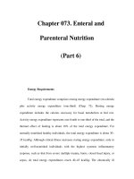
Table 1 The Permeability of Sclera to Tracers of Varying Molecular Weight and Molecular
Radius
Tracer
Molecular
weight (Da)
Molecular
radius (nm)
Permeability coefficient
(Â10
À6
cm/sec) (mean Æ SD)
Sodium fluorescein 376 0.5 84.5 Æ 16.1
FITC-D-4 kD 4400 1.3 25.2 Æ 5.1
FITC-D-20 kD 19,600 3.2 6.79 Æ 4.18
FITC-D-40 kD 38,900 4.5 2.79 Æ 1.58
FITC-BSA 67,000 3.62 5.49 Æ 2.12
Rhodamine D-70 kD 70,000 6.4 1.35 Æ 0.77
FITC-D-70 kD 71,200 6.4 1.39 Æ 0.88
FITC-IgG 150,000 5.23 4.61 Æ 2.17
FITC-D-150 kD 150,000 8.25 1.34 Æ 0.88
Abbreviations: FITC, fluorescein isothiocyanate; D, dextran; BSA, bovine serum albumin; IgG, immuno-
globulin G.
Transscleral Drug Delivery to the Retina and Choroid 195
molecular radius plays a greater role in determining scleral permeability than
molecular weight or charge, similar to diffusion through extracellular tissue in the
brain (31). Scleral permeability to the 70 kDa dextran is not significantly greater than
to the 150 kDa dextran, suggesting that the hydrodynamic radii of these molecules
within sclera is not identical to their molecular radii in aqueous solution. A similar
situation exists in brain tissue, where diffusion of 40 and 70 kDa dextrans are not
significantly different (32). These data are consistent with the existence of multiple
diffusion passages in sclera with varying size limitations.
Surgical thinning of the sclera predictably increases its permeability; however,
neither cryotherapy nor transscleral diode laser application alters permeability (4).
It also increases with increasing tissue hydration (28). Scleral permeability of small
molecules increases rapidly with increasing temperature (23). This low activation
energy supports the existence of an aqueous pore pathway rather than a transcellular
one. This paracellular pathway presumably traverses the aqueous media of the
mucopolysaccharide matrix.
Various prostaglandins (33) and their ester prodrugs diffuse across human
sclera (34). Hydrolysis of these ester prodrugs is less in the sclera than the cornea.
Thus, the sclera is relatively deficient in protease activity compared to the cornea.
Prostaglandins have been found to enhance human scleral permeability (25),
apparently via release of matrix metalloproteinases and subsequent collagen matrix
remodeling (35). Increased permeability also may result from reduction of intraocular
pressure. An increase of 15 mmHg pressure reduces scleral permeability to small mole-
cules roughly by half (24). These authors demonstrated, using Peclet number analysis
(36), that intraocular pressure reduces permeability not by inducing counteraction
hydrostatic flow, but by compressing scleral fibers and narrowing diffusion pathways.
IN VIVO STUDIES OF SCLERAL PERMEABILITY
Anders Bill performed many of the initial studies demonstrating the movement
of macromolecules across the sclera (37,38). Following injection of radio- or dye-
labeled albumin, or red dextran of molecular weight 40 kDa into the suprachoroidal
space of rabbits, both substances passed out through the sclera and accumulated
in the extraocular tissues, suggesting that they diffused through the sclera via peri-
vascular and interfibrillary spaces. In studies of uveoscleral outflow in the cynomol-
gus monkey, Thorotrast particles (10 nm) were observed to enter the sclera but not
latex spheres larger than 100 nm (39). Thus, the sclera has a large pore area with little
restriction to flow.
The diffusion of small molecules across the sclera has been well characterized.
Various sulfonamides diffuse across rabbit sclera (40,41). In patients undergoing
cataract surgery, methazolamide, a hydrophilic compound, penetrated the sclera
much faster than the cornea, but ethoxzolamide, which is lipophilic, had similar
diffusion across the sclera and cornea (40). Other studies also have found greater
scleral permeability to hydrophilic molecules than lipophilic ones (42,43).
The importance of the transscleral route in the penetration of topically admin-
istered macromolecules was demonstrated by denying corneal access by affixing a
glass cylinder to the corneoscleral junction with cyanoacrylate adhesive (44). Signifi-
cant intraocular levels of inulin (5 kDa) were obtained by topical administration
outside the cornea in their paradigm. Neither reentry from the general circulation
nor delivery by local vasculature accounted for the intraocular levels of the drug.
196 Ambati
Contralateral tissue drug levels were <1% of those in the treated eye. Further, intra-
venous administration achieved <5% of the tissue levels attained after topical dosing.
These results imply that syst emic absorption and intraocular reentry are not significant
routes of delivery. In animals where topical dosage was applied after sacrifice, there
was no significant difference in intraocular drug levels compared to live animals.
This implied that delivery by local vasculature was not a major component.
We studied the in vivo pharmacokinetics of transscleral delivery of IgG. We
used an osmotic pump, the tip of which was secured flush against bare sclera in
rabbits to facilitate unidirectional movement, to deliver fluorescently labeled IgG
(150 kDa) at rates on the order of mL/hr. Biologically relevant concentrations in
the choroid and retina were attained for periods of up to four weeks with negligible
systemic absorption (6). Levels in the vitreous and aqueous humors, and orbit were
negligible. Although there was a spatial concentration gradient, the IgG concentration
in the choroidal hemisphere distal to the footprint of the osmotic pump tip was half of
that in the proximal hemisphere. The elimination of IgG from the choroid and retina
followed first-order kinetics with half-lives of approximately two to three days.
Transscleral delivery is but a means to the end of achieving pharamacologic
effect. To confirm biological activity of an agent delivered via the transscleral route,
we used the following paradigm. Quite apart from its angiogenic action, VEGF
induces a phen omenon known as leukostasis, the adhesion of leukocytes to the vascu-
lar endothelium (45). This process is mediated by intracellular adhesion molecule-1
(ICAM-1), an endothelial cell surface receptor. Using an osmotic pump, we deliv-
ered a monoclonal antibody directed against ICAM-1 onto the scleral surface of
one eye of pigmented rabbits, an d a control isotype antibody to the other eye.
In treated eyes, VEGF-induced leukostasis, as measured by tissue myeloperoxidase
(an enzyme abundant in leukocytes) activity, was inhibited in the choroid by 80%
and in the retina by 70%. We also determined that there was remarkable selectivity
of delivery, with an ocular concentration that was 700 times the plasma concentra-
tion (6). The delivery of drug to the retina may be somewhat surprising in the face
of the classical belief that the retinal pigment epithelium (RPE) forms a strict
barrier to drug penetration; however, a recent study has shown that even molecules
as large as 80 kDa dextrans can diffuse through this tissue (46).
Periocular (subconj unctival, peribulbar, retrobulbar) injections can deliver
drugs to intraocular tissues via transcorneal or transscleral diffusion, or systemic
absorption and recirculation. Small molecules such as hydrocortisone diffuse across
the rabbit sclera after subconjunctival injection (47). There is one report of a large
molecule (tissue plasminogen activator –70kDa) reaching the vitreous cavity after
subconjunctival injection in rabbits (48). These authors suggested that most of the
penetration was via transscleral diffusion as levels in the plasma and fellow eye were
much lower than in the treated eye. However, other reports in humans suggest that
much of the intraocular delivery of subconjunctival or peribulbar injections is via
systemic absorption (49,50).
The high ocular selectivity in our experimental design may be due to the
sponge-like nature of the sclera. While bolus injections of drugs into the subconjunc-
tival space may overwhelm the absorptive capacity of the sclera, leading to systemic
absorption, the gradual delivery employed in our experiments may permit more
complete scleral absorption as the absorptive capacity of the sclera is more closely
matched to the rate of drug delivery.
Another drug delivery modality is iontophoresis, the application of an electrical
current to facilitate diffusion of an ionized drug across tissue barriers. von Sallmann
Transscleral Drug Delivery to the Retina and Choroid 197
(51,52) provided the first reports on transcorneal iontophoresis. This approach
was used to deliver various antibiotics into the anterior chamber (53,54) and vitr-
eous cavity (55).
Maurice was the first to report successful transscleral iontophoretic delivery of
fluorescein into the rabbit v itreous humor (56 ). Potentially therap eutic intraocular
levels of antibiotics (57–63), steroids (64), and anti-cytomegaloviral d rugs (65,66), and
anti-fungals (67) have been achieved via transscleral iontophoresis in animal models.
The intensity and duration of the applied current, as well as the configuration
of the electrode de termine the degree of intraocular penetration. Drug characteristics
such as its ionization and molecular shape also affect the rate of transscleral diffu-
sion. Hydrophilic drugs with low molecular weight that are ionized at physiological
pH are best suited for iontophoretic delivery. Negativel y charged molecules pene-
trate much better than positively charged ones (58,63).
Retinal damage appears to be an undesirable companion of transscleral ionto-
phoresis. Retinal edema occurs within seconds of current application (56). Disrup-
tion of the normal retinal architecture, hemorrhagic necrosis, fibrosis, and RPE
hyperplasia are evident beneath the iontophoretic site (68,69), and multiple applica-
tions exacerbate these injuries (70). Loss of tissue integrity, although spatially con-
fined, may permit drug entry into the vitreous cavity or even the anterior segment
tissues. Drug stability to possible electrochemical reactions is another possible
limitation of this approach.
Several types of drug formulations have been reported to enhance sustained
delivery of various drugs with a view to clinical use. Poly(lactic-co-glycolic) acid micro-
spheres have been used as an effective vehicle to deliver an anti-VEGF RNA aptamer
in a sustained fashion over 20 days, while preserving the drug’s bioactivity (71). Fibrin
sealants have been used to deliver carboplatin to the eye for up to two weeks (72). A
collagen matrix gel has been used to deliver cisplatin in a controlled-release fashion
that achieves higher intraocular concentrations than with the native drug (73). A bio-
degradable polymeric scleral plug delivering FK506 or betamethasone was found to
be highly effective in suppressing inflammation in experi mental uveitis for up to one
month (74–76). Oligonucleotides have also been reported to traverse the sclera, with
obvious implications for the burgeoning field of gene therapy (77). The clinically used
anesthetic benzalkonium chloride was found to enhance transscleral penetration of
molecules as large as 20 kDa dextran into the retina and choroid (78).
FUTURE DIRECTIONS
The eye presents unique challenges to the delivery of drugs. When the demand for
sustained delivery to the target tissue is coupled with the desire to avoid systemic
exposure, circumstances are ripe for creative approaches. Transscleral delivery is a
nondestructive and minimally invasive method that achieves targeted delivery of
high molecular weight compounds to the posterior segment. This drug delivery
modality exhibits linear kinetics of absorption and elimination, with the potential to
deliver constant doses of medication. By bridging the potential of our deepening under-
standing of mediators of disease to the need for focused pharmacologic interven-
tion, transscleral delivery sits at the crossroads of molecular medicine and
ophthalmology. It is robust as it is not limited to delivering anti-angiogenic drugs,
but can be extended to neuroprotective agents or vectors for gene transfer, which
hold promise in the treatment of glaucoma and other chorioretinal degenerations.
198 Ambati
A long-term transscleral delivery device may be clinically feasible because the
human eye is remarkably tolerant of foreign bodies overlying the sclera, such as
scleral buckles used in treating retinal detachment, even for years. Moreover, human
sclera is hypocellular and has a large surface area, both of which facilitate diffusion.
While orbital clearance, intraocular pressure, uveoscleral outflow, choroidal blood
flow, and the blood–retinal barri ers pose formidable hurdles, sustained, targeted
drug delivery to the choroid and retina in the clinical setting soon may be a reality.
REFERENCES
1. Kamei M, Misono K, Lewis H. A study of the ability of tissue plasminogen activator to
diffuse into the subretinal space after intravitreal injection in rabbits. Am J Ophthalmol
1999; 128:739–746.
2. Mordenti J, Cuthbertson RA, Ferrara N, et al. Comparisons of the intraocular tissue dis-
tribution, pharmacokinetics, and safety of 125I-labeled full-length and Fab antibodies in
rhesus monkeys following intravitreal administration. Toxicol Pathol 1999; 27:536–544.
3. Maurice DM, Polgar J. Diffusion across the sclera. Exp Eye Res 1977; 25:577–582.
4. Olsen TW, Edelhauser HF, Lim JI, Geroski DH. Human scleral permeability. Effects of
age, cryotherapy, transscleral diode laser, and surgical thinning. Invest Ophthalmol Vis
Sci 1995; 36:1893–1903.
5. Ambati J, Canakis CS, Miller JW, et al. Diffusion of high molecular weight compounds
through sclera. Invest Ophthalmol Vis Sci 2000; 41:1181–1185.
6. Ambati J, Gragoudas ES, Miller JW, et al. Transscleral delivery of bioactive protein to
the choroid and retina. Invest Ophthalmol Vis Sci 2000; 41:1186–1191.
7. Keeley FW, Morin JD, Vesely S. Characterization of collagen from normal human
sclera. Exp Eye Res 1984; 39:533–542.
8. Shuttleworth CA. Type VIII collagen. Int J Biochem Cell Biol 1997; 29:1145–1148.
9. Wessel H, Anderson S, Fite D, Halvas E, Hempel J, SundarRaj N. Type XII collagen
contributes to diversities in human corneal and limbal extracellular matrices. Invest
Ophthalmol Vis Sci 1997; 38:2408–2422.
10. Chapman SA, Ayad S, O’Donoghue E, Bonshek RE. Glycoproteins of trabecular mesh-
work, cornea and sclera. Eye 1998; 12:440–448.
11. White J, Werkmeister JA, Ramshaw JA, Birk DE. Organization of fibrillar collagen in the
human and bovine cornea: collagen types V and III. Connect Tissue Res 1997; 36:165–174.
12. Kimura S, Kobayashi M, Nakamura M, Hirano K, Awaya S, Hoshino T. Immunoelec-
tron microscopic localization of decorin in aged human corneal and scleral stroma.
J Electron Microsc (Tokyo) 1995; 44:445–449.
13. Marshall GE, Konstas AG, Lee WR. Collagens in the aged human macular sclera. Curr
Eye Res 1993; 12:143–153.
14. Komai Y, Ushiki T. The three-dimensional organization of collagen fibrils in the human
cornea and sclera. Invest Ophthalmol Vis Sci 1991; 32:2244–2258.
15. Thale A, Tillmann B, Rochels R. Scanning electron-microscopic studies of the collagen
architecture of the human sclera—normal and pathological findings. Ophthalmologica
1996; 210:137–141.
16. Raspanti M, Marchini M, Della Pasqua V, Strocchi R, Ruggeri A. Ultrastructure of the
extracellular matrix of bovine dura mater, optic nerve sheath and sclera. J Anat 1992;
181:181–187.
17. Fullwood NJ, Hammiche A, Pollock HM, Hourston DJ, Song M. Atomic force micro-
scopy of the cornea and sclera. Curr Eye Res 1995; 14:529–535.
18. Trier K, Olsen EB, Kobayashi T, Ribel-Madsen SM. Biochemical and ultrastructural
changes in rabbit sclera after treatment with 7-methylxanthine, theobromine, acetazo-
lamide, or l-ornithine. Br J Ophthalmol 1999; 83:1370–1375.
Transscleral Drug Delivery to the Retina and Choroid 199
19. Awetissow ES. The role of the sclera in the pathogenesis of progressive myopia (author’s
transl). Klin Monatsbl Augenheilkd 1980; 176:777–781.
20. Norn M. Topography of scleral emissaries and sclera-perforating blood vessels. Acta
Ophthalmol (Copenh) 1985; 63:320–322.
21. Olsen TW, Aaberg SY, Geroski DH, Edelhauser HF. Human sclera: thickness and sur-
face area. Am J Ophthalmol 1998; 125:237–241.
22. Sasaki H, Yamamura K, Tei C, Nishida K, Nakamura J. Ocular permeability of FITC-
dextran with absorption promoter for ocular delivery of peptide drug. J Drug Target
1995; 3:129–135.
23. Unlu N, Robinson JR. Scleral permeability to hydrocortisone and mannitol in the albino
rabbit eye. J Ocul Pharmacol Ther 1998; 14:273–281.
24. Rudnick DE, Noonan JS, Geroski DH, Prausnitz MR, Edelhauser HF. The effect of
intraocular pressure on human and rabbit scleral permeability. Invest Ophthalmol Vis
Sci 1999; 40:3054–3058.
25. Kim JW, Lindsey JD, Wang N, Weinreb RN. Increased human scleral permeability with
prostaglandin exposure. Invest Ophthalmol Vis Sci 2001; 42:1514–1521.
26. Crank J. The Mathematics of Diffusion. Oxford: Clarendon, 1975.
27. Carslaw HS, Jaeger JC. Conduction of heat in solids. London: Oxford University Press,
1959.
28. Boubriak OA, Urban JP, Akhtar S, Meek KM, Bron AJ. The effect of hydration and
matrix composition on solute diffusion in rabbit sclera. Exp Eye Res 2000; 71:503–514.
29. Cooper ER, Kasting G. Transport across epithelial membranes. J Control Release 1987;
6:23–35.
30. Edwards A, Prausnitz MR. Fiber matrix model of sclera and corneal stroma for drug
delivery to the eye. AIChE J 1998; 44:214–225.
31. Tao L, Nicholson C. Diffusion of albumins in rat cortical slices and relevance to volume
transmission. Neuroscience 1996; 75:839–847.
32. Nicholson C, Tao L. Hindered diffusion of high molecular weight compounds in brain
extracellular microenvironment measured with integrative optical imaging. Biophys
J 1993; 65:2277–2290.
33. Bito LZ, Baroody RA. The penetration of exogenous prostaglandin and arachidonic acid
into, and their distribution within, the mammalian eye. Curr Eye Res 1981; 1:659–669.
34. Madhu C, Rix P, Nguyen T, Chien DS, Woodward DF, Tang-Liu DD. Penetration of
natural prostaglandins and their ester prodrugs and analogs across human ocular tissues
in vitro. J Ocul Pharmacol Ther 1998; 14:389–399.
35. Gaton DD, Sagara T, Lindsey JD, Gabelt BT, Kaufman PL, Weinreb RN. Increased
matrix metalloproteinases 1, 2, and 3 in the monkey uveoscleral outflow pathway after
topical prostaglandin F(2 alpha)-isopropyl ester treatment. Arch Ophthalmol 2001;
119:1165–1170.
36. Incropera FP, DeWitt DP. Fundamentals of Heat and Mass transfer. New York: John
Wiley, 1996.
37. Bill A. The drainage of albumin from the uvea. Exp Eye Res 1964; 3:179–187.
38. Bill A. Movement of albumin and dextran through the sclera. Arch Ophthalmol 1965;
74:248–252.
39. Inomata H, Bill A. Exit sites of uveoscleral flow of aqueous humor in cynomolgus mon-
key eyes. Exp Eye Res 1977; 25:113–118.
40. Edelhauser HF, Maren TH. Permeability of human cornea and sclera to sulfonamide
carbonic anhydrase inhibitors. Arch Ophthalmol 1988; 106:1110–1115.
41. Kao KD, Lu DW, Chiang CH, Huang HS. Corneal and scleral penetration studies of
6-hydroxyethoxy-2-benzothiazole sulfonamide: a topical carbonic anhydrase inhibitor.
J Ocul Pharmacol 1990; 6:313–320.
42. Chien DS, Homsy JJ, Gluchowski C, Tang-Liu DD. Corneal and conjunctival/scleral
penetration of p-aminoclonidine, AGN 190342, and clonidine in rabbit eyes. Curr Eye
Res 1990; 9:1051–1059.
200 Ambati
43. Ahmed I, Gokhale RD, Shah MV, Patton TF. Physicochemical determinants of drug
diffusion across the conjunctiva, sclera, and cornea. J Pharm Sci 1987; 76:583–586.
44. Ahmed I, Patton TF. Importance of the noncorneal absorption route in topical ophthal-
mic drug delivery. Invest Ophthalmol Vis Sci 1985; 26:584–587.
45. Miyamoto K, Khosrof S, Bursell SE, et al. Vascular endothelial growth factor (VEGF)-
induced retinal vascular permeability is mediated by intercellular adhesion molecule-1
(ICAM-1). Am J Pathol 2000; 156:1733–1739.
46. Pitkanen L, Ranta V-P, Moilanen H, Urtti A. Permeability of retinal pigment epithelium:
effects of permeant molecular weight and lipophilicity. Invest Ophthalmol Vis Sci 2005;
46:641–646.
47. McCartney HJ, Drysdale IO, Gornall AG, Basu PK. An autoradiographic study of the
penetration of subconjunctivally injected hydrocortisone into the normal and inflamed
rabbit eye. Invest Ophthalmol 1965; 4:297–302.
48. Lim JI, Maguire AM, John G, Mohler MA, Fiscella RG. Intraocular tissue plasminogen
activator concentrations after subconjunctival delivery. Ophthalmology 1993; 100:
373–376.
49. Weijtens O, van der Sluijs FA, Schoemaker RC, et al. Peribulbar corticosteroid injection:
vitreal and serum concentrations after dexamethasone disodium phosphate injection. Am
J Ophthalmol 1997; 123:358–363.
50. Weijtens O, Feron EJ, Schoemaker RC, et al. High concentration of dexamethasone
in aqueous and vitreous after subconjunctival injection. Am J Ophthalmol 1999; 128:
192–197.
51. von Sallmann L. Sulfadizine iontophoresis in pyocyaneus infection of rabbit cornea. Am
J Ophthalmol 1942; 25:1292–1300.
52. von Sallmann L. Iontophoretic introduction of atropine and scopolamine into the rabbit
eye. Arch Ophthalmol 1943; 29:711–719.
53. Witzel SH, Fielding IZ, Ormsby HL. Ocular penetration of antibiotics by iontophoresis.
Am J Ophthalmol 1956; 42(no. 2, pt. 2):89–95.
54. Hughes L, Maurice DM. A fresh look at iontophoresis. Arch Ophthalmol 1984;
102:1825–1829.
55. Fishman PH, Jay WM, Rissing JP, Hill JM, Shockley RK. Iontophoresis of gentamicin
into aphakic rabbit eyes. Sustained vitreal levels. Invest Ophthalmol Vis Sci 1984;
25:343–345.
56. Maurice DM. Iontophoresis of fluorescein into the posterior segment of the rabbit eye.
Ophthalmology 1986; 93:128–132.
57. Choi TB, Lee DA. Transscleral and transcorneal iontophoresis of vancomycin in rabbit
eyes. J Ocul Pharmacol 1988; 4:153–164.
58. Church AL, Barza M, Baum J. An improved apparatus for transscleral iontophoresis of
gentamicin. Invest Ophthalmol Vis Sci 1992; 33:3543–3545.
59. Grossman RE, Chu DF, Lee DA. Regional ocular gentamicin levels after transcorneal
and transscleral iontophoresis. Invest Ophthalmol Vis Sci 1990; 31:909–916.
60. Barza M, Peckman C, Baum J. Transscleral iontophoresis of gentamicin in monkeys.
Invest Ophthalmol Vis Sci 1987; 28:1033–1036.
61. Barza M, Peckman C, Baum J. Transscleral iontophoresis of cefazolin, ticarcillin, and
gentamicin in the rabbit. Ophthalmology 1986; 93:133–139.
62. Burstein NL, Leopold LH, Bernacchi DB. Trans-scleral iontophoresis of gentamicin.
J Ocul Pharmacol 1985; 1:363–368.
63. Yoshizumi MO, Cohen D, Verbukh I, Leinwand M, Kim J, Lee DA. Experimental trans-
scleral iontophoresis of ciprofloxacin. J Ocul Pharmacol 1991; 7:163–167.
64. Lam TT, Edward DP, Zhu XA, Tso MO. Transscleral iontophoresis of dexamethasone.
Arch Ophthalmol 1989; 107:1368–1371.
65. Lam TT, Fu J, Chu R, Stojack K, Siew E, Tso MO. Intravitreal delivery of ganciclovir in
rabbits by transscleral iontophoresis. J Ocul Pharmacol 1994; 10:571–575.
Transscleral Drug Delivery to the Retina and Choroid 201