Neurological Differential Diagnosis - part 8 pps
Bạn đang xem bản rút gọn của tài liệu. Xem và tải ngay bản đầy đủ của tài liệu tại đây (382.1 KB, 56 trang )
378 Chapter 11
3 Albinism
◆
Albinism is a genetically determined abnormality in melanin synthesis that is
associated with congenital nystagmus, foveal hypoplasia, and impaired visual
acuity.
◆
The ocular fundus can be totally devoid of pigment or have a blond appear-
ance. The degree of visual impairment is usually inversely related to the degree
of ocular pigmentation.
4 Compressive lesions
◆
Craniopharyngiomas
◆
Optic nerve or chiasmatic gliomas – associated with neurofi bromatosis.
5 Hereditary optic atrophies
Nystagmus in infants
Features Spasmus nutans Congenital nystagmus
Age of onset 4 months–3 years Birth
Family history Negative Positive or negative
Nystagmus Asymmetric (30% unilateral) Bilateral and symmetric
Head movement Usually previous to nystagmus Simultaneous with nystagmus
Natural history Disappears in 36 months Usually persists
Other
Guidelines for the determination of brain death in children
•
Nystagmus in infants can be diffi cult to detect. Although the onset may be at
birth but it can be detected later.
•
The common forms are spasmus nutans and congenital nystagmus. The
common distinguishing features are provided in the table below.
•
Spasmus nutans is a self-limited disorder of infants, characterized by a slow
cephalic tremor associated with pendular horizontal and rarely vertical
nystagmus that is often monocular. Abnormal head positions are frequently
present.
•
Optic nerve and chiasmatic gliomas can simulate spasmus nutans.
Therefore, neuroimaging should always be obtained in such cases.
•
The guidelines for determination of brain death in children are similar to
adults, although they have some unique features, dealing specifi cally with
the age group from full-term newborn to the 5-year-old.
Pediatric Neurology 379
1 Coma and apnea must co-exist.
2 Absence of brainstem function
2.1 Pupils unreactive to light (midposition or dilated).
2.2 Absence of spontaneous eye movement, or in response to oculocephalic
and oculovestibular testing.
2.3 Absence of movement of bulbar musculature, including facial and oro-
pharyngeal muscles (corneal, gag, cough, sucking, and rooting refl exes).
2.4 Respiratory movements are absent with patient off the respirator.
2.5 Apnea testing using ‘standardized methods’ can be performed.
3 Absence of hypotension for age or hypothermia.
4 Flaccid muscle tone, absence of spontaneous movements (excluding spinal re-
fl exes).
5 Examination consistent with brain death throughout the period of testing and
observation.
6 Observation and testing according to age
6.1 7 days to 2 months: two examinations and EEGs separated by 48 hours.
6.2 2 months to 1 year: two examinations and EEGs separated by 24 hours.
6.3 Older than 1 year: when an irreversible cause exists, laboratory testing
is not required and an observation period of at least 12 hours is recom-
mended. A more prolonged period of at least 24 hours of observation is
recommended if it is diffi cult to assess the extent and reversibility of brain
damage (e.g. following an hypoxic-ischemic event). The observation pe-
riod may be reduced if an EEG demonstrates electrocerebral silence, or the
cerebral radionuclide and angiographic study does not visualize cerebral
arteries.
(Ref: Guidelines for the determination of brain death in children. Pediatrics 1987;
80: 298–300.)
Macrocephaly
•
These features are mainly focused on longer periods of recommended
observation relative to the patient’s age as outlined below.
•
Macrocephaly means enlargement of the head >2 standard deviations from
normal.
•
Statistically, most enlarged heads in children are due to either non-
pathological familial large head size or less commonly, hydrocephalus.
•
There are certain conditions with a large head without signifi cantly enlarged
ventricles that occur in the setting of serious neurological abnormalities.
Most neurodegenerative conditions of children cause small heads.
380 Chapter 11
1 Benign familial macrocephaly: large parental head size, child with normal de-
velopment
2 Hydrocephalus
◆
Features include frontal protuberance, bossing, sunset sign (a tendency for the
eyes to turn down so that the sclera is visible between the upper eyelids and iris),
thinning and/or prominence of scalp veins, and separation of cranial sutures.
2.1 Obstructive/noncommunicating hydrocephalus
2.1.1 Congenital malformation: aqueductal stenosis, Dandy-Walker,
Klippel-Feil syndrome, Chiari II.
2.1.2 Brain tumors: rare; particularly posterior fossa tumors (medulloblas-
toma, cerebellar, or brainstem astrocytoma, ependymoma, choroid
plexus papilloma), and pineal region tumors.
2.1.3 Vein of Galen malformation: may present with neonatal heart failure.
2.2 Communicating hydrocephalus
■
Secondary to subarachnoid hemorrhage or meningitis.
■
Meningeal malignancy.
3 Benign enlargement of subarachnoid space: also known as benign subdural
effusions, etc.
◆
More often in males, associated with large paternal head size.
◆
Distinguished by soft fontanelle, large head, normal development.
◆
Normal ventricular size on CT scan.
4 Subdural hematoma
◆
Signs include bulging fontanelle, vomiting, altered mental status.
◆
Unusual bruising, retinal hemorrhages, or fractures may point to child abuse.
5 Megalencephaly (large brain size)
5.1 Achondroplasia: AD, most are new mutations. True, often alarming, meg-
alencephaly. Usually normal cognitive development.
5.2 Sotos syndrome: variable inheritance, megalencephaly with gigantism.
5.3 Hemimegalencephaly: unilateral cerebral enlargement, associated with
poor development and intractable seizures/infantile spasms.
5.4 Neurocutaneous disorders: hypomelanosis of Ito, incontinentia pigmenti,
neurofi bromatosis, tuberous sclerosis, epidermal nevus syndrome (see
Chapter 12: Neurogenetics – Neurocutaneous syndromes).
5.5 Metabolic megalencephaly
5.5.1 Alexander disease (see Developmental regression in toddlers/chil-
dren, p. 366)
5.5.2 Canavan disease (see Developmental regression in infants, p. 363)
5.5.3 Glutaric aciduria type I: normal development with macrocephaly
until experiencing an encephalopathic event around age 2–3. After
this, spasticity and movement disorders may be prominent, with
variable cognition.
Pediatric Neurology 381
5.5.4 Storage disorders: gangliosidoses (Tay-Sachs, etc.), Krabbe, maple
syrup urine disease, metachromatic leukodystrophy, mucopolysac-
charidoses (see Developmental regression, pp. 364 and 366).
6 Hydranencephaly
◆
Means hydrocephalus plus destruction or failure of development of parts of
the cerebrum, often associated with enlargement of the skull.
◆
The fl uid-fi lled region of the cranium is seen when transilluminated.
7 Conditions with a thickened skull: include anemia, cleidocranial dysostosis, os-
teogenesis imperfecta, osteopetrosis, rickets, etc.
Nightmares vs. night terrors
Nightmare Night terror
Repeated, clinically signifi cant awakening
from sleep with a detailed recall of disturbing
dreams
Abrupt, recurrent, and clinically signifi cant
awakening from sleep, accompanied by panic
and autonomic arousal
Individuals rapidly become alert and
oriented after awakening from nightmares
Individuals are usually unresponsive to the
environment and have subsequent amnesia
for the episode
Polysomnographic recording shows sudden
awakening from REM sleep at the time the
individual reports nightmares
Polysomnographic recording shows sudden
partial awakening from NON-REM sleep
Often begins in childhood and resolves
quickly
Onset is usually middle childhood and resolves
during late childhood or early adolescence
Reassurance only. No therapy or medications
are required
Reassurance is helpful. Diazepam or clonidine
have been used to treat, but are not necessary
•
Individuals, especially children, sometimes have isolated episodes of sudden
arousal from sleep in a condition of terror and confusion.
•
Common disorders include nightmare and night terror. The important clues
for differentiation are provided below. Other differential diagnoses include
sleepwalking disorder, other parasomnias, hypnagogic hallucinations in
narcolepsy, substance-induced sleep disorders, and nocturnal seizures.
•
Normal neurological exam
382
Chapter 12
Neurogenetics
When to suspect genetic disease 383
Patterns of inheritance 383
Examples of autosomal dominant disorders 384
Examples of autosomal recessive disorders (many others) 385
Examples of X-linked disorders 385
Examples of chromosomal autosomal disorders 385
Examples of sex chromosome disorders 386
Mitochondrial encephalomyopathies 386
Diseases due to trinucleotide repeat expansions 387
Heritable human prion diseases 389
Clinical syndromes 389
Hereditary/genetic ataxias 389
Hereditary/genetic epilepsy syndromes 390
Alzheimer disease genetics 393
Hereditary/genetic movement disorders 393
Hereditary/genetic myopathies 395
Neurocutaneous syndromes 398
Hereditary/genetic peripheral neuropathies 399
Neurological Differential Diagnosis: A Prioritized Approach
Roongroj Bhidayasiri, Michael F. Waters, Christopher C. Giza,
Copyright © 2005 Roongroj Bhidayasiri, Michael F. Waters and Christopher C. Giza
Neurogenetics 383
When to suspect genetic disease
1 Positive family history. This should be explored in detail, as many patients will
initially deny any known history of hereditary disease.
2 Unusual morphologic features, especially:
◆
facial dysmorphology
◆
atypical body habitus
3 Absence of obvious alternative etiologies (such as ischemia, infection, and trauma).
4 Clinical constellation with known neurogenetic association, such as:
◆
ataxia
◆
neuropathy
◆
ophthalmoplegia
◆
muscle weakness
◆
progressive myoclonic seizures
◆
developmental regression in children
5 Neurologic disease with additional organ system involvement, such as:
◆
cardiomyopathy
◆
hepatosplenomegaly
◆
cutaneous manifestations
◆
renal disease
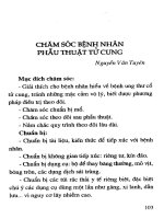
Patterns of inheritance
Patterns of inheritance Risk to offspring Gender bias Transmission
Autosomal dominant 50% Males and females
equally affected
Multiple affected generations
and multiple individuals in
one generation: includes father
to son transmission.
Autosomal recessive 25% Males and females
equally affected
May ‘skip’ a generation.
Carriers may be asymptomatic
X-linked recessive 50% risk to males
or female carriers
Males Multiple affected generations
and multiple individuals in
one generation: father to son
transmission is not seen
Mitochondrial All children at risk Males and females
equally affected
Variable expression and
disease severity, maternal
transmission only
384 Chapter 12
Patterns of inheritance
Examples of autosomal dominant disorders
•
Autosomal dominant nocturnal frontal lobe epilepsy
•
Benign familial neonatal seizures
•
Bethlem myopathy
•
Central core myopathy
•
Charcot-Marie-Tooth disease (HMSN I)
•
Childhood absence epilepsy
•
Dentatorubro-pallidoluysian atrophy
•
Dopa-responsive dystonia
•
Essential tremor
•
Familial amyloid polyneuropathy
•
Familial episodic ataxia
•
Familial hyperkalemic periodic paralysis
•
Familial hypokalemic periodic paralysis
•
Familial paroxysmal choreoathetosis
•
Fascioscapulohumeral dystrophy
•
Hereditary neuralgic amyotrophy
•
Hereditary neuropathy with pressure palsies
•
Huntington disease
•
Hyperekplexia
•
Juvenile myoclonic epilepsy
•
Myotonic dystrophy
•
Neurofi bromatosis 1
•
Spinocerebellar ataxias (many types)
•
Tuberous sclerosis
•
Von Hippel Lindau disease
Autosomal Dominant Autosomal Recessive
X-Linked Mitochondrial
Neurogenetics 385
Examples of autosomal recessive disorders (many others)
•
Ataxia telangiectasia
•
Canavan disease
•
Cerebrotendinous xanthomatosis
•
Dejerine-Sottas disease (HMSN III)
•
Friedreich ataxia
•
Galactosemia
•
Gaucher disease
•
Homocystinuria
•
Isovaleric acidemia
•
Krabbe disease
•
Maple syrup urine disease
•
Metachromatic leukodystrophy
•
Mucopolysaccharidosis type I (Hurler)
•
Mucopolysaccharidosis type III
•
Neuronal ceroid lipofuscinosis, infantile
•
Niemann-Pick disease
•
Phenylketonuria
•
Propionic academia
•
Refsum disease, infantile
•
Sandhoff disease
•
Spinal muscular atrophy type I (Werdnig-Hoffman)
•
Tay-Sachs disease
•
Most congenital metabolic disorders
Examples of X-linked disorders
•
Adrenoleukodystrophy, juvenile
•
Becker muscular dystrophy
•
Duchenne muscular dystrophy
•
Emery-Dreifuss muscular dystrophy
•
Fragile X syndrome
•
Lesch-Nyhan disease
•
Menkes disease
•
Mucopolysaccharidosis type II (Hunter, X-linked recessive)
•
Rett syndrome (X-linked dominant, lethal in males?)
•
Ornithine transcarbamylase defi ciency
•
X-linked hydrocephalus
Examples of chromosomal autosomal disorders
•
Angelman syndrome (deletion of part of long arm of maternal 15q)
•
Cri-du-chat syndrome (deletion of short arm of 5p)
•
Down syndrome (trisomy 21)
386 Chapter 12
•
Prader-Willi syndrome (deletion of part of long arm of paternal 15q)
•
Trisomy 13
•
Trisomy 18
Examples of sex chromosome disorders
•
Klinefelter syndrome (XXY)
•
XYY karyotype
•
Turner syndrome (45,X)
Mitochondrial encephalomyopathies
Disorder Disease features
Mitochondrial encephalomyopathy, lactic
acidosis, and stroke-like episodes (MELAS)
Seizures, developmental delay, growth
retardation, headaches, and stroke-like episodes
with focal neurologic defi cits
Myoclonic epilepsy and ragged red fi bers
(MERRF)
Myoclonus, ataxia, seizures, myopathy, and
peripheral neuropathy
Leber hereditary optic neuropathy (LHON) Optic disc swelling with visual fi eld loss
progressing to involve central vision
Neurogenic weakness, ataxia, and retinitis
pigmentosa syndrome (NARP)
Developmental delay, seizures, dementia, ataxia,
sensory neuropathy, proximal weakness, and
retinitis pigmentosa
Maternally inherited Leigh syndrome (MILS) Dementia, spasticity, optic atrophy
Chronic progressive external ophthalmoplegia
(CPEO)
Ptosis, extra-occular ophthalmoplegia, proximal
limb myopathy
Kearns-Sayre syndrome (KSS) A CPEO-plus syndrome which includes the
above as well as onset prior to age 20 years, heart
block, ataxia, and pigmentary retinopathy
•
Mitochondrial DNA (mtDNA) is nearly exclusively maternally inherited.
•
mtDNA is a closed, circular DNA molecule consisting of ~16,000
nucleotides coding for 37 genes.
•
Every cell contains multiple mitochondria with multiple copies of DNA, a
condition known as polyplasmy.
•
Mitochondrial diseases demonstrate a threshold effect, whereby disease
onset and severity are a function of the balance between the proportion of
mutant and wild-type mtDNAs.
•
May involve multiple tissues with high energy requirements – such as brain,
retina, muscle, cochlea, etc.
Neurogenetics 387
Diseases due to trinucleotide repeat expansions
Disorder Gene and
locus
Protein Repeat
sequence
and location
Disease features Inheritance
Fragile X syndrome
1/1,500 males
FMR1
Xq27.3
FMRP CGG
5’-UTR
Mental
retardation,
elongated facies,
large ears,
macroorchidism
X-linked
Friedreich ataxia
1/50,000
X25
9q13–21.1
Frataxin GAA
1st intron
Ataxia, dysarthria,
extensor plantar
response, arefl exia
AR
Huntington disease
1/20,000 Caucasians
1/100,000 African
Americans
IT15
4p16.3
Huntington CAG coding Personality
change, motor
abnormalities,
extrapyramidal
signs, dysarthria
AlzD
Myotonic dystrophy
1/7500
DMPK
19q13.3
Myotonic
dystrophy
protein
kinase
CTG
3’-UTR
Ptosis, facial
atrophy, proximal
weakness, cardiac
conduction
abnormalities
AlzD
Dentatorubro-
pallidoluysian
atrophy
DRPLA
12p13.31
Atrophin-1 CAG coding Ataxia, personality
changes, chorea,
tonic-clonic
seizures, dementia
AlzD
Spinobulbar
muscular atrophy
(Kennedy disease)
AR
Xq13.21
Androgen
receptor
CAG coding Lower motor
neuron disease,
feminization
X-linked
•
Unstable expansion of trinucleotide repeats is the mechanism underlying
disorders featuring anticipation, the tendency for subsequent generations to
be affected at an earlier age and with increased severity.
•
The molecular mechanisms of disease are poorly understood as the triplet
expansions are known to be located in coding sequences (Huntington
disease), non-coding sequences (Friedreich ataxia), 5’-untranslated regions
(fragile X syndrome), and 3’-untranslated regions (myotonic dystrophy).
•
CAG repeat expansions that encode polyglutamines appear to result in
protein misfolding, a recuring theme in many of these disorders.
•
DNA molecular diagnostic tests are available. However, estimating disease
onset and severity within an individual is not accurately predicted by
absolute repeat number. Therefore, great care must be taken to ensure
accurate information is provided when using molecular data in genetic
counseling, particularly in asymptomatic individuals.
Continued
388 Chapter 12
Disorder Gene and
locus
Protein Repeat
sequence
and location
Disease features Inheritance
Spinocerebellar
ataxia type 6
CACNA1A
19p13
α
1A
subunit
of voltage-
gated Ca
channel
CAG coding Ataxia, cerebellar
symptoms
AlzD
Spinocerebellar
ataxia type 3
(Machdo-Joseph
disease)
SCA3/MJD1
14q32.1
Ataxin-3 CAG coding Ataxia, peripheral
sensorimotor
neuropathy,
pyramidal, and
extrapyramidal
signs
AlzD
Spinocerebellar
ataxia type 2
SCA2
12q24.1
Ataxin-2 CAG coding Ataxia, arefl exia,
dementia, slow
saccades
AlzD
Spinocerebellar
ataxia type 1
SCA1
6p23
Ataxin-1 CAG coding Ataxia, pyramidal
signs, vibratory
loss, dysphagia
AlzD
Spinocerebellar
ataxia type 7
SCA7
3p12–13
Ataxin-7 CAG coding Ataxia, retinal
degeneration
AlzD
Spinocerebellar
ataxia type 8
SCA8
13q21
KLH1 CTG
5’-UTR
Ataxia, cerebellar
signs
AlzD
Spinocerebellar
ataxia type 10
SCA10
22q13
Ataxin-10 ATTCT
9th intron
Ataxia, cerebellar
signs with epilepsy
AlzD
Spinocerebellar
ataxia type 12
PPP2R2B
5q31–33
PP2A
regulatory
protein
CAG
5’-UTR
Ataxia, upper
extremity and
head tremor,
cerebellar signs,
hyperrefl exia,
dementia
AlzD
Spinocerebellar
ataxia type 17
SCA17
6q27
TATA
binding
protein
CAG coding Ataxia,
hyperrefl exia,
plantar responses,
seizures, cognitive
decline
AlzD
Oculopharyngeal
dystrophy
PABP2
14q11
Poly-A
binding
protein
GCG coding Ptosis, diplopia,
dysphagia,
proximal
weakness
AlzD
Fragile XE mental
retardation
FMR2
Xq28
FMR2 GCC
5’-UTR
Mental
retardation
X-linked
Myotonic dystrophy
type 2
ZNF9
3q13–24
Zinc fi nger
protein 9
CTG
1st intron
Ptosis, facial
atrophy, proximal
weakness, cardiac
conduction
abnormalities
AlzD
Neurogenetics 389
Heritable human prion diseases
Disorder Disease features
Creutzfeldt-Jakob disease Rapidly progressive (over weeks/months) dementia with cerebellar
dysfunction, myoclonus, and combinations of pyramidal and
extrapyramidal pathology. Characteristic EEG evolution to either
1–2 Hz triphasic spikes or ‘burst suppression’
Gerstmann-Sträussler-
Scheinker syndrome
Predominant ataxia with dysphagia, dysarthria, hyporefl exia, and
dementia. Slower course (months/years). Familial
Fatal familial insomnia Predominant intractable insomnia, endocrine dysfunction, and
dysautonomia with progressive dementia, rigidity, and dystonia.
Slower course (months/years). Familial
Clinical syndromes
Hereditary/genetic ataxias
•
Poorly understood pathogenesis mediated by prion protein.
•
Multiple mutations characterized.
•
All show autosomal dominant inheritance.
•
Amyloid plaques and spongiform changes seen histologically.
Caused by mutations in the human prion protein gene, PRNP (unknown
function), located on chromosome 20pter-12.
•
The hereditary ataxias collectively share many clinical features, including
gait ataxia, limb dysmetria, intention tremor, and dysphagia. Most of these
pathological features arise by virtue of cerebellar dysfunction.
•
Though there is great overlap amongst the hereditary ataxias, some carry
defi ning features such as bulbar involvement, peripheral neuropathies,
oculomotor palsies, and dementia.
•
Typically present around the third decade, though onset varies from 1st to
7th decade.
•
Slow, though relentless, progression over the course of approximately 15
years.
•
Important to rule out other more common causes of ataxia when making
diagnosis.
•
Molecular diagnostic testing available for disease confi rmation in many
hereditary ataxias.
•
See Chapter 6: Movement Disorders and Diseases due to trinucleotide repeat
expansions, pp. 387–8.
390 Chapter 12
Hereditary/genetic epilepsy syndromes
Disorder Location Gene/protein Disease features Inheritance
Angelman syndrome 15q11–13
maternal
deletion
UBE3A/
E6-AP ubiquitin-
protein ligase
Seizures, mental
retardation, ‘happy
puppet syndrome’
Maternally
inherited
deletion
Autosomal dominant
nocturnal frontal lobe
epilepsy
20q13,
15q14?
1?
CHRNA4/α4
subunit AChR
Unknown
CHRNB2/-2
subunit AChR
Nocturnal seizures,
onset 10 years, normal
development
AlzD
Benign childhood
epilepsy with centro-
temporal spikes
1?
15q14?
Unknown
CHRNA7/-7
subunit AChR?
Simple partial
orofacial seizures,
onset 3–13 years,
associated with sleep
or awakening
AlzD
Benign familial
infantile convulsions
19q
16p12-q12?
Unknown Partial seizures, onset
4–7 months, normal
development
AlzD
Benign familial
neonatal convulsions
20q13, 8q24 KCNQ2 or
KCNQ3/KQT-
like K
+
channel
Multifocal clonic
seizures that self-
resolve, onset 1st
postnatal week,
normal development
AlzD
•
Family history is not uncommon in epilepsy syndromes. Improved genetic
testing is delineating a genetic etiology to many previously idiopathic seizure
syndromes.
•
Some genetic syndromes are associated primarily with epilepsy, with few
additional neurological or behavioral symptoms. Mutations in ion channels
are increasingly being identifi ed as the etiology for these syndromes.
•
Many genetic syndromes have seizures and developmental delay as
components of multisystem dysfunction due to abnormal metabolism or
storage. Mutations in metabolic enzymes are often responsible for these
disorders.
•
Structural chromosomal abnormalities are also associated with seizures and
developmental delay. Dysmorphic features and structural abnormalities of
the brain and other organs occur more commonly in these patients.
•
Improved neuroimaging techniques can detect subtle cerebral dysgenesis in
epilepsy patients that might have previously been classifi ed as idiopathic.
Neurogenetics 391
Disorder Location Gene/protein Disease features Inheritance
Bilateral
periventricular
nodular heterotopia
Xq28 Unknown Lethal in males,
seizures and normal
development in
females
XL
Childhood absence
epilepsy
20q?
8q24?
CHRNA4/-4
subunit AChR?
Unknown
Absence seizures,
onset 3–12 years, 3 Hz
spike wave on EEG,
normal development
AlzD
Down syndrome Trisomy 21 Unknown Mental retardation,
seizures, hypotonia,
characteristic facies
Triploidy
Familial temporal
lobe epilepsy
10q? Unknown Complex partial
seizures, some febrile
seizures
AlzD
Febrile seizures
with temporal lobe
epilepsy
8q? Unknown Febrile and complex
partial seizures
AlzD
Febrile seizures alone 19p13 FEB2/? Febrile seizures alone AlzD
Fragile X syndrome Xq27.3 FMR-1/frataxin Mental retardation,
seizures, large
ears, hypotonia,
macrocephaly,
macroorchidism
XL
Generalized epilepsy
with febrile seizures
plus (GEFS+)
19q?
2q?
SCN1A, 1B, 2A/
sodium channel
subunits
Febrile seizures, later
afebrile seizures
AlzD
Incontinentia
pigmenti
X Unknown Pigmented skin
lesions, mental
retardation,
polymicrogyria
XL
Juvenile absence
epilepsy
8q24?
21q22?
GRIK1?/GluR5?
GABRA1/
GABA
A
R subunit
Absence seizures,
onset 8–17 years,
>3 Hz spike wave,
normal development
AlzD
Juvenile myoclonic
epilepsy
6p?
15q14?
15q11–13?
EJM1?
CHRNA7/-7
subunit AchR
GABA
A
/GABAR
Generalized seizures
(tonic-clonic,
myoclonic, absence),
onset puberty, 4–6 Hz
spike wave, normal
development
AlzD
Continued
392 Chapter 12
Disorder Location Gene/protein Disease features Inheritance
Lissencephaly and
band heterotopia
Xq22 XSCLH or LIS/
double cortin
Lissencephaly (lethal
in males), band
heterotopia (in
females)
XL
MERRF mt DNA tRNA
Lys
/tRNA
Lys
Myoclonic epilepsy,
myopathy
Mitochondiral
Miller-Dieker
syndrome
17q13
hemizygous
deletion
LIS-1/
homologue of G-
protein subunit
Lissencephaly,
severe seizures and
developmental delay
?
Neuronal ceroid
lipofuscinosis, type 1
1q32 PPT/palmitoyl
protein
thioesterase
Progressive myoclonic
seizures, dementia,
retinal degeneration
AR
Neurofi bromatosis 1 17q12–22 neurofi bromin Macrocephaly,
café-au-lait, axillary
freckles, seizures
AlzD
Nonketotic
hyperglycinemia
? Unknown Hypotonia, neonatal
seizures, opisthotonus,
myoclonus, apnea
AR
Partial epilepsy with
auditory features
10q LGI1/? Partial seizures
with ictal auditory
disturbances, onset
8–19 years, normal
development
AlzD
Primary reading
epilepsy
? Unknown Simple partial jaw
seizures, triggered by
reading
AlzD
Progressive epilepsy
with mental
retardation
8p Unknown Progressive seizures,
onset 5–10 years,
mental retardation
AR
Pyridoxine
dependency
? Unknown Neonatal seizures/
status epilepticus
AR
Tuberous sclerosis 9q343.3
16p13.3
TSC1/Hamartin
TSC2/Tuberin
Ash leaf spot, other
skin lesions, mental
retardation, seizures,
visceral tumors, brain
tumors
AlzD
Unverricht-Lundborg 21q22.3 EPM1/cystatin B Generalized tonic-
clonic seizures,
myoclonus, onset 8–13
years, mild dementia
AR
Neurogenetics 393
Alzheimer disease genetics
Gene Chromosome Age at onset % of AlzD
Amyloid precursor protein (APP) 21 45–60 < 1
Presenilin-1 (PS-1) 14 30–60 1–5
Presenilin-2 (PS-2) 1 50–65 < 1
Apolipoprotein E (APOE-4 alleles) 19 60+ 50–60
Hereditary/genetic movement disorders
For hereditary ataxias, see p. 389 and Chapter 6: Movement Disorders.
•
The most important risk factor for AlzD is age, with family history as the
second most important risk factor.
•
Familial AlzD is characterized by multiple affected individuals in which the
disease segregates in a manner consistent with fully penetrant autosomal
dominant inheritance. They probably comprise 5% or less of all cases with
AlzD.
•
Familial AlzD is divided into early and late onset categories with 60–65 years
as the dividing line. Thus far, familial AlzD has been shown to be caused by
three different genes.
•
Recently, apolipoprotein E (APOE) has been considered as an important
genetic susceptibility risk factor for the development of sporadic AlzD.
Individuals who are homozygous and carry two APOE-4 alleles have an
increased probability (>90%) of developing AlzD by the age of 85 and do so
about 10 years earlier than individuals carrying the -2 or -3 allelic variants.
•
Some movement disorders are clearly hereditary, and family history should
be sought as a clue to the underlying diagnosis. Incomplete penetrance may
obscure autosomal dominantly inherited disorders.
•
Particularly for paroxysmal movement disorders, family history may be
incomplete or unknown by the patient.
•
Many of the hereditary movement disorders demonstrate variable but
nonspecifi c symptoms of basal ganglia involvement, including dystonia,
Parkinsonism, choreoathetosis, tics, and dyskinesias.
•
Neuroimaging abnormalities may also be seen in the basal ganglia (e.g. Fahr
disease with calcifi cation, Hallervorden-Spatz with iron deposition, Wilson
disease with copper deposition).
394 Chapter 12
Disorder Gene/location Clinical features Inheritance
Essential (familial)
tremor
3q13
2p
Limb or head tremor, normal
development
AlzD
Huntington disease 4p16.3 Choreoathetosis, dementia,
psychosis
AlzD
Tics/Tourette syndrome Unknown Tics, ADHD, obsessive compulsive
behavior
AlzD
Benign familial chorea Unknown Mild continuous childhood
chorea, normal development
AlzD
Chorea-acanthocytosis 9q21 Tics, self-mutilating orofacial
dyskinesia, dystonia, dementia,
normal lipid profi le
AR
Dementia-Parkinsonism-
amyotrophy complex
17q21–23 Parkinsonism, dementia, weakness,
spasticity
AlzD
Dopa-responsive dystonia 14q11-q24.3 Dystonia, Parkinsonism, diurnal
worsening of symptoms
AlzD
DRPLA 12p12 Chorea, myoclonus, ataxia,
seizures, dementia
AlzD
Fahr disease 14q Dementia, choreoathetosis,
focal dystonia, Parkinsonism,
calcifi cation of basal ganglia
AlzD
Familial CJD 20p terminal
p12
Dementia, ataxia, myoclonus AlzD
Glutaric aciduria 19p13.2 Megalencephaly, dystonia,
choreoathetosis
AR
Hallervorden-Spatz or
PKAN
20p12.3-p13 Dystonia, retinitis pigmentosum,
dementia
AR
Hereditary hyperekplexia 5q Exaggerated startle response AlzD
Idiopathic torsion
dystonia
9q32-q34 Dystonia, initially focal, but later
generalized
AlzD
Juvenile myoclonic
epilepsy
6p Seizures (tonic-clonic, myoclonic
and/or absence), myoclonus
AlzD
LHON mito 3460, mito
11778, mito
14484l
Optic neuropathy, dystonia Maternal
Lubag disease Xq13 Dystonia, parkinsonism X-linked
MERRF tRNA gene Encephalomyopathy Maternal
Neurogenetics 395
Disorder Gene/location Clinical features Inheritance
McLeod syndrome Xp21 Arefl exia, dystonia, chorea,
cardiomyopathy, muscular
dystrophy, dementia, seizures, tics,
normal lipid profi le
X-linked
NARP mito 8993 Neuropathy, ataxia, retinitis
pigmentosa
Maternal
Paroxysmal kinesigenic
choreoathetosis
16p11.2-q12.1 Episodic choreoathetosis or
dystonia, triggered by movement
AlzD
Paroxysmal
nonkinesigenic
choreoathetosis
2q Episodic choreoathetosis or
dystonia, not triggered by
movement
AlzD
Unverricht-Lundborg
disease
21q22.3 Progressive myoclonic epilepsy AlzD
Wilson disease 13q14.3 Tremor, dementia, hepatic failure AR
CJD – Creutzfeldt-Jakob disease, DRPLA – dentatorubral pallidoluysian atrophy, LHON – Leber hereditary
optic neuropathy, NARP – neuropathy, ataxia, retinitis pigmentosa, MERRF – mitochondrial encephalopathy
with ragged red fi bers, PKAN – pantothenate kinase-associated neurodegeneration, SCA – spinocerebellar
ataxia.
Ref: Modifi ed from Fahn S., Greene P.E., Ford B., Bressman S.B. Handbook of Movement Disorders.
Hereditary/genetic myopathies
Disorder Gene and
locus
Protein Disease features Inheritance
pattern
Duchenne MD
1/3,300 male births
DMD
Xp21
Dystrophin
(absent)
Severe, progressive
proximal muscle
weakness, loss of
ambulation by age
10, cardiomyopathy,
mental retardation
X-linked
Continued
•
The muscular dystrophies are primarily genetic myopathies resulting from
disturbances in structural proteins. Many of the actual gene defects are
known, as is the pathophysiology in some cases.
•
Clinically, patients present with weakness and atrophy.
•
The childhood dystrophies may present with failure to achieve milestones.
•
In most instances there is a slow and progressive decline in function.
•
Patterns of weakness vary and may be predominantly distal, proximal, or
involve specifi c muscle groups such as extraocular muscles.
396 Chapter 12
Disorder Gene and
locus
Protein Disease features Inheritance
pattern
Myotonic dystrophy
1/7500 live births
DMPK
19q13.3
Myotonic
dystrophy
protein kinase
Ptosis, facial atrophy,
proximal weakness,
cardiac conduction
abnormalities,
cataracts, testicular
atrophy, diabetes
mellitus
AlzD
Becker MD
1/20,000 male births
BMD
Xp21
Dystrophin
(reduced)
Similar to DMD,
though less severe
and later onset,
ambulation after
age 10
X-linked
Facioscapulohumeral
MD
1/320,000 live births,
higher in Dutch
ancestry
FSHD
4q35
Unknown Facial weakness as
well as shoulder
girdle and mild leg
weakness
AlzD
Emery-Dreifuss MD EDMD
Xq28
Emerin Similar to BMD,
though less severe;
joint contractures
and cardiac
conduction defects
X-linked
Fukyama type MD FCMD
9q31–33
Fukutin Hypotonia, atrophy,
weakness, intellectual
delay with seizures
and cortical dysplasia
AR
Limb-girdle 1A MD,
prevalence of ALL
LGMD 1/100,000
LGMD 1A
5q22–34
Unknown Progressive weakness
of shoulder and
pelvic girdle muscles
AlzD
Limb-girdle 1B MD LGMD 1B
1q11–21
Unknown Same as LGMD 1A AlzD
Limb-girdle 1C MD LGMD 1C
3p25
Caveolin-3 Same as LGMD 1A AlzD
Limb-girdle 2A MD LGMD 2A
15q15.1
Calpain 3 Same as LGMD 1A AR
Limb-girdle 2B MD LGMD 2B
2p13
Dysferlin Same as LGMD 1A AR
Limb-girdle 2C MD LGMD 2C
13q12
γ-sarcoglycan Same as LGMD 1A AR
Limb-girdle 2D MD LGMD 2D
17q21
α-sarcoglycan Same as LGMD 1A AR
Neurogenetics 397
Disorder Gene and
locus
Protein Disease features Inheritance
pattern
Limb-girdle 2E MD LGMD 2E
4q12
β-sarcoglycan Same as LGMD 1A AR
Limb-girdle 2F MD LGMD 2F
5q31
δ-sarcoglycan Same as LGMD 1A AR
Limb-girdle 2G MD LGMD 2G Unknown Same as LGMD 1A AR
Miyoshi distal
myopathy
2p13 Dysferlin Plantar fl exion
weakness with
gastrocnemius
atrophy
AR
Epidermolysis bullosa
simplex with MD
MD-EBS
8q24
Plectin Proximal muscle
weakness with
blistering disorder
AR
Scapuloperoneal MD SPMD
12q21
Unknown Foot drop and
shoulder girdle
weakness
AlzD
Oculopharyngeal MD OPMD
14q11.2-q13
Poly(A)binding
protein
Ptosis, dysphagia,
diplopia, shoulder
girdle weakness
AlzD
Bethlem myopathy 1 21q22 α1(VI) collagen Flexion contractures
with mild proximal
muscle weakness and
atrophy
AlzD
Bethlem myopathy 2 21q22 α2(VI) collagen Same as Bethlem
myopathy 1
AlzD
Bethlem myopathy 3 2q37 α3(VI) collagen Same as Bethlem
myopathy 1
AlzD
Hyperkalemic periodic
paralysis
17q23.1-q25.3 Sodium channel
subunit, SCN4A
Episodic paralysis,
often after exertion,
myotonia
AlzD
Paramyotonia
congenita
17q23.1-q25.3 Sodium channel
α subunit,
SCN4A
Mytonia precipitated
by cold or exertion
AlzD
Myotonia congenita
(Thomsen disease or
Becker disease)
7q32 Voltage-gated
chloride channel,
CLCN1
Myotonia, muscle
stiffness, muscular
hypertrophy
AlzD
(Thomsen)
AR (Becker)
MD – Muscular dystrophy, DMD – Duchenne MD, BMD – Becker MD.
398 Chapter 12
Neurocutaneous syndromes
Disorder Gene and
locus
Protein Disease features Inheritance
Neurofi bromatosis
type 1 (NF1)
1/3,500
NF1
17q11.2
Neuro-
fi bromin
Café-au-lait spots, lisch
nodules, neurofi bromas,
bony abnormalities,
learning disabilities,
epilepsy, vascular
malformations, benign
and malignant tumors
AlzD
Tuberous sclerosis
1/10,000
Tuberous
sclerosis
complex 1
(TSC1)
9q34.1–34.2
Hamartin Cortical tubers with
developmental delay
and epilepsy, facial
sebaceous angiofi bromas,
periungual fi bromas,
shagreen patch, ash-leaf
spots, subependymal
giant cell astrocytoma,
atrial myxoma, renal
angiomyolipoma
AlzD
Tuberous sclerosis TSC2
16p13.3
Tuberin Same as TSC1 AlzD
Neurofi bromatosis
type 2 (NF2)
1/35,000
NF2
chrom22
Schwannomin
(merlin)
Bilateral vestibular
schwannomas,
neurofi bromas,
retinal hamartomas,
meningiomas,
ependymomas
AlzD
Sturge-Weber
syndrome
Unknown Unknown Facial port-wine stain,
seizures, hemiparesis/
plegia, glaucoma, mental
retardation
Variable
Von Hippel-Lindau
disease
VHL
3p25–26
pVHL Retinal angioma,
cerebellar hemangioma,
spinal hemangioma,
pheochromocytoma,
pancreatic cysts
AlzD
•
The hereditary phakomatoses derive their name from the shared features of
either cutaneous or neurological abnormalities, or both.
•
Most share autosomal dominant transmission, high penetrance, and
variable phenotypic expression.
•
Many carry a predisposition to the formation of various tumors.
Neurogenetics 399
Disorder Gene and
locus
Protein Disease features Inheritance
Incontinentia
pigmenti
Xq28 Unknown Erythematous and
vesicular neonatal
rash, seizures, bizarre
polymorphic pigmentary
skin patterns in infancy,
mental retardation,
hydrocephalus
X-linked,
lethal in
males
Hypomelanosis
of Ito
Xp11 Unknown Hypopigmented
cutaneous whorls in
infancy, hypotonia,
pyramidal signs, mental
retardation, seizures,
ophthalmologic signs
AlzD or X-
linked
Hereditary/genetic peripheral neuropathies
Disorder Gene and
locus
Protein Disease features Inheritance
pattern
Charcot-Marie-
Tooth (CMT)
type 1A
PMP22
17p11.2–12
Peripheral myelin
protein 22
Onset in 1st or 2nd
decade, foot deformity
with ambulation
diffi culties, distal
weakness and atrophy,
mild sensory loss, pes
cavus
AlzD
•
Inherited disorders of peripheral nerves represent a fairly common group
of heterogeneous neurologic diseases. These conditions roughly fall into
one of three categories termed hereditary motor neuropathy, hereditary
sensorimotor neuropathy, and hereditary sensory and autonomic
neuropathy.
•
Typically present with an insidious and indolent course, progressive over
years to decades.
•
Frequently, there is a poor appreciation on the part of the patient for a
familial component.
•
When considering a hereditary peripheral neuropathy, it is important to
rule out other potential causes. Many etiologies are treatable (diabetes,
B
12
defi ciency), and others are harbingers of serious systemic disease
(paraneoplastic, heavy metal intoxication).
Continued
400 Chapter 12
Disorder Gene and
locus
Protein Disease features Inheritance
pattern
CMT type 1B P
0
1q22–23
Myelin protein zero Similar to CMT type 1A AlzD
CMT type 1C Unknown Unknown Similar to CMT type 1A AlzD
CMT type 2A Unknown
1p35–36
Unknown Similar to CMT type
1A with later onset, less
severe symptoms, and
lower occurrence of
skeletal deformities
AlzD
CMT type 2B Unknown
3q
Unknown Similar to CMT type 2A AlzD
CMT type 2C Unknown Unknown Weakness of vocal
cords, diaphragm, and
intercostal muscles with
respiratory failure
AlzD
CMT type 2D Unknown
7p14
Unknown Similar to CMT type 2A AlzD
CMT type X CX32
Xq13.1
Connexin 32 Demyelinating neuropathy,
males more severely
affected at earlier age
X-linked
CMT type 4A Unknown
8q13–21
Unknown Onset in childhood
with progressive distal
weakness and ambulation
diffi culties
AR
CMT type 4B Unknown
11q23
Unknown Similar to CMT type 4A AR
CMT type 4B Unknown
5q23–33
Unknown Similar to CMT type 4A AR
Dejerine-Sottas
Disease (DSD)
type A
(CMT 3A)
PMP22
17p11.2–12
Peripheral myelin
protein 22
Onset in childhood with
severe disability, proximal
weakness, arefl exia
AlzD
DSD type B
(CMT 3B)
P
0
1q22–23
Myelin protein zero Similar to DSD type A AlzD
Congenital
hypomyelination
P
0
1q22–23
Myelin protein zero Similar to DSD type A
with severe, congenital
onset
AlzD
Neurogenetics 401
Disorder Gene and
locus
Protein Disease features Inheritance
pattern
Hereditary
neuropathy with
pressure palsies
(HNPP) type A
PMP22
17p11.2–12
Peripheral myelin
protein 22
Onset typically 2nd
to 3rd decade with
recurrent isolated
mononeuropathies
frequently provoked by
compression, traction,
or minor trauma, usually
with complete resolution
AlzD
HNPP type B Unknown Unknown Similar to HNPP type A AlzD
Hereditary
neuralgic
amyotrophy
Unknown
17q23–25
Unknown Onset in childhood with
attacks of parasthesias,
pain, and weakness
involving the brachial
plexus
AlzD
Hereditary
sensory and
autonomic
neuropathy
(HSAN) type I
Unknown
9q22.1–22.3
Unknown Onset 2nd or later decade,
protopathic sensory loss,
variable neural hearing
loss
AlzD
HSAN type II Unknown Unknown Onset in infancy, distal
multimodality sensory
loss, bladder dysfunction,
impotence
AR
HSAN type III Unknown
9q31–33
Unknown Onset at birth with
prominent autonomic
features including
episodic vomiting,
pulmonary infections,
fever, episodic
hypertension, sweating,
cutaneous blotching, and
defective lacrimation,
also pain insensitivity and
hyporefl exia
AR
HSAN type IV NGF-trkA
1q21–22
Nerve growth
factor tyrosine
kinase
Congenital insensitivity
to pain, impaired
temperature regulation,
anhidriosis, mild mental
retardation
AR
Continued
402 Chapter 12
Disorder Gene and
locus
Protein Disease features Inheritance
pattern
Familial amyloid
polyneuropathy
(FAP) type I
TTR
18q11.2–
12.1
Transthyretin Onset in 3rd to 4th
decade with distal
sensory loss progressing
to weakness and atrophy
with impotence, postural
hypotension, bladder
dysfunction, anhidriosis,
diarrhea, and liver failure
AlzD
FAP type II TTR
18q11.2–
12.1
Transthyretin Onset 4th to 5th decade
with carpal tunnel
syndrome
AlzD
FAP type III ApoA-1
11q23–24
Apolipoprotein-1 Similar to type I with
renal involvement
AlzD
FAP type IV Gelsolin
Chrom9
Gelsolin actin-
binding protein
Onset in 3rd decade with
corneal clouding (lattice
corneal dystrophy) and
cranial nerve involvement
AlzD