NEUROLOGICAL FOUNDATIONS OF COGNITIVE NEUROSCIENCE - PART 6 ppt
Bạn đang xem bản rút gọn của tài liệu. Xem và tải ngay bản đầy đủ của tài liệu tại đây (541.48 KB, 30 trang )
Darren R. Gitelman
produced a large number of activations (fortyseven) overlying frontal (precentral and prefrontal),
parietal, occipital, fusiform, and cingulate cortices
and the thalamus. Notational effects were seen in
the right fusiform gyrus (greater activation for
Arabic numerals than spelled-out numbers) and the
left superior, precentral gyrus (slight prolongation
of the hemodynamic response for spelled-out
numbers than for Arabic numerals) (Pinel et al.,
1999).
Although lesion information and brain mapping
data for numerical processing are limited, the available information suggests that the fusiform gyrus
and nearby regions of bilateral visual association
cortex are closely associated with support of numerical notation and numerical lexical access. It is also
tempting to speculate that the syntactic aspects of
number processing are served by left posterior
frontal regions, perhaps in the superior precentral
gyrus (by analogy with syntactic processing
areas for language), but this has not been shown
conclusively.
Calculation Operations
Aside from mechanisms for processing numbers,
a separate set of functions has been posited for
performing arithmetical operations. Deficits in this
area were formerly described as anarithmetia or
primary acalculia (Boller & Grafman, 1985). The
major neuropsychological abnormalities of this
subsystem have been hypothesized to consist of
deficits in (1) processing operational symbols or
words, (2) retrieving memorized mathematical
facts, (3) performing simple rule-based operations,
and (4) executing multistep calculation procedures
(McCloskey et al., 1985). Patients showing dissociated abilities for each of these operations have provided support for this organizational scheme.
Numerical Symbol Processing
Grewel was one of the first authors to codify deficits
in comprehending the operational symbols of calculation. A disorder that he called “asymbolia,”
138
which had been documented in patients as early as
1908, was characterized by difficulty recognizing
operational symbols, but no deficits in understanding the operations themselves (Lewandowsky
& Stadelmann, 1908; Eliasberg & Feuchtwanger,
1922; Grewel, 1952, 1969). A separate deficit also
noted by Grewel in the patients of Sittig and Berger
was a loss of conceptual understanding of mathematical operations (i.e., an inability to describe the
meaning of an operation) (Sittig, 1921; Berger,
1926; Grewel, 1952).
Ferro and Bothelho described a patient who
developed a deficit corresponding to Grewel’s
asymbolia following a left occipitotemporal lesion
(Ferro & Botelho, 1980). Although the patient had
an anomic aphasia, reading and writing of words
were preserved. The patient could also read and
write single and multidigit numerals, and had no
difficulty performing verbally presented calculations. This performance demonstrated intact conceptual knowledge of basic arithmetical operations.
Although the patient frequently misnamed operational symbols in visually presented operations, she
could then perform the misnamed operation correctly. Thus, when presented with 3 ¥ 5, she said
“three plus five,” and responded “eight.”
Retrieval of Mathematical Facts
Remarkably, patients can show deficits in retrievals
of arithmetical facts (impaired recall of “rote”
values for multiplication on division tables) despite
an intact knowledge of calculation procedures.
Warrington (1982) first described a patient (D.R.C.)
with this dissociation. Following a left parietooccipital hemorrhage, patient D.R.C. had difficulty
performing even simple calculations despite preservation of other numerical abilities, such as accurately reading and writing numbers, comparing
numbers, estimating quantities, and properly defining arithmetical operations that he could not
perform correctly. D.R.C.’s primary deficit therefore appeared to be in the recall of memorized
computational facts. Patients with similar deficits
had been alluded to in earlier reports by Grewel
Acalculia
(1952, 1969) and Cohn (1961), but their analyses
did not exclude possible disturbances in number
processing.
Patient M.W. reported by McCloskey et al.
(1985) also showed deficits in the retrieval of facts
from memorized tables. This patient’s performance
was particularly striking because he retrieved incorrect values for operations using single digits
even though multistep calculations were performed
flawlessly (e.g., carrying operations and rule-based
procedures were correct despite difficulties in performing single-digit operations). He further demonstrated intact knowledge for arithmetical procedures
by using table information that he could remember,
to derive other answers. For example, he could not
spontaneously recall the answer to 7 ¥ 7. However,
he could recall the answers to 7 ¥ 3 and 7 ¥ 10, and
was able to use these results to calculate the solution to 7 ¥ 7. Comprehension of both numerals and
simple procedural rules was shown by his nearly
flawless performance on problems such as 1 ¥ N
despite numerous errors for other computations
(e.g., 9 ¥ N).
One interesting aspect of M.W.’s performance on
multiplication problems, and also the performance
of similar patients, is that errors tend to be both
“within table” and related to the problem being calculated. “Within table” refers to responses coming
from the set of possible answers to commonly memorized single-digit multiplication problems. For
example, a related, within-table error for 6 ¥ 8 is 56
(i.e., the answer to 7 ¥ 8). Errors that are not within
table (e.g., 59 or 47), or not related to the problem
(e.g., 55 or 45), are much less likely to occur.
Another important issue in the pattern of common
deficits is that the errors vary across the range of
table facts. Thus the patient may have great difficulty retrieving 8 ¥ 8 or 8 ¥ 7, while having no difficulty retrieving 8 ¥ 6 or 9 ¥ 7. The variability of
deficits following brain injury (e.g., impairment of
8 ¥ 9 = 72 but not 7 ¥ 9 = 63) may somehow reflect
the independent mental representations of these
facts (Dehaene, 1992; McCloskey, 1992).
One model for the storage of arithmetical facts,
which attempts to account for these types of deficits,
139
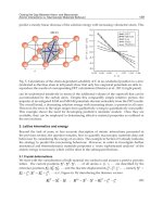
Figure 7.4
Schematic of a tabular representation for storing multiplication facts. Activation of a particular answer occurs by
searching the corresponding rows and columns of the table
to their point of intersection, as indicated by the bold
numbers and lines. (Adapted from McCloskey, Aliminosa,
& Sokol, 1991.)
is that of a tabular lexicon (figure 7.4). The figure
shows that during recall, activation is hypothesized
to spread among related facts (the bold lines in
figure 7.4). This mechanism may account for both
the within-table and the relatedness errors noted
earlier (Stazyk, Ashcraft, & Hamann, 1982). Two
other behaviors are also consistent with a “tabular”
organization of numerical facts: (1) repetition priming, or responding more quickly to an identical previously seen problem and (2) error priming, which
describes the increased probability of responding
incorrectly after seeing a problem that is related but
not identical to one shown previously (Dehaene,
1992).
Other calculation error types are noted in table
7.1. The nomenclature used in the table is derived
from the classification scheme suggested by
Sokol et al., although the taxonomy has not been
universally accepted (Sokol, McCloskey, Cohen, &
Aliminosa, 1991). Two general categories of errors
Darren R. Gitelman
140
Table 7.1
Types of calculation errors
Error type
Description
Example
The correct answer to the problem shares
an operand with the original equation.
5 ¥ 8 = 48. The answer is correct for 6 ¥ 8, which
shares the operand 8 with the original equation.
Operation
The answer is correct for a different
mathematical operation on the operands.
3 ¥ 5 = 8. The answer is correct for addition.
Indeterminate
The answer could be classified as either
an operand or an operational error.
4 ¥ 4 = 8. The answer is true for 2 ¥ 4 or 4 + 4.
Table
The answer comes from the range of
possible results for a particular operation,
but is not related to the problem.
4 ¥ 8 = 30. The answer comes from the “table” of
single-digit multiplication answers.
Nontable
The answer does not come from the
range of results for that operation.
5 ¥ 6 = 23. There are no single-digit multiplication
problems whose answer is 23.
The answer is not given.
3¥7=
Commission
Operand
Omission
are errors of omission (i.e., failing to respond) and
errors of commission (i.e., responding with the
incorrect answer). As shown in table 7.1, there are
several types of commission errors, some of which
seem to predominate in different groups. Operand
errors are the most common error type seen in
normal subjects (Miller, Perlmutter, & Keating,
1984; Campbell & Graham, 1985). Patients can
show a variety of dissociated error types. For
example, Sokol et al. (1991) described patient P.S.,
who primarily made operand errors, while patient
G.E. made operation errors. Although the occurrences of these errors were generally linked to left
hemisphere lesions, there has been no comprehensive framework linking error type to particular
lesion locations.
Rules and Procedures
An abnormality in the procedures of calculation is
the third type of deficit leading to anarithmetia. Procedural deficits can take several forms, including
errors in simple rules, in complex rules, or in
complex multistep procedures. Examples of simple
rules would include 0 ¥ N = 0, 0 + N = N, and 1 ¥
N = N operations.4 An example of a complex rule
would be knowledge of the steps involved in
multiplication by 0 in the context of executing
a multidigit multiplication. Complex procedures
would include the organization of intermediate
products in multiplication or division problems,
and multiple carrying or borrowing operations in
multidigit addition and subtraction problems,
respectively.
Several authors have shown that in normal subjects, rule-based problems are solved more quickly
than nonrule-based types (Parkman & Groen, 1971;
Groen & Parkman, 1972; Parkman, 1972; Miller
et al., 1984), although occasional slower responses
have been found (Parkman, 1972; Stazyk et al.,
1982). Nevertheless, the available evidence suggests that rule-based and nonrule-based problems
are solved differently, and can show dissociations
in a subject’s performance (Sokol et al., 1991;
Ashcraft, 1992).
Patient P.S., who had a large left hemisphere
hemorrhage, was reported by Sokol et al. (1991) as
showing evidence for a deficit in simple rules,
specifically multiplication by 0. This patient made
Acalculia
patchy errors in the retrieval of table facts (0%
errors for 9 ¥ 8, to 52% errors for 4 ¥ 4), but missed
100% of the 0 ¥ N problems. This performance suggested that the patient no longer had access to the
rule for solving 0 ¥ N problems. Remarkably, during
the last part of testing, the patient appeared to
recover knowledge of this rule and began to perform
0 ¥ N operations flawlessly. During the same time
period, performance on calculations of the M ¥ N
type showed only minimal improvement across
blocks.
Patient G.E., reported by Sokol et al. (1991), suffered a left frontal contusion and demonstrated a
dissociation in simple versus complex rule-based
computations. This patient made errors when performing the simple rule computation of 0 ¥ N
(always reporting the result as 0 ¥ N = N), but he
was able to multiply by 0 correctly within a multidigit calculation. In this setting he recalled the
complex rule of using 0 as a placeholder in the
partial products of multiplication problems.
More complex procedural deficits are illustrated
in figure 7.5. Patient 1373, cited by McCloskey et
al. (1985), showed good retrieval of table facts, but
impaired performance of multiplication procedures.
In one case, shown in figure 7.5A, he failed to
shift the intermediate multiplication products one
column to the left. Note that the individual arith-
141
metical operations in figure 7.5A are performed
correctly, but the answer is nonetheless incorrect
because of this procedural error.
Other deficits in calculation procedures have
included incorrect performance of carrying and/or
borrowing operations, as shown by patients V.O.
and D.L. of McCloskey et al. (1985) (figure 7.5B),
and confusing steps in one calculation procedure
with those of another, as in patients W.W. and H.Y.
of McCloskey et al. (1985) (figure 7.5C).
Arithmetical Dissociations
Individual arithmetical operations have also
revealed dissociations among patients. For
example, patients have been described with intact
division, but impaired multiplication (patient 1373)
(McCloskey et al., 1985) and intact multiplication
and addition, but impaired subtraction and/or division (Berger, 1926), among other dissociations
(Dehaene & Cohen, 1997). Several theories have
tried to account for the apparent random dissociations among operations. One explanation is that
separate processing streams underlie each arithmetical operation (Dagenbach & McCloskey, 1992).
Another possibility is that each operation may be
differentially linked to verbal, quantification (see
later discussion), or other cognitive domains (e.g.,
working memory) (Dehaene & Cohen, 1995, 1997).
Figure 7.5
Examples of various calculation errors. (A) Multiplication: failure to shift the second intermediate product. (B) Multiplication: omission of the carrying operation and each partial product is written in full. (C) Addition: addend not properly
carried, i.e., 8 is added to 5 and then incorrectly again added to 4. Each partial addend has then been placed on a single
line. (Adapted from McCloskey, Caramazza, & Basili, 1985.)
Darren R. Gitelman
Based on this concept, each arithmetical operation may require different operational strategies for
a solution. These cognitive links may depend partly
on previous experience (e.g., knowledge of multiplication tables) and partly on the strategies used to
arrive at a solution. For example, multiplication and
addition procedures are often retrieved through the
recall of memorized facts. Simple addition operations can also be solved by counting strategies, an
option not readily applicable to multiplication. Subtraction and division problems, on the other hand,
are more frequently solved de novo, and therefore
require access to several cognitive processes, such
as verbal mechanisms (e.g., recalling multiplication
facts to perform division), quantification operations
(counting), and working memory. Differential injury to these cognitive domains may be manifest as
a focal deficit for a particular arithmetical operation. The deficits in patient M.A.R. reported by
Dehaene and Cohen (1997) support this cognitive
organization.
This patient had a left inferior parietal lesion and
could recall simple memorized facts for solving
addition and multiplication problems, but did not
perform as well when calculating subtractions. This
performance suggested that M.A.R. had access to
some memorized table facts, but that the inferior
parietal lesion may have led to deficits in the calculation process itself. Patient B.O.O., also reported
by Dehaene and Cohen (1997), had a lesion in the
left basal ganglia and demonstrated greater deficits
in multiplication than in either addition or subtraction. In this case, recall of rote-learned table facts
was impaired, leading to multiplication deficits,
but the patient was able to use other strategies for
solving addition and subtraction problems.
Despite these examples, functional associations
are not able to easily explain the dramatic dissociations reported in some patients, such as the one
described by Lampl et al. Their patient had a left
parietotemporal hemorrhage and had a near inability to perform addition, multiplication, or division,
but provided 100% correct responses on subtraction
problems (Lampl, Eshel, Gilad, & Sarova-Pinhas,
1994).
142
Anatomical Relationships and Functional
Imaging
The most frequent cortical site of damage causing
anarithmetia is the left inferior parietal cortex
(Dehaene & Cohen, 1995). While several roles have
been proposed for this region (access to numerical memories, quantification operations, semantic
numerical relations) (Warrington, 1982; Dehaene
& Cohen, 1995), one general way to conceive of
this area is that it may provide a link between verbal
processes and magnitude or spatial numerical
relations.
Other lesion sites reported to cause anarithmetia
include the left basal ganglia (Whitaker, Habinger,
& Ivers, 1985; Corbett, McCusker, & Davidson,
1986; Hittmair-Delazer, Semenza, & Denes, 1994)
and more rarely the left frontal cortex (Lucchelli &
DeRenzi, 1992). The patient reported by HittmairDelazer and colleagues had a left basal ganglia
lesion and particular difficulty mentally calculating
multiplication and division problems (with increasing deficits for larger operands) despite 90%
accuracy on mental addition and subtraction
(Hittmair-Delazer et al., 1994). He was able to use
complex strategies to solve multiplication problems
in writing (e.g., solving 8 ¥ 6 = 48 as 8 ¥ 10 = 80
∏ 2 = 40 + 8 = 48), demonstrating an intact conceptual knowledge of arithmetic and an ability to
sequence several operations. However, automaticity
for recall of multiplication and division facts was
reduced and was the primary disturbance that
interfered with overall calculation performance.
Similarly, patients with aphasia following left
basal ganglia lesions may show deficits in the recall
of highly automatized knowledge (Aglioti &
Fabbro, 1993). Brown and Marsden (1998) have
hypothesized that one role of the basal ganglia may
be to enhance response automaticity through the
linking of sensory inputs to “programmed” outputs
(either thoughts or actions). Such automated or programmed recall may be necessary for the online
manipulation of rote-learned arithmetical facts such
as multiplication tables.
Acalculia
Deficits in working memory and sequencing
behaviors have also been seen following basal
ganglia lesions. The patient reported by Corbett et
al. (1986), for example, had a left caudate infarction, and was able to perform single but not multidigit operations. The patient also had particular
difficulty with calculations involving sequential
processing and the use of working memory. The
patient of Whitaker et al., who also had a left basal
ganglia lesion, demonstrated deficits for both simple
and multistep operations (Whitaker, Habiger, &
Ivers, 1985). Thus basal ganglia lesions may interfere with calculations via several potentially
dissociable mechanisms that include (1) deficits
in automatic recall, (2) impairments in sequencing, and (3) disturbances in operations requiring
working memory.
Calculation deficits following frontal lesions
have been difficult to characterize precisely, possibly because these lesions often result in deficits in
several interacting cognitive domains (e.g., deficits
in language, working memory, attention, or executive functions). Grewel, in fact, insisted that “frontal
acalculia must be regarded as a secondary acalculia” (Grewel, 1969, p. 189) precisely because of
the concurrent intellectual impairments with these
lesions. However, when relatively pure deficits have
been seen following frontal lesions, they appear
to involve more complex aspects of calculations,
such as the execution of multistep procedures or
understanding the concepts underlying particular
operations such as the calculation of percentages
(Lucchelli and DeRenzi, 1992). Studies by Fasotti
and colleagues have suggested that patients with
frontal lesions have difficulty translating arithmetical word problems into an internal representation,
although they did not find significant differences
in performance among patients with left, right, or
bilateral frontal lesions (Fasotti, Eling, & Bremer,
1992). Functional imaging studies, detailed later,
strongly support the involvement of various frontal
sites in calculations, but these analyses have also
not excluded frontal activations that are due to
associated task requirements (e.g., working memory
or eye movements).
143
In contrast to the significant calculation abnormalities seen with left hemisphere lesions, deficits
in calculations are rare following right hemisphere
injuries. However, when groups of patients with
right and left hemisphere lesions were compared,
there was evidence that comparisons of numerical
magnitude are more affected by right hemisphere
injuries (Dahmen, Hartje et al., 1982; Rosselli &
Ardila, 1989). Patients with right hemisphere
lesions may at times demonstrate “spatial acalculia.” Hécaen defined this as difficulty in the spatial
organization of digits (Hécaen et al., 1961). Nevertheless, the calculation deficits after right hemisphere lesions tend to be mild and the performance
of patients with these lesions may not be distinguishable from that of normal persons (Jackson &
Warrington, 1986).
Using an 133Xe nontomographic scanner, Roland
and Friberg in 1985 provided the first demonstration of functional brain activations for a calculation
task (serial subtractions of 3 beginning at 50 compared with rest) (Roland & Friberg, 1985). All subjects had activations on the left, over the middle and
superior prefrontal cortex, the posterior inferior
frontal gyrus, and the angular gyrus. On the right,
activations were seen over the inferior frontal gyrus,
the rostral middle and superior frontal gyri, and the
angular gyrus (figure 7.6) (lightest gray areas).
Because the task and control conditions were not
designed to isolate specific cognitive aspects of
calculations (i.e., by subtractive, parametric, or factorial design), it is difficult to ascribe specific neurocognitive functions to each of the activated areas
in this experiment. Nevertheless, the overall pattern
of activations, which include parietal and frontal
regions, anticipated the results in subsequent
studies, and constituted the only functional imaging
study to investigate calculations until 1996 (Grewel,
1952, 1969; Boller & Grafman, 1983; Roland &
Friberg, 1985; Dehaene & Cohen, 1995).
The past 5 years have seen a large increase in the
number of studies examining this cognitive domain.
However, one difficulty in comparing the results has
been that individual functional imaging calculation
studies have tended to differ from one another along
Darren R. Gitelman
144
Figure 7.6
Cortical and subcortical regions activated by calculation tasks. Symbols are used to specify activations when the original
publications either indicated the exact sites of activation on a figure, or provided precise coordinates. Broader areas of
shading represent either activations in large regions of interest (Dehaene, Tzourio, Frak, Raynaud, Cohen, Mehler, &
Mazoyer, 1996), or the low resolution of early imaging techniques (Roland & Friberg, 1985). Key: Light gray areas: serial
3 subtractions versus rest (Roland & Friberg, 1985). Triangle: calculations (addition or subtraction) versus reading of
equations (Sakurai, Momose, Iwata, Sasaki, & Kanazeu, 1996). Dark gray areas: multiplication versus rest (Dehaene,
Tzourio, Frak, Raynaud, Cohen, Mehler, Mazoyer, 1996). Circle: exact versus approximate calculations (addition)
(Dehaene, Spelke, Pinel, Stanescu, & Tsivikin, 1999). Diamond: multiplication of two single digits versus reading numbers
composed of 0 and 1 (Zago, Pesenti, Mellet, Crevello, Mazoyer, & Tzourio-Mazoyer, 2000). Asterisk: verification of addition and subtraction problems versus identifying numbers containing a 0 (Menon, Rivera, White, Glover, & Reiss, 2000).
Cross: addition, subtraction, or division of two numbers (one to two digits) versus number repetition (Cowell, Egan, Code,
Harasty, 2000). More complete task descriptions are listed in tables 7.2 and 7.3. The brain outline for figures 7.6 and 7.8
was adapted from Dehaene, Tzourio, Frak, Raynaud, Cohen, Mehler, & Mazoyer, 1996. Activations are plotted bilaterally if they are within ±3 mm of the midline or are cited as bilateral in the original text. The studies generally reported
coordinates in Montreal Neurological Institute space. Only Cowell, Egan, Code, Harasty, Watso (2000), and Sathian,
Simon, Peterson, Patel, Hoffman, & Grafton (1999) for figure 7.8, reported locations in Talairach coordinates (Talairach
& Tournoux, 1988). Talairach coordinates were converted to MNI space using the algorithms defined by Matthew Brett
( (Duncan, Seitz, Kolodny, Bor, Herzog, Ahmed, Newell, &
Emsile, 2000). Note that the symbol sizes do not reflect the activation sizes. Thus hemispheric asymmetries, particularly
those based on activation size, are not demonstrated in this figure or in figure 7.8.
Acalculia
multiple methodological dimensions: imaging
modality (PET versus fMRI), acquisition technique
(block versus event-related fMRI), arithmetical
operation (addition, subtraction, multiplication,
etc.), mode and type of response (oral versus button
press, generating an answer versus verifying a
result), etc. These differences have at least partly
contributed to the seemingly disparate functional–
anatomical correlations among studies (figure 7.7).
However, rather than focusing on the disparities in
these reports and trying to relate activation differences post hoc to methodological variations, a more
informative approach may be to look for areas of
commonality (Démonet, Fiez Paulesu, Petersen,
Zatorre, 1996; Poeppel, 1996).
As indicated in figures 7.6 and 7.7, the set of
regions showing the most frequent activations
across studies included the bilateral dorsal lateral
prefrontal cortex, the premotor cortex (precentral
gyrus and sulcus), the supplementary motor cortex,
the inferior parietal lobule, the intraparietal sulcus,
and the posterior occipital cortex-fusiform gyrus
(Roland & Friberg, 1985; Dehaene et al., 1996;
Sakurai, Momose, Iwata, Sasaki, & Kanazawa,
1996; Pinel et al., 1999; Cowell, Egan, Code,
Harasty, & Watson, 2000; Menon, Rivera, White,
Glouer, & Reiss, 2000; Zago et al., 2000). When
examined regionally, six out of eight studies demonstrated dorsal lateral prefrontal or premotor activations, and seven of eight had activations in the
posterior parietal cortex. In addition, ten out of
sixteen areas were more frequently activated on the
left across studies, which is consistent with lesiondeficit correlations indicating the importance of the
left hemisphere for performing exact calculations.
Other evidence regarding the left hemisphere’s
importance to calculations comes from a study by
Dehaene and colleagues (Dehaene, Spelke Pinel,
Staneszu, & Tsivikin, 1999). In their initial psychophysics task, bilingual subjects were taught
exact or approximate sums involving two, two-digit
numbers in one of their languages (native or nonnative language training was randomized). They
were then tested again in the language used for
initial training or in the “untrained” language on a
145
subset of the learned problems and on a new set of
problems. The subjects showed a reaction time cost
(i.e., a slower reaction time) when answering previously learned problems in the untrained language
regardless of whether this was the subject’s native
or non-native language.
There was also a reaction time cost for solving
novel problems. The presence of a reaction time
cost when performing learned calculations in a
language different from training or when solving
novel problems is consistent with the hypothesis
that exact arithmetical knowledge is accessed in a
language-specific manner, and thus is most likely
related to left-hemisphere linguistic or symbolic
abilities.
In contrast, when they were performing approximate calculations, subjects showed neither a
language-based nor a novel problem-related effect
on reaction times. This result suggests that approximate calculations may take place via a languageindependent route and thus may be more bilaterally
distributed.
The fMRI activation results from Dehaene
et al. (1999) were consistent with these behavioral
results in that exact calculations activated a
left-hemisphere predominant network of regions
(figures 7.6–7.7), while approximate calculations
(figures 7.8–7.9) showed a more bilateral distribution of activations. An additional ERP experiment
in this study confirmed this pattern of hemispheric
asymmetry, with exact calculations showing an
earlier (216–248 ms) left frontal negativity, while
approximate calculations produced a slightly later
(256–280 ms) bilateral parietal negativity (Dehaene
et al., 1999).
In a calculation study using PET imaging, which
compared multiplying two, two-digit numbers with
reading numbers composed of 1 or 0 or recalling
memorized multiplication facts, Zago et al. (2000)
made the specific point that perisylvian language
regions, including Broca’s and Wernicke’s areas,
were actually deactivated as calculation-related task
requirements increased. This finding was felt to be
consistent with other studies showing relative independence between language and calculation deficits
Darren R. Gitelman
146
Figure 7.7
Number of studies showing activations for exact calculations organized by region and by hemisphere. Ten out of sixteen
areas have a greater number of studies showing activation in the left hemisphere as opposed to the right. The graph also
indicates that the frontal, posterior parietal, and, to a lesser extent, occipital cortices are most commonly activated in exact
computational tasks. The small bar near 0 for the right cingulate gyrus region is for display purposes. The value was actually 0. Key: DLPFC, dorsal lateral prefrontal cortex; PrM, premotor cortex (precentral gyrus and precentral sulcus); FP,
prefrontal cortex near frontal pole; IFG, posterior inferior frontal gyrus overlapping Broca’s region on the left and the
homologous area on the right; SMA, supplementary motor cortex; Ins, insula; Cg, cingulate gyrus; BG, basal ganglia,
including caudate nucleus and/or putamen; Th, thalamus; LatT, lateral temporal cortex; IPL, inferior parietal lobule; IPS,
intraparietal sulcus; PCu, precuneus; InfT-O, posterior lateral inferior temporal gyrus near occipital junction; FG, fusiform
or lingual gyrus region; Occ, occipital cortex.
Acalculia
147
Figure 7.8
Cortical and subcortical activations for tasks of quantification, estimation, or number comparison. See figure 7.6 for details
of figure design. Key: Dark gray areas: number comparison versus rest (Dehaene, Tzourio, Frak, Raynaud, Cohen, Mehler,
& Mazoyer, 1996). Squares: number comparison with specific inferences for distance effects; closed squares are for
numbers closer to the target, open squares are for numbers farther from the target (Pinel Le Clec’h, van der Moortele,
Naccache, Le Bihan, & Dehaene, 1999). Open diamond: subitizing versus single-target identification (Sathian, Simon,
Peterson, Patel, Hoffman, Graftor, 1999). Closed diamond: counting multiple targets versus subitizing (Sathian, Simon,
Peterson, Patel, Hoffman, Grafton, 1999). Closed article: approximate versus exact calculations (addition) (Dehaene,
Spelke, Pinel, Starescu, Tsivikin, 1999). Star: estimating numerosity versus estimating shape (Fink, Marshall, Gurd, Weiss,
Zafiris, Shah, Zilles, 2000).
in some patients (Warrington, 1982; Whetstone,
1998).
Zago et al. (2000) also noted that the left precentral gyrus, intraparietal sulcus, bilateral cerebellar
cortex, and right superior occipital cortex were activated in several contrasts and that similar activations had been reported in previous calculation
studies (Dehaene et al., 1996, Dehaene et al., 1999;
Pinel et al., 1999; Pesenti et al., 2000). Because
of these results, Zago and colleagues (2000) suggested that given the motor or spatial functions
of several of these areas, they could represent a
developmental trace of a learning strategy based on
counting fingers. As support for this argument, the
authors noted that certain types of acalculia, such as
Gerstmann’s syndrome, also produce finger identi-
fication deficits, dysgraphia, and right-left confusion, and that these deficits are consistent with
the potential role of these regions in hand movements and acquisition of information in numerical
magnitude.
However, these areas are also important for
visual-somatic transformations, working memory,
spatial attention, and eye movements, which were
not controlled for in this experiment (Jonides et al.,
1993; Paus, 1996; Nobre et al., 1997; Courtney,
Petit, Maisog, Ungerleider, & Haxby, 1998;
Gitelman et al., 1999; LaBar, Gitelman, Parrish,
& Mesulam, 1999; Gitelman, Parrish, LaBar, &
Mesulam, 2000; Zago et al., 2000). Also, because
covert finger movements and eye movements were
not monitored, it is difficult to confidently ascribe
Darren R. Gitelman
148
Figure 7.9
Number of studies showing activations for quantification and approximation operations organized by region and by hemisphere. Activations are more bilaterally distributed, by study, than for exact calculations (figure 7.7). In addition, the posterior parietal and occipital cortices now show the predominant activations, with lesser activations frontally. The small
bars near 0 for several of the regions were added for display purposes. The values were actually 0. See figure 7.7 for
abbreviations.
activations in these regions solely to the representation of finger movements.
One region not displayed in figure 7.6 is the cerebellum. Activation of the cerebellum was seen in
only two studies reviewed here. Menon et al. (2000)
saw bilateral midcerebellar activations when their
subjects performed the most difficult computational
task (table 7.2). Zago et al. (2000) saw right cerebellar activation for the combined contrasts (conjunction) of retrieving multiplication facts and de
novo computations versus reading the digits 1 or 0.
Thus cerebellar activations are most likely to be
seen when relatively complex or novel computations are compared with simpler numerical perception tasks. Cerebellar activation may therefore
represent a difficulty effect.
Quantification and Approximation
Quantification is the assessment of a measurable
numerical quantity (numerosity) of a set of items. It
is among the most basic of arithmetical operations
and may play a role in both the childhood development of calculation abilities and the numerical
Acalculia
149
Table 7.2
Description of functional imaging tasks for exact calculations
Study
Modality
Paradigm
Response
Roland, Fribery
1985
133
Serial three subtractions from 50 versus rest
Silent
Rueckert et al.,
1996
fMRI
Block
design
Serial three subtractions from a 3-digit integer versus counting
forward by ones
Silent
Sakurai et al.,
1996
PET
Addition or subtraction of two numbers (2 digits and 1 digit)
versus reading calculation problems
Oral
Dehaene et al.,
1996
PET
Multiply two 1-digit numbers versus compare two numbers
Silent
Dehaene et al.,
1999
fMRI
Block
design
Subjects pretrained on sums of two 2-digit numbers
During the task, subjects selected correct answer (two choices).
For exact calculations, one answer was correct and the tens
digit was off by one in the other. For approximate calculations,
the correct answer was rounded to the nearest multiple of ten.
The incorrect answer was 30 units off.
Silent: two-choice
button press
Cowell et al.,
2000
PET
Addition or subtraction or division of two numbers (1–2 digits)
versus number repetition
Oral
Menon et al.,
2000
PET
Verify addition and subtraction of problems with three operands
versus identify numbers containing the numeral 0
Silent: single-choice
button press
Zago et al.,
2000
PET
Multiply two 2-digit numbers versus reading numbers
consisting of just zeros and ones
Oral
Xe
processes of adults (Spiers, 1987). Despite the basic
nature of quantification operations, they were not
included in some early models of calculations
(McCloskey et al., 1985). Three quantification
processes have been described: counting, subitizing,
and estimation (Dehaene, 1992). Counting is the
assignment of an ordered representation of quantity
to any arbitrary collection of objects (Gelman &
Gallistel, 1978; Dehaene, 1992). Subitizing is the
rapid quantification of small sets of items (usually
less than five); and estimation is the “less accurate” rapid quantification of larger sets (Dehaene,
1992).
Subitizing and Counting
Because subitizing and to an extent counting operations appear to be largely distinct from language
abilities, these operations may be of considerable
importance for understanding the calculation abilities of prelinguistic human infants and even (nonlinguistic) animals. Jokes about Clever Hans aside,5
there is ample evidence that animals possess simple
counting abilities (Dehaene, 1992; Gallistel &
Gelman, 1992).
More important, young children possess counting
abilities from an early age, and there is good evidence that even very young infants can subitize,
suggesting that this ability may be closely associated with the operation of basic perceptual
processes (Dehaene, 1992). Four-day-old infants,
for example, can discriminate between one and
two and two and three displayed objects (BijeljacBabic, Bertoncini, Mehler, 1993), and 6–8-monthold infants demonstrate detection of cross-modal
Darren R. Gitelman
(visual and auditory) numerical correspondence
(Starkey, Spelke, & Gelman, 1983; Starkey, Spelke,
& Gelman, 1990). Although quantification abilities
may be bilaterally represented in the brain, the right
hemisphere is thought to demonstrate some advantage for these operations (Dehaene & Cohen, 1995).
Estimation and Approximation
The use of estimation operations in simple calculations may have a role in performing these operations
nonlinguistically or in allowing the rapid rejection
of “obviously” incorrect answers. For example, if
quantification can be conceived as encoding numbers on a mental “number line,” then addition can
be likened to mentally joining the number line segments and examining the total line length to arrive
at the answer (Gallistel and Gelman, 1992). As with
a physical line, the precision of the measurement is
hypothesized to decline with increasing line length
(Weber’s law6) (Dehaene, 1992).
An example of the role of estimation in calculations is provided by examining subjects’ performance in verification tasks. In these tasks, the subjects
are asked to verify an answer to an arithmetic
problem (e.g., 5 ¥ 7 = 36?). The speed of classifying answers as incorrect increases (i.e., decreased
reaction time) with increasing separation between
the proposed and correct results (“split effect”)
(Ashcraft & Battaglia, 1978; Ashcraft & Stazyk,
1981). The response to glaringly incorrect answers
(e.g., 4 ¥ 5 = 1000?) can be so rapid as to suggest
that estimation processes may be operating in parallel with exact “fact-based” calculations (Dehaene,
Dehaene-Lambertz, & Cohen, 1998).
Further evidence that some magnitude operations
can be approximated by a spatially extended mental
number line comes from numerical comparison
tasks. During these tasks, subjects judge whether
two numbers are the same or different while reaction times are measured. Experiments show that the
time to make this judgment varies inversely with the
distance between the numbers. Longer reaction
times are seen as numbers approach each other. In
one experiment, Hinrichs et al. showed that it was
quicker to compare 51 and 65 than to compare 59
150
and 65 (Hinrichs, Yurko, & Hu, 1981). If numbers
were simply compared symbolically, there should
have been no reaction time difference in this comparison since it should have been sufficient to
compare the tens digits in both cases. This finding
has been interpreted as showing that numbers can
be compared as defined quantities and not just at a
symbolic level (Dehaene, Dupoux, & Mehler,
1990).
Case studies of several patients have provided
further support for the importance of quantification
processes and the independence of these processes
from exact calculations. Patient D.R.C. of Warrington suffered a left temporoparieto-occipital junction
hemorrhage (~3 cm diameter) and subsequently had
difficulty recalling arithmetical facts for addition,
subtraction, and multiplication, yet he usually gave
answers of reasonable magnitude when asked to
solve problems. For example, he said “13 roughly”
for the problem “5 + 7” (Warrington, 1982). A
similar phenomenon occurred in the patient N.A.U.
of Dehaene and Cohen (1991). This patient sustained head trauma, which produced a very large
left temporoparieto-occipital hemorrhage (affecting
most of the parietal, posterior temporal, and anterior occipital cortex). Although N.A.U. could not
directly calculate 2 + 2, he could reject 9 but not
3 as a possible answer, which is consistent with
access to an estimation process. N.A.U. could also
compare numbers (possibly by using magnitude
comparison), even ones he could not read explicitly,
if they were separated by more than one digit.
However, he performed at chance level when
deciding if a number was odd or even. Although
this dissociation may seem incongruous, one
hypothesis is that parity decisions require exact and
not approximate numerical knowledge, consistent
with the inability of this patient to perform exact
calculations.
Grafman et al. described a patient who suffered
near total destruction of the left hemisphere from
a gunshot wound, leaving only the occipital and
parasagittal cortex remaining on the left (Grafman,
Kampen, Rosenberg, & Salazar, 1989). Despite
an inability to perform multidigit calculations, he
Acalculia
151
could compare multidigit numerals with excellent
accuracy, suggesting that intact right hemisphere
mechanisms were sufficient for performing this
comparison task. The opposite dissociation
(increased deficits in approximation despite some
preservation of rote-learned arithmetic) was seen in
patient H.Ba. reported by Guttmann (1937). H.Ba.
was able to perform simple calculations, but had
difficulty with number comparisons and quantity
estimation. Unfortunately, no anatomical information regarding H.Ba.’s lesions was provided since
his deficits were developmental.
Overall, these studies strongly support the
hypotheses that the cognitive processes underlying
exact calculations and those related to estimating
magnitude can be dissociated. In addition, left
hemisphere regions seem clearly necessary for the
performance of exact calculations, while estimation
tasks may be more closely associated with the right
hemisphere or possibly are bilaterally represented.
Anatomical Relationships and Functional
Imaging
Figure 7.8 shows the combined activations from
five studies of quantification or approximation operations, including subitizing, counting, number comparison, and approximate computations (Dehaene
et al., 1996; Dehaene et al., 1999; Pinel et al., 1999;
Sathian et al., 1999; Fink et al., 2000). The paradigms for these studies are summarized in table 7.3.
In comparison with the data from studies of exact
calculations (figures 7.6 and 7.7), approximation
and magnitude operations (figures 7.8 and 7.9)
show relatively more parietal and occipital and less
Table 7.3
Description of functional imaging tasks for approximation and quantification
Study
Modality
Paradigm
Response
Dehaene et al.,
1996
PET
Multiply two 1-digit numbers versus compare two numbers
Silent
Dehaene et al.,
1999
fMRI
Block
design
Subjects were pretrained on sums of two 2-digit numbers.
During fMRI, subjects were shown a two-operand addition
problem and a single answer. They pressed buttons to choose
if the answer was correct or incorrect. For exact calculations,
one answer was correct, while the tens digit was off by one in
the other. For approximate calculations, the correct answer was
was the actual result rounded to the nearest multiple of 10
(e.g., 25 + 28 = 53, so 50 was shown to subjects). The
incorrect answer was 30 units off.
Silent: dual-choice
button press
Pinel et al.,
1999
fMRI
Event
related
Number comparison: Is a target number (shown as a word or a
numeral) larger or smaller than 5?
Silent: single-choice
button press
Sathian et al.,
1999
PET
Subjects saw an array of 16 bars and reported the number of
vertical bars. When 1–4 vertical bars were present, the subjects
were assumed to identify magnitude by subitizing; when 5–8
vertical bars were present, they were assumed to be counting.
Oral
Fink et al., 2000
fMRI
Block
design
In the numerosity condition, subjects indicated if four dots
were present. In the shape condition, subjects indicated
if the dots formed a square.
Silent: dual-choice
button press
Darren R. Gitelman
frontal activity. In addition, the left-right asymmetry seen in figure 7.7 is no longer apparent.
Sathian et al. (1999) examined regions activated
by tasks of counting and subitizing. Subitizing,
which has been linked to preattentive and “pop-out”
types of processes, resulted in activation of the right
middle and inferior occipital gyrus (figure 7.8). The
left hemisphere showed a homologous activation,
which did not quite reach the threshold for significance, and is not shown in the figure. A small right
cerebellar activation was also found just below
threshold. Similar occipital predominant activations
were also obtained by Fink et al. (2000) for a task
that basically involved subitizing (deciding if four
dots were present when shown three, four, or five
dots) versus estimating shape.
Counting, in contrast to subitizing, according to
Sathian et al. (1999), activated broad regions of
the bilateral occipitotemporal, superior parietal, and
right premotor cortices (figure 7.8). Based on these
results, Sathian et al. suggested that counting
processes may involve spatial shifts of attention
(among the objects to be counted) and attentionmediated top-down modulation of the visual cortex.
Although the parietal cortex has been hypothesized to support numerical comparison operations
(Dehaene and Cohen, 1995), this area was nonsignificantly activated (p = 0.078) in a PET study
examining comparison operations (Dehaene et al.,
1996). Instead, the contrast between number comparison and resting state conditions demonstrated
significant activations in the bilateral occipital, premotor, and supplementary motor cortices (figure
7.8) (dark gray areas) (Dehaene et al., 1996). One
possible explanation for the minimal parietal activation in this study is that the task involved repeated
comparisons of two numerals between 1 and 9. In
the case of small numerosities, it has been suggested
that seeing a numeral may evoke quantity representations that are similar to seeing the same number
of objects, and may engender automatic subitization. Hence, the task may have stressed operations
related to number identification and covert subitizing processes more than the authors anticipated.
Therefore the occiptotemporal cortex rather than the
152
parietal cortex may have been preferentially activated (Sathian et al., 1999; Fink et al., 2000).
A subsequent study of number comparison used
event-related fMRI while the subjects decided
whether a target numeral (between 1 and 9) was
larger or smaller than the number 5 (Pinel et al.,
1999). Distance effects (i.e., whether the numbers
were closer to or farther from 5) were seen in the
left intraparietal sulcus and the bilateral inferior,
posterior parietal cortices, which is consistent with
the hypothesized parietal involvement in magnitude
processing (figure 7.8). The authors also noted that
this study showed an apparent greater left hemisphere involvement for number comparison, while
a previous study had suggested more involvement
of the right hemisphere (Dehaene, 1996).
Numerical Representations
One issue of considerable debate has been the
manner in which numerical relations are internally
encoded. For example, are problems handled differently if they are presented as Arabic numerals
(2 + 6 = 8), Roman numerals (II + VI = 8), or words
(two plus six equals eight)? McCloskey and
colleagues have maintained that the various
number-processing and calculation mechanisms
communicate via a single abstract representation of
quantity (Sokol et al., 1991). Others have strongly
disagreed with this approach and have suggested
that internal representational codes may vary
(encoding complex theory) according to input or
output modality, task requirements, learning strategies, etc. (Campbell & Clark, 1988), or even according to the subject’s experience (preferred entry
code hypothesis) (Noël & Seron, 1993). Another
approach, discussed later, is that there are specific
representations (words, numerals, or magnitude)
linked to particular calculation processes, and this
suggestion is embodied by the triple-code model of
Dehaene (Dehaene, 1992).
Considerable evidence exists attesting to the
importance of an internal representation of magnitude. One example is the presence of the numerical
distance effect. As previously noted, this effect is
Acalculia
demonstrated by subjects taking longer to make
comparison judgments for numbers that are closer
in magnitude to one another. The effect has been
demonstrated across a variety stimulus input types,
including Arabic numerals (Moyer & Landauer,
1967; Sekuler, Rubin, & Armstrong, 1971), spelledout numbers (Foltz, Poltrock, & Potts, 1984), dot
patterns (Buckley & Gillman, 1974), and Japanese
kana and kanji ideograms (Takahashi & Green,
1983). The occurrence of this effect regardless of
the format of the stimulus has suggested that it is
not mediated by different input codes for each
format, but rather through a common representation
of magnitude (Sokol et al., 1991).
Evidence for an opposing set of views, i.e., that
numerical processing can take place via a variety
of representational codes, has also been amassed.
One prediction of “multicode” models is that input
and/or response formats may influence the underlying calculations beyond effects attributable to
simple sensory mechanisms. In the single-code
model, since all calculations are based on an amodal
representation of the number, it should not matter
how the number is presented once this transcoding
has taken place. A single-code model would suggest
that differences in adding 5 + 6 and V + VI would
be solely attributable to the transcoding operation.
In support of additional codes, Gonzalez and
Kolers (1982, 1987) found that differences in reaction times to Arabic and Roman numerals showed
an interaction with number size (i.e., there was a
greater differential for IV + 5 = IX, than for II + 1
= III). This difference implied that the calculation
process might have been affected by a combination
of the input format and the numerical magnitude of
the operands. A single-code model would predict
that while calculations might be slower for a given
input format, they should not be disproportionately
slower for larger numbers in that format.
A second set of experiments addressed the possibility that the slower reaction time for Roman
numerals was simply due to slowed numerical comprehension of this format. The subjects were trained
in naming Roman numerals for several days, until
they showed no more than a 10% difference in
153
naming times between Arabic and Roman numerals. Despite this additional training, differences in
reaction time remained beyond the time differences
attributable to numerical comprehension alone. This
result again suggested that numerical codes may
depend on the input format, and may influence
calculations differentially. Countering these arguments, Sokol and colleagues (1991) have noted that
naming numbers and comprehending them for use
in calculations are different processes and may
proceed via different initial mechanisms.
Synthesizing the various views for numerical representation, Dehaene (1992) has proposed that three
codes can account for differences in input, output,
and processing of numbers. These representations
include a visual Arabic numeral, an auditory word
frame, and an analog magnitude code. Each of these
codes has its own input and output procedures and
is interfaced with preferred aspects of calculations.
The visual Arabic numeral can be conceived of as
a string of digits, which can be held in a visualspatial scratchpad. This code is necessary for
multidigit operations and parity judgments. The
auditory word frame consists of the syntactic and
lexical elements that describe a number. This code
is manipulated by language processing systems
and is important for counting and the recall of
memorized arithmetical facts. Finally, the analog
magnitude code contains semantic information
about the physical quantity of a number and can
be conceived of as a spatially oriented number
line. This code provides information, for example,
that 20 is greater than 10 as a matter of quantity
and is not just based on a symbolic relationship (Dehaene, 1992). The magnitude code is particularly important for estimation, comparison,
approximate calculations, and subitizing operations
(Dehaene, 1992).
Several lines of evidence make a compelling
argument for this organization over that of a singlecode model. (1) Multidigit operations appear to
involve the manipulation of spatially oriented
numbers (Dahmen et al., 1982; Dehaene, 1992), and
experiments have suggested that parity judgments
are strongly influenced by Arabic numeral formats
Darren R. Gitelman
(Dehaene, 1992; Dehaene, Bossini, & Giraux,
1993). (2) The preference of bilingual subjects for
performing calculations in their native language
is consistent with the storage of (at least) addition
and multiplication tables in some linguistic format
(Gonzalez & Kolers, 1987; Dehaene, 1992; Noël &
Seron, 1993). (3) The presence of distance effects
on reaction time when comparing numbers and the
presence of the “SNARC” effect both suggest that
magnitude codes play a significant role in certain
approximation processes (Buckley & Gillman,
1974; Dehaene et al., 1993). SNARC is an acronym
for spatial-numerical association of response codes
and refers to an interaction between number size
and the hand used for response when making
various numerical judgments. Responses to relatively small numbers are quicker with the left hand,
while responses to relatively large numbers are
quicker with the right hand. (Relative in this case
refers to the set of given numbers for a particular
judgment task, Fias, Brysbaert, Geypens, &
d’Ydewalle, 1996).
This effect has been interpreted as evidence for a
mental number line (spatially extended from left to
right in left-to-right reading cultures). Thus small
numbers are associated with the left end of a virtual
number line and would be perceived by the right
hemisphere, resulting in faster left-hand reaction
times. The opposite would be true for large
numbers. This effect has been confirmed by several
authors, and argues for the existence of representation of magnitude at some level (Fias et al., 1996;
Bächtold, Baumüller, & Brugger, 1998). Fias et al.
(1996) have also found evidence for the SNARC
effect when subjects transcode numbers from
Arabic numerals to verbal formats. This effect,
some might argue, demonstrates the existence of an
obligatory magnitude representation in what should
be an asemantic task (i.e., one would presume that
the transcoding operation of eight Ỉ 8 should not
require the representation of quantity for its
success). However, Dehaene (1992) has suggested
that even though one code may be necessary for the
performance of a task (in this case the visual Arabic
numeral form), other codes (such as the magnitude
154
representation) may be “incidentally” activated
simply as a consequence of numerical processing,
and then could influence performance (Deloche and
Seron 1982a,b, 1987; McCloskey et al., 1985).
Network Models of Calculations
Despite the explanatory power of current models for
some aspects of calculations, they all have tended
to take a modular rather than a network approach
to the organization of this higher cortical function.
One description of the triple-code model, for
example, was that it represented a “layered modular
architecture” (Dehaene, 1992). Because they resort
to modularity, current models ultimately fail at
some level to provide a flexible architecture for
understanding numerical cognition. The distinctions
between modular and network models of cognition
are subtle, however, and on first pass it may not be
clear to the reader how or why this distinction is so
important. An example will illustrate this point.
The triple-code model proposes that calculations
are subserved by several functional-anatomical
groups of cortical regions. One group centered in
the parietal lobe serves quantification; a group centered around the perisylvian cortex serves linguistic
functions; another group centered in the dorsolateral
prefrontal cortex serves working memory; and so
on. The discreteness of these functional groups
potentially engenders a (false) sense of distinctness
in how these regions are proposed to interact with
numbers. Thus magnitude codes are proposed to be
necessary for number comparisons while memorized linguistic codes are proposed to underlie multiplication. The result is a nearly endless debate
about the right code for a particular job, with investigators proposing ever more clever tasks whose
purpose is to finally identify the specific psychophysiological code (re: “center”) underlying a
particular task.
Similar distinctions have been proposed in
other domains and found to be wanting. For
example, in the realm of spatial attention, it had
long been argued whether neglect was due to
Acalculia
sensory-representational or motor-exploratory disturbances (Heilman and Valenstein, 1972; Bisiach,
Luzzatti, & Perani, 1979). In fact, as suggested by
large-scale network theories, the exploratory and
representational deficits of neglect go hand in
hand, since one’s exploration of space actually
takes place within the mind’s representational
schema (Droogleever-Fortuyn, 1979; Mesulam,
1981, 1999).
An alternative view of the codes underlying
numerical operations is that they are innumerable
and therefore, in a sense, unknowable (Campbell &
Clark, 1988). This viewpoint is also not tenable
because the brain must make decisions based on
abstractions from basic, and fundamentally measurable, sensory and motor processes (Mesulam 1981,
1998).
Thus one important concept of a large-scale
network theory is that while cortical regions may be
specialized for a particular operation, they participate in higher cognitive functions, not as autonomously operating modules, but rather as interactive
epicenters. Use of the term epicenter, in this case,
implies that complex cortical functions arise as a
consequence of brain regions being both specialized
for various operations and integrated with other cortical and subcortical areas. There are several consequences for a cerebral organization based on these
concepts (Mesulam 1981, 1990):
1. Cortical regions are unlikely to interact with
only a single large-scale network. They are more
likely to participate in several cognitive networks,
so damage to any particular region may affect a
number of intellectual functions. (Only the primary
sensory and motor cortices appear to have a one-toone mapping of structure to function, e.g., V1 and
specific areas of the visual field.)
Thus areas of the parietal and frontal cortices participating in calculations are unlikely to serve only
the computation of quantities or the recall of rote
arithmetical answers, respectively. Instead, lesions
of the left inferior parietal cortex, for example, are
likely to disrupt calculation operations as well as
other aspects of spatial and/or linguistic processing.
Likewise, the apparently rare association of frontal
155
injury with pure anarithmetia may occur because
lesions of the frontal lobes so often interfere with
a broad array of linguistic, working memory, and
executive functions that they give the appearance
that any calculation deficit is secondary.
2. Disruptions of any part of a large-scale
network can lead to deficits that were not originally
considered to be part of the lesioned area’s repertoire of operations. For example, in the realm of language, although nonfluent aphasias are more likely
to be associated with lesions in Broca’s area, this
type of aphasia can also follow from lesions in the
posterior perisylvian cortex (Caplan, Hildebrandt,
& Makris, 1996). Similarly, while calculation
deficits most commonly follow left parietal cortex
lesions, they can also be seen after left basal ganglia
lesions (Whitaker et al., 1985; Hittmair-Delazer
et al., 1994; Dehaene & Cohen, 1995). This result
seems less mysterious when it is realized that the
basal ganglia participate in large-scale networks
that include frontal, temporal, and parietal cortices
(Alexander et al., 1990).
3. The psychophysical codes or representations
of a cognitive operation are all potentially activated
during performance of a function. A corollary to this
statement is that the activation of a particular cognitive code is dynamic and highly dependent on
shifting task contingencies for a particular cognitive
operation. Thus the codes underlying calculations
are neither unbounded nor constrained to be
activated individually. Rather, activation of specific
representations is dependent on spatial, linguistic,
and perceptual processes, among others, which
interact to give rise to various cognitive functions.
The activation of a specific representational code
depends on the task requirements and a subject’s
computational strategy. Similar dependence of brain
activations on varying contingencies has also been
found in studies of facial processing (Wojciulik,
Kanwisher, & Driver, 1998).
An attempt to organize the large-scale neural
network for calculations could therefore proceed
along the following lines: There are likely to be
areas in the visual unimodal association cortex
(around the fusiform and lingual gyri) whose
Darren R. Gitelman
function is specialized for discriminating various
forms of numbers (numerals or words). Evidence
suggests that areas for identifying numerals or
verbal forms of numbers are likely to be closely
allied, but are probably not completely overlapping.
There are also data to suggest that their separation
may arise as a natural consequence of various perceptual processes (Polk & Farah, 1998). These
sensory object-form regions are then linked with
higher-order areas supporting the linguistic or symbolic associations necessary for calculations, and
also areas supporting concepts of numerical quantity (Dehaene & Cohen, 1995). The latter “magnitude” areas may be organized to reflect mechanisms
associated with spatial and/or object processing and
thereby provide a nonverbal sense of amount or
quantity. Magnitude regions may be located within
the posterior parietal cortex as part of areas that
assess spatial extent and distributed quantities.
Finally, the linguistic aspects of number processing
are almost certainly linked at some level to language
networks or areas involved with processing symbolic representations, such as the dorsolateral prefrontal cortex and/or the parietal cortex.
Links among the areas supporting the visualverbal, linguistic, and magnitude aspects of numbers thereby form a large-scale neural network from
which all other numerical processes are derived.
The cortical epicenters of this network are likely to
be located in the inferior parietal cortex (most likely
intersecting with the intraparietal sulcus), the dorsolateral prefrontal cortex (probably close to the
precentral gyrus), and the temporoparietal-occipital
junction. Similar connections are likely to exist in
both hemispheres, although the left hemisphere
is proposed to coordinate calculations overall, particularly when the task requires some form of linguistic (verbal or numeral) response or requires
symbolic manipulation. Additional connections of
this network with different parts of the limbic
system could provide episodic numerical memories
or even emotional associations.
Other important connections would include those
involving the frontal poles. This is an area that
appears critical for organizing complex executive
156
functions, particularly when the task involves
branching contingencies, and may be necessary for
complex calculations (Koechlin, Basso, Pietrini,
Panzer, & Grafman, 1999). Subcortical connections
would include the basal ganglia (particularly on the
left) and thalamus. The critical difference between
this proposed model and the triple-code model
would be the a priori constraint of various “codes”
based on specific brain-behavior relationships, and
the distributed nature of the representations.
Bedside Testing
Based on the preceding discussion, testing for acalculia should focus on several areas of numerical
cognition and should also document deficits in other
cognitive domains. Clearly, deficits in attention,
working memory, language, and visual-spatial skills
should be sought. Testing for these functions is
reviewed elsewhere in this volume. More specific
testing for calculation deficits should cover the
areas of numerical processing, quantification, and
calculations proper. The test originally proposed by
Boller and Faglioni (see Grafman et al., 1982;
Boller & Grafman, 1985) represents a good starting
point for the clinician. It contains problems testing
numerical comparison and the four basic mathematical operations. Recommended tests for examining calculations are outlined below.
1. Numerical processing
a. Reading Arabic numerals and spelled-out
numbers (words)
b. Writing Arabic numerals and spelled-out
numbers to dictation
c. Transcoding from Arabic numerals to spelledout numbers and vice versa
2. Quantification
a. Counting the number of several small (1–9)
sets of dots or other objects
b. Estimating the quantity of larger collections of
objects
3. Calculations
Acalculia
Testing should include both single-digit and
multidigit problems. Multidigit operations should
include carrying and borrowing procedures. Simple
rules such as 0 ¥ N, 0 + N, and 1 ¥ N should be
tested as well.
a. Addition
b. Multiplication
c. Subtraction
d. Division
Other tests, such as solving word problems (e.g.,
Jane had one dollar and bought two apples costing
thirty cents each. How much money does she have
left?), more abstract problems (e.g., a ¥ (b + c) =
(a ¥ b) + (a ¥ c), and higher mathematical concepts
such as square root and logarithms can be tested,
although the clinical associations are less clear.
Conclusions and Future Directions
Although this chapter began with a simple case
report outlining some general aspects of acalculia
and associated deficits, subsequent sections have
illustrated the dissection of this function into a rich
array of cognitive operations. Many questions about
this cognitive function remain, however, including
the nature of developmental deficits in calculations.
For example, a patient reported by Romero et al.
had developmental dyscalculia and dysgraphia and
particular difficulty recalling multiplication facts
despite normal intelligence and normal visualspatial abilities (Romero, Granetz, Makale, Manly,
& Grafman, 2000). Magnetic resonance spectroscopy demonstrated reduced N-acetyl-aspartate,
creatine, and choline in the left inferior parietal
lobule, suggesting some type of injury to this area
although no structural lesion could be seen.
While parietal lesions can certainly disrupt
learned calculations, current theories are not able to
fully explain why this patient could not adopt an
alternative means of learning the multiplication
tables, such as remembering multiplication facts as
individual items of verbal material. Based on this
157
case, it is clear that at some point in the learning
process, multiplication facts are not just isolated
verbal memories, as suggested by Dehaene and
Cohen (1997), but must be learned within the
context of other processes subserved by the left
parietal lobe (possibly quantification). This hypothesis would also be consistent with a large-scale
network approach to this function.
The functional–anatomical relationships underlying the most basic aspects of calculations and
numerical processing are also far from being definitively settled, while those related to more abstract
mathematical procedures have not yet been
explored. Furthermore, to what extent eye movements, working memory, or even basic motor
processes (i.e., counting fingers) could be contributing to calculations is also unclear. The range
of processes participating in calculations suggests
that this function has few equals among cognitive
operations in terms of integration across a multiplicity of cognitive domains. By viewing the brain
areas underlying these functions as part of intersecting large-scale neural networks, it is hoped that
it will be possible to understand how their interactions support this complex cognitive function.
Acknowledgments
This work was supported by National Institute of Aging
grant AG00940.
Notes
1. One overview of large-scale neural networks and their
application to several cognitive domains can be found in
Mesulam (1990).
2. In this case, critical refers to directly affecting calculations, as opposed to some other indirect relationship. For
example, patients with frontal lesions can have profound
deficits in attention and responsiveness. This will impair
calculation performance in a secondary, but not necessarily in a primary, fashion (Grewel, 1969).
3. Deficits in production refer to writing the incorrect
number, not to dysgraphia.
Darren R. Gitelman
4. 0 ¥ N refers to multiplication of any number by 0. This
notation also includes the commutated problem of N ¥ 0.
The result is a rule because it is true for all numbers, N.
5. Clever Hans was a horse who supposedly could calculate and perform a variety of linguistic tasks. It was eventually shown that Clever Hans possessed no particular
mathematical abilities, but primarily intuited his owner’s
nonconscious body language, which communicated the
answers (Hediger, 1981).
6. In this context, Weber’s law essentially says that objective numerical differences may seem subjectively smaller
when they are contrasted with larger numbers (Dehaene,
1992).
References
Aglioti, S., & Fabbro, F. (1993). Paradoxical selective
recovery in a bilingual aphasic following subcortical
lesions. NeuroReport, 4, 1359–1362.
Alexander, G. E., Crutcher, M. D., & DeLong, M. R.
(1990). Basal ganglia-thalamocortical circuits: Parallel
substrates for motor, oculomotor, “prefrontal” and
“limbic” functions. Progress in Brain Research 85,
119–146.
Allison, T., McCarthy, G., Nobre, A., Puce, A., & Belger,
A. (1994). Human extrastriate visual cortex and the perception of faces, words, numbers, and colors. Cerebral
Cortex, 4, 544–554.
Ashcraft, M. H. (1987). Children’s knowledge of simple
arithmetic: A developmental model and simulation. In
J. Bisanz, C. J. Brainerd, & R. Kail (Eds.), Formal
methods in developmental psychology: Progress in cognitive developmental research (pp. 302–338). New York:
Springer-Verlag.
Ashcraft, M. H. (1992). Cognitive arithmetic: A review of
data and theory. Cognition, 44, 75–106.
Ashcraft, M. H., & Battaglia, J. (1978). Cognitive arithmetic: Evidence for retrieval and decision processes in
mental addition. Journal of Experimental Psychology:
Human Learning and Memory, 4, 527–538.
Ashcraft, M. H., & Stazyk, E. H. (1981). Mental addition:
A test of three verification models. Memory and Cognition, 9, 185–196.
Bächtold, D., Baumüller, M., & Brugger, P. (1998).
Stimulus-response compatibility in representational space.
Neuropsychologia, 36, 731–735.
158
Benson, D. F., & Denckla, M. B. (1969). Verbal paraphasia as a source of calculation disturbance. Archives of
Neurology, 21, 96–102.
Benton, A. L. (1961). The fiction of the “Gerstmann
syndrome”. Journal of Neurology, Neurosurgery and
Psychiatry, 24, 176–181.
Benton, A. L. (1992). Gerstmann’s syndrome. Archives of
Neurology, 49, 445–447.
Berger, H. (1926). Über rechenstörungen bei herderkrankungen des grosshirns. Archiv für Psychiatrie und
Nervenkrankheiten, 78, 238–263.
Bijeljac-Babic, R., Bertoncini, J., & Mehler, J. (1993).
How do 4-day-old infants categorize multisyllabic utterances? Developmental Psychology, 29, 711–721.
Bisiach, E., Luzzatti, C., & Perani, D. (1979). Unilateral
neglect, representational schema and consciousness.
Brain, 102, 609–618.
Boller, F., & Grafman, J. (1983). Acalculia: Historical
development and current significance. Brain and Cognition, 2, 205–223.
Boller, F., & Grafman, J. (1985). Acalculia. In P. J. Vinker,
G. W. Bruyn, H. L. Klawans, & J. A. M. Frederiks (Eds.),
Handbook of clinical neurology (vol. 45, pp. 473–481).
Amsterdam: Elsevier.
Brown, P., & Marsden, C. D. (1998). What do the basal
ganglia do? Lancet, 351, 1801–1804.
Buckley, P. B., & Gillman, C. B. (1974). Comparisons
of digits and dot patterns. Journal of Experimental
Psychology, 103, 1131–1136.
Campbell, J. I. D., & Clark, J. M. (1988). An encodingcomplex view of cognitive number processing: Comment
on McCloskey, Sokol, and Goodman (1986). Journal of
Experimental Psychology: General, 117, 204–214.
Campbell, J. I. D., & Graham, D. J. (1985). Mental
multiplication skill: Structure, process, and acquisition.
Canadian Journal of Psychology, 39, 338–366.
Caplan, D., Hildebrandt, N., & Makris, N. (1996).
Location of lesions in stroke patients with deficits in syntactic processing in sentence comprehension. Brain, 119,
933–949.
Cohen, L., & Dehaene, S. (1995). Number processing in
pure alexia: The effect of hemispheric asymmetries and
task demands. Neurocase: Case Studies in Neuropsychology, Neuropsychiatry & Behavioral Neurology, 1,
121–137.
Cohn, R. (1961). Dyscalculia. Archives of Neurology, 4,
301–307.
Acalculia
Collingnon, R., Leclercq, C., & Mahy, J. (1977). Etude de
la sémiologie des troubles de calcul observés aú cours des
lésions corticales. Acta Neurologica Belgica, 77, 257–275.
Corbett, A. J., McCusker, E. A., & Davidson, O. R. (1986).
Acalculia following a dominant-hemisphere subcortical
infarct. Archives of Neurology, 43, 964–966.
Courtney, S. M., Petit, L., Maisog, J. M., Ungerleider,
L. G., & Haxby, J. V. (1998). An area specialized for
spatial working memory in human frontal cortex. Science,
279, 1347–1351.
Cowell, S. F., Egan, G. F., Code, C., Harasty, J., & Watson,
J. D. (2000). The functional neuroanatomy of simple
calculation and number repetition: A parametric PET
activation study. NeuroImage, 12, 565–573.
Critchley, M. (1953). The parietal lobes. London: Edward
Arnold.
Dagenbach, D., & McCloskey, M. (1992). The organization of arithmetic facts in memory: Evidence from a braindamaged patient. Brain and Cognition, 20, 345–366.
Dahmen, W., Hartje, W., Bussing, A., & Sturm, W. (1982).
Disorders of calculation in aphasic patients—spatial and
verbal components. Neuropsychologia, 20, 145–153.
Dehaene, S. (1992). Varieties of numerical abilities.
Cognition, 44, 1–42.
Dehaene, S. (1996). The organization of brain activations
in number comparison: Event-related potentials and the
additive-factors method. Journal of Cognitive Neuroscience, 8, 47–68.
Dehaene, S., Bossini, S., & Giraux, P. (1993). The mental
representation of parity and number magnitude. Journal of
Experimental Psychology: General, 122, 371–396.
Dehaene, S., & Cohen, L. (1991). Two mental calculation
systems: A case study of severe acalculia with preserved
approximation. Neuropsychologia, 29, 1045–1074.
Dehaene, S., & Cohen, L. (1995). Towards an anatomical
and functional model of number processing. Mathematical Cognition, 1, 83–120.
Dehaene, S., & Cohen, L. (1997). Cerebral pathways
for calculation: Double dissociation between rote verbal
and quantitative knowledge of arithmetic. Cortex, 33,
219–250.
Dehaene, S., Dehaene-Lambertz, G., & Cohen, L. (1998).
Abstract representations of numbers in the animal and
human brain. Trends in Neurosciences, 21, 355–361.
Dehaene, S., Dupoux, E., & Mehler, J. (1990). Is numerical comparison digital? Analogical and symbolic effects
159
in two-digit number comparison. Journal of Experimental
Psychology: Human Perception and Performance, 16,
626–641.
Dehaene, S., Spelke, E., Pinel, P., Stanescu, R., & Tsivikin,
S. (1999). Sources of mathematical thinking: Behavioral
and brain-imaging evidence. Science, 284, 970–974.
Dehaene, S., Tzourio, N., Frak, V., Raynaud, L., Cohen,
L., Mehler, J., & Mazoyer, B. (1996). Cerebral activations
during number multiplication and comparison: A PET
study. Neuropsychologia, 34, 1097–1106.
Deloche, G., & Seron, X. (1982a). From three to 3: A
differential analysis of skills in transcoding quantities
between patients with Broca’s and Wernicke’s aphasia.
Brain, 105, 719–733.
Deloche, G., & Seron, X. (1982b). From one to 1: An
analysis of a transcoding process by means of neuropsychological data. Cognition, 12, 119–149.
Deloche, G., & Seron, X. (1987). Numerical transcoding:
A general production model. In G. Deloche & X. Seron
(Eds.), Mathematical disabilities: A cognitive neuropsychological perspective (pp. 137–170). Hillsdalte, NJ:
Lawrence Erlbaum Associates.
Démonet, J. F., Fiez, J. A., Paulesu, E., Petersen, S. E., &
Zatorre, R. J. (1996). PET studies of phonological processing: A critical reply to Poeppel. Brain and Language,
55, 352–379.
Droogleever-Fortuyn, J. (1979). On the neurology of
perception. Clinical Neurology and Neurosurgery, 81,
97–107.
Duncan, J., Seitz, R. J., Kolodny, J., Bor, D., Herzog, H.,
Ahmed, A., Newell, F. N., & Emslie, H. (2000). A neural
basis for general intelligence. Science, 289, 457–460.
Eliasberg, W., & Feuchtwanger, E. (1922). Zur psychologischen und psychopathologischen untersuchung und
theorie des erworbenen schwachsinns. Zeitschrift für die
gesamte Neurologie und Psychiatrie, 75, 516.
Fasotti, L., Eling, P. A. T. M., & Bremer, J. J. (1992). The
internal representation of arithmetical word problem sentences: Frontal and posterior-injured patients compared.
Brain and Cognition, 20, 245–263.
Ferro, J. M., & Botelho, M. A. S. (1980). Alexia for arithmetical signs. A cause of disturbed calculation. Cortex, 16,
175–180.
Fias, W., Brysbaert, M., Geypens, F., & d’Ydewalle, G.
(1996). The importance of magnitude information in
numerical processing: Evidence from the SNARC effect.
Mathematical Cognition, 2, 95–110.
Darren R. Gitelman
Fink, G. R., Marshall, J. C., Gurd, J., Weiss, P. H., Zafiris,
O., Shah, N. J., & Zilles, K. (2000). Deriving numerosity
and shape from identical visual displays. NeuroImage,
13(1), 46–55.
Foltz, G. S., Poltrock, S. E., & Potts, G. R. (1984). Mental
comparison of size and magnitude: Size congruity effects.
Journal of Experimental Psychology: Learning, Memory,
and Cognition, 10, 442–453.
Gallistel, C. R., & Gelman, R. (1992). Preverbal and
verbal counting and computation. Cognition, 44, 43–74.
Gazzaniga, M. S., & Smylie, C. E. (1984). Dissociation of
language and cognition: A psychological profile of two
disconnected right hemispheres. Brain, 107, 145–153.
Gelman, R., & Gallistel, C. R. (1978). The child’s understanding of number. Cambridge, MA: Harvard University
Press.
Gerstmann, J. (1924). Fingeragnosie: Eine umschriebene
storung der orientierung am eigenen korper. Wiener
Klinische Wochenschrift, 37, 1010–1012.
Gerstmann, J. (1927). Fingeragnosie und isolierte agraphiein neues syndrom. Zeitschrift für die gesamte Neurologie
und Psychiatrie, 108, 152–177.
Gerstmann, J. (1930). Zur Symptomatologie der hirnlasionen im ubergangsgebiet der unteren parietal-und
mittleren occipital windung. Nervenarzt, 3, 691–695.
Geschwind, N. (1965). Disconnection syndromes in
animals and man. Brain, 88, 237–294.
Gitelman, D. R., Nobre, A. C., Parrish, T. B., Labar, K. S.,
Kim, Y. H., Meyer, J. R., & Mesulam, M. M. (1999). A
large-scale distributed network for covert spatial attention:
Further anatomical delineation based on stringent behavioral and cognitive controls. Brain, 122, 1093–1106.
Gitelman, D. R., Parrish, T. B., LaBar, K. S., & Mesulam,
M. M. (2000). Real-time monitoring of eye movements
using infrared video-oculography during functional
magnetic resonance imaging of the frontal eye fields.
NeuroImage, 11(1), 58–65.
Goldstein, K. (1948). Language and language disturbances. New York: Grune & Stratton.
Gonzalez, E. G., & Kolers, P. A. (1982). Mental manipulation of arithmetic symbols. Journal of Experimental
Psychology: Learning, Memory, and Cognition, 8,
308–319.
Gonzalez, E. G., & Kolers, P. A. (1987). Notational
constraints on mental operations. In G. Deloche &
X. Seron (Eds.), Mathematical disabilities: A cognitive
160
neuropsychological perspective (pp. 27–42). Hillsdale,
NJ: Lawrence Erlbaum Associates.
Grafman, J., Kampen, D., Rosenberg, J., & Salazar, A.
(1989). Calculation abilities in a patient with a virtual left
hemispherectomy. Behavioral Neurology, 2, 183–194.
Grafman, J., Passafiume, D., Faglioni, P., & Boller, F.
(1982). Calculation disturbances in adults with focal
hemispheric damage. Cortex, 18, 37–50.
Grewel, F. (1952). Acalculia. Brain, 75, 397–407.
Grewel, F. (1969). The acalculias. In J. A. M. Frederiks
(Ed.), Handbook of clinical neurology (Vol. 4, pp.
181–194), P. J. Vinken, G. W. Bruyn, & H. L. Klawans
Elsevier, Amsterdam.
Groen, G. J., & Parkman, J. M. (1972). A chronometric
analysis of simple addition. Psychological Review, 79,
329–343.
Guttmann, E. (1937). Congenital arithmetic disability
and acalculia (Henschen). British Journal of Medical
Psychology, 16, 16–35.
Hécaen, H. (1962). Clinical symptomatology in right
and left hemispheric lesions. In V. B. Mountcastle (Ed.),
Interhemispheric relations and cerebral dominance (pp.
215–243). Baltimore: Johns Hopkins University Press.
Hécaen, H., & Angelergues, R. (1961). Etude anatomoclinique de 280 cas de lésions rétrorolandiques unilatérales
des hémiphères cérébraux. Encéphale, 6, 533–562.
Hécaen, H., Angelergues, R., & Houillier, S. (1961). Les
variétés cliniques des acalculies au cours des lésions
rétrorolandiques: Approche statistique du probléme. Revue
Neurologique, 105, 85–103.
Hediger, H. K. P. (1981). The Clever Hans phenomenon
from an animal psychologist’s point of view. Annals of the
New York Academy of Sciences, 364, 1–17.
Heilman, K. M., & Valenstein, E. (1972). Frontal lobe
neglect in man. Neurology, 22, 660–664.
Henschen, S. E. (1919). Über sprach-, musik-, und
rechenmechanismen und ihre lokalisation im großhirn.”
Zeitschrift für die gesamte Neurologie und Psychiatrie,
27, 52–57.
Henschen, S. E. (1920). Klinische und anatomische
beiträge zu pathologie des gehirns. Stockholm: Nordiska
Bokhandeln.
Hinrichs, J. V., Yurko, D. S., & Hu, J. (1981). Two-digit
number comparison: Use of place information. Journal of
Experimental Psychology: Human Perception and Performance, 7, 890–901.
Acalculia
Hittmair-Delazer, M., Semenza, C., & Denes, G. (1994).
Concepts and facts in calculation. Brain, 117, 715–728.
Jackson, M., & Warrington, E. K. (1986). Arithmetic skills
in patients with unilateral cerebral lesions. Cortex, 22,
611–620.
Jonides, J., Smith, E. E., Koeppe, R. A., Awh, E.,
Minoshima, S., & Mintun, M. A. (1993). Spatial working
memory in humans as revealed by PET. Nature, 363,
623–625.
Kahn, H. J., & Whitaker, H. A. (1991). Acalculia: An
historical review of localization. Brain and Cognition,
17, 102–115.
Koechlin, E., Basso, G., Pietrini, P., Panzer, S., &
Grafman, J. (1999). The role of the anterior prefrontal
cortex in human cognition. Nature, 399, 148–151.
Krapf, E. (1937). Ueber akalkulie. Sweizerische Archiv für
Neurologie und Psychiatrie, 39, 330–334.
LaBar, K. S., Gitelman, D. R., Parrish, T. B., & Mesulam,
M. M. (1999). Neuroanatomic overlap of working
memory and spatial attention networks: A functional MRI
comparison within subjects. NeuroImage, 10, 695–704.
Lampl, Y., Eshel, Y., Gilad, R., & Sarova-Pinhas, I. (1994).
Selective acalculia with sparing of the subtraction process
in a patient with left parietotemporal hemorrhage. Neurology, 44, 1759–1761.
Leonhard, K. (1939). Die bedeutung optisch-räumlicher
vorstellungen für das elementaire rechnen. Zeitschrift für
die gesamte Neurologie und Psychiatrie, 164, 321–351.
Levine, D. N., Mani, R. B., & Calvanio, R. (1988). Pure
agraphia and Gerstmann’s syndrome as a visuospatiallanguage dissociation: An experimental case study. Brain
& Language, 35, 172–196.
Lewandowsky, M., & Stadelmann, E. (1908). Ueber einen
bemerkenswerten fall von hirnbluntung und über rechenstörungen bei herderkrankung des gehirns. Journal für
Psychologie und Neurologie, 11, 249–265.
Lucchelli, F., & DeRenzi, E. (1992). Primary dyscalculia
after a medial frontal lesion of the left hemisphere. Journal
of Neurology, Neurosurgery and Psychiatry, 56, 304–307.
McCloskey, M. (1992). Cognitive mechanisms in numerical processing: Evidence from acquired dyscalculia.
Cognition, 44, 107–157.
McCloskey, M., Aliminosa, D., & Sokol, S. M. (1991).
Facts, rules and procedures in normal calculation:
Evidence from multiple single-patient studies of impaired
arithmetic fact retrieval. Brain and Cognition, 17,
154–203.
161
McCloskey, M., Caramazza, A., & Basili, A. (1985).
Cognitive mechanisms in number processing and calculation: Evidence from dyscalculia. Brain and Cognition, 4,
171–196.
McNeil, J. E., & Warrington, E. K. (1994). A dissociation
between addition and subtraction with written calculation.
Neuropsychologia, 32, 717–728.
Menon, V., Rivera, S. M., White, C. D., Glover, G. H., &
Reiss, A. L. (2000). Dissociating prefrontal and parietal
cortex activation during arithmetic processing. NeuroImage, 12, 357–365.
Mesulam, M.-M. (1981). A cortical network for directed
attention and unilateral neglect. Annals of Neurology, 10,
309–325.
Mesulam, M.-M. (1990). Large-scale neurocognitive networks and distributed processing for attention, language,
and memory. Annals of Neurology, 28, 598–613.
Mesulam, M.-M. (1998). From sensation to cognition.
Brain, 121, 1013–1052.
Mesulam, M.-M. (1999). Spatial attention and neglect:
Parietal, frontal, and cingulate contributions to the mental
representation and attentional targeting of salient extrapersonal events. Philosophical Transactions of the Royal
Society of London, Ser B, 354, 1325–1346.
Miller, K., Perlmutter, M., & Keating, D. (1984).
Cognitive arithmetic: Comparison of operations. Journal
of Experimental Psychology: Learning, Memory, and
Cognition, 10, 46–60.
Moyer, R. S., & Landauer, T. K. (1967). Time required
for judgments of numerical inequality. Nature, 215,
1519–1520.
Nobre, A. C., Sebestyen, G. N., Gitelman, D. R., Mesulam,
M. M., Frackowiak, R. S., & Frith, C. D. (1997). Functional localisation of the neural network for visual spatial
attention by positron-emission tomography. Brain, 120,
515–533.
Noël, M., & Seron, X. (1993). Arabic number reading
deficit: A single case study or when 236 is read (2306) and
judged superior to 1258. Cognitive Neuropsychology, 10,
317–339.
Parkman, J. M. (1972). Temporal aspects of simple multiplication and comparison. Journal of Experimental
Psychology, 95, 437–444.
Parkman, J. M., & Groen, G. J. (1971). Temporal aspects
of simple addition and comparison. Journal of Experimental Psychology, 89, 335–342.
Darren R. Gitelman
Paus, T. (1996). Location and function of the human
frontal eye-field: A selective review. Neuropsychologia,
34, 475–483.
Peritz, G. (1918). Zur Pathopsychologie des rechnens.
Deutsche Zeitschrift für Nervenheilkunde, 61, 234–340.
Pesenti, M., Zago, L., Crivello, F., Mellet, E., Salmon, D.,
Duroux, B., Seron, X., Mazoyer, B., & Tzourio-Mazoyer,
N. (2000). Mental calculation in a prodigy is sustained by
right prefrontal and medial temporal areas. Nature Neuroscience, 4, 103–107.
Pinel, P., Le Clec’h, G., van de Moortele, P. F., Naccache,
L., Le Bihan, D., & Dehaene, S. (1999). Event-related
fMRI analysis of the cerebral network for number comparison. NeuroReport, 10, 1473–1479.
Poeck, K., & Orgass, B. (1966). Gerstmann’s syndrome
and aphasia. Cortex, 2, 421–437.
Poeppel, D. (1996). A critical review of PET studies
of phonological processing. Brain and Language, 55,
317–385.
Polk, T. A., & Farah, M. J. (1998). The neural development and organization of letter recognition: Evidence from
functional neuroimaging, computational modeling, and
behavioral studies. Proceedings of the National Academy
of Sciences U.S.A., 95, 847–852.
Poppelreuter, W. (1917). Die psychischen schaedigungen
durch kopfschuss im kriege 1914–1916. Leipzig: Voss.
Posner, M. I. (1986). Chronometric explorations of Mind.
New York: Oxford University Press.
Reuckert, L., Lange, L., Partlot, A., Appollonio, I., Lituan,
I., Le Bihan, O., & Grafman, J. (1996). Visualizing cortical activation during mental calculation with functional
MRI. NeuroImage, 3, 97–103.
Roland, P. E., & Friberg, L. (1985). Localization of cortical areas activated by thinking. Journal of Neurophysiology, 53, 1219–1243.
Romero, S. G., Granetz, J., Makale, M., Manly, C., &
Grafman, J. (2000). Learning and memory in developmental dyscalculia. Journal of Cognitive Neuroscience
(abstract) (Supplement): 104.
Rosselli, M., & Ardila, A. (1989). Calculation deficits
in patients with right and left hemisphere damage. Neuropsychologia, 27, 607–617.
Sakurai, Y., Momose, T., Iwata, M., Sasaki, Y., &
Kanazawa, I. (1996). Activation of prefrontal and posterior superior temporal areas in visual calculation. Journal
of the Neurological Sciences, 139, 89–94.
162
Sathian, K., Simon, T. J., Peterson, S., Patel, G. A.,
Hofftman, J. M., & Grafton, S. T. (1999). Neural evidence linking visual object enumeration and attention.
Journal of Cognitive Neuroscience, 11, 36–51.
Sekuler, R., Rubin, E., & Armstrong, R. (1971). Processing numerical information: A choice time analysis.
Journal of Experimental Psychology, 90, 75–80.
Selemon, L. D., & Goldman-Rakic, P. D. (1988). Common
cortical and subcortical targets of the dorsolateral prefrontal and posterior parietal cortices in the rhesus
monkey: Evidence for a distributed neural network
subserving spatially guided behavior. Journal of
Neuroscience, 8, 4049–4068.
Singer, H. D., & Low, A. A. (1933). Acalculia. Archives
of Neurology and Psychiatry, 29, 467–498.
Sittig, O. (1917). Uber störungen des ziffernschreibens
bei aphasischen. Zeitschrift für Pathopsychologie, 3,
298–306.
Sittig, O. (1921). Störungen des ziffernschreibens und
rechnens. Monatsschrift fuer Psychiatrie und Neurologie,
49, 299.
Sokol, S. M., McCloskey, M., Cohen, N. J., & Aliminosa,
D. (1991). Cognitive representations and processes in
arithmetic: Inferences from the performance of braindamaged subjects. Journal of Experimental Psychology:
Learning, Memory and Cognition, 17, 355–376.
Spiers, P. A. (1987). Acalculia revisited: Current issues. In
G. Deloche & X. Seron (Eds.), Mathematical disabilities:
A cognitive neuropsychological perspective (pp. 1–25).
Hillsdale, NJ: Lawrence Erlbaum Associates.
Starkey, P., Spelke, E. S., & Gelman, R. (1983). Detection
of intermodal numerical correspondences by human
infants. Science, 222, 179–181.
Starkey, P., Spelke, E. S., & Gelman, R. (1990). Numerical abstraction by human infants. Cognition, 36, 97–127.
Stazyk, E. H., Ashcraft, M. H., & Hamann, M. S. (1982).
A network approach to mental multiplication. Journal of
Experimental Psychology: Learning, Memory, and Cognition, 8, 320–335.
Strub, R., & Geschwind, N. (1974). Gerstmann syndrome
without aphasia. Cortex, 10, 378–387.
Takahashi, A., & Green, D. (1983). Numerical judgments
with kanji and kana. Neuropsychologia, 21, 259–263.
Talairach, J., & Tournoux, P. (1988). Co-planar stereotaxic atlas of the human brain. New York: Thieme
Medical.