báo cáo khoa học: "Search for age-dependent as compared to mutagen-induced mutations on the X-chromosome affecting viability in" pdf
Bạn đang xem bản rút gọn của tài liệu. Xem và tải ngay bản đầy đủ của tài liệu tại đây (351.91 KB, 7 trang )
Search
for
age-dependent
as
compared
to
mutagen-induced
mutations
on
the
X-chromosome
affecting
viability
in
Drosophila
melanogaster
males
T.
BJÖRKLUND,
G.
ENGSTRÖM,
Trudy
McKAY
L.E.
LILJEDAHL
Swedish
University
of
Agricultural
Sciences,
Department
of
Animal
Breeding
and
Genetics,
Box
7023,
750 07
Uppsala,
Sweden
*
Institute
of
Animal
Genetics,
King’s
Buildings,
West
Mains
Road,
Edinburgh
EH9
3JN,
United
Kingdom
Summary
The
effect
of
ageing
on
the
possible
accumulation
of
deleterious
genetic
changes
compared
to
ethyl
methane
sulfonate
(EMS)
induced
mutations
on
the
X-chromosome
in
gametes
was
investiga-
ted
in
a
laboratory
population
of
Drosophila
melanogaster
males.
To
detect
X-linked
mutations
affecting
viability,
differences
in
sex
ratio
(number
of
male
offspring/number
of
female
offspring)
were
measured
in
the
progeny
of
these
males
taken
at
three
different
ages,
or
after
EMS
exposure,
and
mated
to
attached-X
females.
There
were
no
significant
differences
in
sex
ratio
for
the
three
age
periods
but
a
strong
significant
difference
in
sex
ratio
after
EMS
exposure
was
obtained.
This
indicates
that
no
age-dependent
accumulation
of
mutations
takes
place
in
the
germ
cells.
Key
words :
ageing,
mutagen
(ethyl
methane
sulfonate)
exposure,
mutations,
X-chromosome,
Drosophila
melanogaster.
Résumé
Recherche
sur
le
chromosome X
de
mutations
dépendant
de
l’âge et
affectant
la
viabilité
chez
les
mâles
Drosophila
melanogaster :
comparaison
avec
des
mutations
induites
par
mutagenèse
L’effet
du
vieillissement
sur
l’accumulation
possible
de
changements
génétiques
sur
le
chromo-
some
X
de
Drosophila
melanogaster
a
été
comparé
à
l’effet
de
l’éthyl-méthane-sulfonate
(EMS)
chez
des
mâles
d’une
population
de
laboratoire.
Nous
avons
mesuré
le
sex
ratio
(nombre
de
descendants
mâles/nombre
de
descendants
femelles)
dans
la
descendance
de
ces
mâles
pris
à
trois
âges
différents
ou
traités
à
l’EMS,
et
croisés
à
des
femelles
à
X-attachés.
Le
sex
ratio
dans
ce
type
de
croisement
permet
de
mesurer
les
fréquences
de
mutations
létales
portées
par
le
chromosome
X.
Aucune
différence
de
sex
ratio
n’a été
trouvée
chez
les
mâles
d’âges
différents ;
en
revanche,
une
différence
hautement
significative
apparaît
après
exposition
à
l’EMS.
Ceci
indique
qu’il
n’existe
aucune
accumulation
de
mutations
létales
au
cours
du
vieillissement
dans
les
gamètes
mâles.
Mots
clés :
vieillissement,
exposition
à
un
mutagène
(éthyl-méthane-sulfonate),
mutations,
chro-
mosome
X,
Drosophila
melanogaster.
I.
Introduction
Many
authors
(S
ZILARD
,
1959 ;
F
AILLA
,
1960 ;
S
TREHLER
,
1962 ;
M
EDVEDEV
,
1964 ;
A
LEXANDER
,
1967)
claim
that
changes
in
the
genome
take
place
in
ageing
cells.
These
age-dependent
changes
have
been
observed
at
different
levels
of
DNA
organization,
from
alteration
in
DNA
sequences
to
chromosomal
rearrangements :
for
a
review
see
G
ENSLER
&
B
ERNSTEIN
(1981)
and
M
ACIEIRA
-C
OELHO
(1984).
There
is
also
evidence
available
on
increased
mutation
rate
at
specific
loci
in
the
later
part
of
the
life
span
of
fibroblast
cultures
(B
EARDMORE
,
1976).
Physiological
data
further
support
the
view
that
DNA
is
a
primary
target
for
and
accumulates
various
forms
of
genetic
damage,
which
arise
partly
as
a
result
of
the
interaction
of
free
radicals
and
electrophilic
molecules
within
cellular
DNA.
The
ability
of
an
organism
to
maintain
the
stability
of
its
genome
depends
upon
the
different
systems
that
protect
DNA
from
damaging
agents
or
remove
damaged
portions
of
DNA,
and
to
what
extent
damaged
regions
of
the
genome
are
expressed
(HART
&
M
ODAK
,
1980).
Different
repair
mechanisms
exist
according
to
the
nature
of
the
genetic
damage.
Investigations
made
on
the
relation
between
DNA
repair
and
ageing
have
shown
contradictory
results.
Thus,
K
EMPF
et
al.
(1984)
and
N
IEDERM
Ü
LLER
et
al.
(1985)
have
found
an
age-dependent
decline
in
DNA
repair
capacity,
while
others
(for
example
G
OLDSTEIN
,
1971 ;
PAINTER
et
al.,
1973)
have
found
no
such
decline.
M
ARINKOVI
E
et
al.
(1973,
1975)
presented
experimental
support
that
ageing
of
Drosophila
melanogaster
results
in
an
increase
in
viability
genetic
load,
which
was
expressed
in
second-chromosome
homozygotes
but
not
in
heterozygous
combinations
of
the
same
chromosomes.
In
another
study,
showing
similar
results,
A
NDJELKO
vit et
al.
(1979)
attempted
to
determine
whether
or
not
age-related
changes
in
viability
and
longevity
genetic
loads
in
the
second
chromosome
are
inherited,
i.e.,
are
transmittable
through
young
progenies
of
old
+lCyL
parental
individuals.
The
results
showed
that
the
longevity
genetic
load
was,
according
to
the
authors,
heritable
and
probably
due
to
accumulated
recessive
mutations
in
the
gametes
of
aged parents.
L
ANSING
(1954)
using
the
rotifer
Philodina
citrina
showed
that
the
mean
life
span
of
offspring
decreased
successively
in
each
generation
in
lines
derived
from
middle-aged
mothers.
Rapid
extinction
occurred
in
the
line
maintained
by
senile
mothers.
In
spite
of
an
extensive
search
for
such
«
Lansing
effects
in
Drosophila
(COMFORT,
1953 ;
O’B
RIAN
,
1961 ;
W
ATTIAUX
,
1968)
and
other
organisms
(C
ALLAHAN
,
1962 ;
FLEMINGS
&
L
UDWIG
,
1964)
no
such
clear-cut
effects
have
since
been
observed
in
any
species.
The
object
of
this
study
is
to
explore
whether
mutations
on
the
X-chromosome
are
accumulating
as
a
result
of
the
process
of
ageing
compared
with
artificial
induction
by
the
chemical
mutagen
ethyl
methane
sulfonate
(EMS)
in
Drosophila
melanogaster.
II.
Material
and
methods
The
population
of
Drosophila
melanogaster
used
in
this
experiment
was
a
cross
between
four
different
equally
contributing
strains.
The
four-way
strain
was
allowed
to
attain
linkage
equilibrium
through
more
than
20
generations
of
random
mating.
From
the
four-way
strain,
1500
males,
all
hatched
within
three
days,
were
chosen
at
random.
All
males
were
kept
in
vials
with
10
males
per
vial,
containing
3
cm
standard
medium
(10
g
agar,
60
g
syrup,
50
g
bakers
yeast,
40
g
powdered
mashed
potatoes,
0.75
g
ascorbic
acid
and
2
ml
propionic
acid
per
liter
of
water).
The
flies
were
kept
in
the
same
incubator
at
25
°C
and
45
%
relative
humidity.
A
lighting
scheme
comprised
of 16
hours
light
and
8
hours
dark
was
used.
All
handling
was
performed
at
room
temperature
using
C0
2
anaesthesia.
The
males
were
transferred
to
new
vials
twice
a
week
throughout
the
experimental
period.
At
the
ages
of
5,
21
and
35
days
(age
periods
1,
2
and
3
respectively),
about
300
sires
were
randomly
chosen
and
each
sire
mated
with
3
virgin
«
attached-X
»
(XX)
females.
These
age
periods
were
chosen
to
obtain
sire
survival
rates
of
approximately
100,
85
and
35
%.
The
!X-females
were
no
older
than
5
days
when
mated
with
the
males
at
the
different
age
periods.
The
sire
groups
consisting
of
one
male
and
three
XX-femates
were
kept
in
separate
vials
for
six
consecutive
egglaying
days
during
each
of
the
three
age
periods.
After
10
and
14
days
from
the
first
day
of
egglaying
in
each
age
period,
the
number
of
male
and
female
offspring
from
each
sire
group
were
recorded.
Male
and
female
offspring
from
the
two
recordings
in
each
sire
group
were
combined
and
used
as
a
single
observation
of
total
number
of
male
and
female
offspring.
In
addition
the
sex
ratio
in
each
sire
group
was
calculated
as
the
total
number
of
male
offspring
divided
by
the
total
number
of
female
offspring.
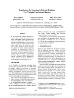
Sex
chromosomes
of
!,X-females
consist
of
two
X-chromosomes
and
one
Y-
chromosome.
The
two
X-chromosomes
are
attached
to
each
other
and
segregate
together
during
meiosis.
Due
to
the
genetic
constitution
of
the
kx-female,
male
offspring
of
these
females
get
their
X-chromosome
from
the
sire
(see
fig.
1).
Thus,
the
proportion
of
male-to-female
offspring
from
the
cross
between
a
wild-type
male
and
an
XX-femate
reflects
the
genetic
load
in
the
sire
X-chromosome.
Another
random
sample
of
100
males
was
taken
from
the
same
base
population
as
described
above,
all
males
hatched
within
one
day.
Five
days
after
eclosion
each
male
was
subjected
to
EMS
treatment
for
24
hours
according
to
the
method
described
by
L
EWIS
&
B
ACHER
(1968)
and
were
then
immediately
mated
with
three
!X
females.
The
same
traits
were
recorded
in
the
manner
described
above
and
the
sex
ratio
was
calculated
for
each
sire
group.
A
phenotypic
analysis
was
made
on
the
following
traits :
number
of
male
offspring,
number
of
female
offspring,
total
number
of
offspring,
sex
ratio
and
percentage
of
sterile
sires.
Sire
sterility
was
calculated
as
the
percentage
of
sires
with
no
offspring
from
the
total
number
of
sires
(301,
300
and
327)
in
each
age
period.
The
single
restriction
imposed
on
the
data
(except
for
calculating
sire
sterility)
was
that
only
sire
groups
having
more
than
15
offspring
were
included
in
the
analysis.
As
a
result
of
this
restriction
1,
4,
13
and
0
observations
(sire
groups)
were
excluded
from
age
periods
1,
2,
3
and
EMS
treatment
respectively.
The
means
and
standard
deviations
of
the
traits
were
calculated
for
the
different
age
periods
and
for
the
EMS
treatment.
III.
Results
The
results
are
presented
in
table
1.
The
mean
values
for
number
of
male
offspring,
number
of
female
offspring
and
total
number
of
offspring
decreased
throu-
ghout
the
three
age
periods,
with
highly
significant
differences
between
them
(P
0.001).
For
the
trait
sex
ratio
the
value
remained
constant
over
the
three
age
periods.
The
mean
values
for
number
of
male
offspring
from
EMS-treated
sires
was
approximately
29
%,
35
%
and
40
%
of
the
mean
from
untreated
(ageing)
sires
in
age
period
1,
2
and
3
respectively.
Number
of
female
offspring
from
EMS
treated
sires
compared
to
number
of
female
offspring
from
sires
in
age
period
1
was
not
greatly
affected,
but
the
difference
was
significant
(P 5
0.01).
Thus,
the
differences
in
total
number
of
offspring
and
sex
ratio
between
EMS
treated
and
untreated
(ageing)
sires
were
highly
significant
(P ;
0.001).
The
percentage
of
sterile
sires
increased
throughout
the
three
age
periods
and
was
most
pronounced
in
the
last
age
period,
whereas
the
percentage
of
sterile
sires
after
the
EMS
treatment
was
approximately
twice
that
of
the
sires
in
age
period
1.
IV.
Discussion
This
study
deals
with
the
effect
of
ageing
on
the
possible
accumulation
of
deleterious
mutations
on
the
X-chromosome
in
gametes
of
male
Drosophila
melanogas-
ter.
The
effects
of
X-linked
recessive
deleterious
mutations
affecting
viability
will
be
perceived
as
a
reduction
in
the
proportion
of
male
progeny
from
XX-females,
as
these
males
carry
the
paternal
X-chromosome.
Various
forms
of
genetic
damage
and
muta-
tions
may
accumulate
in
ageing
cells.
To
compare
any
effect
of
ageing
with
that
of
a
known
mutagen,
a
sample
of
males
was
treated
with
EMS,
and
X-chromosomes
exposed
to
EMS
were
assessed
in
the
same
manner
as
those
from
the
ageing
males.
It
must
be
stressed
that
the
effect
of
mutagen
exposure
is
not
considered
to
act
in
the
same
way
on
the
organism
as
normal
ageing.
It
can
not,
however,
be
excluded
that
age-dependent
genetic
changes,
to
a
certain
extent,
are
of
the
same
kind
as
EMS-
induced
damages.
Since
the
sex
ratio
did
not
change
with
age
of
the
male
parent,
it
is
assumed
that
no
or a
very
small
and
undetectable
accumulation
of
deleterious
mutations
occur
in
the
X-chromosome
of
the
germ
cells.
However,
the
result
is
inconsistent
with
the
findings
of
A
NDJELKOVI
C
et
al.
(1979)
indicating
that
such
an
age-related
accumulation
of
harmful
mutations
does
occur.
The
observed
decline
in
both
male
and
female
offspring
with
increased
paternal
age
and
the
dramatic
increase
in
sterility,
may
reflect
the
general
decline
in
vitality
and
increased
accumulation
of
DNA
damage
in
the
various
stages
of
spermatogenesis
of
ageing
males.
Accumulation
of
DNA
damage
with
advancing
age
in
the
early
stage
of
spermatogenesis
leads
to
increased
sterility.
If
DNA
damage
occurs
in
spermatids/
sperms
both
number
of
male
and
female
progeny
should
be
reduced
whether
the
damage
is
present
in
autosomes
or
in
sex
chromosomes,
due
to
the
lethal
effects
through
the
blockage
of
DNA
replication
produced
by
DNA
damage.
The
observed
decline
in
both
number
of
male
and
female
progeny
and
increase
in
sterility
with
age
is
consistent
with
an
accumulation
of
genetic
damage
in
germ
line
with
age.
Males
exposed
to
EMS
showed
a
drastically
reduced
number
of
male
offspring,
a
slight
but
significant
reduction
in
number
of
female
offspring,
and
a
small
effect
on
sterility.
The
high
frequency
of
harmful
recessive
mutations
implied
by
the
drastically
reduced
number
of
male
offspring
did
not
greatly
affect
the
number
of
female
offspring
nor
sire
sterility.
This
further
emphasizes
the
importance
of
interpreting
the
expected
effects
of
mutations
and
DNA
damage
separately.
The
discrepancy
between
our
results
and
those
obtained
by
Arrna
ELK
OVu
et
al.
(1979)
is
difficult
to
interpret.
In
the
latter
investigation
the
authors
examined
age-
related
changes
in
viability
and
longevity
due
to
recessive
mutations
in
homozygous
second
chromosomes.
Both
female
and
male
parents
and/or
grandparents
were
old
and
+lCyL
z
individuals.
The
present
study
deals
with
age-related
recessive
mutations
in
the
X-chromosome
of
old
wild-type
males.
Whether
differences
in
the
D.
melanogaster
stocks
used
and/or
in
the
experimental
design
account
for
the
discrepancy
is
questiona-
ble.
However,
a
significant
decline
in
the
number
of
male
as
well
as
female
offspring
with
advancing
age
of
the
sire
was
obtained
in
both
investigations.
If
this
result
is
interpreted
as
being
due
to
an
age-related
accumulation
of
DNA
damage
and/or
a
decline
in
vitality,
there
is
no
disagreement
in
this
respect.
In
order
to
examine
the
importance
of
the
protective
mechanisms
of
DNA
repair
and
its
correlation
with
ageing
more
thoroughly,
this
study
will
be
continued
by
a
project
for
selection
of
resistance
to
a
mutagen
in
positive
and
negative
directions.
Received
May
6,
1987.
Accepted
January
14,
1988.
References
A
LEXANDER
P.,
1967.
The
role
of
DNA
lesions
in
processes
leading
to
ageing
in
mice.
Symp.
Soc.
Exp.
Biol.,
21,
29-50.
A
NDJELKOVI
C
M.,
M
ARINKOVI
é
P.,
Tucu
N.,
Tocn
M.,
1979.
Age-affected
changes
in
viability
and
longevity
loads
of
Drosophila
melanogaster.
Am.
Nat.,
114,
915-939.
B
EARDMORE
J.A.,
1976.
Genetics
and
ageing.
Eugen.
Soc.
Bull.,
8,
39-42.
C
ALLAHAN
R.F.,
1962.
Effects
of
parental
age
in
the
life
cycle
of
the
house
fly
Musca
domestica.
J.
N.Y.
Entomol.
Soc.,
70,
150.
COMFORT
A.,
1953.
Absence
of
a
Lansing
effect
in
Drosophila
subobscura.
Nature,
172,
83-83.
FmLLn
G.,
1960.
The
ageing
process
and
somatic
mutation.
In :
S
TREHLER
B.L.
(ed.),
The
biology
of
ageing,
170-175,
American
Institute
of
Biological
Sciences,
Washington
D.C.
FLEMINGS
M.B.,
L
UDWIG
D.,
1964.
Effects
of
temperature
and
parental
age
on
the
life
cycle
of
the
body
louse,
Pediculus
humanus
humanus.
Ann.
Entomol.
Soc.
Amer.,
57,
560-563.
G
ENSLER
H.L.,
B
ERNSTEIN
H.,
1981.
DNA
damages
as
the
primary
cause
of
ageing.
Quart.
Rev.
Biol.,
56,
279-303.
GotusTEirr
S.,
1971.
The
role
of
DNA
repair
in
ageing
of
cultured
fibroblast
from
xeroderma
pigmentation
and
normals.
Proc.
Soc.
Exp.
Biol.
Med.,
137,
730.
HART
R.W.,
M
ODAK
S.P.,
1980.
Ageing
and
changes
in
genetic
information.
Adv.
Exp.
Med.
Biol.,
129,
123-137.
K
EMPF
C.,
S
CHMITT
M.,
P
ANSE
J M.,
KE
ntrF
J.,
1984.
Correlation
of
DNA
repair
synthesis
with
ageing
in
mice,
evidence
by
quantitative
autoradiography.
Mech.
Ageing
Dev.,
26,
183-194.
L
ANSING
A.I.,
1954.
A
non-genetic
factor
in
the
longevity
of
rotifers.
Ann.
N.Y.
Acad.
Sci.,
57,
455-464.
L
EWIS
E.B.,
B
ACHER
F.,
1968.
Method
of
feeding
ethyl
methane
sulfonate
(EMS)
to
Drosophila
males.
Dros.
Inf.
Serv.,
43,
193.
Mnc!E!xn-CoELHO
A.,
1984.
Genome
reorganization
during
cellular
senescence.
Mech.
Ageing
Dev.,
27,
257-262.
M
ARINKOVIT
P.,
Tuoc
N.,
ANDJELKOV14!
M.,
1973.
Age
associated
changes
in
viability
genetic
loads
of
Drosophila
melanogaster.
Exp.
Gerontol.,
8,
199.
M
ARINKOVI
E
P.,
T
UCI
é
N.,
A
NDJELKOV
it
M.,
1975.
The
effect
of
streptomycin
on
age-associated
viability
genetic
loads
in
Drosophila
melanogaster.
Acta
Biol.,
7/8,
339-343.
M
EDVEDEV
Z.A.,
1964.
The
nuclei
acids
in
development
and
ageing.
Adv.
Gerontol.
Res.,
1,
181-
206.
N
IDERM
Ü
LLER
H.,
H
OTECHER
G.,
S
KALICKY
M.,
1985.
Changes
of
DNA
repair
mechanisms
during
the
ageing
of
the
rat.
Mech.
Ageing
Dev.,
29,
221-238.
O’B
RIAN
D.M.,
1961.
Effects
of
parental
age
on
the
life
cycle
of
Drosophila
melanogaster.
Ann.
Entomol.
Soc.
America,
54,
412-416.
PAINTER
R.B.,
C
LARKSON
J.M.,
YOUNG
B.R.,
1973.
Ultraviolet-induced
repair
replication
of
ageing
diploid
human
cells
(UWI-38).
Radiat.
Res.,
56,
560.
S
TREHLER
B.L.,
1962.
Further
studies
on
the
thermal-induced
ageing
of
Drosophila
melanogaster.
J.
Gerontol.,
17,
347-353.
S
ZILARD
L.,
1959.
On
the
nature
of
ageing
process.
Proc.
Natl.
Acad.
Sci.,
45,
1145-1147.
WA
TT
IAUX
J.M.,
1968.
Parental
age
effects
in
Drosophila
pseudoobscura.
Exp.
Gerontol.,
3, 55-61.