Báo cáo y học: "A scalable, fully automated process for construction of sequence-ready human exome targeted capture libraries" pot
Bạn đang xem bản rút gọn của tài liệu. Xem và tải ngay bản đầy đủ của tài liệu tại đây (1.07 MB, 15 trang )
METH O D Open Access
A scalable, fully automated process for
construction of sequence-ready human exome
targeted capture libraries
Sheila Fisher
1
, Andrew Barry
1
, Justin Abreu
1
, Brian Minie
1
, Jillian Nolan
1
, Toni M Delorey
1
, Geneva Young
1
,
Timothy J Fennell
1
, Alexander Allen
1
, Lauren Ambrogio
1
, Aaron M Berlin
2
, Brendan Blumenstiel
3
, Kristian Cibulskis
3
,
Dennis Friedrich
1
, Ryan Johnson
1
, Frank Juhn
4
, Brian Reilly
1
, Ramy Shammas
1
, John Stalker
1
, Sean M Sykes
2
,
Jon Thompson
1
, John Walsh
1
, Andrew Zimmer
1
, Zac Zwirko
1,4
, Stacey Gabriel
2
, Robert Nicol
1
, Chad Nusbaum
2*
Abstract
Genome targeting methods enable cost-effective capture of specific subsets of the genome for sequencing. We
present here an automated, highly scalable method for carrying out the Solution Hybrid Selection capture
approach that provides a dramatic increase in scale and throughput of sequence-ready libraries produced.
Significant process improvements and a series of in-process quality control checkpoints are also added. These
process improvements can also be used in a manual version of the protocol.
Background
The cost of DNA sequencing continues to fall, driven by
ongoing innovation in sequencing technology [1-4]. As a
result, it has recently become feasible to sequence non-
trivial numbers of whole human genomes [3,5-10].
Many more such projects are planned and commercial
genome sequencing services ar e now becoming available
[11,12]. At the same time, there is growing interest in
sequencing specific portions of genomes, and several
affordable methods for sampl e preparation of targeted
region s have been recently published [13-17]. Key appli-
cations for targeted approac hes include sequencing of
exons or sets of protein-co ding genes implicated in spe-
cific diseases [18-21], whole human ex ome sequencing
(for example, in cancer or disease cohorts) [22-24]
(reviewed in [25]), and resequencing of specific regions
as a follow-up to genome-wide association studies [26].
The economics of whole exome sequencing have made
targeted enrichment approaches an attra ctive option for
discovery of rare m utations in a variety of diseases as
the price tag is substantially lower than for sequencing
an entire human genome. For example, using list prices
and including the targeted capture step, the all-in cost
of sequencing a whole exome (roughly 30 Mb), is
13-fold less than for the whole g enome (Table S1a in
Additional file 1). This tra nsla tes directly into a budget
that can include more than ten times as many samples,
greatly increasing the statistical power of the data to be
generat ed. The effect is even greater for smaller sequen-
cing targets, which further scale down the re quired
sequencing, although costs of targeting scale down more
slowly. Ultimately, as long as the expense of the
required sample preparation does not dominate, target-
ing will continue to be a cost-effective approach. To
date, however, no targeting method has been described
that can handle the many thousands of samples that are
becoming available. To fill this need, we set out to
develop such a method.
Solution hybrid selection (SHS), developed by Gnirke
et al. [14], was created as a tool to cheaply and effec-
tively target multiple regions in the genome in a way
that is compatible with next generation sequencing tech-
nologies (Figure 1). The published protocol performs
well in terms of efficiency of enrichment (selectivity),
reproducibility, evenness of coverage, and sensitivity to
detect single-base changes [14]. Using this method, a
single technician can process six samples simultaneously
from genomic DNA to sequence-ready library in
* Correspondence:
2
Genome Sequencing and Analysis Program, Broad Institute of MIT and
Harvard, 320 Charles Street, Cambridge, MA 02141, USA
Full list of author information is available at the end of the article
Fisher et al. Genome Biology 2011, 12:R1
/>© 2011 Fisher et al.; lic ensee BioMed Central Ltd. This is an open access article distributed under the terms of the Creative Comm ons
Attribution Licens e ( nses/by/2.0), which pe rmits unrestricted use, distribution, and reproduction in
any medium, provided the original work is properly cited.
approximately one week. This process was designed
purely as a series of liquid handling steps and incuba-
tions, with the specific intention of making it amenable
to scale up and automation. Given the demonstrated
success of this and other methods, demand for targeted
sequencing has increased sharply. To accommodate the
increased demand, keep costs down, and limit the
requirements for human labor, we have adapted SHS to
an automated high-throughput process. This SHS
method includes improvement s designed to increase the
efficiency of the target selection process through optimi-
zation of reactions and automation of the library and
capture procedures using liquid handling robots. Several
aspects of this method, in particular the ‘ with-bead’
sample preparation method, are amenable to sample
preparat ion steps for a range of next generation sequen-
cing applications, including alternative in-solution and
solid-phase capture strategies.
To support high-throughput SHS for targeted sequen-
cing, we set out to devise a lab oratory process that
would handle very large numbers of samples in parallel
for targeting and preparation of sequence-ready libraries
at a low cost per sample. This process was designed to
carry out whole exome targeting but also yields good
results in targeting subsets of genes or regions for rese-
quencing. Results described here come from whole
exome targeting using the Agilent SureSelect Human
All Exon v2 kit, which is a commercially available imple-
mentation of the optimized capture reagent we have
described previously [14].
A number of challe nges were overcom e in developing
a robust, automated, and highly scalable process for
selection of exomes and other targets. Beyond the need
for processing large numbers of samples, modifications
of the protocol were made to achieve or maintain the
following: elimination of manual, agarose gel-based size
selection, which has now been replaced by fully auto-
mated, bead-based steps; high selectivity, with a high
number of sequenced bases on or near the target region
of interest; evenness of sequence coverage among cap-
tured targets, avoiding highly overrepresented targets
and dropouts; high library complexity, or low molecular
duplication, so that libraries contain large numbers of
unique genome fragments; reproducibility, so that per-
formance of the process is highly predictable; low cost
of the targeting process relative to sequencing; detailed
process tracking to reduce errors and provide sample
history; quality control checkpoints built into the
T7
Elute, amplify,
add T7 promoter
Biotin–UTP
transcription
Biotinylated
bait probes
Adaptored
pond of targets
Solution
hybridization
Bead
capture
Amplify by
off-bead PCR
SEQUENC
E
Generation of RNA bait capture probes
Solution hybrid selection
(
a
)
(b)
Universal amplification sequences
T7 RNA polymerase promoter
Illumina sequencing adaptors
Biotin-labeled UTP
Streptavidin bead
RNA bait probes
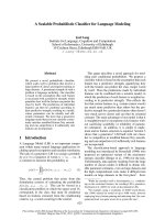
Figure 1 Overview of the hybrid selection method. Two specific sequencing targets and their respective capture baits are indicated in blue
and red. (a) Generation of RNA bait capture probes. 150mer oligos are synthesized on array in batches of 55,000 and cleaved off. They are
made double stranded by PCR amplification and tailed with a T7 RNA polymerase promoter, and RNA capture baits are made by transcription in
the presence of biotinylated UTP. (b) Solution hybrid selection. RNA baits (from the top line) are mixed with a size selected pond library of
fragments modified with sequencing adaptors. Hybridized fragments are then captured to streptavidin beads and eluted by the with-bead
protocol for sequencing. See text for details.
Fisher et al. Genome Biology 2011, 12:R1
/>Page 2 of 15
process to identify poor performers prior to sequencing;
and limited human labor.
We present here a scalable, automated SHS method
that operates at a throughput far higher than achieved
by other methods. The process can also be carried out
by hand using a multichannel pipetter. This method has
not only been scaled but also optimized to improve
selectivity and evenness of target coverage and to mini-
mize artifactual duplication to consistently deliver
greater than 94% of the alignable exome (Additional file
2). The automated p rotocol has a capacity to process
over 1,200 SHS samples in less than a week with four
technicians (one technician can generate 1,200 pond
libraries per week, and three technicians can each gener-
ate 384 SHS captures per week). For ease of explanation,
we employ a fishing-based terminology in SHS, where
the biotinylated RNA capture reagent is referred to as
the ‘bait’, the genomic DNA library from which targets
are captured as the ‘pond’ in which we are ‘fishing ’,and
the DNA targets from the pond that are captured by the
bait are referred to as the ‘catch’.
Results
Building a high-throughput solution hybrid selection
process
SHS is a method used to selectively enrich for regions
of interest within the human genome [14] (Figure 1).
Briefl y, a library (or ‘pond’) of adapter-ligated fragme nts
of randomly sheared DNA is hyb ridized to biotinylated
RNA (or ‘baits’ ) that are complementary to the target
sequences. Hybridized molecules (the ‘catch’)arethen
captured using streptavidin-coa ted beads. Once the cap-
tured DNA fragments are PCR amplified off the capture
reagent, they are available to be sequenced using next
generation sequencing technologies. The standard SHS
protocol was redesigned from a ma nual, bench scale
process to an a utomated proc ess, in muc h the sa me
way as our recent work to scale library construction for
454 sequencing [ 27], and is ca pable of far gre ater
throughput than demonstrated for other methods
(Additional fil e 2). A series of process innovations were
required to facilitate reimplementation of this process at
large scale. In particular, all manual pipetting steps were
converted to automation-amenable liquid handling
steps, and these liquid handling steps were extensively
optimized to maximize yield efficiency. As part of this,
the electrophoretic size selection step has been replaced
by fully automated bead-based sizing. Other optimiza-
tions are described below. Table 1 shows a comparison
of the o riginal published method and the new protocol
with a description of each step and the improvements
in the new method. Table 2 describes a set of key
sequencing metrics by which we measure SHS process
performance.
The automated SHS process is implemented on the
Bravo liquid handling workstation (Agilent Automation
Solutions), a commercially available small-footprint,
liquid handling platform, but can be implemented on
many commercially available liquid handlers. The pro-
cess can also be carried out manually using a multichan-
nel pipette. An overview map of the process can be
found in Additio nal file 3 and the manual protocol ver-
sion can be found in Additional file 4.
Optimization of acoustic shearing
The process begins with fragmentation of genomic DNA
using the Covaris E210 adapt ive focused acousti cs
instrument. Maximizing the yield of DNA fragments in
the desired size range is a key step in minimizing overall
sample loss. The Covaris E210 instrument focuses
acoustic energy into a small, localized zone to create
cavitation, thereby producing breaks in double-s tranded
DNA. A number of variables control mean fragment
length and distribution, including duty cycle, cycles per
burst, and time. The Covaris adaptive focused acoustics
system has several advantages over other methods such
as nebulization or hydrodynamic force. First, DNA is
sheared in a small closed environment and is not
handled in large volume vessels or in tubing, greatly
reducing sample loss. Second, the closed, i ndependent
vessels greatly reduce sample cross-contamination.
Third, the Covaris machine can operate automatically
on up to 96 samples per run, eliminating significant
sample handling labor and eliminati ng shearing as a
process bottleneck. Fourth, improvements to the shear-
ing protoco l in combination with removal of small frag-
ments in subsequent bead-based clean up steps (see
below) eliminates the need to size select and extract
samples from agarose gels, a critical bottleneck in the
overall process.
Shearing performance was extensively optimized for
increased sample yield, narrower insert size distribution,
and robust and reproducible handling of large numbers
of samples in parallel. Optimizations focused on the fol-
lowing factors: shearing volume, tube type, elimination
of tube breakage, shearing pulse time, water degassing,
and positioning of tubes in the water bath (see Materials
and methods for details). In order to accommodate
automated handling of the samples, volumes were
reduced from 100 μlto50μl without any effect on
shearing profiles or sample loss (Additional file 5).
Importantly, proper fit of the shearing rack (Covaris,
catalogue number 500111) into custom adapters (see
Additional file 6 for CAD drawing) prevents movement,
allowing transfers to occur via automated liquid hand-
ling. In addition, specific tubes available from Covaris
(Cov aris, catalogue number 500114) virtu ally eliminated
the problem of tube breakage. Only a single sample in
Fisher et al. Genome Biology 2011, 12:R1
/>Page 3 of 15
the most recent 5,000 processed suffered a broken tube.
Through a systematically desig ned and controlled set of
experiments, optimal pulse time parameters were chosen
to provide a mean fragment length of 150 bp with a
rangeof75to300bp(Materials and metho ds). Addi-
tional file 5 shows the contrast between unoptimized
and optimized size profiles of sheared DNA. In addition
to regular maintenance, careful degassing of the water
bath and proper water levels are critical for reproducible
results. In a nondegassed water bath dissolved oxygen
reduces cavitation and disperses energy, reducing shear-
ing efficiency.
Modified bead-based cleanups enable scale-up to 96 wells
A key requirement in scaling SHS was to implement
processing of samples in a standard 96-well microtiter
plate. This was facilitated by development of a novel
modification to solid-phase reversible immobilization
(SPRI) magnetic bead reaction cleanup methodology
[27,28] we have termed ‘with-bead’ SPRI (Figure 2),
which is highly scalable due to its amenability to liquid
handling automation. Implementation of with-bead SPRI
in SHS offers significant advantages. First, it replaces
single tube spin-column-based cleanups with liquid
handling-c ompatible magnetic be ad-based cleanups; sec-
ond, it enables selection of molecular weight ranges,
eliminating the need for ag arose gel-based sizing; thir d,
it simplifies the process by allowing elimination or com-
bin ing of several steps, which resul ts in a higher overall
DNA yield.
The innovation of the with-bead SPRI method is as
follows. Rather than employing a series of discrete
cleanup steps in the library construction process, the
cleanups are effectively integrated. The SPRI beads are
added to the sample after the shearing step, and remain
in the reaction vessel throughout the sample preparation
protocol. By allowing each cleanup step to employ the
same beads, the with-bead method greatly reduces the
number of liquid transfer steps required. The ‘ cleaned
up’ DNA is then eluted at the conclusion of the process.
This methodology increases the overall DNA yield
(Figure 3), primarily because it allowed us to eliminate
six of the ten sample transfer steps, avoiding the loss of
DNA sticking to the sides of the vessel or loss o f
volume in pipetting. Briefly, following each process step,
DNA is selectively bound to the iron beads, already pre-
sent, through the addition of a 20% polyethylene glycol
(PEG), 2.5 M NaCl buffer. The mixture is placed on a
magnet, which pulls the beads and bound DNA to the
sides of the well so that the reagents, washes and/or
unwanted fragments can be removed with the superna-
tant. Molecular weight exclusion, which is essentially a
size selection, of unwanted lower molecular weight
DNA fragments can be controlled through the volume
of the PEG NaCl buffer that is added to the reaction,
changin g the final concentration of PEG in the resulting
mixture and altering the size range of fragments bound
to the beads [27,28]. DNA fragments that have been
cleaned or size selected are eluted from the beads, ready
Table 1 Comparison of standard versus improved solution hybrid selection methods
Manual standard SHS protocol Automated improved SHS protocol
Process step Standard
method
Drawbacks Improved
method
Advantages
Shearing of genomic
DNA
Covaris S2 Single sample Optimized
Covaris E210
Multi-sample, improved yield, tight size range
Enzymatic cleanups Individual spin
columns
Low throughput, 50 to 60%
recovery, manual
’With-bead’ SPRI High throughput, 80 to 90% recovery, automated
Solution hybrid selection
capture
Manual, column-
based
Labor intensive (6 samples/
FTE/week)
Fully
automated
Walkaway, high throughput (1,200 samples/4FTE/
week)
Final PCR enrichment Denature,
followed by PCR
Sample loss through transfers Direct ‘off-bead’
PCR
Improved final yield
In process quality control
checkpoints
Agilent
Bioanalyzer
Limited visibility until
sequence results
Many In process results: key predictors of sample, library
and sequencing quality
FTE, full time employee; SHS, solution hybrid selection; SPRI, solid phase reversible immobilization.
Table 2 Automated solution hybrid selection
performance
Performance factor 3 μg input average (n = 1,117
exomes)
Median target coverage 131.0×
Percentage bases > 2× 96.0%
Percentage bases > 10× 91.9%
Percentage bases > 20× 87.6%
Percentage selected bases (on
target)
83.7%
Percentage duplicated reads 4.4%
Fold 80 penalty
a
3.17
Estimated library size of captured
fragments
278 million
See Additional file 12 for metric definitions.
a
Fold 80 penalty is a measure of
the non-uniformity of sequence coverage, defined as the amount of
additional coverage (in fold coverage of the genome) required so that 80% of
the target bases will be covered at the current mean coverage (see Additional
file 12 for details).
Fisher et al. Genome Biology 2011, 12:R1
/>Page 4 of 15
for t he next step; however, the eluate is not transferred
into a new reaction vessel. Rather, the reagents for the
next step are added dire ctly to the reaction vessel con-
taining samples and beads. The presence of beads does
not interfere with any of the steps in the process
(Table 3). This with-bead protocol has great ly increased
the number of unique fragments entering the pond PCR
step, increasing the complexity of libraries made by
roughly 12-fold (Table 3).
This increase in yield with the with-bead SPRI proto-
col has the added benefits of reducing both the input
DNA requirement to the process and the number of
PCR cycles required. Efficient with-bead targeted cap-
tures c an be achieved with pond libraries made with as
little as 100 ng of input DNA and six to eight cycles of
PCR, a major improvement over the commercialized
SHS method, which requires 3 μg of starting genomic
DNA and 14 cycles (Table 3). We note here that PCR
Sheared
DNA
Add SPRI
beads
Place on
magnet
Remove from magnet
Elute DNA from beads
Remove
supernatan
t
Add PEG
buffer
1
.
E
n
d
r
e
p
a
i
r
2
.
A
-
b
a
s
e
3
.
A
d
a
p
t
o
r
l
i
g
a
t
i
o
n
4. PCR enrichment
Hybridization
reaction
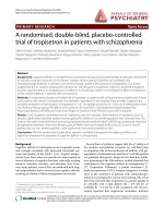
Figure 2 Wi th-bead SPRI method f or pond library construction. SPRI magnetic beads are added to the sheared DNA sample. DNA is
selectively bound to SPRI beads, which are immobilized when the sample plate is placed on a magnet, leaving other molecules in the liquid
phase. The liquid phase is removed and discarded. The sample plate is then removed from the magnet and DNA is eluted from the beads.
Library construction master mixes are then added to eluant/bead solution. The DNA and SPRI beads then pass through three cycles of reaction,
binding to beads (in the presence of polyethylene glycol (PEG)/NaCl solution) and cleanup/washing. The cycles carry out end repair, A-base
addition and adaptor ligation, respectively. A final elution is then followed by PCR amplification.
Fisher et al. Genome Biology 2011, 12:R1
/>Page 5 of 15
cannot be completely eliminated because the efficiency
of adaptor ligation varies between samples, probably
because of variation in input DNA quality. PCR cycle
number was optimized to maximize the number of
unique fragments in the library while minimizing the
duplication rate (Additional file 7). T his resulted in a
modest number of cycles that enriches fragments con-
taining an adapter at each end but not fragments with
either no adapters or an adapter at one end only. These
incomplete constructs compete with two-adapter frag-
mentsinthehybridizationreactionbutcannotbe
sequenced.
Pre-mixed reagents for automated library construction
Currently available commercial library reagent kits are
packaged for bench-level processing of eight to ten
samples. In order to accommodate the increase in scale
and automated processing of samples, large-scale
reagent kits were developed and optimized for the high-
throughput SHS pond construction proce ss. All buffers
and non-enzyme components are premixed and ali-
quoted at volumes appropriate for 96 samples, including
necessary dead volume. Prior to use, the premixed
reagents only need to be thawed and placed on the deck
where enzymes are added imme diately before dispense
into reaction plates. To accomplish this, we developed a
custom reservoir in combination with optimized aspira-
tion and dispense protocols. The custom reservoir is
designed to limit dead volume, thereby minimizing the
reagent volume required, thus reducing reagent waste.
Details, including the dimensions of the reservoir, can
be found in Additional file 8.
Column
cleanups
Automated
standard bead-based
cleanups
Automated
with-bead
SPRI cleanups
Recovery
(
%
)
Output of pond library
construction process (mg)
50
45
40
35
30
25
20
15
10
5
0
0
1
.
6
1.4
1.2
1.0
0.8
0.6
0.4
0.2
23
28
47
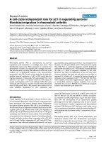
Figure 3 Yield output from pond library construction methods. Data are shown left to right, for pond libraries constructed with three
methods: the widely used standard column-based cleanups [14], an automated implementation of standard bead cleanups and our
implementation of with-bead SPRI cleanups. Each library was constructed with 3 μg input of NA12878 genomic DNA, in triplicate. Bars: total
DNA output from pond library construction before PCR amplification. Blue diamonds: percentage recovery of input DNA for duplicates of 3 μg
of the same input DNA. With-bead-based cleanups increased the amount of DNA retained throughout library construction compared to the
standard column or SPRI cleanup methods.
Table 3 Performance comparison of manual versus automated solution hybrid selection
Factor Column based Automated (with-bead SPRI) Automated (with-bead SPRI) low input
Input DNA 3 μg3μg 0.1 μg
Samples/FTE/week 6-12 384 384
Number of sample transfer steps 10 4 4
Output DNA prior to PCR 720 ng 1,330 ng Below limit of detection
Number of pond PCR cycles 12-16 6 6
Percentage duplicated reads 19.8 2.2 10
Percentage selected bases 84.7 88.6 83.76
Estimated library size 43 million 516 million 223 million
FTE, Full time employee; SHS, solution hybrid selection; SPRI, solid-phase reversible immobilization.
Fisher et al. Genome Biology 2011, 12:R1
/>Page 6 of 15
Automation of capture protocol to process 96 samples
simultaneously
The most labor-intensive step in the manual selection
processisthe‘capture’ protocol (Table 1), where hybri-
dized DNA-RNA bait duplexes are separated from
unbound fragments. The separation is performed using
streptavidin beads that bind to the biotin molecule s that
are covalently linked to the RNA b ait. Fragments that
are not hybridized to the biotinylated RNA baits are
removed through a series of washes.
Wash conditions were redesigned for compatibility
with automated liquid handling and optimized for maxi-
mal yield (Additional file 9). Since microtiter wells are
of much smaller volume than the standard microtubes
used in the manual process, the number of wash cycles
was increased as the volume of each wash had to be
decreased to fit the wells while maintaining the proper
level of stringency. Wash buffers are precisely controlled
for temperature by storing the buffer-containing vessels
in 65°C temperature-calibrated heating blocks (V&P
scientific, VP-741BW MICA) integrated onto the deck
of the liquid handler robot. This automation provides a
hands-off capture protocol capable of consistently set-
ting up capture reactions for 96 samples in 4 hours; in
comparison, the manual (and somewhat variable) pro-
cess handled 6 samples in 4 hours. Additionally, the
automated process delivers output of a more consistent
quality, and eliminates manual tracking a nd pipetting
errors (Additional file 10).
Off-bead PCR to increase yield of captured product
In the manual protocol [14], the elution of desired DNA
fragments fro m the RNA bait-streptadavidin bead com-
plex is accomplished by denaturation using 0.1 N
sodium hydroxide followed by a cleanup step prior to
PCR amplification. This series of steps requires large
volumes and is therefore difficult to scale in a microtiter
plate format. In addition, variability at this step can
result in loss of captured DNA. We have replaced elu-
tion through denaturatio n by amplifying the captured
sequences directly by PCR, by a process we term ‘ off-
bead’ PCR, as the target is PCR amplified off the bead
directly in the capture plate. This allows scaling in a
microtiter plate format, simplifies the process by remov-
ing a pipetting step, eliminates process variability and
improves the yield of captured product roughly three-
fold (Additional file 11). Briefly, PCR enzyme, PCR
primers, and dNTPs are added directly to the bead-bait-
DNA complex, and the mix is amplified via thermalcy-
cling (see Materials and methods for details). Bait
RNAs, which lack Illumina adapter sequences, and pond
fragments with fewer than t wo adapters are not ampli-
fied. The amplified fragments are then separated from
the beads through a modified SPRI bead cleanup
(Materials and methods) . This off-bead PCR protocol, in
combination with improvements described above, signif-
icantly improves yield at this step in the process
(Table 3). This simple, automation-friendly, cost-effec-
tive protocol can be used to process up to 1,200 samples
per week in batches of 96 (Table 2).
Development and automation of in-process quality
control checkpoints
As the process increases in scale, readouts of sample
quality and process success become increasingly impor-
tant as indicators of the likelihood of producing high
quality sequencing results. To this end we have imple-
mented a series of in-line quality control checkpoints.
This enables granular reporting of metrics during the
SHS process and, importantly, allows poorly performing
samples to be quickly identified and removed, avoiding
the associated costs of downstream processing and
sequencing (Figure 4). Central to this is the development
of critical quality control assays, both in terms of their
sensitivity to the samples at the point at which they are
assayed, as well as their utility as a predictor of sequen-
cing quality. The ei ght key quality control checkpoints
that add immediate value to the process are outlined
below (see Materials and methods for details on each).
Volume check
Volumes are checked for every sample by visual inspec-
tion to ensure predictable performance in shearing
(Figure 4a). If volumes are outside of specification (50 μl
± 20%), samples are either concentrated or diluted to
reach the appro priate range. Low volumes cause inaccu-
rate automated transfer of sample into shearing vessels.
Sample concentration check by PicoGreen
Concentrations for all samples are measured via an
automated PicoGreen assay (see Materials and methods)
and are specified to be within 2.0 to 60 ng/μl(Figure
4b). Samples above this range are normalized and re-ali-
quoted to appropriate volumes since excess input DNA
can actually inhibit the enzymatic pond reactions (data
not shown). Samples above the 2.0 ng/μl threshold are
considered to pass. Those below this range can be run
on risk.
Size quality control of sheared DNA
Sheared samples are assayed on an automated microflui-
dic electrophoresis instrument, the Caliper GX system,
using the 1K DNA Chip to evaluate the size distribution
produced by the Covaris instrument (Figure 4c). Frag-
ment sizes should be between 75 and 300 bp with the
distribution centered on 150 bp. Samples that shear
above this range can decrease the specificity and effi-
ciency of the selections. S amples sheared to less than a
mean of 110 bp will be suffer losses during the various
with-bead cleanups, greatly reducing the complexity of
the library before selection.
Fisher et al. Genome Biology 2011, 12:R1
/>Page 7 of 15
Performance quality control of automation
The Bravo automated liquid handling platform is assayed
daily for dispense accuracy and precision using a quantita-
tive fluorescent dye assay (Figure 4d). Standard liquid
handling sequences are run using sulforhodamine dye, and
relative fluorescent units of the dispensed dye are assayed
on a Perkin Elmer Victor3 plate reader. Coefficients of var-
iation (%CV) are calculated between wells and must be
within three standard deviations of the mean. If the robot
is out of specification, maintenance is performed on the
system followed by repeat of the quality cont rol until the
coefficients of variance are back within acceptable ranges.
Covaris shearing
End repair
A-base addition
Pond PCR
Hybridization
Capture on bead
Off-bead catch PCR
Illumina sequencing
2 6 10 14 18 22 26 30 34 384 8 12 16 20 24 28 32 36
0.01
0.1
1
Cycles
Fluorescence (dRn)
20 30 50 70 100 200 300 500700 1000 2000 3000 5000
0
20
40
60
80
100
120
Size (bp)
Fluorescence
Total DNA (μg)
1.5 1.6 1.7 1.8 1.9 2.0 2.1 2.2 2.3 2.4 2.5 2.6 2.7 2.8 2.9 3.0 3.1 3.2 3.3 3.4 3.5 3.6 3.7 3.8 3.9 4.0 4.1 4.2 4.3 4.4 4.5
Distributions
A
B
C
D
E
F
G
H
123456789101112
0 – 50,000
50,000 – 100,000
100,000 – 150,000
150,000 – 200,000
50
40
30
20
10
5046 51 53 54 5655 57 58 59
LC set #
46
50
51
53
54
55
56
57
58
59
LC set #
Enriched library (ng/μl)
0 102030405060708090100
0
20
Concentrations
40
Sample mean (B/B0)
60
ng/μl
80
Linear regression
100
(a) Input DNA quantification by pico
(f) Pond quantification
(g) Catch quantification by pico
(b) Pre-flight pico
(c) Shearing profile
(d) Automation performance
(h) Catch quantification by qPCR
(e) Deck layout
Sample
binding
plate
Magnet
100 ml
70% ethanol
Shearing holder
with product
in AFA tubes
165 ml SPRI XP beads 165 ml filter tips
50 ml
elution
buffer
Adaptor ligation
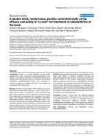
Figure 4 Qua lity control checkpoints. (a-h) Eight different quality control checkpoints for the scaled SHS process are schematized. Quality is
assayed at key steps to quickly identify failed samples and also to provide ability to troubleshoot process failures. See text for details. AFA,
adaptive focused acoustics.
Fisher et al. Genome Biology 2011, 12:R1
/>Page 8 of 15
Confirmation of deck configuration
To confirm proper set up of the Bravo platform before
each step in the protocol, the software requests the
operator to confirm the proper deck layout by compar-
ing the deck positions to a picture shown on
screen (Figure 4e). This prevents users from starting
programs without the proper materials in place or from
running the wrong combination of program and deck
configuration.
Quantification of pond libraries and catch libraries
Prior to selection, pond libraries are assayed for concen-
tration by an automated PicoGreen assay (Materi als and
methods) and are specified to be within a range of 25 to
60 ng/μlinavolumeof40μl (Figure 4f). Samples at
concentrations greater than 25 ng/μl are normalized to
25 ng/μl prior to hybri dization. Samples below this 25
ng/μl generally produce sequence data with high
amounts of duplication. After capture, samples are again
assayed in a similar fashion (Figure 4g). All catches with
concentrations greater than 5 ng/μl are passed on to the
next step in the process. Catches with concentrations
less than 5 ng/μl are considered failures and can be sent
for re-selection.
Quantitative PCR quantification of catch
Final evaluation of the catch material employs an auto-
mated quantitative PCR assay developed in conjunction
with Kapa Biosystems (KAPA Library Quantification Kit,
catalogue number KK4832) designed to accurately quan-
tify the fragments containing two Illumina adapters
(Figure 4h). This step is critical for determining th e cor-
rect concentration of the library to be loaded for
sequencing on the Illumina platform, to maximize clus-
ter densities and sequencing quality. Samples at concen-
trations greater than 2 nM have been found to produc e
sequencing data with sufficient complexity.
In addition to the in-line assays, each 96-well plate of
samples contains control DNAs (two positives and one
negative) that are used for quality assessment (see Mate-
rials and methods). The control checkpoints established
throughout the process provide early warning of issues
with performance of each step and overall quality. In
addition to these in-process lab assays, we have devel-
oped a number of key sequencing metrics that allow us
to gauge the success of each selection (Table 3) as well
as the performance of the process over time (Additional
file 10) in support of continuous process improvement
and optimization (see Additional file 12 for fu rther defi-
nition of sequencing metrics).
Sample tracking and integrity
Any process that handles large numbers of samples must
have a supporting sample tracking system that preserves
sample identification and manages association of critical
process data necessary for analysis. As part of the scaled
SHS process, we developed and implemented a compre-
hensive tracking system that associates sample informa-
tion with a unique barcode on each sample tube and
microt iter plate. Every step takes place in barcoded plas-
ticware, and each step where samples are moved is asso-
ciated with a barcode scan that is reported to the
database so that data trails across all sample handling
events are complete. Microtiter plates are labeled with
unique code 128 barcodes, and individual sample tubes
are labeled with two-dimensional data matrix barcodes.
This system provides flexibility to associate unique infor-
mation with samples, providing granular tracking and the
ability to track sample progress at the plate level. Samples
can thus enter the process from static 96-well plates or
from individually barcoded two-dimensional tubes in a
96-well rack la yout. Two-dimensional barcodes are read
by a flatbed data-matrix barcode scanner (BioRead-A6,
Ziath Ltd, part number 2002Z), integrated into both our
custom laboratory information management system and
the Bravo 96-channel liquid handling robot.
In addition to comprehensive tracking of sample hand-
ling, for human DNA samples we have developed an
additional layer of control to ensure that the DNA
sequence data ultimately delivered matches the exact
input DNA sample. Briefly, 24 baits that specifically cap -
ture well-characterized human polymorphic sites are sup-
plemented into the Agilent SureSelect Human Exon v2
bait reagent before SHS. SNP calls derived from resulting
exome sequencing data are then compared to previously
generated genotype data for absolute validation of biolo-
gical sample identity. The baits capture 22 SNPs on the
autosomes, one SNP on chromosome X and an indel on
chromosome Y that acts as a gender assay (one allele
being fixed on X and the other fixed on Y), and tog ether
are highly diagnostic of identity. The sequences of the 24
baits are available in Additional file 13.
After sequencing and mapping of data to the genome,
the genotypes of these 24 loci are determined using a
simple quality-aware Bay esian genotyping algorithm
similar to published tools [29,30] and compared to those
previously ascertained using a genotyping technology
such as the Sequenom HME platform or the Affymetrix
SNP 6.0 platform. These results are used to confidently
confirm or reject sample identity, ensuring that the likeli-
hood of having incorrectly confirmed sample identity is
on the order 1/100,000 at worst and several orders of
magnitude less likely at best. Human samples for which
identity has been rejected are checked against all human
samples in our genotype database, and in virtually all
cases the mistaken identity can be clarified.
Discussion
Targeted sequencing is a powerful approach. By
enabling sequencing of only the desired regions of a
Fisher et al. Genome Biology 2011, 12:R1
/>Page 9 of 15
genome it provides a significant reduction in cost per
sample over whole genome shotgun sequencing. For
example, capture and sequencing of a complete human
exome can be done at a cost of roughly 10- to 20-fold
less per sample than whole genome shotgun sequencing.
Early success of targeted sequencing methods
[13,18-23,26] has created a rapidly growing demand for
targeted sequencing in areas such as canc er, human
genetic disease, and validation of genome-wide associa-
tion studies. In such projects the number of samples
required to get meaningful statistical power, often hun-
dreds or thousands, makes whole genome sequencing
prohibitively costly. To meet this demand, we have
adapted the SHS method of Gnirke et al. [14] so that it
can be performed at high scale on an automated plat-
form allowing a single technician to perform 96 simulta-
neous capture events in standard microtiter plate
format. The method maintains the high selectivity and
high library complexity of the original manual process,
delivering selected sequence reads with a high on-target
rate of > 83%, and a median rate of duplicated reads of
approximately 4%, similar to that of whole genome shot-
gun sequencing (Table 2). Figure 5 shows the increase
in capacity of the SHS process over time, to a current
level of 1,200 samples per week, and also shows output
for the automated process, with a cumulative total of
over 14,000 samples processed.
SHS is particularly amenable to scaling and automa-
tion because the entire protocol is a series of liquid
handling events. We have successfully implemented it as
a highly scaled process on a standard laboratory liquid
handling platform. A utomated protocols can be found
in Additional files 14 and 15. As part of automation and
scaling of SHS, we have introduced a series of innova-
tions and optimizations to the original manual process,
including: optimization of shearing, g el-free size selec-
tion, ‘ wit h-bead’ sample preparation, ‘off-bead’ PCR and
a series of in-process quality control checkpoints. The
shearing step was optimized to maximize yield of frag-
ments in the desir ed size range, to be compatible with
the subsequent gel-free size selection step and config-
ured to be carried out in a 96-well format. For sample
cleanup and removal of unwanted s mall fragments, w e
devised a novel ‘with-bead’ method, in which the mag-
netic beads used for isolating the DNA remain in the
well with the sample through a series of steps. This is a
key innovation, as it eliminates a large number of liquid
handling steps, greatly reducing sample loss.
The improvements described here are not limited in
application to SHS. Each can be applied to a wide vari-
ety o f sample preparation processes for next generation
sequencing, and to any of the sequencing technology
platforms available. This ‘ with-bead’ protocol in
particular is a widely applicable approach as it can be
used to increase scale and reproducibility, and to reduce
input DNA requiremen ts. In particular we are using it
for production library construction for both Illumina
and 454 sequencing, and for construction of libraries for
ChIPseq. It can also be used for other capture methods
such as the NimbleGen liquid phase (SeqCap EZ)
method.
PCR enrichment and hybridization capture steps were
optimized to greatly increase yield and to minimize
amount of off target and duplicated sequences delivered.
A series of in-process quality control checkpoints has
been added to permit detailed monitoring of the process
and support continued optimization. These granular
quality control checkpoints allow easy identification of
problems, such as bad reagent lots, robot performance
issues or poor quality samples, before the expensive
sequencing step takes place. Finally, the process includes
comprehensive sample tracking via end-to-end sample
barcoding, virtually eliminating sample handling and
tracking errors. Importantly, the scalability of the SHS
method means that we can comfortably produce
libraries at a higher rate than they can typically be
sequenced, preventing sample preparation from becom-
ing a bottleneck.
The scaled SHS process, as currently implemented,
utilizes a 96-well format in the hands of a single trained
laboratory technician, but can easily be scaled to larger
numbers with the addition of plate stacker hardware.
For example, using this configuration our group cur-
rently has the capacity to carry out roughly 1,200 sam-
ple preparations per week with a team of four
technicians. For modest throughput, the extensive tech-
nical improvements of the optimized SHS process can
also be carried out by hand with a multichannel pipette.
Though not approaching the scale of the automated
process, this still represents a significant improvement
in ease of use, scale and efficiency over the standard
process.
Application of targeted sequencing is becoming wide-
spread, and has been successfully demonstrated as
describ ed in recent publications [13,18-23,26]. Following
close on the heels of these early successes, large numbers
of studies are now ready to apply targeted sequencing,
particularly in the areas of cancer and human genetic dis-
ease. For efficient and cost-effective targeted sequencing
of large numbers of samples, an automated, large scale
and fully tracked targeted sequencing process is essential.
We have described here the first such process, which
mak es this approach straightforward for very large num-
bers of samples. Partly as a result of this, targeted
sequencing is poised to have a transforming effect on
medical and cancer genomics in the near future.
Fisher et al. Genome Biology 2011, 12:R1
/>Page 10 of 15
Materials and methods
Shearing of genomic DNA
In sets of 96, 50 μl aliquots of purified genomic DNA
were transferred using the Bravo liquid handling plat-
form (Agilent Automation, Santa Clara CA, USA, cata-
logue number 5400A) from 0.5 ml two-dimensional
barcoded tubes (ThermoFisher Matrix, Hudson NH,
USA, catalogue number 3744) into glass microtubes
(Covaris, Inc., Woburn MA, USA, catalo gue number
500114) held in a 96-wel l rack (Cov aris, Inc., cat alogue
number 500111). A specially designed adapter to hold
the 96-well rack was used (CAD design available in
Additional file 6) to prevent disposable tips from lifting
the rack off the plate pad of the Bravo platform, which
makes them susceptible to breakage. Samples were
sheared for 165 s at Duty cycle = 20%, Intensity = 5,
Cycles per burst (CPB) = 200, Z-axis = 0 mm). The
water bath level should come halfway up the tube. Com-
plete degassing of the water (coupling fluid) prior to
shearing is critica l. The degas pump should be turned
on 30 minutes prior to shearing.
Pond library construction
All liquid handling steps were carried out on the Bravo
liquid handling platform using VWorks Automation
Control Software (Agilent Automation, Santa Cla ra, CA,
USA). Enzyme mastermix dispenses were performed
using the Bravo configured with the 96ST pipetting
head using 70-μl disposable tips (Agilent Technologies,
catalogue number 19133-102), and a custom adapter
(see Addi tional file 8 for CAD designs) to hold disposa-
ble reagent reservoirs (Labcyte, Inc. Sunnyvale, CA,
USA, catalogue number ALL031-01). All SPRI cleanup
steps were performed using the Bravo configured with
the 96LT pipetting head and 180 μl disposable tips (Agi-
lent Technologies, catalogue number 08585-002).
Sheared fragments were cleaned up using SPRI
Ampure c leanup by adding 150 μlofSPRIAMPureXP
(Beckman Coulter Genomics, Danvers, MA, USA, cata-
logue number A63881) beads to the shearing vessel.
After mixing, the bead-DNA mixture was transferred to
a standard 96-well PCR plate (Eppendorf, Hamburg
Germany, catalogue number 47744-116) for the remain-
der of the library construction process. A general SPRI
cleanup involves addition of SPRI beads suspended in
buffer containing 20% PEG and 2.5 M NaCl to DNA
reaction products. After thorough tip mixing and a
2-minute incubation at ambient temperature, the plate
was transferred to a magnet plate (Life Technologies,
Carlsbad, CA, USA, catalogue number DYNAL MPC-
96S), incubated for 4 minutes at ambient temperature,
and the supernatant was removed. Beads were washed
with 100 μl 70% ethanol, the plate was moved off the
magnet, and the b eads were dried for 6 minutes at
room temperature. Desired DNA fragments were eluted
off the beads through the addition of 40 μl 10 mM Tris-
HCl pH 8.0. Additional details, including specific
reagent volumes, are included in Additional file 14.
Reagent kits are prepared in advance for enzymatic
steps including end repair (New England Biolabs
0
2000
4000
6000
8000
10000
12000
14000
16000
0
200
400
600
800
1000
1200
1400
C
umulative selections performed
Capacity (selections/week)
Jan ‘09
Feb ‘09
Mar ‘09
Apr ‘09
M
ay ‘09
Jun ‘09
Jul ‘09
Aug ‘09
Sep ‘09
Oct ‘09
Nov ‘09
Dec ‘09
Jan ‘10
Feb ‘10
Mar ‘10
Apr ‘10
M
ay ‘10
Jun ‘10
Jul ‘10
Aug ‘10
Sep ‘10
Figure 5 Increasing capacity over time and cumulative output. Bars show capacity for selections per week of protocols by date. Line shows
cumulative hybrid selection captures performed.
Fisher et al. Genome Biology 2011, 12:R1
/>Page 11 of 15
Ipswich,MA,USA,cataloguenumberM0201B-96,
M0203B-96), A-base addition (New England Biolabs,
catalogue number M0212B-96), and ligation reactions
(New England Biolabs, catalogue nu mber M2200B-96).
See supplementary material for detailed protocols for
the manual and autom ated implementations of the pro-
cess (Additional files 4, 14).
Optimization of pond PCR to enrich for fragments with
proper adapters
Optimized PCR enrichment conditions were performed
by adding the following to 40 μlofelutedDNAfrom
the adapter ligation reaction: 4 μl of Illumina F&R PE
Enrichment Primers (Illumina, Inc., San Diego, CA,
USA, catalogue number 1002290), 1 μl 100-mM dNTP
mix (25 mM each; Agilent Technologies 200415), 6 μl
10× buffer (0.1 M KCl, 0.01 M MgSO
4
.7H
2
O, 0.01 M
bovine serum albumin, 0.01 M (NH
4
)
2
SO
4
,0.2%Tris-
HCl, 0.001% Triton X-100), 2 μlPfuUltraIIFusionHS
DNA Polymerase (Agilent Technologies, catalogue num-
ber 600852) and 7 μl nuclease free water (VWR, Radnor,
PA, USA, catalogue number PAP1193). Reactions were
incubated on Eppendorf Mastercycler Pro thermalcyclers
(Eppendorf, catalogue number 6321 000.515) for 120 s
at 95°C, and cycled six times for 30 s at 95°C, 30 s at
65°C and 60 s at 72°C.
Hybridization and capture of pond fragments to RNA
baits
Twenty microliters of pond libraries dil uted to 25 ng/μl
were hybridized using whole exome baits (Agilent Sure-
Select Human All Exon Kit v 2). The reaction was car-
ried out according to manufacturer’sspecificationsfor
the SureSelect Target Enrichment System Sequencing
Platform Library Prep v2.0 (Agilent Technologies, cata-
logue number G3360-90000). Additional fingerprint
baits used to check sample identity were prepared
according to the published protocol [14] and spiked into
the whole exome bait reagent prior to hybridization.
Hybridization buffer , pond libraries with spiked in
blocking agents, and bait aliquots were aliquotted to
separate 96 well Eppendorf Twintec plates (catalogue
number 128.648). This was carried out on the Bravo
liquid handling platform outfitted with the 96ST pipet-
ting head using 70-μl disposable tips (Agilent Technolo-
gies, catalogue number 19133-102).
Hybridization w as carried out by denaturing the plate
for 95°C for 5 minutes and then incubating for 72 hours
at 65°C on an Eppendorf Mastercycler Pro thermalcycler
(Eppendorf, catalogue number 6321 000.515).
M280 Streptavidin Dynabeads (Life Technologies,
Carlsbad, CA, USA, catalogue number112-05D) were
prepared for use by buffer exchange using a modified,
scaled protocol that utilized a magnetic separator (Life
Technologies, Dynamag-15, catalo gue number 123-01D)
designed to hold 15-ml t est tubes (VWR, catalogue
number 21008-917). See automated protocol in supple-
mentary material for details (Additional file 14).
Automation of capture protocol
Capture of DNA-RNA complexes was performed using
the Bravo configured with the 96LT pipetting head, one
low plate pad at position 2, and plate heaters (V&P
scientific,SanDiego,CA,USA,VP-741BWMICA)at
positions 2 and 7. All liquid handling steps used 180-μl
disposable tips (Agilent Technologies , catalogue number
08585-002). Reactions were carried out according to
manufacturer’ s specifications in the SureSelect Target
Enrichment System Sequencing Platform Library Prep
v2.0 (Agilent Technologie s, catalogue number G3360-
90000). Wash protocols were modified to increase the
number of wash iterations while decreasing wash buffer
volumes to allow wash steps to take place in microtiter
plates. See automated protocol in supplementary mate-
rial for details (Additional file 14).
Off-bead catch PCR
DNA fragments were released from the biotinylated
RNA baits t hrough off-bead PCR amplification. Reac-
tions were carried out by adding 50 μlPCRMastermix
(41.5 μl Ultrapure water, 2 μl Illumina PE enrichment
primers, 0.5 μl100-mMdNTPmix,5μl10×buffer(0.1
MKCl,0.01MMgSO
4
.7H
2
O, 0.01 M b ovine serum
albumin, 0.01 M (NH
4
)
2
SO
4
, 0.2% Tris-HCl, 0.001% Tri-
ton X-100), and 1 μlofPfuUltraIIFusionHSDNA
Polym erase) to Dynabead M280 Streptavidin beads (Life
Technologies, catalogue number 112-05D) and incu-
bated on Eppendorf Mastercycler Pro thermalcycler
(Eppendorf, catalogue number 6321 000.515) for 120 s
at 95°C, cycled 20× for 30 s at 95°C, 30 s at 65°C and 60
s at 72°C and then incubated for 10 minutes at 7 2°C.
PCR reaction products were again purified using SPRI
protocol.
Quality control checkpoints
All quality control assays involved the auto mated trans-
fer of sample aliquots to 96-well plates using Bravo
Liquid Handling platform outfitted with 96ST pipetting
head.
DNA quantification by PicoGreen fluorescence
DNA samples were quantified at several poin ts through-
out the process using PicoGreen fluorescence using
Molecular Probes Quant-IT broad range dsDNA kit
(Life Technologies, catalogue number Q33120#). Ali-
quots (1 μl) were transferred into Costar 96-well fluor-
escence plates (Corning Corp., Corning, NY, USA,
catalogue number 3915) along with manufacturer-
supplied DNA standards. Fluorescence was measured
Fisher et al. Genome Biology 2011, 12:R1
/>Page 12 of 15
using a Victorx3 Plate reader (Perkin Elmer, Waltham,
MA, USA, catalogue number 2030-0030) with integrated
stacker and barcode reader, compared to the standard
curve p rovided in the Quant-IT kit, and analyzed using
Workout software (Perkin Elmer, Waltham, MA, USA).
Caliper GX DNA sizing assay
Following fragmentation with the Covaris instrument,
3-μl sample aliquots were diluted with 12 μl of Tris-HCl
pH 8.0 for a total volume of 15 μl. Aliquots w ere ana-
lyzed for fragment size distribution relative to supplied
marker, which is also diluted 1:5 on the Caliper Labchip
GX System and v2 software (Caliper LifeSciences,
Hopkinton, MA, USA, catalogue number 122000) using
a HT DNA 1K LabChip (Caliper LifeSciences , catalogue
number 760517).
Quantitative PCR
Quantification of adapter-ligated fragments was per-
formed according to the KAPA Library Quantification
Kit ( KAPA Biosystems, Cape Town, South Africa, cata-
logue number KK4832). Samples were analyzed in tripli-
cate along with manufacturer-supplied standards in 384
fluorescence plates (Costar, catalogue number 8281)
using an Applied Biosystems Prism 7900HT Fast Real
Time QPCR system and supplied SDS software (Life
Technologies, catalogue number 4329001).
Robot performance quality control by dye handling
Precision performance of the liquid handling robot is
maintained by regular quality control. A dummy run is
performed daily in which 5 μl of a 0.1-M solution of
sulforhodamine dye (Life Technolog ies, catalogue num-
ber S-359) is dispensed into each well of a 96-well plate
(Eppendorf Twintec). Accuracy is evaluated by measur-
ing fluorescence on the Perkin Elmer Victor ×3 Plate
reader (Perkin Elmer, catalogue number 2030-0030).
Coefficients of variation are measured for each plate
tested, data are stored for trending analysis, and outlying
wells (> 3 standard deviations from the mean) are iden-
tified. Corresponding barrels on the pipetting head are
visually inspected for wear and replaced when necessary.
Control samples
Each 96-well plate of samples to be processed contains
three samples that serve as process controls. These aid
in the characterization of potential fail modes. During
the sample preparation process, 3 μgofhumanDNA
(Coriell Institute, Camden, NJ, USA, catalog number
NA12878) is added to one well in each plate. This
highly sequenced individual serv es as a positive control.
Similarly, 500 ng of a known high performing SHS pond
library is added to one well to serve as a control sample
for the hybridization process. Finally, one well contains
no DNA and serves as a control for cross-contamination
in the process.
Additional material
Additional file 1: Table S1a and S1b - cost comparison. (a) Cost
model comparison of whole genome shotgun to whole exome
sequencing. (b) Performance metrics of whole genome shotgun
compared to whole exome sequencing with a control sample.
Additional file 2: Comparison of targeted capture methods. Table
comparing scaled solution hybrid selection to other approaches.
Additional file 3: Automated SHS process map. A powerpoint file
showing a process map for the solution hybrid selection method.
Additional file 4: Manual SHS protocol. A word document outlining
the manual protocol.
Additional file 5: DNA shearing optimization. Profiles of sheared
genomic DNA from unoptimized (blue) and optimized (red) conditions
are shown. The size distribution from optimized conditions has a larger
fraction of product DNA in the desired size range of 120 to 150 bases.
The sharp peaks at approximately 20 and approximately 1,500 bases
represent size standards.
Additional file 6: Shearing rack CAD drawing. A PDF showing the
CAD drawing and dimensions for the shearing rack adapter for the
Covaris unit.
Additional file 7: Optimization of pond PCR cycle number. For each
number of PCR cycles tested, red bars (left-hand y-axis) show number of
unique molecules per library, in millions; green bars (right-hand y-axis)
show percent duplicated sequences. Data were generated in a controlled
experiment using high quality human female DNA purchased from
Promega (Madison WI, USA, catalogue number G1521). Patient samples
typically demonstrate lower performance likely due to lower sample
quality.
Additional file 8: Reagent reservoir CAD drawing. A PDF showing the
CAD drawing and dimensions for the low volume custom reservoir used
for reagent dispensing.
Additional file 9: Optimization of hybrid selection wash conditions.
Results for three sets of conditions are shown: manual protocol from
Gnirke et al. [14], with three 500-μl washes; unoptimized automated
protocol, with three 150-μl washes; optimized automated protocol, with
six 150-μl washes. Shown are percent sequenced bases on target for a
controlled bait set.
Additional file 10: Improved process control with transition from
manual to automated capture. Implementation of the automated
capture protocol greatly reduced sample to sample variability as
measured by the percent of bases on or near the target. Data from 550
samples from the production process are shown. Samples in the gray
box (the first 110) were performed manually, and samples on the white
background represent the first group run with the automated protocol.
Additional file 11: Comparison of DNA recovery between manual
NaOH denaturation and automated ‘off-bead’ enrichment. Total yield
of DNA in nanograms is shown.
Additional file 12: Sequencing metrics definitions. A Word document
that defines the sequencing metrics used to measure process
performance.
Additional file 13: Fingerprint bait sequences. A Word document
listing the sequences of baits used in the fingerprint panel.
Additional file 14: Automated SHS library construction protocol .A
Word document detailing the automated SHS library construction
protocol.
Additional file 15: Automated SHS hybridization and capture
protocol. A Word document detailing the automated hybridization and
capture protocols.
Fisher et al. Genome Biology 2011, 12:R1
/>Page 13 of 15
Abbreviations
bp: base-pair; CAD: computer aided design; PEG: polyethylene glycol; SHS:
solution hybrid selection; SNP: single nucleotide polymorphism; SPRI: solid-
phase reversible immobilization.
Acknowledgments
We thank the Broad Institute Sequencing Platform for data generation, Peter
Kisner and Erin Dooley for in-house library kits, Carrie Sougnez for sample
acquisition, Jim Meldrim and Maura Costello for troubleshooting expertise
and comments on the manuscript, Mark Depristo and Kiran Garimella for
help with 1000 Genomes data, Jennifer Wineski for help with editing, Leslie
Gaffney for help with figures and tables, Andreas Gnirke and Alexander
Melnikov for advice on reaction optimization, Niall Lennon and Andreas
Gnirke for comments on the manuscript and Emily LeProust (Agilent
Technologies) for technical advice and collaboration. We acknowledge the
1000 Genomes Consortium SNP calls from NA12878. Work was funded by a
grant from the National Human Genome Research Institute HG03067-05
(CN).
Author details
1
Genome Sequencing Platform, Broad Institute of MIT and Harvard, 320
Charles Street, Cambridge, MA 02141, USA.
2
Genome Sequencing and
Analysis Program, Broad Institute of MIT and Harvard, 320 Charles Street,
Cambridge, MA 02141, USA.
3
Genetic Analysis Platform, Broad Institute of
MIT and Harvard, 320 Charles Street, Cambridge, MA 02141, USA.
4
Foundation Medicine, One Kendall Square, Suite B6501, Cambridge, MA
02139, USA.
Authors’ contributions
SF and AB managed the process development, process implementation, and
jointly drafted the manuscript. JA supervised implementation and drafted
the Materials and methods section. BM worked on automation scripts,
developed with-bead cleanup protocols, and contributed significantly to the
manuscript and figures. AA, TD, CF, FJ, JN, and ZZ participated in the
process development and implementation of protocols and all contributed
significantly to the manuscript. LA managed samples and project-specific
deliverables. AB and SS provided analysis support and details for Table S1B
in Additional file 1. BB advised and participated in development efforts. KC
advised and provided analysis on early development efforts. TF managed
the development of the analysis Picard pipeline, fingerprinting process
controls, and advised on development and implementation of protocols. RS
developed custom adapters and machined parts. JS, JW, BR, JT, and AZ
developed LIMs automation and sample tracking. RJ and GY managed bait
development and contributed to the manuscript. SG oversaw the project
and managed samples entering the pipeline. RN oversaw the project, and
advised on development work and implementation. CN oversaw the project
and managed the writing process. All authors read and approved the final
manuscript.
Received: 5 August 2010 Revised: 25 September 2010
Accepted: 4 January 2011 Published: 4 January 2011
References
1. Margulies M, Egholm M, Altman WE, Attiya S, Bader JS, Bemben LA, Berka J,
Braverman MS, Chen YJ, Chen Z, Dewell SB, Du L, Fierro JM, Gomes XV,
Godwin BC, He W, Helgesen S, Ho CH, Irzyk GP, Jando SC, Alenquer ML,
Jarvie TP, Jirage KB, Kim JB, Knight JR, Lanza JR, Leamon JH, Lefkowitz SM,
Lei M, Li J, et al: Genome sequencing in microfabricated high-density
picolitre reactors. Nature 2005, 437:376-380.
2. Bentley DR, Balasubramanian S, Swerdlow HP, Smith GP, Milton J,
Brown CG, Hall KP, Evers DJ, Barnes CL, Bignell HR, Boutell JM, Bryant J,
Carter RJ, Keira Cheetham R, Cox AJ, Ellis DJ, Flatbush MR, Gormley NA,
Humphray SJ, Irving LJ, Karbelashvili MS, Kirk SM, Li H, Liu X, Maisinger KS,
Murray LJ, Obradovic B, Ost T, Parkinson ML, Pratt MR, et al: Accurate
whole human genome sequencing using reversible terminator
chemistry. Nature 2008, 456:53-59.
3. Pushkarev D, Neff NF, Quake SR: Single-molecule sequencing of an
individual human genome. Nat Biotechnol 2009, 27:847-852.
4. Schuster SC: Next-generation sequencing transforms today’s biology. Nat
Methods 2008, 5:16-18.
5. Wheeler DA, Srinivasan M, Egholm M, Shen Y, Chen L, McGuire A, He W,
Chen YJ, Makhijani V, Roth GT, Gomes X, Tartaro K, Niazi F, Turcotte CL,
Irzyk GP, Lupski JR, Chinault C, Song XZ, Liu Y, Yuan Y, Nazareth L, Qin X,
Muzny DM, Margulies M, Weinstock GM, Gibbs RA, Rothberg JM: The
complete genome of an individual by massively parallel DNA
sequencing. Nature 2008, 452:872-876.
6. Mardis ER, Ding L, Dooling DJ, Larson DE, McLellan MD, Chen K,
Koboldt DC, Fulton RS, Delehaunty KD, McGrath SD, Fulton LA, Locke DP,
Magrini VJ, Abbott RM, Vickery TL, Reed JS, Robinson JS, Wylie T, Smith SM,
Carmichael L, Eldred JM, Harris CC, Walker J, Peck JB, Du F, Dukes AF,
Sanderson GE, Brummett AM, Clark E, McMichael JF, et al: Recurring
mutations found by sequencing an acute myeloid leukemia genome. N
Engl J Med 2009, 361:1058-1066.
7. Li R, Zhu H, Ruan J, Qian W, Fang X, Shi Z, Li Y, Li S, Shan G, Kristiansen K,
Li S, Yang H, Wang J, Wang J: De novo assembly of human genomes with
massively parallel short read sequencing. Genome Res 2009, 20:265-272.
8. Drmanac R, Sparks AB, Callow MJ, Halpern AL, Burns NL, Kermani BG,
Carnevali P, Nazarenko I, Nilsen GB, Yeung G, Dahl F, Fernandez A, Staker B,
Pant KP, Baccash J, Borcherding AP, Brownley A, Cedeno R, Chen L,
Chernikoff D, Cheung A, Chirita R, Curson B, Ebert JC, Hacker CR, Hartlage R,
Hauser B, Huang S, Jiang Y, Karpinchyk V, et al: Human genome
sequencing using unchained base reads on self-assembling DNA
nanoarrays. Science 2010, 327:78-81.
9. Kaiser J: DNA sequencing: a plan to capture human diversity in 1000
genomes. Science 2008, 319:395.
10. 1000 Genomes. [ />11. Complete Genomics, Inc. [ />12. Illumina, Inc., Individual Genome Sequencing Service. [http://www.
everygenome.com/].
13. Choi M, Scholl UI, Ji W, Liu T, Tikhonova IR, Zumbo P, Nayir A, Bakkaloğlu A,
Ozen S, Sanjad S, Nelson-Williams C, Farhi A, Mane S, Lifton RP: Genetic
diagnosis by whole exome capture and massively parallel DNA
sequencing. Proc
Natl Acad Sci USA 2009, 106:19096-19101.
14. Gnirke A, Melnikov A, Maguire J, Rogov P, LeProust EM, Brockman W,
Fennell T, Giannoukos G, Fisher S, Russ C, Gabriel S, Jaffe DB, Lander ES,
Nusbaum C: Solution hybrid selection with ultra-long oligonucleotides
for massively parallel targeted sequencing. Nat Biotechnol 2009,
27:182-189.
15. Hodges E, Smith AD, Kendall J, Xuan Z, Ravi K, Rooks M, Zhang MQ, Ye K,
Bhattacharjee A, Brizuela L, McCombie WR, Wigler M, Hannon GJ, Hicks JB:
High definition profiling of mammalian DNA methylation by array
capture and single molecule bisulfite sequencing. Genome Res 2009,
19:1593-1605.
16. Porreca GJ, Zhang K, Li JB, Xie B, Austin D, Vassallo SL, LeProust EM,
Peck BJ, Emig CJ, Dahl F, Gao Y, Church GM, Shendure J: Multiplex
amplification of large sets of human exons. Nat Methods 2007, 4:931-936.
17. Herman DS, Hovingh GK, Iartchouk O, Rehm HL, Kucherlapati R,
Seidman JG, Seidman CE: Filter-based hybridization capture of
subgenomes enables resequencing and copy-number detection. Nat
Methods 2009, 6:507-510.
18. Nikopoulos K, Gilissen C, Hoischen A, van Nouhuys CE, Boonstra FN,
Blokland EA, Arts P, Wieskamp N, Strom TM, Ayuso C, Tilanus MA,
Bouwhuis S, Mukhopadhyay A, Scheffer H, Hoefsloot LH, Veltman JA,
Cremers FP, Collin RW: Next-generation sequencing of a 40 Mb linkage
interval reveals TSPAN12 mutations in patients with familial exudative
vitreoretinopathy. Am J Hum Genet 2010, 86:240-247.
19. Daiger SP, Sullivan LS, Bowne SJ, Birch DG, Heckenlively JR, Pierce EA,
Weinstock GM: Targeted high-throughput DNA sequencing for gene
discovery in retinitis pigmentosa. Adv Exp Med Biol 2010, 664:325-331.
20. Summerer D, Schracke N, Wu H, Cheng Y, Bau S, Stähler CF, Stähler PF,
Beier M: Targeted high throughput sequencing of a cancer-related
exome subset by specific sequence capture with a fully automated
microarray platform. Genomics 2010, 95:241-246.
21. Hoischen A, Gilissen C, Arts P, Wieskamp N, van der Vliet W, Vermeer S,
Steehouwer M, de Vries P, Meijer R, Seiqueros J, Knoers NV, Buckley MF,
Scheffer H, Veltman JA: Massively parallel sequencing of ataxia genes
after array-based enrichment. Hum Mutat 2011, 31:494-499.
22. Ng SB, Turner EH, Robertson PD, Flygare SD, Bigham AW, Lee C, Shaffer T,
Wong M, Bhattacharjee A, Eichler EE, Bamshad M, Nickerson DA,
Shendure J: Targeted capture and massively parallel sequencing of 12
human exomes. Nature 2009, 461:272-276.
Fisher et al. Genome Biology 2011, 12:R1
/>Page 14 of 15
23. Ng SB, Buckingham KJ, Lee C, Bigham AW, Tabor HK, Dent KM, Huff CD,
Shannon PT, Jabs EW, Nickerson DA, Shendure J, Bamshad MJ: Exome
sequencing identifies the cause of a mendelian disorder. Nat Genet 2010,
42:30-35.
24. Hedges DJ, Burges D, Powell E, Almonte C, Huang J, Young S, Boese B,
Schmidt M, Pericak-Vance MA, Martin E, Zhang X, Harkins TT, Züchner S:
Exome sequencing of a multigenerational human pedigree. PLoS One
2009, 4:e8232.
25. Bleeker LG: Exome sequencing makes medical genomics a reality. Nat
Genet 2010, 42:13-14.
26. Rehman AU, Morell RJ, Belyantseva IA, Khan SY, Boger ET, Shahzad M,
Ahmed ZM, Riazuddin S, Khan SN, Riazuddin S, Friedman TB: Targeted
capture and next-generation sequencing identifies C9orf75, encoding
taperin, as the mutated gene in nonsyndromic deafness DFNB79. Am J
Hum Genet 2010, 86:378-388.
27. Lennon NJ, Lintner RE, Anderson S, Alvarez P, Barry A, Brockman W, Daza R,
Erlich RL, Giannoukos G, Green L, Hollinger A, Hoover CA, Jaffe DB, Juhn F,
McCarthy D, Perrin D, Ponchner K, Powers TL, Rizzolo K, Robbins D, Ryan E,
Russ C, Sparrow T, Stalker J, Steelman S, Weiand M, Zimmer A, Henn MR,
Nusbaum C, Nicol R: A scalable, fully automated process for construction
of sequence-ready barcoded libraries for 454. Genome Biol 2010, 11:R15.
28. Hawkins TL, O’Connor-Morin T, Roy A, Santillan C: DNA purification and
isolation using a solid-phase. Nucleic Acids Res 1994, 22:4543-4544.
29. Marth GT, Korf I, Yandell MD, Yeh RT, Gu Z, Zakeri H, Stitziel NO, Hillier L,
Kwok PY, Gish WR: A general approach to single-nucleotide
polymorphism discovery. Nat Genet 1999, 23:452-456.
30. Li H, Ruan J, Durbin R: Mapping short DNA sequencing reads and calling
variants using mapping quality scores. Genome Res 2008, 18:1851-1858.
31. Picard analysis pipeline source code. [].
32. Lander ES, Waterman MS: Genomic mapping by fingerprinting random
clones: a mathematical analysis. Genomics 1988, 2:231-239.
33. NCBI Truth calls. [ />release/2010_07/trio/snps/].
34. dbSNP. [ />35. Bainbridge MN, Wang M, Burgess DL, Kovar C, Rodesch MJ, D’Ascenzo M,
Kitzman J, Wu YQ, Newsham I, Richmond TA, Jeddeloh JA, Muzny D,
Albert TJ, Gibbs RA: Whole exome capture in solution with 3 Gbp of
data. Genome Biol 2010, 11:R62.
doi:10.1186/gb-2011-12-1-r1
Cite this article as: Fisher et al.: A scalable, fully automated process for
construction of sequence-ready human exome targeted capture
libraries. Genome Biology 2011 12:R1.
Submit your next manuscript to BioMed Central
and take full advantage of:
• Convenient online submission
• Thorough peer review
• No space constraints or color figure charges
• Immediate publication on acceptance
• Inclusion in PubMed, CAS, Scopus and Google Scholar
• Research which is freely available for redistribution
Submit your manuscript at
www.biomedcentral.com/submit
Fisher et al. Genome Biology 2011, 12:R1
/>Page 15 of 15