Báo cáo y học: "omparative and functional genomics provide insights into the pathogenicity of dermatophytic fungi" pps
Bạn đang xem bản rút gọn của tài liệu. Xem và tải ngay bản đầy đủ của tài liệu tại đây (1012.44 KB, 16 trang )
RESEARCH Open Access
Comparative and functional genomics provide
insights into the pathogenicity of dermatophytic
fungi
Anke Burmester
1,2†
, Ekaterina Shelest
3†
, Gernot Glöckner
4†
, Christoph Heddergott
1,2†
, Susann Schindler
5,6
,
Peter Staib
7
, Andrew Heidel
4
, Marius Felder
4,8
, Andreas Petzold
4
, Karol Szafranski
4
, Marc Feuermann
9
, Ivo Pedruzzi
9
,
Steffen Priebe
3
, Marco Groth
4
, Robert Winkler
6,10
, Wenjun Li
11
, Olaf Kniemeyer
1
, Volker Schroeckh
1
,
Christian Hertweck
6,10
, Bernhard Hube
6,12
, Theodore C White
13
, Matthias Platzer
4
, Reinhard Guthke
3
,
Joseph Heitman
11
, Johannes Wöstemeyer
2
, Peter F Zipfel
5,6
, Michel Monod
14
, Axel A Brakhage
1,2*
Abstract
Background: Millions of humans and animals suffer from superficial infe ctions caused by a group of highly
specialized filamentous fungi, the dermatophytes, which exclusively infect keratinized host structures. To provide
broad insights into the molecular basis of the pathogenicity-associated traits, we report the first genome
sequences of two closely phylogenetically related dermatophytes, Arthroderma benhamiae and Trichophyton
verrucosum, both of which induce highly inflammatory infections in humans.
Results: 97% of the 22.5 megabase genome sequences of A. benhamiae and T. verrucosum are unambiguously
alignable and collinear. To unravel dermatophyte-specific virulence-associated traits, we compared sets of
potentially pathogenicity-associated proteins, such as secreted proteases and enzymes involved in secondary
metabolite production, with those of closely related onygenales (Coccidioides species) and the mould Aspergillus
fumigatus. The comparisons revealed expansion of several gene families in dermatophytes and disclosed the
peculiarities of the dermatophyte secondary metabolite gene sets. Secretion of proteases and other hydrolytic
enzymes by A. benhamiae was proven experimentally by a global secretome analysis during keratin degr adation.
Molecular insights into the interaction of A. benhamiae with human keratinocytes were obtained for the first time
by global transcriptome profiling. Given that A. benhamiae is able to undergo mating, a detailed comparison of the
genomes further unraveled the genetic basis of sexual reproduction in this species.
Conclusions: Our results enlighten the genetic basis of fundamental and putatively virulence-related traits of
dermatophytes, advancing future research on these medically important pathogens.
Background
Dermatophytes are highly specialized pathogenic fungi
and the most common cause of superficial mycoses in
humans and animals [1]. During disease, these microor -
ganisms exclusively infect and multiply within kerati-
nized host structures - for example, the epidermal
stratum corneum, nails or ha ir - a characteristic that is
putatively related to their common keratinolytic activity
[2] (Figure 1; Additional file 1). Consistent with this
assumption, during in vitro cultivation with keratin as
the s ole source of carbon and nitrogen, dermatophytes
were proven to secrete multiple proteases, some of
which have been identified and discussed a s potential
virulence determinants [2]. Little is known, however,
about the general basis of pathogenicity in these fungi, a
drawback that might be explained by the fact that these
microorganisms have so far not been intensively studied
at the molecular level. Dermatophytes are comparatively
slow growing under laboratory conditions and
* Correspondence:
† Contributed equally
1
Department of Molecular and Applied Microbiology, Leibniz Institute for
Natural Product Research and Infection Biology - Hans Knöll Institute (HKI),
Beutenbergstrasse 11a, Jena, 07745, Germany
Full list of author information is available at the end of the article
Burmester et al. Genome Biology 2011, 12:R7
/>© 2011 Burmester et al.; licensee BioMed Central Ltd. This is an open access article distributed under the terms of the Creative
Commons Attribution License ( which permits unrestricted use, distribution, and
reproduction in any medium, provided the original work is properly cited.
genetically less amenable than other clinically relevant
fungal pathogens such as Candida albicans or Aspergil-
lus fumigatus [3]. Recent advances in dermatophyte
research allowed the first broad-scale transcriptional and
proteomic analyses [4-8], and some selected genes have
been functionally characterized [9-11]. However, gen-
ome-wide an alyses have been hampered by a lack of full
genome sequences, thereby precluding the gener ation of
principle hypotheses on dermatophyte pathogenicity in a
comparative genomic context.
The two dermatophyte species Arthroderma benhamiae
and Trichophyton verrucosum are both zoophilic, yet the
natural reservoir of T. verrucosum is almost exclusively
cattle, whereas A. benhamiae is usually found on
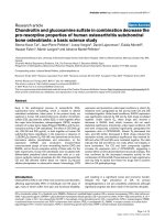
(a) (b)
(c) (d)
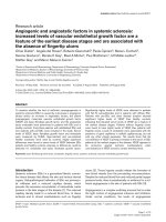
Figure 1 Hyphae and microconidia of A. benhamiae on human hair and human keratinocytes. (a) Fluorescence microscopic picture (laser
scanning microscope LSM 5 LIVE, Zeiss, Jena) of hyphae and microconidia stained with fluorescent brightener 28 (Sigma, USA). Scale bar: 5 μm.
(b) Colonization of human hair. Cyan, fluorescence brightener 28-stained fungal hyphae; orange, hair autofluorescence. Scale bar: 20 μm. (c)
Attachment of microconidia to human keratinocytes. Cyan, fluorescence brightener 28-stained fungal hyphae, red, wheat-germ agglutinin
stained keratinocytes. Scale bar: 5 μm. (d) Human keratinocytes with germinating A. benhamiae microconidia. Scanning electron microscopy
image. Scale bar: 10 μm. See Additional file 1 for supplementary information pertaining to this figure.
Burmester et al. Genome Biology 2011, 12:R7
/>Page 2 of 16
rodents, in particular guinea pigs [12 ,13]. The two spe-
cies also differ in their ability to grow under laboratory
conditions, with T. verrucosum being very difficult to
cultivate at all [14]. Conversely, A. benhamiae is com-
paratively fast growing and produces abundant microco-
nidia. As a teleomorphic species, the fungus is even able
to undergo sexual development, including the formation
of sexual fructifications (cleistothecia) [15,16]. These
characteristics, together with the recent establishment of
a guinea pig infection model and a genetic system for
targeted gene dele tion (P Staib and colleagues, manu-
script submitted) for this species, suggest A. benhamiae
is a useful model organism to investigate the funda men-
tal biology and pathogenicity of dermatophytes [8].
Despite the above mentioned phenotypic differences, A.
benhamiae and T. verrucosum are phylogenetically very
closely related, and both induce highly inflammatory
cutaneous infections in humans, such as tinea corporis
[15,17]. Therefore, a genome comparison of the two
species should reveal common basic pathogenicity-asso-
ciated traits.
In the present study, we report and compare the gen-
ome sequences of A. benhamiae and T. verrucosum and
refer to potential dermatophyte-specific pathogenicity-
associated factors, a s revealed by comparisons with
groups of proteins important for pathogenicity in other
species of the Onygenales (Coccidioides posadasii and
Coccidioides immitis) and in the mould A. fu migatus.
Applyi ng our insights thereof, we used secretome analy-
sis to reveal secreted factors of A. benhamiae that med-
iate extracellular in vitro keratin degradation. The
interaction between A. benhamiae and the human host
was monitored by global transcriptome profiling of the
fungal cells in contact with human keratinocytes. Inves-
tigating t he molecular basis of sexual reproduction, we
inspected in detail the A. benhamiae mating type locus.
Results and discussion
Comparative genomics of A. benhamiae and
T. verrucosum
The genomes of A. benhamiae and T. verrucosum were
sequenced by a whole-genome shotgun hybrid approach.
The assembly of A. benhamiae spans 22.3 Mb [DDBJ/
EMBL/GenBank:ABSU00000000] and that of T. verruc o-
sum comprises 22.6 Mb [DDBJ/EMBL/GenBank:
ACYE00000000] (Table 1; Additional file 2; both gen-
omes are also deposited in the Broad Institute database
[18]). Thus, these genomes are smaller than those of
phylogenetically related ascomycete s, such as aspergilli
(28 Mb and 37.3 Mb in case of Aspergillus clavatus and
Aspergillus niger, respectively), Co ccidioides species (27
to 29 Mb), and Histoplasma capsulatum (30 to 39 Mb).
The genomes of A. benhamiae and T. verrucosum
contain 7,980 and 8,024 pre dicted protein-encoding
genes, respectively (Table 1). Introns were found in
5,809 of the A. benhamiae and 5,744 of the T. verruco-
sum genes. Both genomes c omprise a mosaic of long
G + C rich, gene-containing portions separated by A +
Trich‘islands’ with a GC content below 40%, ranging
from a few kilobases to more than 25 kb. As expected
from previous reports based on nuclear ribosomal inter-
nal transcribed spacer regions 1 and 2 [15,19-21], the
comparison of the two genome sequences revealed a
strong similarity. Using the software Mummer [22],
approximately 21.8 Mb of the genomes (98.0% of the
available A. benhamiae and 96.7% of the T. verrucosum
genomic sequences) can be aligned to each other, indi-
cating that the vast majority of genes lie in collinear
regions and are shared between the two organisms. The
aver age identity of the alignabl e portion of the genomes
is 94.8%. The alignment of the two genomes points to
only five major genomic rearrangements, one inversion
and four balanced translocations between chromosomes
(Figure S1 in Additional file 2). The presence of only a
few rearrangements between the two genomes suggests
very recent speciation. These findings are reflected
bythephylogenetictreeconstructedbyuseofthe
available genome sequences (Figure 2; Figure S2 in
Additional file 3).
However, we also identified notable dissimilarities
between the genomes of A. benhamiae and T. verruco-
sum. After having detected the o rthologous pairs with
best bidirectional hits, we came up with lists of proteins
that presumably were unique for either species. Since
the best bidirectional hits were identified using protein
Blast, we next applied BlastN to correct for possible
gene prediction errors. We used a filter threshold for
significant hits of 80% identity between sequences over
less than 50% of the query length. There were 238
A. benhamiae sequences that gave no hits or non-signif-
icant hits in T. ve rrucosum, and 219 T. verrucosum
genes were not found in A. benhamiae. Of these, 83 and
78 genes (A. benhamiae and T. verrucosum, respectively)
have assigned names and/or functional domains. A list
Table 1 Genome data of A. benhamiae and T. verrucosum
Length (Mb) Predicted CDS Mean CDS length Genes with introns Predicted tRNAs
A. benhamiae 22.3 7,980 1,482 5,809 80
T. verrucosum 22.6 8,024 1,458 5,744 77
CDS, coding sequence.
Burmester et al. Genome Biology 2011, 12:R7
/>Page 3 of 16
of the predictions is provided in Additional file 4. Given
the overall strong genome sequence similarity, a future
functional investigation of these distinctions appears to
be of interest, in particular with respect to the tremen-
dous differences between the two species in terms of in
vitro growth ability and animal host preference (see also
the ‘Other interesting genes’ section).
We analy zed the A. benhamiae fast-evolving g enes in
comparison to T. verrucosum. Using the dN/dS ratio as
a measure for selective pressure, we obtained a list of
positively selected genes (dN/dS >1) (Additional file 5).
In total we found 132 positively selected genes with
assigned functions, enabling assumptions about their
roles in the cell and, hence, the reasons for their a ccel-
erated evolution. Of particular interest are t he two most
abundant groups of these genes, those encoding tran-
scription factors (18 genes) and MFS transporters (5
genes). The latter are known to be usual constituents of
secondary metabolite (SM) gene clusters.
Both dermatophyte genomes encode the basic meta-
bolic machinery for glycolysis, tricarboxylic acid cycle,
glyoxylate cycle, pentose phosphate shunt, and synthesis
of all 20 standard a mino acids and the five nucleic acid
bases. Moreover, dermatophytes appear to be capable of
producing a wide range of SMs, which is reflected by
thepresenceofpolyketidesynthase(PKS)-andnon-
ribosomal peptide synthetase (NRPS)-encoding genes
(see the ‘ Genetic basis for secondary metabolism
gene clusters’ section). The outstanding ability of
dermatophytes to specifically infect superficial host
structuresmaybesupportedbythepossessionofa
broad repertoire of genes encoding hydrolytic enzymes,
the expression of many of which was also proven
experime ntally (see the next paragrap h and the ‘Identifi-
cation of secreted fungal proteins during keratin degra-
dation by secretome analysis’ section). In addition, the
ability of dermatophytes to assimilate lipids, major con-
stituents of the skin, is putatively reflected by the pre-
sence of 16 lipase genes in eit her genome. A putative
link between the possession of lipases and fungus-
induced skin disease has previously been revealed for
basidiomycetes of the genus Malassezia [23].
Of particular note is the apparent relative paucity of
tRNA genes i n both dermatophytes in comparison with
other closely related ascomycetes. The genomes of A.
benhamiae and T. verrucosum contain 80 and 77 tRNA
genes, respectively, whereas the number of tRNA g enes
varies between approxima tely 100 to 130 in Coccidioides
species and 150 to 370 in aspergilli . However, some
strains of H. capsulatum, representing a compa ratively
closely related pathogen, also possess only 83 to 89
tRNA genes, suggesting that the low number of tRNA
genes is not specific to dermatophytes.
Identification of a broad repertoire of protease genes in
dermatophyte genomes
Dermatophytes are keratinophilic fungi, sharing the abil-
ity to u tilize compact hard keratin as a sole source of
890
0
.1
1000
1000
987
1000
1000
982
1000
519
A
rt
h
ro
d
erma
b
en
h
am
i
ae
Trichophyton verrucosum
1000
Coccidioides immitis
Uncinocarpus reesii
Histoplasma capsulatum
Paracoccidioides brasiliensis
Aspergillus oryzae
Aspergillus flavus
1000
Aspergillus terreus
Aspergillus fumigatus
Aspergillus clavatus
Aspergillus nidulans
Neurospora crassa
Onygenale
s
Eurotiales
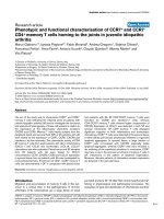
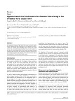
Figure 2 Partial genome-based phylogenetic tree of A. benhamiae and T. verrucosum representing the most closely related clades. The
tree was inferred by the neighbor-joining analysis method using the PHYLIP package [59], with the number of bootstrap trials set to 1,000.
Numbers at the nodes indicate the bootstrap support. See the details and the entire tree in Additional file 3.
Burmester et al. Genome Biology 2011, 12:R7
/>Page 4 of 16
carbon and nitrogen. In line wi th this knowledge, t he
two se quenced genomes reflect a remarkable metabolic
capability for protein degradation. They contain 235
predicted protease-encoding ge nes, 87 of the deduced
proteins possessing a secretion signal (Table S3 in Addi-
tional file 6). We di d not detect an y protease in A. ben-
hamiae or T. verrucosum unique to either species, a
finding that may reflect similar life styles and/or host
adaptation mechanisms, especially with respect to their
common keratinophilic growth. In general, deviations in
the number of proteases per genome are rather large in
the fungal kingdom, ranging from approximately 90 in
Ustilago maydis to approximately 350 in Gibberella zeae
(according to the MEROPS database [24]). Dermato-
phytes belong to the most protease-rich species.
The protein sequence of each protease is highly con-
served across dermatophyte species [25]. Collections of
predicted secreted proteases of A. benhamiae and T.
verrucosum as well as Coccidioides spp. (Onygenales)
were compared to those of A. fumigatus as a member of
the Eurotiales, for which many secreted proteases have
previously been characterized. Most A. fumigatus pro-
teases in A1 (pepsins), M28 ( leucine aminopeptidases),
S9 (dipeptidylpeptidases), S10 (carboxypeptidases) and
S53 (tripeptidylpeptidases) families have an orthologue
in dermatophytes and Coccidioides spp. (Table S4 in
Additional file 7). The major striking differences found
between the secreted protease batteries of A. fumigat us
and Onygenales are the following: subtilisin (S8), deuter-
olysin (M35), and fungalysin (M36), which belong to
endoprotease gene families, have expanded in Onygen-
ales (Table S4 in Additional file 7); the same is true for
exopeptidases o f the M14 family (metallocarboxypepti-
dases) and the M28 family (aminopeptidases) - a major
carboxypeptidase (McpA) homologous to the human
pancreat ic carboxypeptidase A was prev iously character-
ized in dermatophytes [26], and of particular note,
Aspergillus spp. have no McpA orthologue; and genes
encoding acidic glutamic proteases (G1 family) were not
detected in either dermatophytes or Coccidioides spp.
Major differences between dermatophytes and Cocci-
dioides spp. proteases were found in M35, M36 and S8
proteases families (see the phylogenetic trees in Addi-
tional file 8). Proteases of these three families of derma-
tophytes and Cocc idioides spp. form distinct clades in
phylogenetic trees ( Additional file 8). Members of the
S8 and M36 families have undergone additional amplifi-
cations in the dermatophyte lineage, and expansion of
the M35 family appears to be different in Coccidioides
spp. and dermatophytes. In the latter, a clade was appar-
ently lost. In addition, three genes encoding proteases of
the S41 family were found in the dermatophyte genomes
while only one gene encoding a protease of this family
was identified in Coccidioides spp.
Recent comparative genomic analyses of Coccidioides
species with other members of the Onygenales showed
gene family sizes are associated with a host/substrate
shift from plants t o animals in these microorganisms
[27]. Experimentally, the expression of genes encoding
fungalysins and subtilisins was recently moni tored in A.
benhamiae by cDNA microarray analysis during growth
on keratin, and also during cutaneous infection of gui-
nea pigs [8]. Interestingly, the prominent keratin
induced A. benhamiae subtilisin-encoding genes, such
as SUB3 and SUB4, were not observed in this former
analysis to be strongly activated in vivo,incontrastto
others that conversely were not found to be induced
during in vitro growth on keratin. A role for Sub3 was
recently observed in adhesion of the dermatophyte
Microsporum canis to feline epidermis, but not for the
invasion thereof [ 28]. These findings suggest additional
functions of secreted proteases during host adaptation
other than keratin degradation. Since the formerly used
cDNA microarray does not comprise the full genome of
A. benhamiae, the future identification of in vivo specific
dermatophyte proteases on the basis of the presented
genome appears to be of major interest.
Identification of secreted fungal proteins during keratin
degradation by secretome analysis
A potential role of secreted proteases, in particular ser-
ine proteases, in pathogenesis has been widely reported
in many prokaryotes and fungi [2,29-31], including func-
tions as allergens [32]. In order to apply insights from
the present genome sequences to determine putative
virulence gene function, we set out to reveal the basic
panel of factors that are secreted during growth of A.
benhamiae on keratin. To achieve this, secretome analy-
sis was performed, an approach that, to our knowledge,
has not been applied to A. benhamiae before. Experi-
mental analysis (after 2 days of growt h) led to the iden-
tification of 203 single electrophoretic species (Figure
3b). From these entities, 53 different proteins w ere
detected (Table S5 in Additional file 6). By far the lar-
gest group of identified proteins is formed by putative
proteases (approximately 75% relative spot volume). In
addition, we found other, different hydrolases and pro-
teins involved in carbohydrate metabolism (Table S 5 in
Additional file 6). Three of the subtilisin-like serine pro-
teases (Sub3, Sub4, and Sub7), three fungalysine-type
metalloproteases (Mep1, Mep3, and Mep4), the leucine
aminopeptidases Lap1 and Lap2, as well as the dipepti-
dyl-peptidases DppIV and DppV were detected in the
secretome, consistent with gene expression analysis in
A. benhamiae during keratin degradation [8]. Supporting
our r esults, the pattern of proteins secreted by the two
related dermatophyte species Trichophyton rubrum and
T. violaceum during growth on soy protein was
Burmester et al. Genome Biology 2011, 12:R7
/>Page 5 of 16
previously describ ed in [4]. In that study , a gel-based
approach led to the identification of 19 proteins secreted
by at least one of these species. Remarkably, 15 of the
corresponding homologs were also found to be secre ted
inthepresentstudybyA. benhamiae on keratin med-
ium, including major keratinases of the subtilisin family
of secreted proteases (also see Table S6 in Additional
file 6). Individual differences between the present and
formerly observed secretion patterns might be due to
the different dermatophyte species analyzed and/or to
the different protein substrates an d cultivation para-
meters used. In conclusion, the set of dermatophyte
secreted proteases in a protein medium is similar to that
of A. fumigatus, which includes endoproteases such as
the major subtilisin Alp1 and the fungalysin Mep and
exoproteases such as Lap1, Lap2, DppIV and DppV.
Endo- and exoproteases secreted by microorganisms
cooperate very efficiently in protein digestion to produce
oligopeptides and free amino acids that can be incopo-
rated via transporters. During the process of protein
digestion the main function of endoproteases is to pro-
duce a large number of free end peptides on which exo-
proteases may act. At neutral and alkaline pH,
synergistic action of Lap and DppIV was shown in
Aspergil lus spp. [ 13,24 ]. Laps d egrade peptides from the
amino terminus until reaching an X-Pro sequence,
whichactsasastop.Inacomplementary manner, the
X-Pro sequences can be removed by DppIV, thus allow-
ing Laps access to the next residue. Dermatophyte and
Aspergillus spp. Lap1, Lap2, DppIV and DppV have
shown comparable substrate specificity [33]. Therefore,
our proteomi cs approach allows us to hypothesize com-
mon basic mechanisms in dermatophytes during extra-
cellular protei n digestion. However, the presence o f
large protease gene families in dermatophytes reflects
selection d uring evolution and the abilit y of these fungi
to adapt to different environmental conditions during
infection and saprophytic growth.
Differential gene expression in A. benhamiae during
infection of keratinocytes
Growth of A. benhamiae on keratin might mimic
selected in vivo growth substrates, yet may not reflect
the entire process of infection. In order to gain more
insights into basic host adaptation mechanisms, we stu-
died the global transcriptional response of A. benhamiae
during infection of human keratinocytes. After 12 h of
co-cultivation, germinating A. benhamiae microconidia
were observed to be localized and concentrated on the
host cells, suggesti ng that the fungus actively adheres to
the keratinocytes (Figure 1c,d). To perform 454 RNA
sequencing, the fungal cells were harvested after
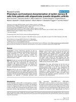
(a)
(
b
)
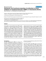
Figure 3 Secretome of A. benhamiae grown on keratin. (a) A. benhamiae grown on keratin particles. Cyan, fluorescence brightener 28-stained
fungal hyphae; orange, keratin particle autofluorescence. Scale bar: 10 μm. (b) Two-dimensional gel of secreted A. benhamiae proteins obtained
from culture supernatant after 48 h cultivation in a shaking flask with 0.9 g/l glucose and 10 g/l keratin. The apparent molecular mass of proteins
and the pI range of the first dimension are indicated. Proteins were identified by mass spectrometry (matrix-assisted laser desorption/ionization-
time of flight/time of flight (MALDI-TOF/TOF)). Identified proteins are given in Table S5 in Additional file 6. See also Additional file 1 for more
details.
Burmester et al. Genome Biology 2011, 12:R7
/>Page 6 of 16
incubation for 96 h with and without keratinocytes.
About 50 A. benhamiae genes showed differential
expressionwithafoldchange>5(P-value < 0.05; Table
S7a in Additional file 6); 45 genes encoding putatively
secreted proteins (Table S5 in Additional file 6) and 13
genes coding fo r proteins in volved in the biosynthesis of
SMs are expressed either o nly with or without keratino-
cytes, or under both conditions. Of the 235 predicted
protease-encoding genes, 158 are expressed under both
conditions. Sixteen potentially secreted proteins, includ-
ing three proteases, are differentially expressed (Table
S7b in Additional file 6). In particular, the expression
profile of the genes encoding carboxypeptidase S1 and
dipeptid yl-peptidase DppV implies their poten tial invol-
vement in the infection process. The transcript levels of
two NRPS genes were reduced during co- cultivation
with keratin ocytes, a finding that is noticeable but can-
not be explained at this stage.
To confirm the RNAseq results, we selected several
genes that were predicted to be differentially expressed
and tested them by Northern blotting. We used
two houseke eping genes, actin (ARB_04092) and glyce r-
aldehyde 3-phosphate dehydrogenase (GAPDH,
ARB_00831), as controls as they are not expected to be
differentially regul ated between the control and co-incu-
bation conditions. All tested genes were regulated as
expected from the RNAseq data (Figure S4 in Additional
file 9). The expression level alterations of metabolic
enzymes (ARB_07 891, ARB_04156, ARB_0 1650 and
ARB_04856) and membrane transporters (ARB_01027)
reflect the adaptation of the fungus to the different
nutrition provided by keratinocytes and their remnants,
whereas the strong up-regulation of the hydrophobin
ARB_06975 indicates altered binding properties and
adhesivity during growth on epithelial cells and during
infection. In conclusion, this independent experimental
method shows that the accuracy of the RNAseq data
was exemplary.
Genetic basis for secondary metabolism gene clusters
The A. benhamiae and T. verrucosum genomes encode a
relatively high number (26 and 25, respectively) of SM
biosynthesis gene clusters (Table 2), a finding that con -
trasts with observations made in other fungi and bac-
teria highly adapted to humans. For comparison,
Candida albicans or Staphylococcus aureus hardly pro-
duce SMs and Histoplasma species have no more than
seven SM gene clusters per genome; more closely
related to dermatophytes is Coccidioides immitis,which
has16SMgeneclusters,themaindifferencebeingin
the number of NRPSs (5 versus 15 in A. benhamiae).
Nine PKS, 15 NRPS and 3 PKS/NRPS hybrid genes
were identified in the A. benhamiae genome, all of
which except for one NRPS gene (ARB_02149) are
conserved in both species (Table 2). Addressing the
question of whether the absence of the latter gene in T.
verrucosum is associated with phen otypic and/or host-
specific differences between the two species will be of
future interest. To see whether only the NRPS or the
entire associated gene cluster is a bsent from T. verrucosum,
we examined the conservation of the other constituents of
the ARB_02149 gene cluster and observed that the ‘miss-
ing’ NRPS belongs to an otherwise very well conserved and
collinear region that spans more than 75 kb (the whole T.
verrucosum supercontig 79). However, one other gene
besides ARB_02149 is missing in T. verrucosum, the MFS
transporter ARB_02151 (Figure 4). Interestingly, the ‘miss-
ing’ genes are separate d by a perfectly conserved ABC mul-
tidrug tra nsporter (ARB_02150 = TRV_01489). Th e
Arthroderma ARB_02149 gene cluster has several traits
typical of func tional SM gene clusters, suc h as the presen ce
of genes for the MFS transporter, feruloyl esterase and C6
transcription factor. This makes us suppose that the NRPS
was lost in Trichophyton rather than acquired by Arthro-
derma. Ho wever, it re mains unclear if the MFS transporter
was deleted simultaneously, and why the deletion did not
capture the ‘middle’ ARB_02150 gene.
All nine PKS genes detected in A. benhamiae have
unequivocal counterparts in the T. verrucosum genome
(Table 2). A n interesting feature of the dermatophyte
PKS set is the unusual proportions of reducing and
non-reducing PKSs. Whereas in all other closely related
ascomycetes (such as aspergilli) most of the PKSs are
non-reducing, in dermatophytes most are reducing
PKSs. A compar ison with t he closest sequenced relative,
C. immitis (Table 2; see more details below), also
revealed substantial differences in the composition of
the PKS set: the ratio of reducing to non-reducing in
dermatophytes is 2:1, whereas in C. immitis it is 2:3.
This observation suggests dermatophytes have an
uncommon SM profile, which deserves future investi-
gation. Particular attention should be paid to the fact
that these fungi are characterized by intense pigmenta-
tion, a phenotype that may be related to their patho-
genicity. For the related species T. rubrum,the
polyketide-derived mycotoxin xanthomegnin has been
suggested to be responsible for the characteristic red
colony reverse pigment. Mo st interestingly, xantho-
megnin production has even been detected in epider-
mal material infected by T. rubrum,incontrastto
non-infected controls [34]. A putative link between SM
production and host adaptation of A. benhamiae might
also be reflected by our observation that several gen es
associated with the synthesis of such molecules were
found to be differential ly regulated during infe ction of
human keratinocytes (see the ‘
Differential gene expres-
sio
n in A. benhamiae during infection of keratinocytes’
section).
Burmester et al. Genome Biology 2011, 12:R7
/>Page 7 of 16
Table 2 Putative PKS and NRPS genes of A. benhamiae, T. verrucosum, and C. immitis
Type LocusLink Arthroderma
benhamiae
LocusLink Trichophyton
verrucosum
LocusLink Coccidioides
immitis
Domain architecture
PKSs
Non-reducing ARB_00538 TRV_00386 - KS-AT-ACP
ARB_03291 TRV_02519 CIMG_13102 KS-AT-ACP-ME
a
- - CIMG_05571 KS-AT-ACP
- - CIMG_04689 KS-AT-ACP-ME
- - CIMG_03162 KS-AT-ACP
ARB_07994 TRV_04611 CIMG_08569 KS-AT-ACP-ACP-TE
- - CIMG_08564 AT-KS-ACP-TE
Reducing ARB_01525 TRV_04236 CIMG_13632 KS-AT-ME-ER-KR-ACP
ARB_05854 TRV_06867 - KS-AT-KR-ACP
b
ARB_06393 TRV_01071 - KS-AT-ME-ER-KR-ACP
ARB_05333 TRV_06912 CIMG_02398 KS-AT-DH-MT-ER-KR-ACP
ARB_07933 TRV_04104 - KS-AT-ME-ER-KR-ACP
ARB_07966 TRV_04285 - KS-AT-ME-KR-ACP
- - CIMG_05569 KS-AT-DH-ER-KR-ACP
- - CIMG_03014 KS-AT-DH-ER-KR-ACP
ARB_00195 TRV_05651 CIMG_07298 A-T-C-T-C
- - CIMG_01429 A-T-C-T
ARB_01698 TRV_01735 CIMG_09750 C-A-T-C-A-T-C-A-T-C-A-T-C-A-T-
C-T-C-T
ARB_02149 - - C-A-T-C-A-T-C-A-T-C-A-T-C
c
ARB_02226 TRV_00553 - A-T-C-A-T-C-A-T-C
ARB_02570 TRV_5508 - A-T-C
ARB_02750 TRV_06186 - A-T-C-A-T-C-A-T-C-A-T-C-A-T-C-T
ARB_03095 TRV_06056 - T-C-A-T-C/T-C-A
NRPSs ARB_03768 TRV_07570 - A-C-A-T-C-A-T
ARB_04984 TRV_06313 CIMG_01861 A-T-C-A-T-C
ARB_05131 TRV_07837 - A-T-C-A-T-C-A-T
ARB_05579 TRV_06828 - T-C-A-T-C-A-T
ARB_06786 TRV_05681 - A-T-C
ARB_07686 TRV_05452 CIMG_00941 A-T-C-A-T-C-T-C-A-T-C-T-C-T-C
ARB_07850 TRV_01776 - A-T-C/A-T-C-A-T
ARB_07862 TRV_04720 - A-T-C-A-T-C-T
ARB_07534 TRV_00508 - KS-AT-DH-ER-KR-ACP-C-A-T
PKS/NRPS hybrids ARB_02973 TRV_03721 CIMG_06629 KS-AT-ME-KR-ACP-C-A-T
ARB_07844 TRV_05146 - A-T-KS-AT-KR-ACP-TE
a
Potential citrinin-like product; similar to pksCT BAD44749.1.
b
Product 6-methyl-salicylic acid; similar to 6-MSA synthase CAA39295.1.
c
Unique for A. benhamiae.A,
adenylation domain; ACP(PP), acyl carrier protein, or phosphopantetheine domain; AT, acetyltransferase domain; C, condensation domain; DH, dehydratase
domain; E, epimerization domain; ER, enoyl reductase domain; KR, ketoacyl reductase domain; KS, beta-ketoacyl synthase domain; ME, methyltransferase domain;
T, thiolation domain; TE, thioesterase domain.
NRPS
C6 TF
MFS
TRV_01489
TRV_01486
TRV_01488 TRV_01487
TRV_01490
ARB_02150
ARB_0215
4
ARB_02151
ARB_02148
ARB_02149
ARB_02153 ARB_02152
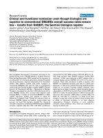
Figure 4 A. benhamiae NRPS ARB_02149 gene cluster and the corresponding region in the T. verrucosum genome.
Burmester et al. Genome Biology 2011, 12:R7
/>Page 8 of 16
To get an impression of possible expansions of
families and evolutionary relationships, we compared
the sets of SM producers in dermatophytes with that of
C. immitis (Table 2; Figure S5.1 and S5.2 in Additional
file 10). As mentioned above, the total number of SM
gene clusters is higher in dermatophytes, mainly due to
the more abundant NRPSs. However, we observe differ-
ences also in the PKS set as well as in the number of
PKS/NRPS hybrids: C. immitis possesses only one
hybrid, whereas each dermatophyte has three. The
higher number of non-reducing PKSs in C. immitis is
mainly due to the expansion of one clade; most likely
we are seeing the results of duplication of some ancestor
genes with a domain architecture of a beta -ketoacyl
synthase domain, an acetyltransferase domain, an acyl
carrier protein domain, and a methyltransferase domain
(KS-AT-ACP-ME). Four of s ix C. immitis non-reducing
PKSs belong to this clade. Of the other two, one has a
clear ortholog in dermatophytes, and the other has an
unusual structure (AT-KS-ACP-thioesterase domain
(TE)) without an orthologous dermatophyte gene. In
comparison to C. immitis, dermatophytes possess two
additional non-reducing clades, which means that, in
spite of the lower number of non-reducing PKSs, they
have more various potential capacities. The reducing C.
immitis PKSs also cannot boast great variety: two of
four C. immitis genes are most likely the result of a
duplication (they form a separate clade and do not have
derma tophyte orthologs), one PKS has orthologs in der-
matophytes, and one is only a probable homolog ( see
below). On the other hand, in dermatophytes we see an
expansion of the group with a fumonisin synthase-like
structure (KS-AT-ME-e noyl reductase domain (ER)-
ketoacyl reductase domain (KR)-ACP): three ortholo-
gous pairs formed by out-paralogs in each species have
only one close homolog in C. immitis.SincetheC.
immitis gene lacks one of the domains (methyltransfer-
ase), we cannot consider it as a fumonisin-like ortholog.
Besides the 6-methyl-sa licylic acid synthase, completely
lacking in C. immitis, another not completely reducing
PKS (KS-AT-ME-KR-ACP), as well a s two PKS/NRPS
hybrids, do not have homologs in C. immitis.Taken
together, these data agree with our hypothesis that
highly adapted parasites such as Coccidioides do not
require a large arsenal of SMs.
Sexuality in dermatophytes
Sexual reproduct ion is known for A. benhamiae but not
for T. verrucosum [35,36]. The A. benhamiae and T. ver-
rucosum genomes revea led the w hole sets of genes for
mating and meiosis in both species, suggesting that the
lack of a known sexual cycle in T. verrucosum is not
due to major deletions of genes essential for sexual
reproduction and meiosis (Table S8 in Additional file 6).
Both sequenced strains showed a single mating type
encoding an HMG box transcription factor. To identify
the complementary mating type, we sequenced the cor-
responding region of an A. benhamia e mating partner
strain (strain CBS 809.72; Figure 5). The newly identified
region encodes an alpha-box type transcription factor,
indicating that A. benhamiae exhibits two mating types,
as described for other closely related fungal pathogens
such as H. capsulatum and C. immitis [37]. A. benha-
miae mating type + strains as well as mating type -
strains are often routinely isolated [36]. There is no
apparent disequilibrium between mating type + and
mating type - strain frequencies.
We did not identify a striking defect in the T. verruco-
sum mating type locus, which appe ars to be intact. Sev-
eral strains of T. verrucosum were found to be of the
same mating type as the sequenced strains, suggesting a
strong disequilibrium towards mating type +.
In Aspergillus (Eurotiales), Coccidioides and Histo-
plasma (Onygenales) the mating type (MAT) loci are
flanked by APN2 and the SLA2 genes encoding a DNA
lyase and a cytoskeleton protein, respectively [37]. The
MAT idiomorphs and flanking regions described here for
A. benhamiae and T. verrucosum are essentially ident ical
to those of other closely related dermatophytes [38].
Other interesting genes
Of particular interest are the genes of A. benhamiae that
have no obvious counterpart in T. verrucosum (Addi-
tional file 4) and whose predicted functions suggest
their potential involvement in basi c biological pheno-
types and/or pathogenicity . Two such genes,
ARB_04713 and ARB_02149, encoding a phosphopan-
tetheine-binding domain and an NRPS, respectively,
were found in the transcriptome analysis, although not
expressed differentially. The expression pattern of the A.
benhamiae-specific NRPS ARB_02149 further suggests
that its as yet unidentified product is produced during
infection by the fungal cells.
Another gene of particular interest encodes hydropho-
bin. In A. fumigatus, surface hydrophobin was shown to
prevent immune recognition [39]. The A. benhamiae
hydrophobin gene (ARB_06975) shows 99% similarity
with the respective T. verrucosum gene (TRV_00350)
and displays moderate overexpression (1.6×) under c o-
cultivation conditions (Tabl e S7b in Additional file 6).
The analysis of a potential role of dermatophyte hydro -
phobins in immune response functions and/or adhesion
to host surfaces will be part of future research.
Conclusions
Numerous examples in microbial pathogenicity research
still need to be explained at the genomic level, thus
requiring genome sequences to be made available. Here,
Burmester et al. Genome Biology 2011, 12:R7
/>Page 9 of 16
we present the first genomes of dermatophyte species,
filamentous fungi that cause most superficial infections
in humans and animals. The presence of putative patho-
genicity-related factors, such as numerous secreted pro-
teases, was revealed at the genome level and also
experimentally confirmed during keratin degr adation by
A. benhamiae. Although keratin utilization is tradition-
ally supposed to be of major relevance for the patho-
genicity of these microorganisms, the entire process of
host adaptation during infection seems to be more com-
plex. T ranscriptome analysis showed that only some of
the typically keratin-induced proteases were found to be
strongly expressed during fungus-keratinocyte interac-
tion. Instead, genome and transcriptome analyses draw
attention to so far hardly noticed dermatophyte factors -
for example, putative SMs - the role of which sh ould be
addre ssed in the future. Our research on dermatophytes
was strongly facilitated by the selection of A. benhamiae
as a model species, which provides practical advantages
such as comparatively fast growth and the production of
abundant microconidia. Moreover, future basic studies
on the regulation of m ating, dermatophyte evolution
and host preference will profit from the ability of A.
benhamiae to undergo sexual reproduction. In conclu-
sion, by p resenting dermatophyte genomes and global
insights into major processes of h ost adaptation, we
intend to advance molecular studies on these medically
important microorganisms.
Materials and methods
A. benhamiae and T. verrucosum strains and growth
conditions
A clinical isolate of A. benhamiae strain 2354 was used
(isolate LAU2354) [15]. T. verrucosum stra in 44 [17]
A. benhamiae MAT1-1
A. benhamiae MAT1-2
T. verrucosum MAT1-
1
A. fumigatus MAT1-2
Sla2
Cox13
Apn2
MAT1-1-4
HMG TF
MAT associated
A
-box TF
ORF
Rps4
Figure 5 Mating type gene organization of A. benhamiae and T. verrucosum. Genes constituting the MAT locus: Sla2, putative cytoskeleton
assembly control protein (ARB_07317, TRV_02048, AFUA_3G06140); Cox13, cytochrome C oxidase subunit VIa (ARB_08059, TRV_08208,
AFUA_3G06190); Apn2, DNA lyase (ARB_07318, TRV_02049, AFUA_3G06180); a gene similar to MAT1-1-4 (ARB_07319, TRV_02050); HMG TF, HMG-
box transcription factor (MAT1-2-1; ARB_7320, TRV_02051, AFUA_3G06170); MAT associated protein of unknown function (ARB_07321, TRV_02052,
AFUA_3G06160); a-box transcription factor (MAT1-1-1, GB GQ996965); ORF, glycine rich protein of unknown function; Rps4, protein S4 of the 40S
ribosomal subunit (ARB_7322, TRV_02053, AFUA_3G06840).
Burmester et al. Genome Biology 2011, 12:R7
/>Page 10 of 16
waskindlyprovidedbyYvonne Gräser (Ch arité, Berlin,
Germany). Strains were cultivated at 28°C on Sabouraud
2% (w/v) glucose agar (SG, Merck, Darmstadt, Germany)
for 12 days; liquid cultures were shaken at 180 rpm at
30°C for 5 to 7 days. Hyphae and conidia were separated
by filtration using a 40 μm cell strainer (BD Bioscien ce,
Heidelberg, Germany). Conidia were counted with a cell
counter (Beckman, Coulter, Krefeld, Germany) or manu-
ally using a Thoma chamber. For crossing experiments
of A. benhamiae LAU2354 with the opposite mating
type CBS 809.72, MAT medium [40] (1/10 SG, 0.1% (w/v)
MgSO
4
and 0.1% (w/v) KH
2
PO
4
) was used.
DNA and RNA preparation for DNA sequencing and cDNA
library
For DNA preparation, mycelia were separated from the
medium by filtration through Miracloth (Calbiochem,
Darmstadt, Germany) and ground in a mortar under
liquid nit rogen. After evaporation, the powder was sus-
pended in a solution containing 150 mM EDTA,
50 mM Tris-HCl, pH 8.0, 1% (w/v) SDS, 20 mM NaCl
and 100 μg/ml proteinase K (Merck). After incubation
for 1 h at 55°C, the solution was gently mixed with 1/
4 v olume of 4 M NaCl and kept on ice for 30 minutes.
After centrifugation for 10 minutes at 6,000 rpm and
4°C, polyethylene glycol 6000 (Serva, Heidelberg,
Germany) was added to the supernatant to a final con-
centration of 10% (w/v). The DNA was precipitated for
1 h on ice and centrifuged for 10 minutes at 10,000 rpm
at 4°C. The pellet was dissolved in a solution containing
25 mM Tris-HCl, pH 8.0, 5 mM EDTA, 10 mM NaCl
and 1% (v/v) Triton X100. For density centrifugation, 1g
CsCl and 12 μl bisbenzimide (10 mg/ml) for each millili-
ter of solution were added [41,42]. Ultracentrifugation
was performed in a vertical rotor at 44,000 rpm for 24 h
at 25°C. DNA was separated into two bands of different
density according to the AT-content of the DNA. The
upper band contained a DNA fraction highly enriched
for mitochondrial DNA. For T. verrucosum,tworounds
of density gradient centrifugation were necessary. In the
first round, eth idium bromide was used instead of bi s-
benzimide. For RNA preparation, SG medium was
inoculated with conidia to a final concentration of 3 ×
10
4
conidia/ml and shaken at 180 rpm for three days at
30°C. Total RNA was isolated using a commercial kit
as described by the manufacturer (Qiagen, Hilden,
Germany). After RNA extraction, a cDNA library
was constructed from this material according to the
manufacturer’s protocols (MINT cDNA synthesis kit,
Evrogen, Moscow, Russia).
Plasmid/fosmid libraries and sequencing
Nuclear DNA of A. benhamiae and T. verrucosum was
sheared, size fractionated (3 to 4 kb), end-repaired, and
cloned into the SmaI site of pUC18. For both fungal
species, two fo smid libraries each were prepare d in
pCC1FOS (Epicentre Biotechnologies, Madison, WI,
USA) as described by the manufacturer, one for the
high-GC chromosomal DNA fraction and one for the
AT-rich mitochondrial DNA fraction. For T. verruco-
sum, 40,000 fosmids from GC-rich and 80,000 fosmids
from AT-rich DNA were obtained. For A. benhamiae,
the corresponding yields were around 50,000 (GC-rich)
and 20,000 (AT-rich), respectively. End sequences of
plasmid and fosmid clones were obtained using dye ter-
minator chemistry and a 3730×l seque ncer (Applied
Biosystems, Foster City, CA, USA). Moreover, a fosmid
library was generated with a GC-rich DNA fraction of
the A. benhamiae strain CBS 809.72 encoding the oppo-
site m ating type locus. We tested 1, 000 fosmids by col-
ony filter hybridizatio n and in PCR experiments.
Fosmids were identified by hybridization experiments
with a digoxygenin-labeled part of the apn2 gene
(ARB_07318) and in P CR experiments using apn2
amplifying primers (5’ -CTTCTAGTGAC TCGCCA-
CAGG-3’ forward and 5’ -GAGTTGGAGGTTGA-
GATGCTGAC-3’ reverse). Three clones were positively
identifie d by both methods. To test whether the fosmids
contained the full length MAT region, the clones were
tested in PCR experiments amplifying parts of other
flanking genes, such as the sla2 gene (ARB_07317) and
the rps4 gene (ARB_07322). For sla2, PCR primer pair
5’-CTTGTTCAGGAGAGCTATGG-3’ and 5’-CAGCTT-
CTCGAGCTCCTCCC-3’ was used; for rps4,PCRpri-
mer p air 5’-CAGCGCCTGGTCAAGGTCGACG-3’ and
5’-GGTCACGCTCCTCAGCAATGG-3’ was used. DNA
of a positive fosmid was shotgun sequenced using dye
terminator chemistry (ABI).
In addition, genomic 454 libraries were generated
according to the manufacturer’s protocol and sequenced
using a GS FLX (Roche, Mannheim, Germany). The
nucleotide sequences were assembled species-specific
using the newbler software. Clone gaps were filled using
a primer walking strategy with custom primers. Isola-
tion, quantification and quality control of total RNA was
performed as described [43]. A cDNA-library was con-
structed according to the manufacturer’ sprotocols
(Evrogen) and 1,411 ABI dye terminator se quences were
obtained mainly from the 5’ end. The sequen ces were
matched to the assembled genomic sequences to deter-
mine exon/intron structures and to obtain an intron sig-
nature for the species.
Next generation sequencing and assembly
The same DNA as for the preparation of the plasmid/
fosmid libraries was used for the preparation of genomic
libraries for the 454/FLX system (Roche) according to
the manufacturer’s protoc ols. Three runs each were
Burmester et al. Genome Biology 2011, 12:R7
/>Page 11 of 16
performed on the 454/FLX sequencing machine. All
454/FLX sequence data were assembled species-specific
using the n ewbler software. The San ger based sequen-
cing reads were assembled on to this ‘ backbone’.Clone
gaps were filled using a primer walking s trategy with
custom primers.
Both genomes are deposited in NCBI with accession
codes [DDBJ/EMBL/GenBank:ABSU00000000] for
A. benhamiae and [DDBJ/EMBL/GenBank:ACYE-
00000000] for T. verrucosum.
Gene prediction
Gene models of both fungi were generated by using in
silico predictions and sequence data from an EST library
constructed from cultured A. benhamiae cells. We
matched 1,411 ABI d ye terminator sequences obtained
from the cDNA library sequencing to the assembled
genomic sequences to determine exon/intron structures
and to obtain an intron signature for the species. The
alignments of the cDNA sequences to the genomic
backbone yielded evidence for 861 introns and at least
653 protein-coding open reading frames (coding
sequences), which were validated by BLAST. These data
were used to train the gene prediction program geneid
[44]. To validate the accuracy of the gene prediction, 47
gene structures in one genomic region were annotated
manually and compared to the automated predictions,
indicating a specificity of 82% at a sensitivity of 97%.
For the annotation and comparative analyses of the gen-
omes a web based genome browser was set up using the
GenColors database/software system [45].
Best bidirectional hits and BlastN analysis
Blast analysis of all coding sequences of one genome
against the other yielded best bidirectional hits. We used a
filter threshold for significant hits of 30% identity between
amino acid sequences over at least 50% of the protein.
A BlastN analysis of the genomic sequences was per-
formed for all protein coding genes of T. verrucosum
against all A. benhamiae contigs. A filter threshold for
significant hits was 80% identity between sequences over
at least 60% of the query length; 239 T. verrucosum
sequences gave no hits or non-significant hits.
Transcriptome analysis
The human keratin ocyte line HaCaT was obtained from
Prof. Fusenig (Deutsches Krebsforschungszentru m, Hei-
delberg, Germany). The cells were cultivated in DMEM
supplemented with 10% (v/v) fetal calf serum, gentamy-
cin (28 μg/ml) and 1% (w/v) ultraglutamine at 37°C in a
humidified atmosphere and 5% (v/v) CO
2
for 2 days.
Medium and supplements were purchased from Lonza
(Basel, Belgium). Human keratinocytes were infected by
A. benhamiae conidia with a mult iplicity of infection
(MOI) of 6. Infected human cells were cultivated in fetal
calf serum-free DMEM supplemented with both genta-
mycin and ultraglutamine for 96 h at 28°C. As a control,
A. benhamiae conidia were grown in the absence of ker-
atinocytes under the same conditions. A fter infection,
the human keratinocytes were lysed by addition of
0.03% (v/v) Triton X for 2 minutes and A. benhamiae
was harvested. Fungal cells were collected by centrifuga-
tion for 3 minutes at 3,500 g. A. benhamiae cells were
washed twice in Dulbecco’s phosphate buffered saline
(Lonza) and stored in aliquots at -80°C. For RNA
sequencing, total RNA was isolated using RiboPure™-
Yeast Kit (Ambion Europe, Huntingdon, UK) according
to the manufacturer’ s instructions from keratinocytes
co-incubated with conidia and conidia only grown in
cell culture medium for 96 h.
RNA was reverse transcribe d using a SMART techni-
que (Evrogen). The single-stranded DNA was then
amplified using SMART primers for 20 cycles to p ro-
duce double-stranded DNA in sufficient quantity for
GS-FLX sequencing (Roche). We generated A. benha-
miae transcriptome data by sequencing parts of indivi-
dual cDNAs after fragmentation by nebulization using
454/FLX sequencing technology. For postprocessing of
these sequences, SMART adapters were identified and
clipped using a combination of perl scripts plus cross_-
match. After further cleaning with seqclean (removal of
polyA tails and low complexity regions), 682,580 ESTs
(98.8 Mb) remained for mapping.
Mapping of the ESTs to the repeat -masked A. benha-
miae genome as a backbo ne was done in two major
steps. First, we used Blat [46] to assign each EST to its
most pr obable position in the genome allowing a maxi-
mum intron length of 10 kb. A valid hit require d a
minimum length o f 30 bp and a minimum identity of
90% to the backbone sequence. In the second step, each
EST was realigned to its most probable position utilizing
a slightly modified version of Exalin [47] that imple-
ments the Smith-Waterman algorithm and information
theory for better alignments and intron predictio n.
Using this approach, w e were able to align 571,963
ESTs to the genome. Finally, EST positions were trans-
lated to positions of known gene models if possible. In
this way, we determined for each gene a set of ESTs
and thereby its raw expression level. The data were nor-
malized to the total number of mapping ESTs. Table S 9
in Additional file 6 shows the total number s of gener-
ated reads, the reads ma pped to a genome, and the
reads in gene models for each technical replicate of
infection and control samples.
The raw counts for the transcripts were analyzed
using the R Statistical Computing Environment and the
Burmester et al. Genome Biology 2011, 12:R7
/>Page 12 of 16
Bioconductor packages DESeq [48] and edgeR [49]. Both
packages provide statistical routines for determining dif-
ferential expression in digital gene expression data using
a model based o n the negative binomial distribution.
The resulting P-values were adjusted using the Benja-
mini and Hochberg’s approach for co ntrolling the false
discovery rate [50]. Genes with an adjusted P-value
<0.05 found by both packages were assigned as differen-
tially expressed.
The RNAseq data ar e submitted to the Sequence read
archive of NCBI and are available with t he accession
numbers [NCBI:SRR070551] and [NCBI:SRR070552]
(sample runs) and [NCBI:SRR070553] and [NCBI:
SRR070554] (control runs).
Northern blotting
Total RNA from mycelial samples was isolated using
RiboPure™-Yeast Kit (Ambi on) according to the manu-
facturer’s instructions. Total RNA was denatured (15
minutes, 60°C; 5% (v/v) formaldehyde, 50% (v/v) forma-
mide, 40 mM MOPS, pH 7) and separated by agarose
gel electrophoresis (1.2% agarose, 40 mM MOPS, 10
mM sodium acetate, 2 mM EDTA, 2% (v/v) formalde-
hyde, pH 7). Blotting, hybridization and chemolumines-
cent signal detection were performed according to the
manufactur er’ s instructions (DIG Application Manual
for Filter Hybridization, Roche). Gel load and blot signal
strength were quantified and normalized using Bio-Rad
(Munich, Germany) Quantity One (v4.6.7) software.
Secretome analysis
For cultivation of A. benhamiae, medium was prepared
as follows: 10 g/l keratin (MP Biomedicals Europe, Ill-
kirch, France) was autoclaved in water and subsequently
20 mM potassium phosphate pH 5.5, 0.4 mM MgSO
4
,
77 mM NaCl, 5 mM glucose and 0.5% (v/v) SL-8 trace
elements [51] were added. Microconidia obtained from
A. benhamiae cultivated for 7 days on MAT agar at 30°
C were used to inoculate shaking flasks at a final spore
concentration of 10
6
per milliliter. After cultivation for
2 days at 200 rpm and 30°C, cultures were filtered
through Miracloth (Calbiochem, Darmstadt, Germany)
and the supernatant was centrifuged at 4,000 g for 20
minutes at 4°C. Secreted proteins were precipitated with
10% (w/v) trichloroacetic acid/6.5 mM DTT overnight
at 4°C. The precipitate was pelleted at 4,000 g for 20
minutes at 4°C and resuspended twice in ice-cold acet-
one/water (9:1)/6.5 mM DTT followed by subsequent
centrifugation steps. The air-dried pellet was dissolved
in lysis buffer 3, as described [52]. Immobiline DryStrips
of 11 cm covering a pH range from 3 to 10 (GE Health-
care Life Sciences) were rehydrated overnight according
to the manufacturer’ s instructions. Isoelectric focusing
was carried out in an Ettan IPGphor II using a 0 to 1
kV gradient for 11 h, 1 to 8 kV for 3 h and finally 8 kV
for 24 kVh. Afterwards, strips were incubated for 15
minutes in equilibration buffer (6 M urea, 2% (w/v)
SDS, 75 mM Tris
.
Cl pH 8.8, 30% (v/v) glycerol) with 65
mM DTT, followed by an alkylation step of the proteins
with 135 mM iodoacetamide in e quilibration buffer
under the same conditions. Separation of proteins by
the second dimension was carried out using pre-cast
Criterion gels (12.5% (w/v), Tris-HCl; Bio-Rad) accord-
ing to the manufacturer’ s instructions. Proteins were
visualized by Colloidal Coomassie Brilliant Blue G-250
staining [53].
Protein identification
Protein spots were excised from the gels and digested
with sequencing-grade Trypsin (Promega, Mannheim,
Germany) as described elsewhere [54]. Eluted peptides
were mixed with an equal amount of a saturated alpha-
cyano -hydroxycinnamic acid (Bruker Daltonics, Bremen,
Germany) solution in aqueous 30% (v/v) acetonitrile and
spotted on an MTP anc hor-chip 800/384 (Bruker Da l-
tonics). Mass spectrometry spectra were acquired with
an Ultraflex I TOF/TOF (Bruker Daltonics) mass spec-
trometer using Peptide Mass Standard II (Bruker Dal-
tonics) as calibrant. The five most intense mass
spectrometry signals were selected for tandem mass
spectrometry analysis. MASCOT (version 2.1.02; Matrix
Science, London, UK) searching against protein predic-
tions from the A. benhamiae genome and the NCBI
database (taxa fungi) was used for protein identification
with the following the parameters: fixed modif ication of
cysteine to S-carbamidomethyl derivatives, variable
methionine oxidation, no missed cleavage and a peptide
mass tolerance of 200 ppm.
PKS and NRPS domain architecture prediction
The PKS and NRPS domain architecture was predicted
using the InterProScan [55] and NRPS-PKS [56] tools.
Generation of the phylogenetic tree
For genome-based phylogeny, 23 proteins from 28 fully
sequenced fungal genomes were used for the recon-
struction of the phylogenetic relationships of A. benha-
miae and T. verrucosum (Additional file 3). The 23
ortholog groups were selected based on the KOG (clus-
ters of orthologous gro ups for eukaryotes) assignments,
as described by Xu et al. [23]. Only KOGs without para-
logs, that is, proteins represented by a single protein in a
species, were taken into consideration. Five proteins
from the publication of Xu et al. [23] were not con-
firmed as fulfilling this requirement. Thus, they were
not included. The gen ome set selected for the survey
Burmester et al. Genome Biology 2011, 12:R7
/>Page 13 of 16
was non-redundant, that is, we did not consider four
closely related Candida speciesaswellassixSaccharo-
myces species, but only representative s of each clade,
that is, C. albicans and S. cerevisiae, respectively. By
contrast, we included all available Pezizomycetes, since
A. benhamiae and T. verrucosum presumably belong to
this phylum. A representative of Zygomycota (Rhizopus
oryzae) was used as an outgroup. The considered gen-
omes were as follws. Eurotiomycetes: Arthroderma ben-
hamiae, Trichophyton verrucosum, Aspergillus clavatus,
Aspergillus flavus, Aspergillus fumigatus, Aspergillus
nidulans, Aspergillus oryzae, Aspergillus terreus, Botryti s
cinerea, Coccidioides immitis, Histoplasma capsulatum,
Paracoccidioides brasiliensis, Sclerotinia sclerotiorum,
Stagonospora nodorum, Uncinocarpus reesii. Sordario-
mycetes: Chaetomium globosum, Fusa rium grami-
nearum, Magnaporthe grisea, Neurospora crassa.
Saccharomycotina: Candida albicans, Lodderomyces
elongisporus, Saccharomyces cerevisiae. Taphrinomyco-
tina: Schizosaccharomyces japonicus. Basidiomycota:
Coprinus cinereus, Cryptococcus neoformans, Puccinia
graminis, Ustilago maydis. Zygomycota: Rhizopus oryzae.
The protein sets for each KOG protein shared among
the 28 genomes were collected. Each set was then
aligned by ClustalX, and the conserved blocks were
extracted using the Gblocks tool [57] with allowance of
smaller final blocks (five amino acids) and gap positions
within the final blocks using otherwise default para-
meters. The extracted blocks were concatenated for
each species. The phylogenetic analysis w as performed
using PHYML [58] for the construction of the maximal
likelihood tree, and PHYLIP for the construction the
neighbor joining tree, with the Jones-Taylor-Thornton
model of the amino acid substitution in bot h cases. The
neighbor joining and maximal likelihood trees had iden-
tical architecture.
The phylogenetic trees for proteases and enzymes
involved in SM production were obtained using PHYLIP
for t he construction of the neighbor joining tree, with
the Jones-Taylor-Thornton model of the amino acid
substitution.
Additional material
Additional file 1: Supplementary information to Figures 1and 3.
Additional file 2: Supplementary data on genome structure of
dermatophytes. Table S1a: a detailed description of the sequencing.
Table S1b: information on combined assembly. Figure S1: found
translocations and the inverson.
Additional file 3: Generation of the phylogenetic tree. The file
contains the whole phylogenetic tree (Figure S2) and a table of genes
used for its construction.
Additional file 4: Species-specific genes. The Excel file contains lists of
genes that do not have counterparts in the other genome.
Additional file 5: Table S2: Fast-evolving A. benhamiae genes (dN/
dS >1).
Additional file 6: supplementary Tables S3, S5, S6, S7, S8, and S9.
Table S3: predicted proteases with marked proteases with secretion
signal according to SignalP predictions. Table S5: identification and
prediction of secretion signals of protein spots shown in Figure 3. Table
S6: comparison of dermatophyte secretome data of Giddey et al. [4 ] and
the present study. Table S7: differentially expressed genes of A.
benhamiae during co-cultivation with human keratinocytes. Table S8:
genes implicated in sexual reproduction and meiosis-specific genes.
Table S9: numbers of reads obtained in the transcriptome analysis of
infection and control samples.
Additional file 7: Table S4: secreted proteases in A. benhamiae, T.
verrucosum, Aspergillus fumigatus and Coccidioides spp.
Additional file 8: Phylogenetic trees of secreted proteases. The file
contains the phylogenies of the A. benhamiae, T. verrucosum, and
Coccidioides secreted proteases of the most distinguishing families S8,
M35, and M36 (Figure S3.1, S3.2, and S3.3, respectively).
Additional file 9: Figure S4: Northern Blot analysis.
Additional file 10: Phylogenetic trees of A. benhamiae, T.
verrucosum, and Coccidioides immitis PKSs and NRPSs. The file
contains phylogenetic trees built for NRPSs (Figure S5.1) and PKSs (Figure
S5.2), comparing the corresponding genes sets of the three species.
Abbreviations
ACP: acyl carrier protein domain; AT: acetyltransferase domain; DMEM:
Dulbecco’s Modified Eagle’s medium; DTT: dithiothreitol; EST: expressed
sequence tag; KR: ketoacyl reductase domain; KS: beta-ketoacyl synthase
domain; Lap: leucine aminopeptidase; MAT locus: mating type locus; ME:
methyltransferase domain; Mep: metalloprotease, fungalysin; NRPS: non-
ribosomal peptide synthetase; PKS: polyketide synthase; SG medium:
Sabouraud 2% glucose medium; SM: secondary metabolite; Sub: subtilisin-
like protease.
Acknowledgements
We thank Nancy Hannwacker, Silke Förster, Christin Heinrich, Sophia Keller,
Ingrid Richter, Maria Pötsch and Silke Steinbach (HKI) for their technical
assistance and advice. We are very grateful to the electron microscopy
center at the University Hospital Jena for electron microscopic photograph s,
Yvonne Gräser (Berlin) for providing strains and Christina Cuomo (Broad
Institute) for helpful discussions. This research was supported by the ‘Pakt für
Forschung und Innovation’ of the Free State of Thuringia and the Federal
Ministry of Science and Technology (BMBF, Germany), the HKI, the DFG
funded excellence graduate school Jena School for Microbial
Communication (JSMC) and the International Leibniz Research School for
Microbial and Biomolecular Interactions Jena (ILRS). The Swiss-Prot group is
part of the Swiss Institute of Bioinformatics (SIB) and of the UniProt
Consortium. Swiss-Prot group activities are supported by the Swiss Federal
Government through the Federal Office of Education and Science and by
the National Institutes of Health (NIH) grant 2 U01 HG02712-04. Additional
support comes from the European Commission contract SLING (226073).
Author details
1
Department of Molecular and Applied Microbiology, Leibniz Institute for
Natural Product Research and Infection Biology - Hans Knöll Institute (HKI),
Beutenbergstrasse 11a, Jena, 07745, Germany.
2
Institute of Microbiology,
Friedrich Schiller University (FSU) Jena, Neugasse 24, Jena, 07743, Germany.
3
Systems Biology/Bioinformatics group, Leibniz Institute for Natural Product
Research and Infection Biology - Hans Knöll Institute (HKI), Beutenbergstrasse
11a, Jena, 07745, Germany.
4
Genome Analysis group, Leibniz Institute for
Age Research - Fritz Lipmann Institute (FLI), Beutenbergstrasse 11, Jena,
07745, Germany.
5
Department of Infection Biology, Leibniz Institute for
Natural Product Research and Infection Biology - Hans Knöll Institute (HKI),
Beutenbergstrasse 11a, Jena, 07745, Germany.
6
Friedrich Schiller University
(FSU) Jena, Fürstengraben 26, Jena, 07743, Germany.
7
Junior Research Group
Fundamental Molecular Biology of Pathogenic Fungi, Leibniz Institute for
Natural Product Research and Infection Biology - Hans Knöll Institute (HKI),
Beutenbergstrasse 11a, Jena, 07745, Germany.
8
Biocomputing group, Leibniz
Institute for Age Research - Fritz Lipmann Institute (FLI), Beutenbergstrasse
Burmester et al. Genome Biology 2011, 12:R7
/>Page 14 of 16
11, Jena, 07745, Germany.
9
Swiss-Prot group, SIB, Swiss Institute of
Bioinformatics, 1 rue Michel Servet, Geneve, 1204, Switzerland.
10
Department
of Biomolecular Chemistry, Leibniz Institute for Natural Product Research and
Infection Biology - Hans Knöll Institute (HKI), Beutenbergstrasse 11a, Jena,
07745, Germany.
11
Department of Molecular Genetics and Microbiology,
Duke University Medical Center, 322 CARL Building, Box 3546 DUMC,
Durham, NC 27710, USA.
12
Department of Microbial Pathogenicity
Mechanisms, Leibniz Institute for Natural Product Research and Infection
Biology - Hans Knöll Institute (HKI), Beutenbergstrasse 11a, Jena, 07745,
Germany.
13
Seattle Biomedical Research Institute, University of Washington,
307 Westlake Ave, N., Suite 500, Seattle, WA 98109-5219, USA.
14
Department
of Dermatology, Centre Hospitalier Universitaire Vaudois, Lausanne, CH-1011,
Switzerland.
Authors’ contributions
AAB initiated the study; AAB, JW, CH, MP, PS, PFZ, and RG designed the
research; AB prepared DNA and fosmid libraries and carried out mating type
analysis; ES, MF, GG, RG, WL, and SP carried out bioinformatic analyses; GG,
AH, KS, MF, AP, MP, ES, MG, and VS performed genome and transcriptome
sequence analysis; CHed, RW, and OK carried out proteome analysis; SS,
CHed and PFZ performed experiments with human keratinocytes; MM
provided fungal materials and critical discussions; MM, MFeu and IP
performed analysis of proteases; AAB, PS, ES, MM, BH, CH, JH, and PFZ
analyzed the results; TCW participated in the design and coordination of
research and provided critical discussions; PS, ES, CHed, and AAB wrote the
paper. All authors read and approved the final manuscript.
Received: 20 July 2010 Revised: 9 November 2010
Accepted: 19 January 2011 Published: 19 January 2011
References
1. Weitzman I, Summerbell RC: The dermatophytes. Clin Microbiol Rev 1995,
8:240-259.
2. Monod M: Secreted proteases from dermatophytes. Mycopathologia 2008,
166:285-294.
3. White TC, Oliver BG, Gräser Y, Henn MR: Generating and testing molecular
hypotheses in the dermatophytes. Eukaryot Cell 2008, 7:1238-1245.
4. Giddey K, Monod M, Barblan J, Potts A, Waridel P, Zaugg C, Quadroni M:
Comprehensive analysis of proteins secreted by Trichophyton rubrum
and Trichophyton violaceum under in vitro conditions. J Proteome Res
2007, 6:3081-3092.
5. Liu T, Zhang Q, Wang L, Yu L, Leng W, Yang J, Chen L, Peng J, Ma L,
Dong J, Xu X, Xue Y, Zhu Y, Zhang W, Yang L, Li W, Sun L, Wan Z, Ding G,
Yu F, Tu K, Qian Z, Li R, Shen Y, Li Y, Jin Q: The use of global
transcriptional analysis to reveal the biological and cellular events
involved in distinct development phases of Trichophyton rubrum
conidial germination. BMC Genomics 2007, 8:100.
6. Zaugg C, Monod M, Weber J, Harshman K, Pradervand S, Thomas J,
Bueno M, Giddey K, Staib P: Gene expression profiling in the human
pathogenic dermatophyte Trichophyton rubrum during growth on
proteins. Eukaryot Cell 2009, 8:241-250.
7. Zhang W, Yu L, Yang J, Wang L, Peng J, Jin Q: Transcriptional profiles of
response to terbinafine in Trichophyton rubrum. Appl Microbiol Biotechnol
2009, 82:1123-1130.
8. Staib P, Zaugg C, Mignon B, Weber J, Grumbt M, Pradervand S,
Harshman K, Monod M: Differential gene expression in the pathogenic
dermatophyte Arthroderma benhamiae in vitro versus during infection.
Microbiology 2010, 156:884-895.
9. Yamada T, Makimura K, Abe S: Isolation, characterization, and disruption
of dnr1, the areA/nit-2-like nitrogen regulatory gene of the zoophilic
dermatophyte, Microsporum canis. Med Mycol 2006, 44:243-252.
10. Fachin AL, Ferreira-Nozawa MS, Maccheroni W Jr, Martinez-Rossi NM: Role
of the ABC transporter TruMDR2 in terbinafine, 4-nitroquinoline N-oxide
and ethidium bromide susceptibility in Trichophyton rubrum. J Med
Microbiol 2006, 55:1093-1099.
11. Ferreira-Nozawa MS, Silveira HC, Ono CJ, Fachin AL, Rossi A, Martinez-
Rossi NM: The pH signaling transcription factor PacC mediates the growth
of
Trichophyton rubrum on
human nail in vitro. Med Mycol 2006,
44:641-645.
12. Chermette R, Ferreiro L, Guillot J: Dermatophytoses in animals.
Mycopathologia 2008, 166:385-405.
13. Drouot S, Mignon B, Fratti M, Roosje P, Monod M: Pets as the main source
of two zoonotic species of the Trichophyton mentagrophytes complex in
Switzerland, Arthroderma vanbreuseghemii and Arthroderma benhamiae
Vet Dermatol 2009, 20:13-18.
14. Kane J, Smitka C: Early detection and identification of Trichophyton
verrucosum. J Clin Microbiol 1978, 8:740-747.
15. Fumeaux J, Mock M, Ninet B, Jan I, Bontems O, Léchenne B, Lew D,
Panizzon RG, Jousson O, Monod M: First report of Arthroderma benhamiae
in Switzerland. Dermatology 2004, 208:244-250.
16. Takahashi Y, Sano A, Takizawa K, Fukushima K, Miyaji M, Nishimura K: The
epidemiology and mating behaviour of Arthroderma benhamiae var.
erinacei in household four-toed hedgehogs (Atelerix albiventris) in Japan.
Nippon Ishinkin Gakkai Zasshi 2003, 44:31-38.
17. Grunewald S, Paasch U, Gräser Y, Glander HJ, Simon JC, Nenoff P: Scarring
tinea profunda in the pubic area due to Trichophyton verrucosum
Hautarzt 2006, 57:811-813.
18. Broad Institute database: Dermatophyte Comparative Database. [http://
www.broadinstitute.org/annotation/genome/dermatophyte_comparative/
MultiHome.html].
19. Makimura K, Tamura Y, Mochizuki T, Hasegawa A, Tajiri Y, Hanazawa R,
Uchida K, Saito H, Yamaguchi H: Phylogenetic classification and species
identification of dermatophyte strains based on DNA sequences of
nuclear ribosomal internal transcribed spacer 1 regions. J Clin Microbiol
1999, 37:920-924.
20. Summerbell RC, Haugland RA, Li A, Gupta AK: rRNA gene internal
transcribed spacer 1 and 2 sequences of asexual, anthropophilic
dermatophytes related to Trichophyton rubrum. J Clin Microbiol 1999,
37:4005-4011.
21. Gräser Y, El Fari M, Vilgalys R, Kuijpers AF, De Hoog GS, Presber W, Tietz H:
Phylogeny and taxonomy of the family Arthrodermataceae
(dermatophytes) using sequence analysis of the ribosomal ITS region.
Med Mycol 1999, 37:105-114.
22. Kurtz S, Phillippy A, Delcher AL, Smoot M, Shumway M, Antonescu C,
Salzberg SL: Versatile and open software for comparing large genomes.
Genome Biol 2004, 5:R12.
23.
Xu J, Saunders CW, Hu P, Grant RA, Boekhout T, Kuramae EE, Kronstad JW,
Deangelis YM, Reeder NL, Johnstone KR, Leland M, Fieno AM, Begley WM,
Sun Y, Lacey MP, Chaudhary T, Keough T, Chu L, Sears R, Yuan B,
Dawson TL Jr: Dandruff-associated Malassezia genomes reveal
convergent and divergent virulence traits shared with plant and human
fungal pathogens. Proc Natl Acad Sci USA 2007, 104:18730-18735.
24. Rawlings ND, Barrett AJ, Bateman A: MEROPS: the peptidase database.
Nucleic Acids Res 2010, 38:D227-D233.
25. Giddey K, Favre B, Quadroni M, Monod M: Closely related dermatophyte
species produce different patterns of secreted proteins. FEMS Microbiol
Lett 2007, 267:95-101.
26. Zaugg C, Jousson O, Léchenne B, Staib P, Monod M: Trichophyton rubrum
secreted and membrane-associated carboxypeptidases. Int J Med
Microbiol 2008, 298:669-682.
27. Sharpton TJ, Stajich JE, Rounsley SD, Gardner MJ, Wortman JR, Jordar VS,
Maiti R, Kodira CD, Neafsey DE, Zeng Q, Hung CY, McMahan C,
Muszewska A, Grynberg M, Mandel MA, Kellner EM, Barker BM, Galgiani JN,
Orbach MJ, Kirkland TN, Cole GT, Henn MR, Birren BW, Taylor JW:
Comparative genomic analyses of the human fungal pathogens
Coccidioides and their relatives. Genome Res 2009, 19:1722-1731.
28. Baldo A, Mathy A, Tabart J, Camponova P, Vermout S, Massart L, Maréchal F,
Galleni M, Mignon B: Secreted subtilisin Sub3 from Microsporum canis is
required for adherence to but not for invasion of the epidermis. Br J
Dermatol 2010, 162:990-997.
29. Segers R, Butt TM, Carder JH, Keen JN, Kerry BR, Peberdy JF: The subtilisins
of fungal pathogens of insects, nematodes and plants: Distribution and
variation. Mycol Res 1999, 103:395-402.
30. Henderson IR, Nataro JP: Virulence functions of autotransporter proteins.
Infect Immun 2001, 69:1231-1243.
31. da Silva BA, dos Santos ALS, Barreto-Bergter E, Pinto MR: Extracellular
peptidase in the fungal pathogen Pseudallescheria boydii. Curr Microbiol
2006, 53:18-22.
32. Woodfolk JA, Slunt JB, Deuell B, Hayden ML, Platts-Mills TA: Definition of a
Trichophyton protein associated with delayed hypersensitivity in
humans. Evidence for immediate (IgE and IgG4) and delayed
hypersensitivity to a single protein. J Immunol 1996, 156:1695-701.
Burmester et al. Genome Biology 2011, 12:R7
/>Page 15 of 16
33. Monod M, Léchenne B , Jousson O, Grand D, Zaugg C, Stöcklin R,
Grouzmann E: Aminopeptidases and dipeptidyl-peptidase s secreted
by the dermatophyte Trichophyton rubrum. Microbiology 2005,
151:145-155.
34. Gupta AK, Ahmad I, Borst I, Summerbell RC: Detection of xanthomegnin in
epidermal materials infected with Trichophyton rubrum. J Invest Dermatol
2000, 115:901-905.
35. Ajello L, Cheng SL: The perfect state of Trichophyton mentagrophytes.
Sabouraudia 1967, 5:230-234.
36. Symoens F, Jousson O, Planard C, Fratti M, Staib P, Mignon B, Monod M:
Molecular analysis and mating behaviour of the Trichophyton
mentagrophytes species complex. Int J Med Microbiol 2010.
37. Fraser JA, Stajich JE, Tarcha EJ, Cole GT, Inglis DO, Sil A, Heitman J:
Evolution of the mating type locus: Insights gained from the dimorphic
primary fungal pathogens Histoplasma capsulatum, Coccidioides immitis
and Coccidioides posadasii. Eukaryot Cell 2007, 6:622-629.
38. Li W, Metin B, White TC, Heitman J: Organization and evolutionary
trajectory of the mating type (MAT) locus in the dermatophyte and
dimorphic fungal pathogens. Eukaryotic Cell 2010, 9:46-58.
39. Aimanianda V, Bayry J, Bozza S, Kniemeyer O, Perruccio K, Elluru SR,
Clavaud C, Paris S, Brakhage AA, Kaveri SV, Romani L, Latgé JP: Surface
hydrophobin prevents immune recognition of airborne fungal spores.
Nature 2009, 460:1117-1121.
40. Takashio M: Sexual reproduction of some Arthroderma and Nannizia on
diluted sabouraud agar with and without salts. Mykosen 1972, 15:11-17.
41. Burmester A, Wöstemeyer J: Cloned mitochondrial DNA from the
zygomycete Absidia glauca promotes autonomous replication in
Saccharomyces cerevisiae. Curr Genet 1986, 10:435-441.
42. Erhard M, Hipler UC, Burmester A, Brakhage AA, Wöstemeyer J:
Identification of dermatophyte species causing onychomycosis and tinea
pedis by MALDI-TOF mass spectrometry. Exp Dermatol 2008, 17:356-361.
43. Schroeckh V, Scherlach K, Nützmann HW, Shelest E, Schmidt-Heck W,
Schuemann J, Martin K, Hertweck C, Brakhage AA: Intimate bacterial-fungal
interaction triggers biosynthesis of archetypical polyketides in Aspergillus
nidulans. Proc Natl Acad Sci USA 2009,
106:14558-14563.
44. Parra G, Blanco E, Guigo R: GeneID in Drosophila. Genome Res 2000,
10:511-515.
45. Romualdi A, Felder M, Rose D, Gausmann U, Schilhabel M, Glöckner G,
Platzer M, Sühnel J, GenColors: Annotation and comparative genomics of
prokaryotes made easy. Methods Mol Biol 2007, 395:75-96.
46. Kent WJ: BLAT-the BLAST-like alignment tool. Genome Res 2002,
12:656-664.
47. Zhang M, Gish W: Improved spliced alignment from an information
theoretic approach. Bioinformatics 2006, 22:13-20.
48. Anders S, Huber W: Differential expression analysis for sequence count
data. Genome Biol 2010, 11:R106.
49. Robinson MD, McCarthy DJ, Smyth GK: edgeR: a Bioconductor package
for differential expression analysis of digital gene expression data.
Bioinformatics 2010, 26:139-140.
50. Benjamini Y, Hochberg Y: Controlling the false discovery rate: a practical
and powerful approach to multiple testing. J R Stat Soc Series B 1995,
57:289-300.
51. Biebl H, Pfennig N: Growth yields of green sulfur bacteria in mixed
cultures with sulfur and sulfate reducing bacteria. Arch Microbiol 1978,
117:9-16.
52. Kniemeyer O, Lessing F, Scheibner O, Hertweck C, Brakhage AA:
Optimisation of a 2-D gel electrophoresis protocol for the human-
pathogenic fungus Aspergillus fumigatus. Curr Genet 2006, 49:178-189.
53. Neuhoff V, Arold N, Taube D, Ehrhardt W: Improved staining of proteins in
polyacrylamide gels including isoelectric focusing gels with clear
background at nanogram sensitivity using Coomassie Brilliant Blue G-
250 and R-250. Electrophoresis 1988, 9:255-262.
54. Vödisch M, Albrecht D, Lessing F, Schmidt AD, Winkler R, Guthke R,
Brakhage AA, Kniemeyer O: Two-dimensional proteome reference maps
for the human pathogenic filamentous fungus Aspergillus fumigatus.
Proteomics 2009, 9:1407-1415.
55. InterProScan. [ />56. NRPS-PKS: [ />57. Talavera G, Castresana J: Improvement of phylogenies after removing
divergent and ambiguously aligned blocks from protein sequence
alignments. Syst Biol 2007, 56:564-577.
58. Guindon S, Gascuel O: A simple, fast, and accurate algorithm to estimate
large phylogenies by maximum likelihood. Syst Biol 2003, 52
:696-704.
59. PHYLIP: [ />doi:10.1186/gb-2011-12-1-r7
Cite this article as: Burmester et al.: Comparative and functional
genomics provide insights into the pathogenicity of dermatophytic
fungi. Genome Biology 2011 12:R7.
Submit your next manuscript to BioMed Central
and take full advantage of:
• Convenient online submission
• Thorough peer review
• No space constraints or color figure charges
• Immediate publication on acceptance
• Inclusion in PubMed, CAS, Scopus and Google Scholar
• Research which is freely available for redistribution
Submit your manuscript at
www.biomedcentral.com/submit
Burmester et al. Genome Biology 2011, 12:R7
/>Page 16 of 16