Fundamentals of Clinical Ophthalmology - part 8 pot
Bạn đang xem bản rút gọn của tài liệu. Xem và tải ngay bản đầy đủ của tài liệu tại đây (406.29 KB, 23 trang )
IOL into the sulcus, endothelial loss is initially
similar to that with posterior chamber lens
implantation
101
and should subsequently be
lower than that with an anterior chamber IOL.
However, the risks associated with a sutured
IOL (see Chapter 8) usually only make this the
preferred option in young patients, in whom
long term preservation of the endothelial cell
count takes priority.
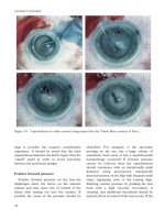
Implant power in triple procedures
The inaccuracy associated with lens implant
power calculation during a triple procedure
reflects the unpredictability of keratometry
following corneal grafting. The options to
minimise this source of error are discussed in
Chapter 6. The variation in refractive outcome
has led to the suggestion that non-simultaneous
penetrating keratoplasty, cataract extraction,
and lens implantation (or two-stage surgery)
should be adopted.
102,103
As mentioned above,
cataract surgery as a second procedure inevitably
causes some endothelial damage and may cause
graft rejection. A two-stage operation also has
the disadvantage that keratometry does not
stabilise until graft sutures are removed (up to
two years after surgery), which delays visual
rehabilitation. In addition, many graft patients
have to wear a contact lens to correct residual
astigmatism irrespective of spherical error. As a
result, two-stage surgery may only be advisable
when early cataract is present and its visual
significance is uncertain.
104
Postoperative management
In patients with dry eyes or cicatrising
conjunctival disease, intensive preservative free
topical lubricants should be used in conjunction
with the usual topical antibiotics and steroids
(also preservative free if available). Close and
regular follow up is essential in these patients,
who have a high rate of serious complications.
Persistent epithelial defects should be treated
with a soft bandage contact lens or tarsorrhaphy.
In cases refractory to this treatment, amniotic
membrane transplantation may be required
and cyanoacrylate glue is useful if perforation
occurs. Dry eyes associated with a systemic
connective tissue disorder have more frequent
complications, such as corneal melting, infective
keratitis, and endophthalmitis, following
cataract extraction. In ocular cicatricial
pemphigoid the disease may reactivate after
surgery. Close review allows systemic
immunosuppression to be commenced early if
necessary.
Herpes simplex keratitis, a common indication
for penetrating keratoplasty, may be reactivated
following intraocular surgery. This is of particular
concern because of the need for topical steroids
after cataract extraction. In such cases
postoperative prophylactic oral antiviral treatment
is advisable (aciclovir 400 mg twice a day).
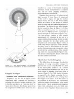
Glaucoma
Glaucoma and cataract may coexist in a wide
variety of situations. This includes patients who
have controlled open angle glaucoma but may
require drainage surgery in the future, or those
who have uncontrolled open angle galucoma
and require drainage. Other glaucoma patients
with cataract may have had a trabeculectomy
to lower intraocular pressure or peripheral
iridotomies to prevent or treat acute angle
closure glaucoma. Glaucoma also occurs in
association with extremes of axial length and
conditions such as pseudoexfoliation. Cataract
surgery in these patients, like in those who have
had previous procedures, presents a surgical
challenge. In addition, phacomorphic and
phacolytic glaucoma are caused by hypermature
cataract and treatment is by lens extraction.
Preoperative management
Miotics such as pilocarpine are in decline as a
topical treatment for glaucoma, but historically
many patients have been treated with these
agents. A small pupil may accentuate the effect of
early cataract, and simply changing to a different
CATARACT SURGERY IN COMPLEX EYES
147
medication may be sufficient to delay the need for
cataract surgery. Stopping miotic treatment may
also improve pupil dilatation if cataract surgery is
planned. When a patient with narrow angles and
cataract is examined at the preoperative stage, the
intraocular pressure should be measured
following dilated fundoscopy. If a significant
increase in pressure occurs, then medical
treatment or peripheral iridotomy to lower it may
be required in the perioperative period.
The presence of cataract may affect the
accuracy of both field testing and optic disc
examination, which complicates the assessment
of glaucoma progression. This may have
implications for the timing of cataract and
drainage surgery. Trabeculectomy may accelerate
the development of cataract because of
intraoperative lens trauma, inflammation, and
the use of topical steroids following surgery.
This should be borne in mind if early cataract
exists and drainage surgery alone is planned.
The patient should be informed of the possible
need for cataract extraction in the future, or that
a combined procedure may be indicated.
Lens induced glaucoma
Lens induced glaucoma is usually caused by
an advanced hypermature cataract. Phacolytic
glaucoma may also follow traumatic capsule
rupture, and is caused by leakage of high
molecular weight lens proteins from the capsular
bag that obstruct the trabecular meshwork.
Phacomorphic glaucoma results from a
tumescent lens that causes pupil block and acute
angle closure (Figure 10.25). In both phacolytic
and phacomorphic glaucoma the intraocular
pressure may be very high in conjunction with a
marked inflammatory response and corneal
oedema. Phacomorphic glaucoma appears
to be more common in patients with
pseudoexfoliation syndrome, reflecting zonular
laxity and anterior movement of the lens–iris
diaphragm.
Treatment in the first instance is medical,
using topical and systemic agents to lower
intraocular pressure as well as to treat
inflammation. Where angle closure exists
temporary success has been reported using
Nd:YAG laser peripheral iridotomy.
105
Topical
miotics may reduce intraocular pressure but they
may also exacerbate pupil block, and dilatation
is required before cataract extraction.
Surgical technique and lens implantation
Controlled open angle glaucoma
Clear corneal phacoemulsification with
posterior chamber IOL implantation is associated
with a significant sustained drop in intraocular
pressure in the order of 1–3 mmHg in normal
patients as well as glaucoma suspect and
glaucoma patients.
106
This may prove to be
beneficial, allowing a reduction in topical
glaucoma medication. Surgery that involves the
conjunctiva is known to compromise the
success of future drainage surgery,
107
and
phacoemulsification through a clear corneal
incision minimises disturbance to the ocular
surface. If patients have been treated with miotics
then the pupil may fail to dilate or dilate only
poorly, and techniques to enlarge the pupil may
be required.
Uncontrolled glaucoma (combined
drainage and cataract surgery)
Patients with progressive glaucoma,
uncontrolled with topical medications, may
CATARACT SURGERY
148
Figure 10.25 Angle closure glaucoma with a
phacomorphic component.
require drainage surgery. When cataract is also
present the surgical options are sequential
trabeculectomy and cataract extraction or
combined surgery. Combined trabeculectomy
and cataract extraction offers the advantage of a
single operation. However, trabeculectomy
combined with ECCE is not as effective as
trabeculectomy alone.
108
Phacoemulsification
combined with trabeculectomy may be performed
at a single site using a modified scleral tunnel
incision, and this has been shown to provide better
long term postoperative control of intraocular
pressure than does ECCE combined with
trabeculectomy.
109
Although phacotrabeculectomy
may be performed under general or local
anaesthesia, topical anaesthesia requires the
addition of subconjunctival anaesthetic.
110
Numerous phacotrabeculectomy techniques have
been described, but a fornix based conjunctival
flap combined with a scleral tunnel incision is
easiest to perform and does not compromise
outcome.
111
To provide an adequate superficial
scleral flap, the tunnelled incision should be
commenced more posteriorly than usual. This
may reduce movement of the phaco probe and
cause compression of the irrigation sleeve, with
heating of the wound and phaco burn. A lateral
scleral relieving incision, partly opening the
superficial scleral flap, reduces these problems
(Figure 10.26). Following phacoemulsification
and folding lens implantation, the scleral flap is
produced by incising anteriorly from the lateral
edges of the incision. A sclerostomy is most easily
produced using a scleral punch (Figure 10.27),
and a peripheral iridectomy is then performed
with scissors. The scleral flap may then be sutured
with adjustable or releasable 10/0 nylon sutures.
The conjunctiva is closed in a manner similar to
any trabeculectomy with either absorbable or
non-absorbable sutures.
Studies of single site phacotrabeculectomy
have suggested that its success may be lower than
that with trabeculectomy performed in isolation.
112
This may be due to trauma, inflammation, and
subsequent scarring caused by phacoemu-
lsification at the trabeculectomy site. A single
intraoperative application of an antimetabolite,
such as 5-fluorouracil (5FU), modifies the
healing response and improves the outcome of
CATARACT SURGERY IN COMPLEX EYES
149
Figure 10.26 Single site phacotrabeculectomy:
lateral relieving incision in a scleral tunnel (arrow) to
aid phaco probe movement and reduce the risk of
phacoburn.
Figure 10.27 Kelly sclerostomy punch (Altomed).
trabeculectomy alone.
113
Antimetabolites have
therefore been used as an adjunct to improve the
performance of phacotrabeculectomy. Comparison
of phacotrabeculectomy and 5FU with
trabeculectomy and 5FU followed later by
phacoemulsification has shown similar long term
results in terms of intraocular pressure.
114
Mitomycin C has also been shown to be effective
in conjunction with phacotrabeculectomy,
115
but
this antimetabolite has more potential for early
and late complications. To minimise tissue
manipulation that occurs with a single site
phacotrabeculectomy, two site surgery may offer
advantages. Typically, a temporal clear corneal
incision is used for phacoemulsification and a
separate trabeculectomy is performed
superiorly.
116
Although good results have been
reported using this approach, it does require the
surgeon to move position during surgery.
Previous glaucoma surgery
Patients who have undergone trabeculectomy
may develop cataract, or pre-existing cataract
may progress following filtration surgery.
Poorly dilating pupils or a shallow anterior
chamber may then complicate cataract
extraction. Cataract surgery must also avoid
damage to a functioning bleb and, as far as
possible, must not compromise long term
control of intraocular pressure. Unless bleb
revision is planned as part of surgery, a corneal
incision anterior to the bleb is usually adopted
during ECCE. This avoids injury to the bleb,
but the anterior position of the incision makes
postoperative astigmatism and endothelial cell
loss more likely. In patients who have had
filtration surgery and subsequently had cataract
extraction, intraocular pressure is better
controlled by phacoemulsification than by
ECCE.
117
Clear corneal phacoemulsification
using a temporal approach minimises the risk
to the filtering bleb and is the operation of
choice.
Lens induced glaucoma
Cataract surgery is the definitive treatment
for lens induced glaucoma, which should ideally
be performed soon after intraocular pressure is
controlled. This is particularly relevant in
phacomorphic glaucoma, in which permanent
peripheral anterior synechiae may develop and
prevent a return to normal pressures. If
permanent peripheral anterior synechiae are
present, then a combined procedure is usually
required. Corneal oedema, the risk of unstable
zonules, and difficulty in obtaining a capsulorhexis
may be indications for an ECCE.
118
Capsulorhexis is complicated both by the lack of
red reflex and the tension a tumescent lens
places on the anterior capsule. Puncture of the
anterior capsule with a standard rhexis needle or
cystotome may then result in a rapidly
propagating radial tear. This can usually be
overcome by using a suitable viscoelastic to
tamponade the anterior chamber and aspiration
of lens material through a narrow (30 G) needle
(see Chapter 3).
119
Although poor pupil
dilatation and unstable zonules may also be
present, phacoemulsification may then be
possible and provide the advantages of small
incision surgery with “in the bag” IOL
implantation.
Lens implantation
In most glaucoma patients anterior chamber
lens implantation should be avoided, and the
ideal position for the IOL is the posterior
chamber within the capsular bag.
Phacoemulsification allows the use of a foldable
posterior chamber lens implanted through a
small incision. During phacotrabeculectomy a
foldable lens can be inserted either through the
trabeculectomy opening or a separate corneal
incision without the need for wound
enlargement. Foldable silicone lens implantation
in conjunction with single site phacotrabe-
culectomy does not appear to impact negatively
CATARACT SURGERY
150
on bleb formation or control of intraocular
pressure when compared with the use of a
PMMA lens.
120
Anterior chamber inflammation,
as measured by the laser flare meter, is more
prolonged after phacoemulsification than after
trabeculectomy.
121
Postoperative inflammation
may be a relevant factor in the failure of
drainage procedures, and the biocompatibility
of the IOL material is therefore of particular
importance in combined procedures (see
Chapter 7).
122
Implant biocompatibility and
IOL selection is also relevant following cataract
surgery in eyes that may be associated with
increased postoperative inflammation, for
example those with phacomorphic or phacolytic
glaucoma.
Postoperative management
It is important that all viscoelastic is removed
from the anterior chamber at the end of surgery
because this is recognised to cause a
postoperative pressure rise.
123
Despite this the
intraocular pressure frequently elevates during
the first 24 hours following cataract surgery and
may exceed 35 mmHg.
124
In patients with
existing glaucoma and optic nerve damage,
medical prophylaxis to prevent this pressure
spike is required, such as a single dose of oral
Diamox SR 250 mg (Wyeth). Six hours after
cataract surgery, intraocular pressure has been
shown to be statistically higher in patients with a
scleral tunnel incision as compared with a clear
corneal incision.
125
Following cataract surgery,
patients with glaucoma may be more likely to
have additional postoperative inflammation,
particularly those that have suffered an episode
of acute angle closure glaucoma. Topical
steroids may be required at a higher
concentration or frequency. These patients
should be carefully followed up in view of the
risk of a steroid response and intraocular
pressure elevation.
Paediatric cataract
The treatment of paediatric cataract is a
complex subspeciality area. It often requires a
multidisciplinary team of doctors and eye
professionals to work closely with the child and
parents. Ocular examination may be difficult
and surgery is technically challenging. At all
stages of treatment it is imperative that the
child’s parents fully understand the relevant
issues and are able to be actively involved in the
decision making process. This is particularly
important because intensive management of
amblyopia and refractive error after surgery are
the key to effective treatment. Despite this, a
successful outcome is not guaranteed,
particularly in unilateral cataract.
Preoperative management
Ophthalmologist, optometrist, orthoptist, and
paediatric anaesthetist all play important roles in
the management of paediatric cataract. A
geneticist and paediatrican may also be required
if a cataract is associated with a systemic
disorder. Clear information should be provided
to the parents of the affected child from the
outset. It is often difficult to determine the visual
impact of a cataract on a preverbal infant. The
CATARACT SURGERY IN COMPLEX EYES
151
Figure 10.28 Altered red reflex in a typical
congenital cataract.
appearance of the red reflex (Figure 10.28) and
fixation pattern may be useful indicators, but
fixed choice preferential looking and visual
evoked potentials provide a subjective
assessment of acuity. Examination under
anaesthesia allows the appraisal of cataract
morphology, which may also be an indicator of
its visual significance. Features that favour
surgery include large, axial, dense, or posterior
cataracts. Pupil dilatation may benefit eyes with
less significant cataract but success can be
limited by loss of accommodation and glare.
Patients with bilateral visually significant
cataracts should undergo surgery within three
months of age to minimise the risk of developing
irreversible amblyopia and nystagmus.
126
The
second eye should have surgery within one week
of the first (intermittently patching the operated
eye in the interim). The management of unilateral
visually significant cataract is more controversial.
127
The results of cataract surgery in these
circumstances are variable and good outcomes are
only obtainable with early surgery (as early as six
weeks of age
128
) and intensive treatment of
amblyopia. This has a risk of inducing amblyopia
in the non-affected eye and requires substantial
long term commitment from the child’s parents.
Surgery is unlikely to be effective if there is a
coexisting ocular disorder such as retinopathy of
prematurity or sclerocornea. The decision to
operate on unilateral cataract should also be
carefully considered if severe systemic disease is
present or if the parents or child are unlikely to
manage amblyopia treatment.
Cataract presenting later in infancy poses a
management problem because surgery may be of
little use if visually significant cataract has existed
since birth but has gone undetected. Lack of
strabismus or nystagmus in an older infant with a
substantial lens opacity may indicate that an
initially insignificant cataract has progressed, and
surgery may be worthwhile in such cases.
Surgical technique
Spin-off techniques from phacoemulsification
have been incorporated into paediatric cataract
extraction, but there are several aspects of this
surgery that differ from that in adults. These
relate to the soft lens, anatomical differences,
and the need to address the high incidence of
posterior capsular and anterior hyaloid opacity
found postoperatively.
129
Scleral or corneal
tunnelled incisions can be used in infants but
have a tendency to leak and should be sutured at
the end of the procedure.
The thin flexible sclera in the paediatric eye is
thought to account for the tendency of the
anterior chamber to collapse during surgery,
particularly when instruments are removed from
the eye. This may be minimised by using an
anterior chamber maintainer (Figure 10.29)
throughout surgery and ensuring that
anaesthesia is deep enough to prevent
extraocular muscle contraction. The lens
capsule is also highly elastic as compared with
that in adults, and this makes anterior
continuous curvilinear capsulorhexis difficult.
Alternative techniques that have been suggested
include radiofrequency diathermy capsulorhexis
130
and central anterior capsulotomy performed
with a vitrector. The vitrector can then be used
to aspirate the lens and perform a posterior
capsulotomy with anterior vitrectomy. This
removes the need for secondary surgical
intervention to clear the visual axis. Posterior
capsulorhexis has been reported as an effective
alternative, which allows “in the bag” IOL
CATARACT SURGERY
152
Figure 10.29 A self-retaining Lewicky anterior
chamber maintainer (BD Ophthalmic Systems).
implantation.
131
Although a phacoemulsification
probe can be used for lens removal, irrigation
and aspiration equipment, especially bimanual
instruments, are probably less traumatic and
safer. An aspiration port with a diameter larger
than that usually found on a standard
instrument (0·35 mm) may be more effective.
Pars plana lensectomy has been used to
remove paediatric cataracts,
132
but the long term
risk of posterior segment complications are largely
unknown and usually little capsule remains to
support an IOL. Intracapsular surgery is not
appropriate in children because of the strong
attachments between the posterior capsular and
the anterior vitreous, which may cause substantial
vitreous loss and risk retinal detachment.
Lens implantation and selection of power
Lens implantation as a primary procedure is
increasingly common in all children.
133
The long
term complications of anterior chamber lenses
preclude their use, and the ideal site for an IOL
is within the capsular bag in the posterior
chamber. PMMA is the only implant material
that has sufficient follow up to allow safe
implantation in infants. Although lenses with
optics constructed from highly biocompatible
foldable materials may offer advantages, at
present their long term outcomes are unknown.
Lenses designed specifically for the paediatic eye
are available but adult lenses can be used,
providing their overall diameter is not greater
than 12 mm.
During the first six to eight years of life the
infant eye undergoes a substantial myopic shift
from hypermetropia to emmetropia.
134
There
is general agreement that an IOL implant
should aim to anticipate this with an initial
hypermetropic over-correction.
133
The extent of
intentional hypermetropia depends on the age of
the child at time of surgery. Residual refractive
error must then be corrected with spectacles
(bifocals), contact lenses, or a combination (to
prevent amblyopia). Relative contraindications
to IOL implantation are anatomical ocular
abnormalities such as microphthalmos or
persistent hyperplastic primary vitreous. Contact
lenses are the main alternative to IOL
implantation, although aphakic spectacles may
be used. Refractive corneal techniques, for
example epikeratophakia, have largely been
abandoned in favour of lens implantation.
Postoperative management
The key to the treatment of paediatric cataract
is the postoperative management of amblyopia
and refractive error. This requires a major input
from the child’s parents that may put a strain on
family life. The parents may need supervision and
help in many aspects of postoperative care
including, for example, contact lens care and
handling. In young infants incremental part time
patching reduces the risk of inducing amblyopia in
the better or normal eye. Daily wear or extended
wear contact lenses can be used to correct
refractive error, usually with a lens power designed
to achieve near vision (i.e. induce a low degree of
myopia).
Refraction and postoperative assessment may
require multiple examinations under general
anaesthesia. Intraocular inflammation commonly
complicates paediatric cataract surgery, and may
require intensive topical steroids and, in some
cases, recombinant TPA. Other frequent
complications include glaucoma and, as
previously mentioned, posterior capsule and
anterior hyaloid opacification.
135
The latter
requires either Nd:YAG capsulotomy or a surgical
procedure to clear the visual axis. Because of the
lifetime risk of glaucoma and retinal detachment,
patients should be monitored in the long term.
136
References
1 Ederer F, Hiller R, Taylor HR. Senile lens changes and
diabetes in two population studies. Am J Ophthalmol
1981;91:381–95.
2 Klein R, Klein BE, Moss SE. Visual impairment in
diabetes. Ophthalmology 1984;91:1–9.
3 Klein BE, Klein R, Moss SE. Incidence of cataract
surgery in the Wisconsin Epidemiologic Study of Diabetic
Retinopathy. Am J Ophthalmol 1995;119:295–300.
CATARACT SURGERY IN COMPLEX EYES
153
4 Dowler JG, Hykin PG, Lightman SL, Hamilton AM.
Visual acuity following extracapsular cataract extraction
in diabetes: a meta-analysis. Eye 1995;9:313–7.
5 Krupsky S, Zalish M, Oliver M, Pollack A. Anterior
segment complications in diabetic patients following
extracapsular cataract extraction and posterior chamber
intraocular lens implantation. Ophthalmic Surg
1991;22:526–30.
6 Ionides A, Dowler JG, Hykin PG, Rosen PH, Hamilton
AM. Posterior capsule opacification following diabetic
extracapsular cataract extraction. Eye 1994;8:535–7.
7 Ulbig MR, Hykin PG, Foss AJ, Schwartz SD, Hamilton
AM. Anterior hyaloidal fibrovascular proliferation after
extracapsular cataract extraction in diabetic eyes. Am J
Ophthalmol 1993;115:321–6.
8 Pollack A, Leiba H, Bukelman A, Oliver M. Cystoid
macular oedema following cataract extraction in
patients with diabetes. Br J Ophthalmol 1992;76:221–4.
9 Jaffe GJ, Burton TC. Progression of nonproliferative
diabetic retinopathy following cataract extraction. Arch
Ophthalmol 1988;106:745–9.
10 Dowler JGF, Sehmi KS, Hykin PG, Hamilton AM. The
natural history of macular edema after cataract surgery
in diabetes. Ophthalmology 1999;106:663–5.
11 Hykin PG, Gregson RM, Stevens JD, Hamilton PA.
Extracapsular cataract extraction in proliferative
diabetic retinopathy. Ophthalmology 1993;100:394–9.
12 Benson WE, Brown GC, Tasman W, McNamara JA,
Vander JF. Extracapsular cataract extraction with
placement of a posterior chamber lens in patients with
diabetic retinopathy. Ophthalmology 1993;100:730–8.
13 Schatz H, Atienza D, McDonald HR, Johnson RN.
Severe diabetic retinopathy after cataract surgery. Am J
Ophthalmol 1994;117:314–21.
14 Borgioli AM, Coster DJ, Fan RFT, Henderson J. Effect of
heparin surface modification of polymethylmethacrylate
intraocular lenses on signs of post-operative inflammation
after extracapsular cataract extraction. Ophthalmology
1992;99:1248–55.
15 Hayashi K, Hayashi H, Nakao F, Hayashi F. Reduction
in the area of the anterior capsule opening after
polymethylmethacrylate, silicone, and soft acrylic
intraocular lens implantation. Am J Ophthalmol 1997;
123:441–7.
16 Francese JE, Christ FR, Buchen SY, Gwon A,
Robertson JE. Moisture droplet formation on the
posterior surface of intraocular lenses during fluid/air
exchange. J Cataract Refract Surg 1995;21:685–9.
17 Apple DJ, Federman JL, Krolicki TJ, et al. Irreversible
silicone oil adhesion to silicone intraocular lenses. A
clinicopathologic analysis. Ophthalmology 1996;103:
1555–61.
18 Apple DJ, Isaacs RT, Kent DG, et al. Silicone oil
adhesion to intraocular lenses: an experimental study
comparing various biomaterials. J Cataract Refract Surg
1997;23:536–44.
19 Hollick EJ, Spalton DJ, Ursell PG, et al. The effect of
polymethylmethacrylate, silicone, and polyacrylic
intraocular lenses on posterior capsular opacification
three years after cataract surgery. Ophthlamology
1999;106:49–54.
20 Dowler JG, Hykin PG, Hamilton AM.
Phakoemulsification versus extracapsular cataract
surgery in diabetes. Ophthalmology 2000:107:457-62.
21 Eifrig DE, Hermsen V, McManus P, Cunningham R.
Rubeosis capsulare. J Cataract Refract Surg 1990;16:633–6.
22 Henricsson M, Heijl A, Janzon L. Diabetic retinopathy
before and after cataract surgery. Br J Ophthalmol
1996;80:789–93.
23 Wagner T, Knaflic D, Rauber M, Mester U. Influence
of cataract surgery on the diabetic eye: a prospective
study. Ger J Ophthalmol 1996;5:79–83.
24 Okhravi N, Lightman SL, Towler HMA. Assessment of
visual outcome after cataract surgery in patients with
uveitis. Ophthalmology 1999;106:703–9.
25 Okhravi N, Towler HMA, Lightman SL. Cataract
surgery in patients with uveitis. Eye 2000;14:689–90.
26 Jones NP. Cataract surgery using heparin surface-
modified intraocular lenses in Fuchs’ heterochromic
uveitis. Ophthalmic Surg 1995;26:49–52.
27 Barton K, Hall AJH, Rosen PH, Cooling RJ, Lightman S.
Systemic steroid prophylaxis for cataract surgery in
patients with uveitis. Ocular Immunol Inflamm
1994;2:207–16.
28 Cheng CK, Berger AS, Pearson PA, Ashton P, Jaffe GJ.
Intravitreal sustained-release dexamethasone device in
the treatment of experimental uveitis. Invest Ophthalmol
Vis Sci 1995;36:442–53.
29 Diamond JG, Kaplan HJ. Lensectomy and vitrectomy
for complicated cataract secondary to uveitis. Arch
Ophthalmol 1978;96:1798–804.
30 Foster CS, Fong LP, Singh G. Cataract surgery and
intraocular lens implantation in patients with uveitis.
Ophthalmology 1989;96:281–8.
31 Ceisler EJ, Foster CS. Juvenile rheumatoid arthritis and
uveitis: minimizing the blinding complications. Int
Ophthalmol Clin 1996;36:91–107.
32 Kanski JJ. Lensectomy for complicated cataract in juvenile
chronic iridocyclitis. Br J Ophthalmol 1992;76:72–5.
33 Flynn HW Jr, Davis JL, Culbertson WW. Pars plana
lensectomy and vitrectomy for complicated cataracts in
juvenile rheumatoid arthritis. Ophthalmology 1988;95:
1114–9.
34 Oshika T, Yoshimura K, Miyata N. Postsurgical
inflammation after phacoemulsification and extracapsular
extraction with soft or conventional intraocular lens
implantation. J Cataract Refrac Surg 1992;18:356–61.
35 Dick HB, Schwenn O, Krummenauer F, Krist R,
Pfeiffer N. Inflammation after sclerocorneal versus clear
corneal tunnel phacoemulsification. Ophthalmology
2000;107:241–7.
36 Davison JA. Capsule contraction syndrome. J Cataract
Refract Surg 1993;19:582–9.
37 Luke C, Dietlein TS, Jacobi PC, Konen W, Krieglstein
GK. Massive anterior capsule shrinkage after plate-
haptic silicone lens implantation in uveitis. J Cataract
Refract Surg 2001;27:333–6.
38 Koenig SB, Mieler WF, Han DP, Abrams GW.
Combined phacoemulsification, pars plana vitrectomy,
and posterior chamber intraocular lens insertion. Arch
Ophthalmol 1992;110:1101–4.
39 Shah SM, Spalton DJ. Comparison of the post-operative
inflammatory response in the normal eye with heparin-
surface-modified and polymethyl methacrylate intraocular
lenses. J Cataract Refract Surg 1995;21:579–85.
40 Percival SPB, Pai V. Heparin-modified lenses for eyes at
risk for breakdown of the blood- aqueous barrier during
cataract surgery. J Cataract Refract Surg 1993;19:760–5
41 Dick B, Kohnen T, Jacobi KW. Alterationen der
heparinbeschichtung auf intraokularlinsen durch
implantationsinstrumente. Klin Monatsbl Augenheilkd
1995;206:460–6.
CATARACT SURGERY
154
42 Rauz S, Stavrou P, Murray PI. Evaluation of foldable
intraocular lenses in patients with uveitis. Ophthalmology
2000;107:909–19.
43 Apple DJ, Solomon KD, Tetz MR, et al. Posterior
capsule opacification. Surv Ophthalmol 1992;37:
73–116.
44 Dana MR, Chatzistefanou K, Schaumberg DA, Foster
CS. Posterior capsule opacification after cataract
surgery in patients with uveitis. Ophthalmology 1997;
104:1387–94.
45 Lemon LC, Shin DH, Song MS, et al. Comparative
study of silicone versus acrylic foldable lens
implantation in primary glaucoma triple procedure.
Ophthalmology 1997;104:1708–13.
46 Estafanous MF, Lowder CY, Meisler DM, Chauhan R.
Phacoemulsification cataract extraction and posterior
chamber lens implantation in patients with uveitis. Am J
Ophthalmol 2001;131:620–5.
47 Tanner V, Casswell AG. A comparative study of the
efficacy of 2⋅5% phenylephrine and 10% phenylephrine
in pre-operative mydriasis for routine cataract surgery.
Eye 1996;10:95–8.
48 Duffin MR, Pettit TH, Straatsma BR. 2⋅5% v 10%
phenylephrine in maintaining mydriasis during cataract
surgery. Arch Ophthalmol 1983;101:1903–9.
49 Roberts CW. Comparison of diclofenac sodium and
flurbiprofen for inhibition of surgically induced miosis.
J Cataract Refract Surg 1996;22(suppl 1):780–7.
50 Solomon KD, Turkalj JW, Whiteside SB, Stewart JA,
Apple DJ. Topical 0⋅5% ketorolac vs 0⋅03% flurbiprofen
for inhibition of miosis during cataract surgery. Arch
Ophthalmol 1997;115:1119–22.
51 Corbett MC, Richards AB. Intraocular adrenaline
maintains mydriasis during cataract surgery. Br J
Ophthalmol 1994;78:95–8.
52 Fell D, Watson AP, Hindocha N. Plasma concentrations
of catecholamines following intraocular irrigation with
adrenaline. Br J Anaesth 1989;62:573–5.
53 Joseph J, Wang HS. Phacoemulsification with poorly
dilated pupils. J Cataract Refract Surg 1993;19:551–6.
54 Shepherd DM. The pupil stretch technique for miotic
pupils in cataract surgery. Ophthalmic Surg 1993;24:
851–2.
55 Dinsmore SC. Modified stretch technique for small
pupil phacoemulsification with topical anesthesia.
J Cataract Refract Surg 1996;22:27–30.
56 Graether JM. Graether pupil expander for managing the
small pupil during surgery. J Cataract Refract Surgery
1996;22:530–5.
57 De Juan E Jr, Hickingbotham D. Flexible iris retractor
[letter]. Am J Ophthalmol 1991;111:776–7.
58 Nichamin LD. Enlarging the pupil for cataract
extraction using flexible nylon iris retractors. J Cataract
Refract Surg 1993;793–6.
59 Novak J. Flexible iris hooks for phacoemulsification.
J Cataract Refract Surg 1997;23:828–31.
60 Smith GT, Liu CS. Flexible iris hooks for
phacoemulsification in patients with iridoschisis.
J Cataract Refract Surg 2000;26:1277–80.
61 Masket S. Avoiding complications associated with iris
retractor use in small pupil cataract extraction.
J Cataract Refract Surg 1996;22:168–71.
62 Yuguchi T, Oshika T, Sawaguchi S, Kaiya T. Pupillary
function after cataract surgery using flexible iris
retractor in patients with small pupil. Jpn J Ophthalmol
1999;43:20–4.
63 Birchall W, Spencer AF. Misalignment of flexible iris
hook retractors for small pupil cataract surgery: effects
on pupil circumference. J Cataract Refract Surg
2001;27:20–4.
64 Fine IH. Pupilloplasty for small pupil phacoemulsification.
J Cataract Refract Surg 1994;20:192–6.
65 Merriam JC, Zheng L. Iris hooks for
phacoemulsification of the subluxed lens. J Cataract
Refract Surg 1997;23:1295–7.
66 Fine IH, Hoffman RS. Phacoemulsification in the
presence of pseudo-exfoliation: Challenges and options.
J Cataract Refract Surg 1997;23:160–5.
67 Menapace R, Findl O, Georgopoulos M, et al. The
capsular tension ring: designs, applications and
techniques. J Cataract Refract Surg 2000;26:898–912.
68 Lam DS, Young AL, Leung AT, et al. Scleral fixation of
a capsular tension ring for severe ectopia lentis.
J Cataract Refract Surg 2000;26:609–612.
69 Fischel JD, Wishart MS. Spontaneous complete
dislocation of the lens in pseudo-exfoliation syndrome.
Eur J Implant Refract Surg 1995;7:31–3.
70 Cionnin RJ, Osher RH. Management of profound
zonular dialysis or weakness with a new endocapsular
ring designed for scleral fixation. J Cararact Refract Surg
1998;24:1299–306.
71 Shastri L, Vasavada A. Phacoemulsification in Indian
eyes with pseudo-exfoliation syndrome. J Cataract
Refract Surg 2001;27:1629–37.
72 Isakov I, Bartov E. Managing inferior zonule tears
during manual extracapsular extraction. J Cataract
Refract Surg 1998;24:300–2.
73 Demler U, Sautter H. Surgery in sub-luxated lenses in
adults. Dev Ophthalmol 1985;11:162–5.
74 Blumenthal M, Kurtz, Assia EI. Hydroexpression of
subluxed lenses using a glide. Ophthalmic Surg 1994;
25:34–7.
75 Hakin KN, Jacobs M, Rosen P, et al. Management of the
subluxed crystalline lens. Ophthalmology 1992;99:542–5.
76 Plager DA, Parks MM, Helveston EM, Ellis FD.
Surgical treatment of subluxed lenses in children.
Ophthalmology 1992;99:1018–21.
77 Hubbard AD, Charteris DG, Cooling RJ.
Vitreolensectomy in Marfan’s syndrome. Eye 1998;3A:
412–6.
78 Gimbel HV. Role of capsular tension rings in preventing
capsule contraction. J Cataract Refract Surg 2000;26:
791–2.
79 Liu C, Eleftheriadis H. Multiple capsular tension rings
for the prevention of capsule contraction syndrome.
J Cataract Refract Surg 2001;27:342–3.
80 Berger RR, Kenyeres A, Van Coller BM, Pretorius CF.
Repositioning a tilted ciliary-sulcus-fixated intraocular
lens. J Cataract Refract Surg 1995;21:497–8.
81 Davison JA. Capsule contraction syndrome. J Cataract
Refract Surg 1993;19:582–9.
82 Blankenship GW. Stability of pars plana vitrectomy
results for diabetic retinopathy complications; a
comparison of five-year and six-month post-vitrectomy
findings. Arch Ophthalmol 1981;99:1009–12.
83 McCuen B, de Juan E, Landers MB, Machemer R.
Silicone oil in vitreoretinal sugery II. Results and
complications. Retina 1985;5:198–205.
84 Wilbrandt HR, Wilbrant TH. Pathogenesis and
management of the lens-diaphragm retropulsion
syndrome during phacoemulsification. J Cataract
Refract Surg 1994;20:48–53.
CATARACT SURGERY IN COMPLEX EYES
155
85 Franks WA, Leaver PK. Removal of silicone oil:
rewards and penalties. Eye 1991;5:333–7.
86 Baer RM, Aylward WG, Leaver PK. Cataract
extraction following vitrectomy and silicone oil
tamponade. Eye 1995;9:309–12.
87 Lacelle VD, Garate FJO, Alday NM, et al.
Phacoemulsification cataract surgery in vitrectomised
eyes. J Cataract Refract Surg 1998;24:806–9.
88 Grey R, Horsborough B. Cataract extraction following
vitrectomy and silicone oil tamponade. Eye
1996;10:151–2.
89 Apple DJ, Federman JL, Krolicki TJ, et al. Irreversible
silicone oil adhesion to silicone intraocular lenses.
A clinicopathologic analysis. Ophthalmology 1996;103:
1555–61.
90 Ravalico G, Tognetto D, Palomba MA, Lovisato A,
Baccara F. Corneal endothelial function after
extracapsular cataract extraction and phacoemulsification.
J Cataract Refract Surg 1997;23:1000–5.
91 Hayashi K, Nakao F, Hayashi F. Corneal endothelial
cell loss after phacoemulsification using nuclear
cracking procedures. J Cataract Refract Surg 1994;
20:44–7.
92 Oshima Y, Tsujikawa K, Oh A, Harino S.
Comparative study of intraocular lens implantation
through 3⋅0mm temporal clear corneal and superior
scleral tunnel self-sealing incisions. J Cataract Refract
Surg 1997;23:347–53.
93 Malbran ES, Malbran E, Buonsanti J, Adrogue E.
Closed-system phacoemulsification and posterior
chamber implant combined with penetrating
keratoplasty. Ophthalmic Surg 1993;24:403–6.
94 Caporossi A, Traversi C, Simi C, Tosi GM. Closed-
system and open-sky capsulorhexis for combined
cataract extraction and corneal transplantation.
J Cataract Refract Surg 2001;27:990–3.
95 Lindquist TD. Open-sky phacoemulsification during
corneal transplantation. Ophthalmic Surg 1994;25:
734–6.
96 Ram J, Sharma A, Pandav SS, Gupta A, Bambery P.
Cataract surgery in patients with dry eyes. J Cataract
Refract Surg 1998;24:1119–24.
97 MacLeod JD, Dart JK, Gray TB. Corneal and cataract
surgery in chronic progressive conjunctival
cicatrisation. Dev Ophthalmol 1997;28:228–39.
98 Lim ES, Apple DJ, Tsai JC, et al. An analysis of
flexible anterior chamber lenses with special reference
to the normalised rate of lens explanation
Ophthalmology 1991;98:243–6.
99 Ohguro N, Matsuda M, Kinoshita S. Effects of
posterior chamber lens implantation on the
endothelium of transplanted corneas. Br J Ophthalmol
1997;81:1056–9.
100 Barrett G, Constable IJ. Corneal endothelial loss with
new intraocular lenses. Am J Ophthalmol 1984;98:
157–65.
101 Lee JH, Oh SY. Corneal endothelial cell loss from
suture fixation of a posterior chamber intraocular lens.
J Cataract Refract Surg 1997;23:1020–2.
102 Rosen ES. Combined or sequential keratoplasty and
cataract surgery? J Cataract Refract Surg 1998;24:
1283–4.
103 Hsiao CH, Chen JJ, Chen PY, Chen HS. Intraocular
lens implantation after penetrating keratoplasty.
Cornea 2001;20:580–5.
104 Epstein RJ. Combining keratoplasty and cataract
surgery. J Cataract Refract Surg 1999;25:603.
105 Tomey KF, Al-Rajhi AA. Neodymium YAG laser
iridotomy in the initial management of phacomorphic
glaucoma. Ophthalmology 1992;99:660–5.
106 Shingleton BJ, Gamell LS, O’Donoghue MW, et al.
Long-term changes in intraocular pressure after clear
corneal phacoemulsification: Normal patients versus
glaucoma suspect and glaucoma patients. J Cataract
Refract Surg 1999;25:885–90.
107 Broadway DC, Grierson I, Hitchings RA. Local effects
of previous conjunctival incisional surgery and the
subsequent outcome of filtration surgery. Am
J Ophthalmol 1998;125:805–18.
108 Murchinson JF Jr, Shields MB. An evaluation of three
surgical approaches for coexisting cataract and
glaucoma. Ophthalmic Surg 1989;20:393–8.
109 Kosmin AS, Wishart PK, Ridges PJG. Long-term
intraocular pressure control after cataract extraction:
phacoemulsification versus extracapsular technique.
J Cataract Refract Surg 1998;249–55.
110 Vicary D, McLennan S, Sun XY. Topical plus
subconjunctival anesthesia for phacotrabeculectomy:
one year follow-up. J Cataract Refract Surg 1998;24:
1247–51.
111 Shingleton BJ, Chaudhry IM, O’Donoghue MW, et al.
Phacotrabeculectomy: limbus-based versus fornix-
based conjunctival flaps in fellow eyes. Ophthalmology
1999;106:1152–5.
112 Naveh N, Kottass R, Glovinsky J, et al. The long-term
effect on intraocular pressure of a procedure combining
trabeculectomy and cataract surgery, as compared with
trabeculectomy alone. Ophthalmic Surg 1990;21:339–45.
113 Smith MF, Sherwood MB, Doyle JW, Khaw PT.
Results of intra-operative 5-fluorouracil supplementation
on trabeculectomy for open-angle glaucoma. Am J
Ophthalmol 1992;114:737–41.
114 Donoso R, Rodriguez A. Combined versus sequential
phacotrabeculectomy with intra-operative 5-fluorouracil.
J Cataract Refract Surg 2000;26:71–4.
115 Cohen JS, Greff LJ, Novack GD, Wind BE. A
placebo-controlled, double-masked evaluation of
mitomycin C in combined glaucoma and cataract
procedures. Ophthalmology 1996;103:1934–42.
116 El Sayyad F, Helal M, El Maghraby A, Khalil M,
El Hamzawey H. One-site versus 2-site
phacotrabeculectomy: a randomized study. J Cataract
Refract Surg 1999;25:77–82.
117 Manoj B, Chako D, Khan MY. Effect of extracapsular
cataract extraction and phacoemulsification performed
after trabeculectomy on intraocular pressure.
J Cataract Refract Surg 2000;26:75–8.
118 McKibbin M, Gupta A, Atkins AD. Cataract
extraction and intraocular lens implantation in eyes
with phacomorphic or phacolytic glaucoma. J Cataract
Refract Surg 1996;22:633–6.
119 Rao SK, Padmanabhan R. Capsulorhexis in eyes with
phacomorphic glaucoma. J Cataract Refract Surg 1998;
24:882–4.
CATARACT SURGERY
156
120 Braga-Mele R, Cohen S, Rootman DS. Foldable
silicone versus polymethyl methacrylate intraocular
lenses in combined phacoemulsification and
trabeculectomy. J Cataract Refract Surgery 2000;26:
1517–22.
121 Siriwardena D, Kotecha A, Minassian D, Dart JK,
Khaw PT. Anterior chamber flare after
trabeculectomy and after phacoemulsification. Br J
Ophthalmol 2000;84:1056–7.
122 Samuelson TW, Chu YR, Kreiger RA. Evaluation of
giant-cell deposits on foldable intraocular lenses after
combined cataract and glaucoma surgery. J Cataract
Refract Surg 2000;26:817–23.
123 Tanaka T, Inoue H, Kudo S, Ogawa T. Relationship
between post-operative intraocular pressure elevation
and residual sodium hyaluronate following
phacoemulsification and aspiration. J Cataract Refract
Surg 1997;23:284–8.
124 Barak A, Desatnik H, Ma-Naim T, et al. Early post-
operative intraocular pressure pattern in glaucomatous
and nonglaucomatous patients. J Cataract Refract Surg
1996;22:607–11.
125 Schwenn O, Dick HB, Krummenauer F, et al.
Intraocular pressure after small incision cataract
surgery: temporal sclerocorneal versus clear corneal
incision. J Cataract Refract Surg 2001;27:421–5.
126 Lesueur LC, Arne JL, Chapotot EC, Thouvenin D,
Malecaze F. Visual outcome after paediatric cataract
surgery: is age a major factor? Br J Ophthalmol
1998;82:1022–5.
127 Taylor D, Wright KW, Amaya L, Cassidy L, Nischal
K, Russell-Eggitt I. Should we aggressively treat
unilateral congenital cataracts? Br J Ophthalmol
2001;85:1120–6.
128 Birch EE, Stager DR, Leffler J, Weakley D. Early
treatment of congenital unilateral cataract minimises
unequal competition. Invest Ophthalmol Vis Sci
1998;39:1560–6.
129 Koch DD, Kohnen T. Retrospective comparison of
techniques to prevent secondary cataract formation
after posterior chamber intraocular lens implantation
in infants and children. J Cataract Refract Surg
1997;23:657–63.
130 Comer RM, Abdulla N, O’Keefe M. Radiofrequency
diathermy capsulorhexis of the anterior and posterior
capsules in paediatric cataract surgery: preliminary
results. J Cataract Refract Surg 1997;23:641–4.
131 Gimbel HV. Posterior continuous curvilinear
capsulorhexis and optic capture of the intraocular lens
to prevent secondary opacification in paediatric
cataract surgery. J Cataract Refract Surg
1997;23:652–6.
132 Ahmadieh H, Javadi MA, Ahmady M, et al. Primary
capsulectomy, anterior vitrectomy, lensectomy and
posterior chamber lens implantation in children:
limbal versus pars plana. J Cataract Surg
1999;25:768–75.
133 Dahan E. Intraocular lens implantation in children.
Curr Opin Ophthalmol 2000;11:51–5.
134 Flitcroft DI, Knight-Nanan D, Bowell R, et al.
Intraocular lenses in children: changes in axial length,
corneal curvature and refraction. Br J Ophthalmol
1999;83:265–9.
135 Keech RV, Tongue AC, Scott WE. Complications
after surgery for congenital and infantile cataracts. Am
J Ophthalmol 1989;108:136–41.
136 Brady KM, Atkinson CS, Kilty LA, Hiles DA.
Glaucoma after cataract extraction and posterior
chamber lens implantation in children. J Cataract
Refract Surg 1997;23:669–74.
CATARACT SURGERY IN COMPLEX EYES
157
158
Vitreous loss is the most common serious
intraoperative complication of phacoemulsification
and extracapsular cataract surgery, occurring
in approximately 2–4% of contemporary
procedures.
1,2
Incidences of up to and around
10% have been reported, particularly from
surgeons in training
3–6
and in the older literature.
Vitreous loss usually results from iatrogenic
intraoperative rupture of the posterior capsule,
although it can also arise from intraoperative
zonule dehiscence or pre-existing injuries or
anomalies of the capsule and zonule.
The importance of vitreous loss is its
association with increased surgical morbidity
and a poorer postoperative visual outcome
7–9
as
compared with uncomplicated cataract surgery
(Box 11.1). If vitreous loss cannot be prevented,
then appropriate and careful management at the
time of initial surgery can ameliorate problems.
It is essential to have a systematic approach to
the variety of causes and consequences of
vitreous loss, and familiarity with the additional
instrumentation that may be required.
Prevention
Identification of eyes that are especially at
risk of capsular rupture or zonular dehiscence
is important (Box 11.2). This may allow
the surgery to be undertaken by a more
experienced surgeon in eyes with, for example,
pseudoexfoliation syndrome (Figure 11.1)
10
or
very dense/white cataracts (Figure 11.2).
Alternatively, it may be more appropriate to
employ a different surgical method, such as pars
plana vitreolensectomy for subluxed cataractous
lenses following ocular injury or in patients with
Marfan’s syndrome (Figure 11.3). Small pupils
(Figure 11.4) are associated with a significantly
11 Vitreous loss
Box 11.1 Consequences of vitreous loss
• Uveitis
• Glaucoma
• Macular oedema
• Corneal oedema
• Rhegmatogenous retinal detachment
• Endophthalmitis
• Pupil irregularity and distortion
• Vitreous wick syndrome
Box 11.2 Risk factors for vitreous loss
• Small pupil (diabetes, uveitis, age, previous
intraocular surgery, chronic pilocarpine)
• Intraoperative miosis (secondary to iris
trauma)
• Lens subluxation (iridodonesis and
phacodonesis)
• Irregularity of capsulorhexis
• Radial tears of anterior capsule
• Very dense cataracts
• Pseudoexfoliation syndrome
• Previous blunt trauma
• Poor wound construction
• Extraocular pressure on globe (lid speculum,
large volume peribulbar local anaesthetic)
• Retrobulbar haemorrhage
• Vitreous loss in previous eye
• Surgical inexperience: the “learning curve”
• Uncooperative patient
VITREOUS LOSS
159
increased risk of capsule rupture and vitreous
loss,
11
and this underlines the value of
intraoperative enlargement of the pupil using
surgical iridotomies, iris hooks (Figure 11.5), or
stretching.
Radial tears of the anterior capsule incurred
during capsulorhexis may extend peripherally
Figure 11.1 Pseudoexfoliation.
Figure 11.2 Mature white cataract.
Figure 11.3 Lens subluxed inferiorly.
Figure 11.4 Posterior synechiae and a small pupil in
an eye with recurrent anterior uveitis.
Figure 11.5 Iris hooks being used during pars plana
vitrectomy and cataract extraction.
through the zonule into the posterior capsule if
subjected to undue pressure. In the absence of
an intact capsulorhexis, phacoemulsification of a
hard nucleus requires a technique that does not
transmit forces to the capsule in a manner that is
likely to extend the radial tear posteriorly. A high
index of suspicion in “at risk” eyes can help to
identify capsular rupture at an earlier stage,
before vitreous loss or dislocation of lens
fragments has occurred, and allow appropriate
remedial action to be taken. The surgeon needs
to be alert for subtle signs that may indicate the
development of capsular rupture, such as
unexpected deepening of the anterior chamber.
If the surgeon is faced with a zonular dehiscence,
or a situation in which zonular support is
suspect, such as in pseudoexfoliation, then the
insertion of an endocapsular tension ring will
redistribute forces throughout the lens equator
and stabilise the situation.
12
General principles of management
During phacoemulsification the combination
of gravity and the posteriorly directed force of
the infusion fluid conspire to encourage lens
material to fall backward into the vitreous cavity
(the “dropped nucleus”). In extracapsular
surgery, however, capsular catastrophes are
usually associated with forward movement of the
vitreous through the pupil into the anterior
chamber, the surgical wound, and beyond.
Once vitreous loss has been recognised, the
immediate priority is to prevent the posterior
loss of the nucleus or its fragments into the
vitreous. The next priority is to clear the wound,
anterior chamber, and pupil of vitreous and lens
material, while preserving the anterior and
posterior lens capsule to allow, if appropriate,
the insertion of an intraocular lens implant.
When topical anaesthesia has been used,
supplementary anaesthesia by intracameral,
subconjunctival, or sub-Tenon’s routes may be
required.
Removal of remaining nuclear fragments
from the anterior chamber or capsular bag
will usually require conversion from
phacoemulsification to an extracapsular
extraction, although it may be possible for the
experienced surgeon to remove these by
phacoemulsification if the vitreous can be
adequately controlled. Vitreous should be
removed using a suction cutter either with an
integral irrigation sleeve or a separate anterior
chamber infusion, and using a high cut rate and
low (<150 mmHg) suction. An experienced
vitreoretinal surgeon should deal with
posteriorly dislocated lens material, and the
timing of this intervention will vary according to
the individual circumstances and available
facilities.
Expulsive choroidal haemorrhage is a very
serious intraoperative complication, but is
fortunately much rarer than vitreous loss. It is
important to remember that vitreous loss may
be the first sign of expulsive choroidal
haemorrhage, and the surgeon must always be
alert to this possibility, especially when no other
cause of vitreous loss can be identified.
Shallowing of the anterior chamber and
disappearance of the red reflex usually precede
expulsive haemorrhage. Rapid wound closure
is required and this may be successful in
preserving some vision if the retina can be
preserved. Expulsive haemorrhage during
phacoemulsification is much more readily
controlled because of the ability to achieve
prompt wound closure.
Vitreous loss during
phacoemulsification surgery
Vitreous loss during phacoemulsification
most commonly arises as a result of the radial
extension of an anterior capsular tear through
the zonule into the posterior capsule, or through
direct injury of the posterior capsule by the tip of
the phaco needle.
13
This latter scenario typically
occurs during the “learning curve”, while the
surgeon in training is learning the volume and
shape of the endocapsular space. It is important
to remember that the posterior lens surface is
CATARACT SURGERY
160
curved and that the tip of the phaco needle must
therefore be lifted anteriorly as the equator is
approached when sculpting a groove. Extension
of capsular tears may occur early during
hydrodissection or at the commencement of
phacoemulsification, allowing the entire lens to
dislocate into the vitreous. Smaller tears of the
capsule may only be apparent after the nucleus
has been cracked, when a lens fragment can
drop through the capsular rent into the vitreous.
If this is not accompanied by forward
displacement of vitreous, loss of small lens
fragments may not be noticed by the surgeon
and not come to light unless complications
ensue. It is therefore imperative that all eyes
complicated by posterior capsule rupture be
examined carefully for retained lens fragments.
Unrecognised loss of lens fragments can result in
patients presenting with severe uveitis, possibly
with hypopyon, which may be mistaken for
endophthalmitis.
14
Capsule rupture in the presence of a complete
nucleus or nuclear fragments is discussed below.
If a capsule tear with vitreous loss occurs during
cortex aspiration or becomes apparent at the end
of phacoemulsification, then all soft lens matter
should be removed while avoiding traction on
the vitreous. In practice this is not achievable
with either an automated or manual irrigation
and aspiration instrument. An alternative is a
dry aspiration technique using a syringe and a
cannula (typically a lacrimal cannula). The soft
lens matter is fully engaged with the cannula tip
and aspirated; if the anterior chamber begins to
collapse then it is reformed using a viscoelastic.
This can be a protracted procedure, and because
vitreous traction may still occur cortical lens
matter may be better removed as part of a
vitrectomy.
An anterior vitrectomy using a separate
cutter and infusion (or bimanual) technique
(Figure 11.6a) is preferable to a suction cutter
with an integral (coaxial) infusion (Figure
11.6b).
15
The anterior chamber may not act as a
closed system when using a coaxial infusion and
cutter, particularly if the main incision has been
enlarged. The delivery of the infusion to the tip
of the cutter and loss of fluid around the
instrument can then force vitreous anteriorly
and out of the eye. A bimanual technique affords
better access, less corneal distortion, and less
forward displacement of vitreous. Using this
technique, the vitreous cutter should be inserted
through a limbal paracentesis, avoiding the main
incision, to produce a closed anterior chamber.
A second paracentesis, opposite that initially
made for the second instrument, enables the
vitreous cutter to access 360° of the capsular
bag. The vitreous cutter can then be used in
aspirate mode to gently remove residual soft lens
material attached to the lens capsule. If vitreous
is aspirated, cutting mode can then be used but
not in proximity to the capsule. The infusion can
either be held in the other paracentesis or a self-
retaining anterior chamber maintainer can be
inserted through a third paracentesis. The
anterior segment and wound should be cleared
VITREOUS LOSS
161
a)
b)
Vitrector
(cutter)
Coxaxial vitrector
and infusion
Anterior chamber
maintainer (infusion)
Figure 11.6 Comparison of anterior vitrectomy
techniques. (a) Separate cutter and infusion (or
bimanual). (b) Suction cutter with an integral
(coaxial) infusion.
CATARACT SURGERY
162
of vitreous, avoiding damage to the iris and
minimising traction on the vitreous in order to
reduce the risk of retinal tears. The lens capsule
should be carefully preserved, unless it is
obvious that neither the anterior or posterior
capsule are capable of supporting a posterior
chamber lens implant, in which case the capsule
remnants can be excised.
To ensure that no vitreous strands persist to
the internal aspect of the wound, an intracameral
injection of acetylcholine or carbachol should be
given to constrict the pupil; if this is peaked then
vitreous is likely to be the cause and can be dealt
with. If a sulcus intraocular lens (IOL) is to be
inserted then this should precede the use of any
miotic agent. Gently sweeping a second
instrument or an iris repositor across the pupil
can then help to identify vitreous. Similarly, the
external aspect of the wound needs to be
meticulously checked with a cellulose sponge to
exclude vitreous herniating through the wound
(even with a round pupil). Again, this should be
undertaken as gently as possible. Any iris
movement implies the presence of vitreous from
the posterior segment through the wound, and
risk of a vitreous wick syndrome or chronic
macular oedema. If there is any doubt that the
incision may not self-seal, it should be sutured.
This prevents a postoperative wound leak, which
may become incarcerated with vitreous.
Small pieces of retained soft lens material may
frequently be reabsorbed spontaneously and do
not always require surgical removal if the eye
remains quiet and the intraocular pressure
normal during follow up. In these cases
outpatient review needs to be frequent and
assiduous. Larger retained pieces of soft lens
matter are more frequently associated with a
moderate to severe uveitis with raised intraocular
pressure that necessitates their removal.
Management of impending
dislocation of nuclear fragment
In the presence of a sudden unexpected
deepening of the anterior chamber or suspicion
that vitreous loss has occurred, the natural
human response is denial. This is of course the
worst possible response because continuing
surgery is likely to further compromise the
outcome. If vitreous loss is suspected then stop
everything and assess the situation. The aims of
the surgeon should now be modified to avoid
losing the entire nucleus or large nuclear
fragments into the vitreous cavity.
The precise action to take will depend on the
stage of surgery and the experience of the
operator. Junior surgeons must never hesitate to
seek advice. Whatever strategy is undertaken,
the bottle height should immediately be lowered
to avoid flushing the nucleus into the posterior
segment. Should the nucleus or nuclear
fragment(s) appear to be dropping posteriorly,
they can first be supported using the second
instrument. Then a high density viscoelastic can
be injected beneath them, pushing back any
vitreous and supporting the lens. If nuclear
sculpting has only just been started, then it may
be appropriate to convert to extracapsular
approach, enlarge the wound (see Chapter 2),
and remove the nucleus with an irrigating vectis
after making relieving incisions in the rhexis.
An experienced surgeon might consider pars
plana transfixation of the lens or passing a
Sheets glide beneath the lens,
16
followed by
phacoemulsification in the anterior chamber.
Continuing with a basic unmodified “Divide
and conquer” technique will inevitably result in
loss of large fragments of nucleus into the
posterior segment. If the nucleus has already
been divided into segments then the danger is
greater because smaller fragments are more
likely to migrate through the capsule defect. In
this situation it may be possible to use a narrow
width irrigating vectis (Figure 11.7) to remove
the fragments through the unenlarged (or
minimally enlarged) incision. Alternatively, after
low infusion pressure vitrectomy, the fragments
may be secured with high vacuum phaco and
removed from the eye (although the anterior
chamber must be completely clear of vitreous).
If the nucleus or a fragment of it falls posteriorly,
VITREOUS LOSS
163
they should not be pursued into the vitreous
cavity by a surgeon without vitreoretinal
experience because this will increase the risk of
retinal detachment, particularly giant retinal
tears.
17
Once all nuclear fragments are removed
from the anterior segment, cortical lens matter
and vitreous can then be dealt with using one of
the methods already described.
Retained lens fragments in the vitreous may
result in raised intraocular pressure, uveitis,
corneal and macular oedema, vitreous
opacification, and retinal detachment.
18,19
Small
fragments (less than 25%) may be reabsorbed
without causing complications, allowing a policy
of observation to be followed. Larger fragments
or the entire nucleus (Figure 11.8) in the
vitreous cavity require removal, the timing of
which will be dictated by a variety of factors (see
below). Retinal detachment is the most
significant complication affecting visual outcome
following retained lens fragments, occurring in
approximately one in six eyes (16%), either
before or after vitrectomy.
19–22
Vitreous loss during extracapsular
cataract surgery
Vitreous loss during planned extracapsular
surgery is infrequently associated with posterior
displacement of the lens nucleus or fragments,
and typically occurs during cortex aspiration.
13
Subsequent vitreoretinal surgical intervention is
uncommon in the absence of the development of
rhegmatogenous retinal detachment or chronic
macular oedema. However, the larger wound
results in more extensive vitreous loss, over a
greater area, which requires meticulous removal
to minimise visual morbidity.
Removal of obvious vitreous from the wound
with scissors and cellulose sponges should be
avoided because this puts traction on the vitreous
base, which increases the risk of retinal tears and
subsequent retinal detachment. A suction cutter
with an integral infusion sleeve or separate
anterior chamber infusion should be used, the
former being more suitable during extracapsular
surgery. When the wound has been cleared of
vitreous, one or two temporary sutures can be
inserted to close the section, which helps reduce
further vitreous loss and corneal endothelial
damage. Vitreous should then be cleared from the
anterior chamber and pupil, working steadily
toward the posterior capsule, while trying to avoid
enlarging any capsular tears or unnecessarily
removing residual lens capsule. If a large tear in
the posterior capsule is present, then the vitrector
should be cautiously advanced through the tear to
Figure 11.7 Pearce small incision irrigating vectis
(BD Ophthalmic Systems).
Figure 11.8 Entire lens nucleus retrieved from the
posterior segment in a patient with Stickler’s
syndrome. Note the nuclear sculpting grooves.
assist removal of all prolapsing vitreous. Care
should be taken to avoid inserting an irrigating
cutter too far into the vitreous cavity because the
infusion fluid may promote forward vitreous
displacement and increase the risk of retinal
damage. Sterile filtered air can be used instead of
an irrigating fluid during anterior vitrectomy,
23
which has the advantage of promoting posterior
displacement of the vitreous due to the surface
tension of the gas, and facilitates identification of
vitreous strands caught in the wound or distorting
the pupil margin. Alternatively, the techniques
discussed above as part of vitrectomy in the
context of phacoemulsification can be used.
Intraocular lens implantation
in the presence of vitreous loss
Before IOL insertion all vitreous must be
cleared from the anterior segment. The style and
position of lens implant will be determined by
the amount of residual lens capsule that is
available for support. When posterior capsule
rupture results in vitreous loss during
extracapsular surgery, there is usually less
residual capsule to support a lens implant than
with phacoemulsification, particularly if a “can
opener” or endocapsular (intercapsular)
capsulotomy has been performed. In most
situations in which phacoemulsification results
in vitreous loss, it should be possible to place a
posterior chamber IOL in the ciliary sulcus.
13
An intact capsulorhexis provides an excellent
platform to support a posterior chamber lens in
the sulcus. Providing the lens haptic diameter is
suitable (≥ 12·5 mm), there is no reason not to
insert a foldable IOL. This allows the patient to
benefit from the advantages of small incision
surgery despite vitreous loss. Whichever IOL is
used, it is important to ensure that the haptics
are located in the sulcus and not posterior to the
capsule. This is aided by first injecting
viscoelastic between the iris and the residual
capsule. When implanting a sulcus placed lens,
reducing the optimal IOL power by 0·5 dioptres
should compensate for the relative anterior
position of the IOL. If a different type of IOL is
to be implanted, then the lens power should be
appropriately modified in accordance with the
difference in A constants.
When part or all of the nucleus has been
dropped into the vitreous, the decision of
whether to implant an IOL depends on the
technique used to remove them, and liaison with
the local vitreoretinal service before problems are
encountered is recommended. In general, if the
entire lens nucleus or large fragments (25% or
more) have fallen into the vitreous cavity, then
insertion of a lens implant may be best deferred
until these have been removed. Pseudophakia in
these circumstances can cause difficulties in
visualising the posterior segment, preclude
transpupillary removal of the nucleus, and may
compromise subsequent vitreoretinal surgery.
In the absence of sufficient capsule to support
a sulcus placed IOL, either an open loop
anterior chamber lens or a sutured posterior
chamber lens can be used (see Chapter 8). In the
context of vitreous loss a sutured lens is best
performed as a secondary elective procedure,
leaving the patient aphakic in the interim. An
anterior chamber lens can be usually be inserted
without the need for a second procedure.
24
This
may first require the incision to be enlarged to
match the optic diameter. The technique for
anterior chamber lens insertion is described in
Chapter 8 and, as mentioned, a peripheral
iridectomy must be performed to prevent pupil
block glaucoma. Diplopia may occur unless this
is placed in the superior peripheral iris. Although
a forceps and scissors can be used, performing a
peripheral iridectomy through a temporal
phacoemulsification wound is easiest using the
vitrector. Iris is aspirated in the selected site for
the peripheral iridectomy and the iris is excised
using a low cut rate.
Surgical management of the
dropped nucleus
The most significant factor in determining the
timing of surgery to remove retained lens
CATARACT SURGERY
164
fragments is the clarity of the fundus view
through the cornea (Table 11.1). This is optimal
at the time of the primary cataract surgery, and
whenever possible vitrectomy should be
undertaken then. However, delay in surgical
intervention to allow control of raised
intraocular pressure and clearing of corneal
oedema does not appear to affect the final visual
outcome adversely.
19–22
Unfortunately, there
have to date been no prospective, randomised
studies comparing early versus delayed
intervention, and the conclusions of published
retrospective series with regard to visual
outcome and complications have often been
contradictory.
When dealing with a dropped nucleus, the
vitreoretinal surgeon is frequently confronted
with the problem of a small pupil. This is most
easily overcome by the use of self-retaining iris
hook retractors inserted through the peripheral
cornea. In addition to improving the view of the
fundus, residual lens material is more readily
identified and removed without compromising
the remaining lens capsule.
A thorough three-port pars plana vitrectomy
should be performed, ensuring that all vitreous is
removed from around the lens fragments, which
can be debulked of any adherent soft lens
material. Residual vitreous gel in the anterior
chamber or around the lens capsule should
be removed, while avoiding any further
capsular damage. If it is planned to deliver the
nucleus through the pupil with the aid of
perfluorocarbon heavy liquid,
25
then residual
anterior vitreous gel may ensnare the nucleus
and increase the risk of damage to peripheral
retina. Hence, the vitreous base should be
cleared as extensively as possible, which also
allows an opportunity to inspect closely the
peripheral retina for any breaks. An attached
posterior hyaloid face can be left to act as a
cushion over the retina at this stage, but should
be removed later when all lens fragments have
been dealt with. If the retina is detached then
heavy liquid can be used to flatten the retina,
which reduces the risk of iatrogenic damage.
Small lens fragments may be crushed between
the cutter and the endoilluminator, but this is a
very laborious means of dealing with larger
fragments, and is inappropriate for very dense
nuclei. These can be either be fragmented with
ultrasound or removed intact via a corneolimbal
incision. Floating the lens fragments on a layer
of heavy liquid allows the use of ultrasound
fragmentation in the mid-vitreous cavity. This
may prove difficult because of the mobility of the
fragments and, in addition, hard nuclear
fragments can tear the retina during ultrasound
fragmentation or careless manipulation. A snare
can be fashioned from a flute needle and a 6/0
prolene suture to catch and stabilise lens
fragments, which can then be safely fragmented
in the mid-vitreous without risk to the retina. It
is critical to ensure that all vitreous has been
removed before using ultrasound fragmentation
because this instrument does not cut vitreous
and can result in serious retinal injury.
If ultrasound fragmentation is not available or
the nucleus is too hard or too large to fragment
safely in the vitreous cavity, then the lens can be
delivered to the anterior chamber through
the pupil by floating it up with heavy liquid.
25
Here, the fragment can be removed by
phacoemulsification, allowing the operator to use
a bimanual technique with direct illumination,
but at risk of trauma to the endothelium and iris.
Alternatively, the entire nuclear fragment can be
removed through a limbal or corneal incision. If
VITREOUS LOSS
165
Table 11.1 Factors influencing the timing of
vitrectomy for retained lens fragments
Timing of vitrectomy Factor
Immediate/early Single procedure for patient
(immediate only)
Possible reduced risk of
glaucoma (<3 weeks)
Uveitis less severe
Macular oedema less severe
Late Better fundus visualisation
Posterior vitreous detachment
more likely
Lens material softer
Possible lower risk of retinal
complications
an anterior or posterior chamber lens implant has
previously been inserted, this must first be
removed. It is important to make the incision
large enough to allow the nuclear fragment to be
removed easily because excessive manipulation
in the anterior chamber will further compromise
the corneal endothelium. Viscoelastic should be
injected into the anterior chamber, after which
the nucleus can be floated into the plane of the
pupil and removed with, for example, an
irrigating vectis.
The peripheral retina should then be carefully
examined and any identifiable breaks treated by
cryopexy or laser photocoagulation. If retinal
detachment has already occurred, then gas
tamponade with or without scleral buckling may
be required. Approximately half of retinal
detachments associated with retained fragments
have been observed to develop after
vitrectomy.
20–22
If no retinal complications have been
identified, then a lens implant can be inserted at
this stage. Should there be insufficient capsule to
support a sulcus fixated posterior chamber lens,
then either an anterior chamber lens or a scleral
sutured posterior chamber lens can be placed. In
the presence of retinal problems, lens
implantation is best avoided at this stage.
Aphakia can be corrected by a contact lens or a
secondary lens implant performed as a separate
procedure when retinal integrity has been
established.
Postoperative management
Following successful removal of dislocated
lens fragments, raised intraocular pressure and
inflammation usually respond well to topical
medication, which can subsequently be
withdrawn. Patients with pre-existing glaucoma
may be at risk of more sustained elevation of
intraocular pressure and require filtration
surgery.
22
There is a significant risk of retinal
detachment during the postoperative period, of
which the patient must be warned and the
ophthalmologist should be alert.
References
1 Courtney P. The National Cataract Surgery Survey:
1. Method and descriptive features. Eye 1992;6:487–92.
2 Desai P, Minassian DC, Reidy A. National cataract
surgery survey 1997–8: a report of the results of the
clinical outcomes. Br J Ophthalmol 1999;83:1336–40.
3 Browning DJ, Cobo LM. Early experience in
extracapsular cataract surgery by residents.
Ophthalmology 1985;92:1647–53.
4 Cruz OA, Wallace GW, Gay CA, Matoba AY, Koch DD.
Visual results and complications of phacoemulsification
with intraocular lens implantation performed by
ophthalmology residents. Ophthalmology 1992;99:448–52.
5 Heaven CJ, Davison CRN, Boase DL. Learning
phacoemulsification; the incidence of complications
and the outcome in these cases. Eur J Implant Refract
Surg 1994;6:324–7.
6 Tabandeh H, Smeets B, Teimory M, Seward H.
Learning phacoemulsification: the surgeon-in-training.
Eye 1994;8:475–7.
7 Claoue C, Steele A. Visual prognosis following
accidental vitreous loss during cataract surgery. Eye
1993;7:735–9.
8 Frost NA, Sparrow JM, Strong NP, Rosenthal AR.
Vitreous loss in planned extracapsular cataract
extraction does lead to a poorer visual outcome. Eye
1995;9:446–51.
9 Ionides A, Minassian D, Tuft S. Visual outcome
following posterior capsule rupture during cataract
surgery. Br J Ophthalmol 2001;85:222–4.
10 Lumme P, Laatikainen L. Exfoliation syndrome and
cataract extraction. Am J Ophthalmol 1993;116:51–5.
11 O’Donuel FE, Santos BA. Prospective study of posterior
capsule-zonular disruption during extracapsular cataract
extraction: eliminating iatrogenic disruption. J Cataract
Refract Surg 1990;16:329–32.
12 Cionni RJ, Osher RH. Endocapsular ring approach to
the subluxed cataractous lens. J Cataract Refract Surg
1995;21:245–9.
13 Ah-Fat FG, Sharma MK, Majid MA, Yang YC.
Vitreous loss during conversion from conventional
extracapsular cataract extraction to phacoemulsification.
J Cataract Refract Surg 1998;24:801–5.
14 Irvine WD, Flynn HW, Murray TG, Rubsamen PE.
Retained lens fragments after phacoemulsification
manifesting as marked intraocular inflammation with
hypopyon. Am J Ophthalmol 1992;114:610–4.
15 Kusaka S, Tsujioka M, Mano T, Tsuboi S, Ohashi Y.
Two-port vitrectomy for vitreous loss during sutureless
cataract surgery. Am J Ophthalmol 1994;117:533–4.
16 Michelson MA. Use of a Sheets glide as a
pseudoposterior capsule in phacoemulsification
complicated by posterior capsule rupture. Eur J Implant
Refract Surg 1993;5:70–2.
17 Aaberg TM Jr, Rubsamen PE, Flynn HW Jr, Chang S,
Mieler WF, Smiddy WE. Giant retinal tear as a
complication of attempted removal of intravitreal lens
fragments during cataract surgery. Am J Ophthalmol
1997;124:222–6.
18 Gilliland GD, Hutton WL, Fuller DG, Topping TM.
Retained intravitreal lens fragments after cataract
surgery. Ophthalmology 1992;99:1263–9.
19 Kim JE, Flynn HW Jr, Smiddy WE, et al. Retained lens
fragments after phacoemulsification. Ophthalmology
1994;101:1827–32.
CATARACT SURGERY
166
20 Borne MJ, Tasman W, Regillo C, Malecha M, Sarin L.
Outcomes of vitrectomy for retained lens fragments.
Ophthalmology 1996;103:971–6.
21 Wang D, Briggs MC, Hickey-Dwyer MU, McGalliard JN.
Removal of lens fragments from the vitreous cavity. Eye
1997;11:37–42.
22 Vilar NF, Flynn HW, Smiddy WE, Murray TG,
Davis JL, Rubsamen PE. Removal of retained lens
fragments after phacoemulsification reverses secondary
glaucoma and restores visual acuity. Ophthalmology
1997;104:787–92.
23 Ansons AM, Atkinson PL, Wang D. A closed
microsurgical technique for anterior vitrectomy using a
continuous air infusion. Eye 1980;3:704–5.
24 Bayramlar HS, Hepsen IF, Cekic O, Gunduz A.
Comparison of the results of primary and secondary
implantation of flexible open-loop anterior chamber
intraocular lens. Eye 1998;12:826–8.
25 Lewis H, Blumenkranz MS, Chang S. Treatment of
dislocated crystalline lens and retinal detachment with
perfluorocarbon liquids. Retina 1992;12:299–304.
VITREOUS LOSS
167
168
The wide variety of postoperative complications
following cataract surgery are summarised in
Table 12.1.
Endophthalmitis
Intraocular infection (endophthalmitis) is a
devastating complication of cataract surgery
(Figure 12.1) that shows no signs of eradication.
Although the incidence is low, occurring in
between 0·1 and 0·3% of cases,
1,2
it remains one
of the most feared complications because of its
unpredictability and variable response to
treatment. Consideration of endophthalmitis as
a possible differential diagnosis in eyes with
postoperative inflammation is crucial to early
diagnosis and the implementation of appropriate
treatment in order to minimise the risk of a poor
visual outcome. Endophthalmitis occurring
within the first 6 weeks is termed “acute”, and
endophthalmitis presenting more than 6 weeks
after surgery is termed “chronic” or “delayed”.
12 Postoperative complications
Table 12.1 Postoperative complications
Complication Examples
Infection Endophthalmitis
Haemorrhage Hyphaema
Uveitis–glaucoma–
hyphaema syndrome
Suprachoroidal
haemorrhage
Raised intraocular Open angle
pressure/glaucoma Viscoelastic induced
Steroid related
α-Chymotrypsin induced
Retained lens matter
Closed angle
Pupil block
Malignant glaucoma
(aqueous misdirection)
Rubeosis
Chronic uveitis
Epithelial ingrowth
Wound related Wound leak/dehiscence
Suture related/surgically
induced astigmatism
Sclerokeratitis/corneal melt
Corneal Oedema
Endothelial trauma
Intraocular lens or
vitreous touch
Brown–McClean
Toxicity
Descemet’s membrane
detachment
Intraocular lens related Capsule opacification
Decentration and
subluxation
Refractive error
Retinal Macula oedema
Retinal detachment
Retinal light toxicity
Miscellaneous Atonic pupil
Ptosis
Figure 12.1 Classical endophthalmitis with hypopyon.
Pathogenesis and risk factors
Risk factors for endophthalmitis are listed in
Table 12.2.
Bacteria can be isolated from the surgical field
during cataract surgery and from intraocular
fluids even in the presence of adequate surface
disinfection and draping of the eyelids.
3
Most
endophthalmitis is due to commensals from the
ocular surface,
4
but on occasion bacteria or
fungi may be introduced from contaminated
fluids,
5
intraocular lenses (IOLs),
6,7
and
cataract equipment,
8
or from distal sites of
infection.
2
Pre-existing ocular surface disease, blepharitis
and eyelid malposition, and nasolacrimal
infection are well recognised risk factors for
endophthalmitis. These should be corrected
where possible before consideration of cataract
surgery. Occult nasolacrimal obstruction without
evidence of infection is common in patients
undergoing cataract surgery and does not
require any additional measures. Systemic
immunosuppressive treatment, diabetes mellitus,
advanced age, and male sex have also been found
to be statistically significant risk factors for
endophthalmitis.
Intraoperative risk factors for endophthalmitis
include capsule rupture with vitreous loss
necessitating anterior vitrectomy, secondary
versus primary IOL implantation,
2
poor wound
construction and wound leak, and ocular surface
contact of the IOL implant during insertion.
7
Intracapsular cataract extraction is associated
with an increased risk of endophthalmitis,
9
but there is no evidence for any significant
difference in risk between small incision
phacoemulsification and extracapsular cataract
surgery.
Incidence
The reported incidence of endophthalmitis in
cataract surgery is variable but most commonly
within the range 0·1–0·5%.
1,2,9
It is likely that
the true incidence of endophthalmitis is
underestimated because of the combination of
failure to isolate the infecting organism, variable
follow up of patients, and on some occasions
self-limiting disease.
Prevention
The most common source of the infecting
organisms in eyes without other identifiable risk
factors is the ocular surface,
4
and hence most
prophylactic measures have been directed
toward this.
Preoperative administration of topical
antibiotics has been shown to reduce the
POSTOPERATIVE COMPLICATIONS
169
Table 12.2 Risk factors for endophthalmitis
Risk factors Examples
Preoperative Ocular surface disease
Keratoconjunctivitis sicca
Blepharitis
Rosacea
Atopic blepharoconjunctivitis
Lid malposition
Entropion
Poor eyelid closure
Previous ocular surgery
Corneal graft
Trabeculectomy
Nasolacrimal disease
Partial obstruction
Subclinical dacryocystitis
Systemic
Diabetes mellitus
Immunosuppressive therapy
Advanced age
Male sex
Intra-operative Inadequate ocular surface
disinfection
Inadequate isolation of the
surgical field
Vitreous loss
Inappropriate handling of
instruments and materials
Inadequate cleansing of
non-disposable equipment
Contamination of intraocular
fluids, viscoelastics, or lens
implants
Poor wound construction
Postoperative Wound leak
Iris prolapse
Vitreous wick
Removal or breakage of corneal
sutures
Systemic infection
Poor personal hygiene