Fundamentals of Clinical Ophthalmology - part 10 pot
Bạn đang xem bản rút gọn của tài liệu. Xem và tải ngay bản đầy đủ của tài liệu tại đây (327.6 KB, 23 trang )
The World Health Organisation (WHO)
estimates that there are 20 million people
blinded by cataract, which is approximately 45%
of all blindness (Figure 13.1). At present this
number is growing by about one million per year
as the world’s population increases and ages.
Around 80% of these people live in the poor
countries of the developing world.
1
If present
trends continue, it is estimated that by 2020
there will be 75 million blind people in the
world, of whom 50 million will be blind from
cataract.
2
Currently, there are approximately ten
million cataract operations per year, of which
about four million are carried out in Third
World countries.
3
To avoid a massive increase in
cataract blindness, the number of operations
must grow to 32 million per year.
2
This requires
an increase in the number of cataract operations
of about 7% per year. Virtually all of this
increase must take place in Third World
countries.
As well as extracting a terrible cost in terms of
human suffering, cataract has major economic
implications. It has been estimated that the cost
of blindness in India is more than four billion
dollars every year. Approximately half of this
cost is due to cataract.
4
The cataract surgical rate (CSR; namely the
number of operations per million people per
year) is a simple measure of the delivery of
cataract surgery to a population. Currently the
CSR varies from over 5000 in parts of North
America to less than 100 in some African
countries. The CSR needed to eliminate
cataract blindness will vary according to the
number of elderly people in a population and
the perceived visual requirements of that
population, but it is thought that the minimum
required is about 2000 operations per million
people per year.
193
13 Cataract surgery
in the Third World
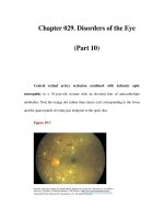
Figure 13.1 This woman had been blind for at least
two years when she came to an eye clinic in Beletwein,
Somalia. She had travelled for more 200 km in order
to have cataract surgery. Her situation is typical of the
millions who are blind from cataract today.
Global situation
Africa
Africa has the highest prevalence of blindness
in the world, estimated by WHO to be
approximately 1%. Half of this is due to
cataract. Africa also has the fewest resources
with which to combat blindness. There is, on
average, only one ophthalmologist for one
million people. Although simple cataract surgery
may cost only $30 per procedure, this is more
than ten times the annual per capita health
budget of many African countries. The CSR is
100–500 in most African countries.
Asia
The prevalence of blindness in Asia is 0·75%,
of which about two thirds is due to cataract.
Most Asian countries are better equipped to deal
with the problems of cataract blindness, having
approximately one ophthalmologist per 100 000
people. However, these resources are at risk of
being overwhelmed by the sheer scale of the
problem. There are now about 3 500 000
cataract operations performed annually in India
alone, representing a CSR of about 3 500.
Unfortunately, this has not yet eliminated the
backlog of cataract blind patients. In China it is
difficult to obtain accurate figures, but it appears
that no more than 250 000 operations are
carried out each year for a population of more
than one billion, yielding a CSR of less than 300.
Latin America
Latin America has a relatively smaller
population, with a prevalence of blindness of
around 0·5%, of which about half is due
to cataract. There is no shortage of
ophthalmologists, but cataract blindness remains
a serious problem. Many ophthalmologists
practise in large towns and cities, where services
are of a high standard. However, these services
are inaccessible to rural people and urban slum
dwellers.
Barriers to cataract surgery
Modern cataract surgery is one of the most
successful medical interventions of all time. Why
is cataract still the world’s leading cause of
blindness? The explanation lies in the barriers
that prevent blind people from coming for
surgery. These can be divided into patient
related (i.e. motivation, mobility, and money)
and provider related factors (i.e. manpower,
materials, management, and marketing).
• Motivation. Patients who have a different
understanding of health and disease may be
reluctant to come for surgery because they do
not believe that cataract is a curable disease.
Cataract blindness may be regarded as a
normal part of ageing. Alternatively, they may
not believe the surgeon’s claims that surgery
will cure their disability.
• Mobility. Travel is difficult in developing
countries. For a blind person it is almost
impossible. In Africa many blind people live
over 100 km from the nearest eye surgeon.
Because cataract blind patients are relatively
immobile, they cannot reach eye clinics.
• Money. Many Third World countries now
require patients to pay for their treatment.
This constitutes a significant barrier for blind
patients, who are already impoverished
because of their disability.
• Manpower. A lack of trained personnel means
that many cataract patients never meet an eye
surgeon. Their condition may not be
recognised by a rural health worker who has
little ophthalmic expertise.
• Materials. Shortages of essential materials are
a recurrent problem for all types of health care
in the Third World. This has been addressed
by encouraging the local manufacture of
essential supplies such as sutures, eye drops,
glasses, and even intraocular lenses (IOLs).
• Management. Mismanagement and poor
marketing of scarce health care resources are
further problems. Resources are concentrated
CATARACT SURGERY
194
in the capital cities of most Third World
countries, although most blind people are
found elsewhere.
With the knowledge and techniques available to
us today, it should be possible to eliminate
cataract blindness. The failure to achieve this
suggests that the problem is not technical but
managerial. It has been suggested that
ophthalmologists might learn from the
MacDonald’s fast food outlets. If cataract
surgery was as universally available, as effectively
marketed, and as efficiently delivered as a “Big
Mac”, then the cataract backlog would rapidly
disappear.
5
Essential resources for cataract
surgery
Human resources
Innovative strategies have been devised to
overcome the lack of trained ophthalmic
personnel in most of the Third World,
particularly in Africa, where the deficit is most
severe.
In many African countries, non-physician
health workers have been trained to deliver basic
eye care, including the diagnosis and referral of
cataract patients. In east and southern Africa,
selected ophthalmic assistants have been trained
to perform cataract surgery. Prospective studies
have shown that, with uncomplicated senile
cataracts, non-physician cataract surgeons can
obtain excellent results.
6
Although training programmes are effective at
providing basic instruction for ophthalmologists
and cataract surgeons, human resources
development is ineffective unless it also includes
mechanisms for providing supervision, continuing
education, and adequate material resources. If
these are not incorporated, then the value of the
training is severely compromised.
At the village level, ordinary members of the
community, and traditional healers, have been
trained to identify blindness. These community
based field workers visit blind people and their
families, and encourage them to come for
surgery. Because those individuals are already
known to the patients, they are more effective at
communicating the benefits of cataract surgery
than are eye care professionals, who may have
no link to the patients’ own communities.
7
However, because the community perceives
blindness as a chronic disability associated with
ageing, rather than as an eye disease that can be
cured, patients may not come to an eye clinic,
which is perceived as treating eye diseases. Most
Third World eye surgeons have had the
experience of finding a patient, blind from
cataract for many years, living within a few
hundred metres of their clinic.
Material resources
Great efforts have been made during the past
two decades to develop simple and appropriate
solutions to overcome the lack of locally
manufactured ophthalmic surgical resources. In
Africa, for example, many centres now make
their own eye drops. It is possible for a small
pharmacy to produce 60 000 bottles of eye
drops per year, at an average cost of about $0·30
per bottle. This not only saves money but also
ensures a reliable supply of effective topical
medications.
8
High quality, single piece polymethyl-
methacrylate lenses are currently made in Eritrea,
Nepal, and India. They are sold for $7–10 each,
and have been found to be of a standard equivalent
to that of similar designs of lens manufactured in
industrialised countries. The availability of well
manufactured, inexpensive lens implants has had an
enormous impact on Third World cataract
surgery.
A lack of inpatient accommodation has been
addressed by “eye camps”, in which cataract
operations are performed outside the usual eye
hospital setting. Although conditions for surgery
are not ideal, eye camps provide cataract surgery
for patients who cannot get to a hospital
(Figure 13.2).
CATARACT SURGERY IN THE THIRD WORLD
195
Intraocular lenses
The use of IOLs in the Third World has been
controversial.
9–11
However, there is now
widespread agreement that IOLs represent the
best solution to cataract blindness in developing
countries.
12
Aphakic spectacles are safe and
inexpensive. Unfortunately, they are frequently
lost or broken. The distortion and magnification
associated with aphakic glasses also militate
against their use.
13
Cataract surgery with
aphakic glasses reduces the number of cataract
blind but increases the number blind from
uncorrected aphakia, leading to little change in
the overall prevalence of blindness.
14
When the other eye sees well, spectacle
correction of unilateral aphakia leads to
intolerable anisometropia, and aniseikonia, and
so surgery must be deferred until the patient has
bilateral visual impairment. With bilateral loss of
vision, travel becomes even harder. The patient’s
remaining savings will have been spent on food
and other essentials, so that there is nothing left
for luxuries such as medical care. The use of an
IOL makes it possible to intervene much earlier,
before the patient is blind in both eyes; this in
effect prevents cataract blindness, with all of its
associated human, social, and economic costs.
Surgical techniques
Intracapsular cataract extraction
and anterior chamber
intraocular lenses
Intracapsular cataract extraction (ICCE)
remains popular in parts of the Third World.
The surgery does not require complex
equipment or expensive irrigating fluids. The
use of loupes with four- to fivefold magnification
gives results that are comparable to those
obtained with an operating microscope.
However, ICCE is associated with serious
posterior segment complications, such as retinal
detachment. The larger incision required leads
to greater astigmatism and prolongs recovery. In
poor countries there are relatively few centres
that can manage aphakic detachments, and
astigmatic spectacle lenses are too expensive for
many people.
Early designs of anterior chamber lens
implants, particularly those with closed loop
haptics, were associated with unacceptably high
complication rates. This has given anterior
chamber IOLs a poor reputation in the
developed world. Recently, it has been shown
that open loop designs, with three or four point
fixation, have fewer complications.
15
The lack of
posterior capsule opacification following ICCE
and anterior chamber IOL implantation is a
distinct advantage in a Third World setting,
where follow up is limited and there are few
neodymivm : yttrium aluminium garnet
(Nd: YAG) lasers. A prospective study
conducted in Nepal has demonstrated the safety
and efficacy of this operation.
16
However, although modern designs of open
loop anterior chamber lenses are safer than their
predecessors, many surgeons are reluctant to use
them in young people for fear of long term
damage to the endothelium and trabecular
meshwork. Moreover, so long as anterior
chamber IOLs are not regarded as the optimum
treatment for aphakia in developed nations, they
will not be received enthusiastically in the Third
World.
CATARACT SURGERY
196
Figure 13.2 A non-physician cataract surgeon
operating in a refugee camp in Kenya. The operating
theatre is a wooden hut, with a corrugated iron roof.
More than 600 successful cataract operations have
been performed here since 1992. The operating
microscope weighs less than 20 kg and can be carried
in a suitcase.
Extracapsular cataract extraction and
posterior chamber intraocular lenses
Uncomplicated extracapsular cataract
extraction (ECCE) carries a much lower risk of
posterior segment complications. However,
there is a significant risk of posterior capsule
opacification. This can easily be treated with a
Nd:YAG laser, but these lasers are expensive and
are not available in most Third World eye
clinics. This is important in developing
countries. It can be difficult for a blind person to
travel once to an eye clinic for surgery. To make
the journey twice may be impossible.
The risk of posterior capsule opacification can
be minimised by good surgical technique, and
by the IOL material and design.
17
Most patients
presenting for surgery in the Third World have
mature cataracts, and the risk of capsule opacity
may be lower in these eyes.
18
Furthermore,
although capsule opacification may occur, it
rarely reduces vision to below 6/60, following
uncomplicated extraction of a senile cataract.
If the capsule does become opaque, then in
the absence of a Nd:YAG laser a surgical
capsulotomy can be performed through the
pars plana.
To obtain good results with extracapsular
surgery, an operating microscope is essential.
Until recently these have been prohibitively
expensive for most eye clinics in poor countries.
It is now possible to obtain a good quality coaxial
microscope, which can be packed in a suitcase
and taken to outlying clinics, for around $3000.
Despite the risk of posterior capsule opacity,
the use of ECCE, with a posterior chamber IOL,
is increasing in Third World countries. The
advent of low cost coaxial microscopes,
inexpensive IOLs, and a desire to achieve the
same standard of care as in developed countries
have all played a role in this trend.
Phacoemulsification and
small incision surgery
Phacoemulsification equipment is costly,
complex, and difficult to maintain. Because
many patients do not present until they are
completely blind, a high proportion of Third
World cataracts are mature or hypermature and
are less amenable to phacoemulsification.
However, small incision surgery offers real
advantages for developing countries. The small
incision causes less inflammation and leaves a
strong eye. Visual rehabilitation is faster, and
there is minimal induced astigmatism. This
means that follow up beyond the immediate
postoperative period is not essential, which is
even more desirable in the Third World than in
an industrialised country.
Unfortunately, foldable IOLs remain too
expensive for most patients in the Third World.
This will change, and there will be intense efforts
to develop safe and reliable methods of removing
the nucleus through a small incision without the
cost or complexity of phacoemulsification.
Cataract surgical outcomes
Although hospital based studies have shown
excellent results from both ICCE and anterior
chamber IOL,
16
and ECCE and posterior
chamber IOL,
19,20
studies in the community
suggest that too many patients have a poor
outcome,
21,22
with as many as 40% of operated
eyes having an acuity of less than 6/60.
21
The
main reasons for the poor outcome are pre-
existing eye disease, complications of surgery,
and uncorrected refractive error. Although the
use of IOLs will reduce the latter, it will not
affect the other causes.
The same studies have shown that quality of
life and visual function measurements are closely
correlated with postoperative visual acuity.
21
If
patients have a poor outcome, it will have an
adverse effect on their quality of life. This will in
turn affect the community’s perception of the
effectiveness of cataract surgery, reducing
demand and raising the barriers to surgery.
The WHO has recently suggested that at least
90% of operated cataract eyes should have a best
corrected acuity of 6/18 or better, and that fewer
than 5% should be worse than 6/60.
23
These
CATARACT SURGERY IN THE THIRD WORLD
197
targets are low compared with expected
outcomes in wealthy countries, but are
ambitious for most Third World eye clinics.
Whether or not the WHO targets are achieved, it
is essential for cataract surgeons to monitor their
outcomes as well as their output, and to set goals
for regular quality control and continuous
improvement.
The aim of outcome monitoring is not
primarily to compare one clinic or surgeon with
another, but to assist all surgeons to identify why
they have poor outcomes and to take the
necessary corrective measures. This will lead to
improved outcomes for all patients.
Cost of surgery
Cataract extraction is thought to be one of the
most cost effective interventions in modern
medicine.
24
However, the communities in
greatest need of surgery are also the least able to
pay for it.
The cost of cataract surgery can be divided
into the cost of consumables (such as the IOL,
drugs, and sutures) and fixed costs (salaries,
depreciation, etc.). The cost of consumables can
be minimised by bulk purchase from suppliers in
Third World countries. However, it is unlikely to
be less than $20–$25 per operation. Fixed costs
remain the same whether the clinic does 10
operations or 100. The best way of minimising
the fixed cost per operation is to increase the
number of operations. If a clinic does 500
operations per year, then the cost per operation
is $20 + (total fixed costs/500). If the clinic
works more efficiently, and doubles its output,
then the cost per operation will be $20 + (total
fixed costs/1000).
Ideally, a clinic should aim to achieve self-
sufficiency, from generating sufficient income
from patient fees and sale of glasses, among
other sources, to cover all their costs. The only
way this can be accomplished in a Third World
situation is to have tiered pricing. Poor patients,
who may have been blind for years, must be
treated for free. Other patients can only pay a
small proportion of the total cost of surgery.
Others can pay the full cost. A minority will be
willing to pay more than the true cost of surgery
if they receive preferential treatment, for
example a private or air conditioned room. This
approach has been very successful in some
hospitals in Nepal and India.
The future
The problem of cataract blindness in the
Third World is so large that there is no single
simple answer. Different circumstances will
require different solutions. In all situations the
quality of the surgery and of the overall patient
care will influence outcome more than variations
in the type of operation.
In training surgeons for developing countries,
the ideal is probably “complete eye surgeons”,
who are equally at home performing high volume
surgery in an eye camp and small incision surgery
at the base hospital. However, in addition to
having technical proficiency, Third World eye
surgeons must be aware that the patients on
whom they operate represent only a fraction of
those in need. The surgeon’s objective should be
to increase the numbers of sight restoring
operations by minimising the barriers that
prevent people from obtaining surgery. This can
be accomplished by actively involving local
communities in the elimination of cataract and
by providing high quality surgery with a good
visual outcome at an affordable price.
References
1 World Health Organisation. The World Health Report. Life
in the 21st century: a vision for all. Geneva: World Health
Organisation, 1998.
2 World Health Organisation. Vision 2020, the global
initiative for the elimination of avoidable blindness. Geneva:
World Health Organisation, 1999.
3 Foster A. Cataract: a global perspective: output, outcome
and outlay. Eye 1999;13:449–53.
4 Shamanna BR, Dandona L, Rao GN. Economic burden
of blindness in India. Indian J Ophthalmol 1998;46:
169–72.
5 Venkataswamy G. Can cataract surgery be marketed like
hamburgers in developing countries? Arch Ophthalmol
1993;111:580.
CATARACT SURGERY
198
6 Foster A. Who will operate on Africa’s 3 million curably
blind people? Lancet 1991;337:1267–9.
7 Yorston D. Accessible eye care: primary health care and
community-based rehabilitation. In: Proceedings of the
Fifth General Assembly. International Agency for
Prevention of Blindness, 1994.
8 Taylor J. Appropriate methods and resources for third
world ophthalmology. In: Tasman W, Jaeger EA, eds.
Duane’s clinical ophthalmology, vol 5. Hagerstown:
Lippincott, 1984.
9 Taylor HR, Sommer A. Cataract surgery. A global
perspective [editorial] Arch Ophthalmol 1990;108:
797–8.
10 World Health Organisation. Use of intraocular lenses in
cataract surgery in developing countries: memorandum
from a WHO meeting. Bull World Health Organ
1991;69:657–66.
11 Young PW, Schwab L. Intraocular lens implantation in
developing countries: an ophthalmic surgical dilemma.
Ophthalmic Surg 1989;20:241–4.
12 Yorston D. Are intraocular lenses the solution to cataract
blindness in Africa? Br J Ophthalmol 1998;82:469–71.
13 Hogeweg M, Sapkota YD, Foster A. Acceptability of
aphakic correction. Results from Karnali eye camps in
Nepal. Acta Ophthalmol 1992;70:407–12.
14 Cook CD, Stulting AA. Impact of a sight-saver clinic on
the prevalence of blindness in northern KwaZulu. S Afr
Med J 1995;85:28–9.
15 Auffarth GU, Wesendahl TA, Brown SJ, Apple DJ. Are
there acceptable anterior chamber intraocular lenses for
clinical use in the 1990’s? Ophthalmology 1994;101:
1913–22.
16 Hennig A, Evans JR, Pradhan D, et al. Randomised
controlled trial of anterior chamber intra-ocular lenses.
Lancet 1997;349:1129–33.
17 Spalton DJ. Posterior capsular opacification after
cataract surgery. Eye 1999;13:489–92.
18 Argento C, Nunez E, Wainsztein R. Incidence of post-
operative posterior capsular opacification with types of
senile cataracts. J Cataract Refract Surg 1992;18:586–8.
19 Yorston D, Foster A. Outcome of ECCE & PC-IOL in
adults in E. Africa. Br J Ophthalmol 1999;83:897–901.
20 Prajna NV, Chandrakanth KS, Kim R, et al. The
Madurai Intraocular Lens Study II: clinical outcomes.
Am J Ophthalmol 1998;125:14–25.
21 Zhao J, Sui R, Jia L, Fletcher AE, Ellwein LB. Visual
acuity and quality of life outcomes in patients with
cataract in Shunyi county, China. Am J Ophthalmol
1998;126:582–5.
22 Limburg H, Foster A, Vaidyanathan K, Murthy GVS.
Monitoring visual outcome of cataract surgery: results
from India. Bull World Health Organ (in press).
23 World Health Organisation. Informal consultation on
analysis of blindness prevention outcomes. Geneva: World
Health Organisation WHO/PBL/98⋅68, 1998.
24 Marseille E. Cost-effectiveness of cataract surgery in a
public health eye care programme in Nepal. Bull World
Health Organ 1996;74:319–24.
CATARACT SURGERY IN THE THIRD WORLD
199
200
When Kelman
1
introduced phacoemulsification
over 30 years ago, he revolutionised cataract
surgery not only by introducing small incision
surgery but also by spurring the development of
new lens technology, namely the foldable
intraocular lens (IOL). The results of these new
developments have greatly improved patient
outcomes by decreasing induced astigmatism
and decreasing wound complications, and thus
enabling quicker rehabilitation.
2
However, this
technique is not without its problems. Issues of
safety related to the release of excess energy at
the probe tip, and the consequent effects on
non-target tissues such as the iris, cornea, and
posterior capsule remain a concern. The
excessive heat generated around the phaco tip
mandate that a sleeve be present to provide a
water bath to prevent subsequent corneal burns
and wound distortion. Until recently this has
limited the incision size to between 2·2 and 3·2
mm (see chapter 4). Thus, there is a drive to
study and develop newer and better technologies
to circumvent these problems. Other techniques
that are currently under investigation include the
use of lasers, warm water jet technology (to melt
the lens), and mechanical instruments such as
Catarex and phacotmesis. The Catarex machine
uses a small impellar to break up the lens,
whereas phacotmesis involves a spinning needle.
Smaller incisions require new solutions to lens
implantation. Development has been directed
toward capsular filling techniques, which may also
provide the answer to restoring accommodation
following surgery.
Lasers for cataract removal
Evolution
In 1975 Kasnov
3
reported the technique of
laser phacopuncture, the first laser procedure for
cataract removal. With a Q switched ruby laser,
microperforations were made in the anterior
capsule, thus enabling gradual reabsorption of
the lens material over time. This technique had
very limited applications because it was only
effective for very soft cataracts. There was also
the problem of induced uveitis. In the ensuing
years, focus was shifted toward four ultraviolet
wavelengths: 193 nm (argon fluoride), 248 nm
(krypton fluoride), 308 nm (xenon chloride),
and 351 nm (xenon fluoride).
4–6
Of these, the
308 nm excimer laser appeared most promising
because of both efficacy of ablation and
transmissibility through fibreoptics.
4–6
However,
the cataractogenic effects of the 308 nm laser
posed a threat to the eyes of the surgeon,
7–9
and
questions of possible retinal toxicity and
carcinogenic effects arose.
7,8,10
Attention was
then redirected toward the infrared wavelengths,
namely the erbium : yttrium aluminium garnet
(Er:YAG)
11–14
and the neodymium : yttrium
aluminium garnet (Nd:YAG)
15–17
lasers.
In 1980, Aron-Rosa and others reported the
use of the Nd:YAG (pulsed 1064 nm) laser for
performing posterior capsulotomy,
18–20
peripheral iridotomy,
20–22
and cutting of
pupillary membranes.
20,21,23
This then evolved
into the next stage in the use of lasers for cataract
removal, namely laser anterior capsulotomy
14 Cataract surgery: the next
frontier
before cataract extraction.
24
This technique
never gained widespread acceptance because of
problems of intraocular pressure rise,
inflammation, and poor mydriasis at the time of
surgery, and the need to perform surgery
promptly after the laser treatment.
25,26
The next procedure to come along in this
evolution was laser photofragmentation,
27–31
which involved the use of the Nd:YAG laser to
photodisrupt the lens nucleus before
phacoemulsification. By firing the laser into
the substance of the lens nucleus while leaving
the anterior and posterior capsules intact, the
nucleus is softened, thus making subsequent
phacoemulsification easier. Although several
studies did demonstrate less phaco time and
power needed in those cases pretreated with
laser, this procedure does carry the risk of
inadvertent perforations of the anterior/posterior
capsules and potential increase in intraoperative
complications. This also had the inconvenience
of a two staged procedure.
Nd:YAG laser systems
Dodick photolysis (ARC Lasers;
Figure 14.1)
Since the early 1990s, Dodick has been
studying the use of the Q switched, pulsed 1064
nm Nd:YAG laser for one stage, direct
photolysis of cataractous lenses.
15
The probe,
similar to a standard irrigation and aspiration
hand piece, consists of an irrigation and
aspiration port chamber, which contains a 300
µm quartz clad fibre. The proximal portion of
the 300 µm fibre is attached via a standard laser
connector to the laser source. The fibre enters
the probe through the infusion cannula and
terminates approximately 2 mm in front of a
titanium target inside the probe tip. The pulsed
laser energy is transmitted via the quartz fibre
and is focused on the titanium target, thus
enabling optical breakdown and plasma
formation to occur at very low energy levels.
This in turn causes the emanation of shock
waves, which propagate within the aspiration
chamber toward the mouth of the probe, where
the nuclear material is held in place by the
suction created by the aspiration port. The
shock waves disrupt the nuclear material and the
fragments are aspirated.
15,32
The titanium target is the key element of this
device because the metal target, with its low
ionization potentials, acts as a transducer in
converting light energy to shock waves at low
laser energy levels. Because there is no direct
contact between the laser energy and the target
tissues, the shock waves generated here are more
controlled, so that only the area in contact with
the tip of the device is disrupted. In effect, the
titanium target shields the non-target tissues
such as the endothelium and the retina, as well
as the surgeon’s eyes, from direct laser light.
33,34
The quartz clad fibre and the titanium targets
are relatively inexpensive, making disposable
CATARACT SURGERY: THE NEXT FRONTIER
201
Figure 14.1 Dodick laser photolysis unit.
hand pieces a possibility. The same tip may be
used for irrigation and aspiration.
Photon (Paradigm Medical Industries)
This is a Nd:YAG system that is partnered
with the manufacturer’s conventional ultrasonic
phaco system. The probe consists of a titanium
tip with a fused silica fibre. It currently has a
repetition rate of 10–50 Hz, which will
eventually be increased to above 50 Hz to
increase its ability to fragment tissue. Its fluidics
system also allows for surge control at all
vacuum levels up to 500 mmHg. It is a uni-
manual unit in which the irrigation and
aspiration system is incorporated into the laser
probe. The probe has a tip diameter ranging
from 1·2 to 1·7 mm, and passes through a
3·0–3·5 mm incision. The unit uses a peristaltic
system with up to 500 mmHg vacuum. The
company has completed phase I US Food and
Drug Administration trials and is currently in
phase II trials, which are being conducted at
seven clinical sites across the USA. To date, over
100 procedures have been performed using this
system, and the results demonstrate quieter eyes
on postoperative day one compared with
ultrasound phaco cases. The reported endothelial
cell loss is 7·6% at 3 months of follow up for all
sites.
Er:YAG laser systems
Another laser currently being developed for
cataract removal is the Er:YAG system.
11–14
Er:YAG emits energy in the mid-infrared region
(2940 nm), and may be transmitted through a
150 µm fibreoptic probe.
13
One advantage of the
erbium system is that the 2940 nm wavelength
corresponds to the maximum peak of water
absorption. This translates into low penetration
(~1 mm), with excess energy absorbed by water
without dispersion to surrounding non-target
tissues. The laser is focused directly into the lens
nucleus to create an optical breakdown in the
nucleus, leading to microfractures of the lens
without heat generation. Fragmentation rate per
pulse is related not only to pulse energy but also
to the repetition frequency. With high pulse
frequency, longitudinal chains of cavitation
bubbles form at the probe tip. Depending on the
pulse energy, these bubbles may extend up to
3 mm or more in water and up to 1 mm in nuclear
material. Because the bubbles allow the laser
energy to travel further than the penetration
depth of the laser radiation (energy travelling
through bubbles rather than absorbed by water),
they facilitate the fragmentation of denser
nuclei. However, this also increases the risk to
damage of adjacent structures (i.e. the posterior
capsule).
There are three companies currently
developing the Er:YAG laser for cataract
removal. All systems presently available use a
conventional irrigation and aspiration system to
remove tissue and debris from the capsular bag.
In addition, because the laser is focused directly
into the lens and not onto a metal target, there is
some exposure of the patient’s and surgeon’s
eyes to direct laser light.
A number of systems are under trial,
including the following:
• Phacolase (Aesculap-Meditec)
• Centauri (EyeSys-Premier)
• Adagio (WaveLight).
Advantages of laser cataract removal
Currently, several laser systems are available
in Europe, while clinical trials continue in the
USA. Although laser is unlikely to replace
ultrasound phaco systems in the near future,
laser phaco systems do have several advantages
over ultrasound systems. Because the laser
probes produce no clinically significant heat,
there is no risk of corneal and scleral burns.
Studies have demonstrated that after 30 seconds
of continual use in standard conditions, a
CATARACT SURGERY
202
temperature increase of 2·6°C was noted with a
laser probe, as compared with an increase of
30°C with an ultrasound probe. Furthermore,
the water temperature in a 2·5 cc closed
chamber increased by 1°C with a laser probe
versus 9·5°C with an ultrasound probe. The
minimal heat generated by the laser probes
eliminates the need for a water bath around the
probe, thus enabling the separation of irrigation
from laser/aspiration, thereby reducing probe
and incision size (Figure 14.2).
Unlike ultrasound phaco hand pieces, the
laser probes do not house motors and do not
require electrical voltage to drive vibrating
needles, both of which are subject to wear and
tear. In addition to being lighter and easier to
handle, the components of the laser probes are
relatively cheap, thus making disposable hand
pieces a possibility (Figure 14.3).
A notable problem with the current laser
systems is that dense nuclei still present a
challenge. One can expect that with further
refinements in fluidics and laser parameters, this
problem will be overcome in the near future.
New lens technology
Just as the introduction of ultrasound
phacoemulsification spurred the development of
foldable IOLs, laser phaco systems have already
brought about revolutions in lens technology. In
July 1999, the first case of IOL insertion through
a 1·8 mm incision was reported by Kanellopoulos
in Greece.
35
The new lens was developed by
Dr Christine Kreiner, of Acritec (Berlin,
Germany). The acrylic IOL has a 6 mm optic, is
12·5 mm in total length, and was prefolded by
27% dehydration. The folded lens has a width of
1·2–1·3 mm and can be implanted through an
incision of less than 2 mm. Once in the capsular
bag, the lens slowly unfolds over 25–30 minutes.
In the future, we can look forward to the next
generation of IOLs to be made of injectable
substances such as silicone, hydrogel, or
collagen that could be used to refill the capsular
bag through the same small opening that is used
to evacuate the cataract. This would facilitate
true endocapsular surgery and enable us to
preserve accommodation.
Accommodative lens technology
In addition to the restoration of
accommodation, the goals of this lens
technology comprise the following:
• A small incision/capsulorhexis
• Injection of a biocompatible material with
appropriate refractive indices/transparency/
elasticity
• Control of posterior capsule opacification.
CATARACT SURGERY: THE NEXT FRONTIER
203
Figure 14.2 Bimanual laser photolysis procedure.
Probe on right delivers infusion. Probe on left delivers
laser and aspiration.
Figure 14.3 Laser photolysis probe: a lightweight
disposable probe made of injection molded plastic.
In the 1960s, Kessler
36
and Agarwal et al.
37
were among the first to report lens refilling with
silicone oil in rabbits. However, silicone oil
leakage from the capsular bag was noted to be a
major problem in these cases.
Use of polymeric gels
In the 1980s, in order to overcome the
problem of leakage, several investigators
attempted to refill lens capsules using precured
silicone polymeric gels, commonly known as
silicone elastomers or silicone rubbers.
38–40
Haefliger et al.
40
found evidence to suggest that
the injected silicone gel undergoes accommodative
change when the ciliary body is stimulated by
pilocarpine. Although the actual amplitude of
accommodation could not be directly measured
because of posterior capsule opacification,
observations such as the forward movement and
increase in curvature of the anterior lens capsule,
and the decrease in anterior chamber depth by
an amount comparable to that of the natural lens
suggested preservation of accommodation in
these eyes. While the use of a precured silicone
gel simplifies the surgical procedure, it is difficult
to prepare a gel with viscosity that is high
enough to prevent leakage and low enough to
enable injection through a small needle. An
alternative method would be to inject liquid
silicone that polymerises in situ. This approach
would, in turn, require some means of sealing
the capsular opening.
The endocapsular balloon
In the late 1980s, Nishi and coworkers
41,42
developed an inflatable endocapsular balloon for
the purpose of lens refilling. In this technique,
lens extraction is performed via a 1·2–1·5 mm
minicapsulorhexis. The capsular bag is then
treated with ethylenediaminetetraacetic acid
(EDTA) to remove any lens epithelial cells
(LECs). The deflated balloon is inserted into the
empty capsular bag, and a mixture of two liquid
silicone polymers is injected into the balloon.
The liquid silicone polymerises in situ in 2
hours, and the resultant inflated balloon fills the
capsular bag. Problems encountered with this
technique included fibrin deposition and
capsular fibrosis. It was also observed that the
postoperative amplitude of accommodation was
a small fraction of that present preoperatively,
and that the amplitude subsequently decreased
over time. A possible explanation for this finding
was that the capsular tension was not effectively
transmitted to the balloon because of discrepancies
in size, shape, and other physical properties.
Progressive capsular fibrosis, which decreases
capsular pliability, may in turn account for the
gradual decrease in postoperative accommodative
amplitude. These studies also demonstrated that
the final shape and the degree of filling of these
endocapsular balloons are important factors in
determining not only the transmission of
capsular tension but also the amplitude of
accommodation. In studies on young monkey
eyes, Nishi et al. reported a mean accommodation
amplitude of 4·6 ± 0·5 diopters (D). Using the
same technique several years later, Sakka et al.
43
reported an average accommodative change of
6·74 D after instillation of 4% pilocarpine.
Endocapsular polymerisation
An alternative approach to injecting liquid
polymers into an endocapsular balloon is to
inject the materials directly into the capsular bag
and allow endocapsular polymerisation. This
eliminates the need for a containment device
such as a balloon. However, such a process may
be associated with endogenous heat production,
which may secondarily affect the lens capsule
and zonules. Hettlich et al.
44
examined this issue
with studies on an acrylate copolymer, and
found that the maximal temperature recorded
was at the posterior capsule (45·1°C) for several
seconds. This temperature rise was not believed
to be significant in causing damage to the
capsular collagen. This study also confirmed an
earlier observation reported by Haefliger et al.
40
that the degree of capsular refilling is inversely
related to the degree of posterior capsule
opacification.
CATARACT SURGERY
204
Direct capsular filling: capsular plug
Of the materials studied to date, silicone
compounds appear to the best currently available
substances for lens refilling because of their
transparency, biocompatibility, refractive index,
and elasticity. After studies on the endocapsular
balloon, Nishi and coworkers
45–51
went on to
develop a direct lens refilling technique, in which
a silicone plug is used to seal the
minicapsulorhexis of 1·2–1·5 mm. The plug
consists of a silicone double plate through which
a thin delivery tube provides access to the
capsular bag. Two liquid silicone polymers are
injected via the delivery tube, which is then cut at
its root. A soft silicone gel is then used to fill the
remaining tube stump, thus preventing reflux.
The injected mixture then polymerises in the
capsular bag within 2 hours. Problems
encountered with this technique include capsular
tears during plug insertion into the capsular bag.
In some cases, mild leakage occurred at the
capsular opening during silicone injection
because of difficulty in maintaining the stability
of the plug–syringe connection. In instances of
leakage, it was noted that the liquid silicone
remained cohesive in aqueous because of its
hydrophobic nature, and that no adhesions to
adjacent tissues occurred. Silicone leakage
occurring at the time of injection was washed out
of the anterior chamber with ease, and that which
had polymerised (detected after surgery) was
removed surgically without difficulty on
postoperative day one. In a series conducted by
Nishi and Nishi,
47
postoperative accommodative
amplitude ranged from 1 to 4·5 D with mean of
2·3 ± 1·3 D (while preoperative accommodative
amplitude ranged from 5·75 to 11·25 D with
mean of 8·0 ± 2·0 D). This demonstrated that
refilling the lens capsule was feasible, although
the postoperative amplitude of accommodation
was only a fraction of that present before surgery.
Determining lens power
Postoperative refraction using the lens
refilling technique is determined by two main
factors: the refractive index of the injected
material and the anterior capsular curvature,
which is determined by the degree of capsular
filling. The greater the degree of capsular filling,
the steeper the anterior capsular curvature
and the greater the power of the implant. However,
the degree of capsular filling also determines the
amplitude of accommodation. At low volumes,
the accommodation amplitude increases as the
degree of capsular refilling increases. The
accommodative amplitude then reaches a
maximum value, after which any further increase
in volume results in a decrease in accommodative
amplitude. Nishi and coworkers
45–47
observed
that the optimal accommodation amplitude with
silicone polymers is achieved by filling the
capsular bag to 60–70% capacity. However,
postoperative emmetropia is not achieved when
the capsule is under-filled to this extent, and
optimal accommodation amplitude is attained at
the expense of refractive outcome. In Nishi’s
series, with capsule filling of 60–70%, an average
accommodation amplitude of 2·3 ± 1·3 D was
achieved, but with a mean hyperopic shift of
+6·4 D in postoperative refraction. This
hyperopic shift is probably due to the relatively
low refractive index (1·405) of injected silicone,
as well as a flatter anterior capsule curvature
secondary to the under-filling. Methods of
correcting the residual refractive error may
include corneal refractive surgery or IOL
implantation. The ideal solution would, of
course, be the development of new injectable
materials with different refractive indices that
would enable the achievement of emmetropia
along with optimal accommodation amplitude.
Posterior capsule opacification
A common finding among all studies,
regardless of the technique of lens refilling, is
posterior capsule opacification in the early
postoperative period. Haefliger et al.
40
noted
proliferation of LECs on the posterior capsule
as early as two weeks postoperatively, with the
equatorial region being the most prominent. In
several other series,
40,47
posterior capsule
CATARACT SURGERY: THE NEXT FRONTIER
205
opacification precluded refraction at three
months. Histopathological studies demonstrated
a thick layer of LECs that had migrated
posteriorly between the lens capsule and the
injected silicone. In Nishi’s series,
46
YAG laser
capsulotomy was performed. Although no
silicone leakage or herniation was noted with the
procedure, it may negate the accommodation
attained previously. To date, various methods of
removing LECs have been reported. Nishi and
coworkers studied the use of ultrasound
aspiration,
48
as well as the use of a high
concentration of a proteolytic enzyme,
49,50
to
loosen LECs at their junction complexes.
Humphrey et al.
51
had described the use of
EDTA and trypsin for the removal of LECs.
Thus far, no method has proven ideal. The
problem of posterior capsule opacification must
be solved before any lens refilling technology
may be introduced clinically. Nevertheless, with
further advances in research and development of
new products, the 21st century promises to be a
very exciting era for cataract surgery.
References
1 Kelman CD. Phaco-emulsification and aspiration: a
new technique of cataract removal, a preliminary report.
Am J Ophthalmol 1967;64:23–35.
2 Leaming DV. Practice styles and preferences of ASCRS
members: 1987 survey. J Cataract Refract Surg 1988;14:
552–9.
3 Krasnov MM. Laser phakopuncture in the treatment of
soft cataracts. Br J Ophthalmol 1975;56:96–8.
4 Maguen E, Martinez M, Grundfest W, et al. Excimer
laser ablation of the human lens at 308 nm with a fiber
delivery system. J Cataract Refract Surg 1989;15:
409–14.
5 Nanevicz T, Prince MR, Gawande AA, et al. Excimer
laser ablation of the lens. Arch Ophthalmol 1986;104:
1825–9.
6 Puliafito CA, Steinert RF, Deutsch TF, et al. Excimer
laser ablation of the cornea and lens: experimental
studies. Ophthalmology 1985;92:741–8.
7 Marshall J, Sliney DH. Endoexcimer laser intraocular
ablative photodecomposition [letter]. Am J Ophthalmol
1986;101:130–1.
8 Zuclich JA. Ultraviolet-induced photochemical damage
in ocular tissues. Health Phys 1989;56:671–82.
9 Borkman RF. Cataracts and photochemical damage in
the lens. Ciba Found Symp 1984;106:88–109.
10 Kochevar IE. Cytotoxicity and mutagenicity of excimer
laser radiation. Lasers Surg Med 1989;9:440–5.
11 Colvard DM. Erbium:YAG laser removal of cataracts.
Presented at the American Society of Cataract and
Refractive Surgery Annual Meeting, Seattle, May 1993.
12 Margolis TI, Farnath DA, Destro M, Puliafito CA.
Erbium-YAG laser surgery on experimental vitreous
membranes. Arch Ophthalmol 1989;107:424–8.
13 Peyman GA, Katoh N. Effects of an erbium:YAG laser
in ocular ablation. Int Ophthalmol 1987;10:245–53.
14 Tsubota K. Application of erbium:YAG laser in ocular
ablation. Ophthalmologica 1990;200:117–22.
15 Dodick JM. Laser phacolysis of the human cataractous
lens. Dev Ophthalmol 1991;22:58–64.
16 Dodick JM, Sperber LTD, Lally JM, Kazlas M.
Neodymium-YAG laser phacolysis of the human
cataractous lens. Arch Ophthalmol 1993;111:903–4.
17 Dodick JM, Christiansen J. Experimental studies on the
development and propagation of shock waves created by
the interaction of short Nd:YAG laser pulses with a
titanium target: possible implication for Nd:YAG laser
phacolysis of the cataractous human lens. J Cataract
Refract Surg 1991;17:794–7.
18 Aron-Rosa D, Aron J, Griesemann M, et al. Use of the
neodymium:YAG laser to open the posterior capsule
after lens implant surgery: a preliminary report. Am
Intraocul Implant Soc J 1980;6:352–4.
19 Dodick JM. Nd:YAG laser treatment of the posterior
capsule. Trans New Orleans Acad Ophthalmol 1988;
169–78.
20 Fankhauser F, Roussel P, Steffen J, et al. Clinical studies
on the efficiency of high power laser radiation upon
some structures of the anterior segment of the eye: first
experiences of the treatment of some pathological
conditions of the anterior segment of the human eye by
means of a Q-switched laser system. Int Ophthalmol Clin
1981;3:129–39.
21 Fankhauser F. The Q-switched laser: principles and
clinical results. In: Trokel SL, ed. YAG laser ophthalmic
microsurgery. Norwalk, CT: Appleton-Century-Crofts,
1983.
22 Klapper RM. Q-switched neodymium:YAG laser
iridotomy. Ophthalmology 1984;91:1017–21.
23 Fankhauser F, Rol P. Microsurgery with the Nd:YAG
laser: an overview. Int Ophthalmol Clin 1985;25:55–8.
24 Aron-Rosa D. Use of a pulsed neodymium-YAG laser
for anterior capsulotomy before extracapsular cataract
extraction. J Am Intraocul Implant Soc 1981;7:332–3.
25 Aron-Rosa DS, Aron JJ, Cohn HC. Use of a pulsed
picosecond Nd:YAG laser in 6,654 cases. Am Intra-
Ocular Implant Soc J 1984;10:35–9.
26 Chambless WS. Neodymium:YAG laser anterior
capsulotomy and a possible new application. J Am
Intraocul Implant Soc 1985;11:33–4.
27 Chambless WS. Neodymium:YAG laser phacofracture:
an aid to phacoemulsification. J Cataract Refract Surg
1988;14:180–1.
28 L’Esperance FA Jr. Ophthalmic lasers, vol. 2, ed 3. St.
Louis: CV Mosby, 1989.
29 Levin ML, Wyatt KD. Prospective analysis of laser
photophacofragmentation. J Cataract Refract Surg
1990;16:96–8.
30 Ryan EH Jr, Logani S. Nd:YAG laser photodisruption
of the lens nucleus before phacoemulsification. Am J
Ophthalmol 1987;104:382–6.
31 Zelman J. Photophaco fragmentation. J Cataract Refract
Surg 1987;13:287–9.
CATARACT SURGERY
206
32 Dodick JM. Can cataracts be removed using laser
technology? Ophthalmol Clin N Am 1991;4:355–64.
33 Dodick JM, Lally JM, Sperber LTD. Lasers in cataract
surgery. Curr Opin Ophthalmol 1993;4:107–9.
34 Dodick JM, Sperber LTD. The future of cataract
surgery. Int Ophthalmol Clin 1994;34:201–10.
35 Charters L. Two-mm incision barrier is broken in
Greece. Ophthalmology Times July 1, 1999;1:24.
36 Kessler J. Experiments in refilling the lens. Arch
Ophthalmol 1964;71:412–7.
37 Agarwal LP, Narsimhan EC, Mohan M. Experimental
lens refilling. Orient Arch Ophthalmol 1967;5:205–12.
38 Gindi JJ, Wan WL, Schanzlin DJ. Endocapsular cataract
surgery, I. Cataract 1985;2:6–10.
39 Parel J-M, Gelender H, Trefers WF, Norton EW.
Phaco-ersatz: cataract surgery designed to preserve
accommodation. Graefes Arch Clin Exp Ophthalmol
1986;224:165–73.
40 Haefliger E, Parel J, Fantes F, et al. Accommodation of
an endocapsular silicone lens (Phaco-Ersatz in the
non-human primate. Ophthalmology 1987;94:471–7.
41 Nishi O, Hara T, Hara T, et al. Refilling the lens with
an inflatable endocapsular balloon: surgical procedure
in animal eyes. Graefes Arch Clin Exp Ophthalmol
1992;230:47–55.
42 Nishi O, Nakai Y, Yamada Y, Mizumoto Y. Amplitudes
of accommodation of primate lenses filled with two
types of inflatable endocapsular balloons. Arch
Ophthalmol 1993;111:1677–84.
43 Sakka Y, Hara T, Yamada Y, Hara T, Hayashi F.
Accommodation in primate eyes after implantation of
refilled endocapsular balloon. Am J Ophthalmol 1996:
121:210–2.
44 Hettlich H, Lucke K, Asiyo-Vogel MN, Schulte M,
Vogel A. Lens refilling and endocapsular polymerization
of an injectable intraocular lens: in vitro and in vivo
study of potential risks and benefits. J Cataract Refract
Surg 1994;20:115–23.
45 Nishi O, Nishi K, Mano C, Ichihara M, Honda T.
Controlling the capsular shape in lens refilling. Arch
Ophthalmol 1997;115:507–10.
46 Nishi O, Nishi K, Mano C, Ichihara M, Honda T. Lens
refilling with injectable silicone in rabbit eyes. J Cataract
Refract Surg 1998;24:975–82.
47 Nishi O, Nishi K. Accommodation amplitude after lens
refilling with injectable silicone by sealing the capsule
with a plug in primates. Arch Ophthalmol 1998;116:
1358–61.
48 Nishi O. Removal of lens epithelial cells by ultrasound
in endocapsular cataract surgery. Ophthalmic Surg
1987;18:577–580.
49 Nishi O, Nishi K. A new approach to remove lens
epithelial cells: dispersion aspiration. Journal of the Eye
1990;7:605–610.
50 Nishi O, Nishi K, Hikida M. Removal of lens epithelial
cells by dispersion with enzymatic treatment followed
by aspiration. Ophthalmic Surgery 1991;22:444–450.
51 Humphry RC, Davies EG, Jacob TJ, Thompson GM.
The human anterior capsule: an attempted
chemical debridement of epithelial cells by
ethylenediaminetetraacetic acid (EDTA) and trypsin.
Br J Ophthalmol 1988;72:406–8.
CATARACT SURGERY: THE NEXT FRONTIER
207
208
acetazolamide 187
acetylcholine vitreous loss 162
acrylic implants 75
axial length measurement, optical interferometry 79
damage to 86 86
diabetic patients 127
high hyperopia correction 92–93
implantation 87
properties 84, 84
uveitis-related cataract 132
AcrySof (Alcon) lenses
forceps folding 87, 88
opacification 184
Africa, cataract surgery 193, 194
“against the rule” astigmatism (ATR) 14, 182
alfentanil 119
amaurosis lack of 123
amblyopia 151, 152
amethocaine 119
amikacin 174
amphotericin B, 174
anaesthesia 115–124
A-mode ultrasound preparation 70
anaesthetist’s role 123–124
comorbidity 116, 116
critical incidents 123, 123
general 119–120
advantages 119
contraindications 118, 118
hazards 116
indications 117
reinforced laryngeal mask airway 119, 120
intracapsular surgery 110
local 116, 120–123
contraindications 117, 117–118
facial nerve block 120
hazards 116
pain 122–123
peribulbar 118, 120, 122
retrobulbar 120
subconjunctival 120
sub-Tenon’s 120–121, 121
topical 118, 120, 121–123, 160
neurolept 118–119
neurovegetative block 119
options 115
paediatric 152
perioperative monitoring 123–124
preoperative preparation 116, 117
safety 115–117, 116
sedation and 117, 118–119
sudden awakening 117, 119
see also specific drugs
analgesia 119, 123
angle closure glaucoma 148, 148
animal eyes 2–3, 3
aniridic IOLs 92, 92
aniridic rings 92, 92
antibiotics in endophthalmitis
prevention 169–171
therapy 174–175
antimetabolites 150
aphakic eye
A-mode ultrasound 74
correction 166
aphakic spectacles 196
aqueous biopsy 172–173
artificial eyes 2, 2–3, 3
Asia, cataract surgery 194
aspiration
bypass tips 43, 43
cortical clean up 61–65
“divide and conquer” technique 7
flow rate 42
irrigation balance 4, 4–5
astigmatic funnel 11, 11
astigmatism
“against the rule” astigmatism
(ATR) 14, 182
coexisting, surgical reduction 14–17, 15, 16
Huber’s myopic 93
irregular, corneal topography 69, 69–70
postoperative 11, 181, 181–182
incision choice 13–14, 14
reduction 11, 11
uveitis-related cataracts 131
Index
Page numbers in bold refer to figures and those in italic type refer to tables or boxed material.
Abbreviations used in sub entries include; IOL, intraocular lens; PCR, polymerase chain reaction.
“with the rule” astigmatism (WTR) 14, 93, 182
axial length
extreme, IOL power calculations 80
measurement 70–78
A-mode ultrasound 72
B-mode ultrasound 73
complex 75–78
optical interferometry 78, 79
see also ultrasound
balanced salt solution (BSS)
hydrodissection 46
incision closure 23
injection devices 89
Bell’s phenomenon 123
benzodiazepines 118
biometry 66–83
axial length measurement 70–78
corneal curvature 66–70
equations 67, 83
IOL calculation formulae 78–81
postoperative errors 81–82
see also keratometry; ultrasound
bleb revision 150
blepharitis 144, 144
blepharoconjunctivitis 144, 146
blindness
economics 193
endophthalmitis 120
global situation 194
“Bowl technique” 54, 55
Brown–McLean syndrome 183
buphthalmic globe, B-mode ultrasound 73
callipers, diamond tipped cutting 22, 22
“can opener” technique 25, 105, 107
capsular block 48
capsular plug 204
capsule polishing 63, 64
capsule tension rings 139, 139–140, 140, 141, 160
capsulophimosis 185
capsulorhexis 25–35
advantages 25
completed 28
complication management 29–31, 159–160
capsule “explosion” 33
diameter enlargement 31
discontinuity 30, 30
zonule involvement 30–31
development 25–26
difficult situations 31–33
corneal/surface disorders 145
fibrosis 33
infantile/juvenile cataract 33
intumescent white cataract 33
no red reflex 31
positive forward pressure 32–33
small pupils 31–32
uveitis-related cataracts 131
zonule weakness 139, 139
experienced extracapsular surgeons 8
radiofrequency diathermy 152
stretch v shear forces 26, 26
surgical techniques 26, 26–29, 105–106, 106
choices 27
forceps technique 28–29, 29, 33
learning 29
mini-capsulorhexis 34, 34
needle technique 27–28, 28
optimal diameter 29
posterior capsulorhexis 33–34
rhexis fixation 34
“special” 33–34
tear propagation/control 26, 26–27, 27
two/three-stage 34
capsulotomy
“can opener” 25, 105, 107, 127
diabetic patients 127, 127
endophthalmitis 176, 176–177
extracapsular surgery 25, 105–106, 106, 107, 108
“letter box” 25
Nd:YAG laser see Nd:YAG laser capsulotomy
paediatric 152
vitrectomised eye 143
carbachol, vitreous loss 162
cataracts
artificial 2, 2–3
dense 72, 75
vitreous loss 158, 159
diabetes as risk factor 125
ethnicity and 130
infantile/juvenile 33, 151
posterior subcapsular 54
soft 140
uveitis-related 129, 130
visual acuity and 49
white 32, 33
WHO figures 193
cataract surgical rate (CSR) 193
Catarex machine 200
cavitation 38, 38–39, 39
ceftazidime 174
central safe zone
“divide and conquer” technique 6
stop and chop 57
chondroitin sulphate, viscoelastics 95
choroidal haemorrhage, expulsive 160
cicatrical conjunctivitis 146, 146
ciprofloxacin 174
clear corneal incision (CCI) 11, 12, 18–20
astigmatism induction 13–14, 14
closure 23, 23
complications 20, 21
limbal relaxing incisions 16–17
pregroove incision 19
scleral tunnel versus 12–13, 13, 14
self-sealing 18
wound profiles 12, 19, 20
“cobra” phaco tip 39
communication, training 10
comorbid disease 116, 116
corneal/surface disorders 143–147
diabetes see diabetes
endophthalmitis risk factors 169
glaucoma see glaucoma
ocular 134
INDEX
209
subluxed lenses/anbnormal zonules 137–138
complex surgical cases 125–157
capsulorhexis 31–33
comorbid disease 116, 116, 134
corneal/surface disorders 143–147
cicatrising conjunctivitis 146, 146
lens implantation 146–147
postoperative management 147
preoperative management 144–145
technique 145–147
diabetics see diabetes
glaucoma see glaucoma
paediatric cataract 151–153
anterior chamber maintainers 152, 152
IOL insertion/choice 153
postoperative management 153
preoperative management 151–152
technique 152–153
small pupils 134–137
capsulorhexis 31–32
chopping techniques 59
iris hooks 135–136, 136
iris spincterotomies 136–137, 137
postoperative management 137
preoperative management 134–135
stretching 135, 135
surgical technique 135, 135–137
uveitis-related cataract 132
vitreous loss 158–159, 159, 165
subluxed lenses/abnormal zonules 137–141
postoperative management 141
preoperative management, 138
syndromes associated 137
techniques 138–141
vitreous loss 158, 159
uveitis-related cataract see uveitis
vitrectomised eyes 141–143
IOl selection 143
postoperative management 143
preoperative management 142
technique 142–143
Concentrix (scroll) pump systems 40, 42, 42
conjunctival cultures 173
conjunctival peritomy, scleral tunnel incisions 12
conjunctival scarring 146
contact lenses
corneal flattening 66
corneal power after refractive surgery 68–69, 69
continuous curvilinear capsulorhexis
(CCC) see capsulorhexis
cornea
cultures 173
curvature
extreme IOL power calculations 80
measurement see keratometry
Descemet’s membrane detachment 183
disorders, surgery on 143–147
flattening 13, 66
graft plus cataract surgery 68
hydration, CCL closure 23, 23
illumination, A-mode ultrasound 71
oedema 182–183, 183
removal 5
structure 66
see also astigmatism
corneal coupling 67
corneal melt 144, 144, 182
corneal power equation 83
corneal video topography 16, 69, 69–70
cortex aspiration
automatic 61–62, 62
complications 64–65
extracapsular surgery 106–107
manual 61, 61
paracentesis 62, 63
phacoemulsification 61–65
technique 61–64, 62, 64
bimanual 62–63, 63, 63
capsule polishing 63, 64
unstable zonules/lens subluxation 140
vitreous loss 161
cryoextraction, vitrectomised eye 143
cystoid macular oedema 187, 187–188, 188
day surgery 116
Descemet’s membrane detachment 183
diabetes
anaesthesia 117
anterior segment complications 127–128
fibrinous uveitis 127, 127
fibrovascular proliferation 128, 128
capsulotomy 127, 127
cataract risk 125
future surgical developments 129
indications/timing of surgery 126
posterior segment complications 126, 128–129
macular oedema 126, 128
retinopathy 129
postoperative management 127–129
preoperative management 126
retinopathy and outcome 125, 125–126, 129
see also diabetic retinopathy
surgical technique 126–127
diabetic retinopathy
laser treatment 126
management algorithm 126
outcome effects 125, 125–126
photocoagulopathy 126, 128, 129
progression following surgery 129
diaphragm pump systems 40, 41, 41
diplopia, vitreous loss and 164
“divide and conquer” technique 6, 49–54
advantages/disadvantages 49
“Bowl technique” 54, 55
capsule protection 54, 54
central safe zone 6
cracking the lens 6, 7, 52–53
techniques 53
IOL insertion 7
irrigation/aspiration 7
learning 5–7
lens density 49–50
machine settings 50, 50
soft nucleus management 54
nucleus sculpting 5, 6, 50–51
“down-sculpting” 50, 50
INDEX
210
grooves 5, 50, 50
instruments 50–51, 51
phaco tip
groove width and 50, 51
rotation 50–51
selection 51–52
quadrant
division 52, 52–53
removal 7, 53, 53–54
rotation and cracking 5, 6, 7
vitreous loss management 162–163
Dodick photolysis unit (ARC Lasers) 201, 201–202
“down-sculpting” 50, 50
draping 170, 170
droperidol 119
dry eye 146, 146
economics 194, 198
cataract-related blindness 193
Ehlers–Danlos syndrome 137
lens subluxation 138
endocapsular balloon 204
endocapsular polymerisation 204
endophthalmitis 133, 168–177
acute v chronic 168
blinding 120
clinical presentation 171, 171–172
delayed postoperative 176, 176–177
diagnosis 171–174
differential diagnosis 172
incidence 168, 169
investigations 172–174
biopsy 172, 172–173
cultures 173, 173
PCR 173–174
management 174–175
capsulotomy 176, 176–177
drug therapy 174–175
postoperative 175–176
protocol 172
opacification 175–176
pathogens 170, 174, 176
prevention 169–171, 170
risk factors 169, 169, 171
Endophthalmitis Vitrectomy Study 171, 173, 174, 175
entropion 144, 144
epinucleus removal 61, 106
epitheliopathy, punctate 146
equipment
availability 194
capsulorhexis 27, 28, 28, 29
diamond tipped cutting callipers 22, 22
keratome see keratome
keratometers 66–67
laser surgery 201, 201–202, 203
phacoemulsification see phaco equipment
simulated surgery 2, 2–3
wet lab see wet laboratory
see also specific instruments
Er:YAG laser systems 202
extracapsular surgery 102–110
complications 104, 109, 109, 197
diabetic patients 127
retinopathy effects 125
future developments 109–110
indications 102–104, 104
vitreous loss 162
intracapsular versus 102
iris spincterotomies 137
technique 103, 104–109
capsulotomy/capsulorhhexis 25, 105–106,
106, 107, 108
chord length 104, 105
cortex aspiration 106–107
incision 104–105
nucleus manipulation 106, 108
rigid IOL insertion 107
wound closure 107, 109
Third World surgery 104, 197
transition to phacoemulsification 8, 8–9
unstable zonules/lens subluxation 140
uveitis-related cataracts 131
vitrectomised eyes 142–143
vitreous loss 163–164
management 160
eye
artificial/animal 2, 2–3, 3
draping 170
length see axial length
normal 72, 79
paediatric 152
“eye camps” 195, 196
eye fixation
A-mode ultrasound 72
keratometry 67–68
eye movements, keratometry and 67, 68
facial nerve block 120
fibrin deposition 175
fibrinolysis, endophthalmitis therapy 175
fibrosis
anterior capsule 33
diabetic patients 127, 127–128, 128
uveitis-related cataracts 131, 133, 134
fibrovascular proliferation 128, 128
flare phaco tip 39, 52
flumazenil 118
fluoroscein angiography, macular oedema 187, 187
forceps 28, 29
fragmatome 112
Fuchs’ endothelial dystrophy 145, 183
Fuchs’ heterochromatic cyclitis 130, 132, 133, 177
gentamicin 174
glaucoma
bleb revision 150
cataract surgery in 147–151
complications 151
IOL implantation 150–151
phacotrabeculectomy 149, 149–150
postoperative management 151
preoperative management 147–148
technique 148–151
closed angle 179–180
controlled 148
lens-induced 148, 148, 150
INDEX
211
malignant 179
open angle 148, 148, 178–179
as postoperative complication 92, 178–180
previous surgery 150
trabeculectomy 147, 148, 150
uncontrolled 148–150
vitreous loss 166
glycosaminoglycans, viscoelastics 95
“golden ring” appearance 48, 51
Goldman applantation tonometry 118
haemorrhage 177, 177–178
choroidal, expulsive 160
hyphaema 177, 177, 178
suprachoroidal 177–178
see also vitreous loss
haloperidol 119
“honey stick” 62, 62
Huber’s myopic astigmatism 93
human resources, Third World surgery 195
hydrodelamination 46, 47–48
complications 48
epinuclear layer 61
“golden ring” appearance 48, 51
technique 47–48, 48
unstable zonules/lens subluxation 139
hydrodissection 46–47
“Bowl technique” and 54
complications 48
fluid wave 47, 47
stop and chop 57
syringe/cannula 46, 46, 47
technique 46–47, 47
unstable zonules/lens subluxation 139
hydrogel implants 84, 85
lens epithelial growth 86, 87
properties 84
uveitis-related cataract 132
Hydroview lenses (Bausch and Lomb),
forceps folding 87, 88
hydroxypropylmethyl cellulose
(HMPC), viscoelastics 95, 97
hyperbaric oxygen 187
hyperopia, IOL choice 92–93
hyphaema 177, 177, 178
hypopyon, endophthalmitis and 171, 176
hypotony 178, 180
incisions
astigmatism induction 13–14
astigmatism reduction 14–17, 15, 16
capsulorhexis 27
choice 12–13
clear corneal see clear corneal incision (CCI)
closure 23, 23–24, 107, 109, 109
complications 20, 21
enlargement 20, 22–23, 105
instruments 22, 22
wound profile 22
extracapsular surgery 104–105, 105
profiles 106
intracapsular surgery 110
Langerhan’s hinge 15
“no go” meridia 15, 15
phacoemulsification 11–24
placement 13
scleral tunnel see scleral tunnel incision (STI)
shape 12, 14, 20
size 11, 104
techniques 17, 17–20, 19
see also astigmatism
infantile/juvenile cataract
capsulorhexis in 33
consequences 151, 152
surgery in 151–153
see also complex surgical cases
intracapsular surgery 110–111
anterior chamber IOL insertion 111, 111, 111, 196–197
complications 196
extracapsular versus 102
indications 110, 110
technique 103, 110–111
Third World surgery 110, 196–197
unstable zonules/lens subluxation 141
vitrectomised eye 143
intraocular lens implants (IOLs) 11
accommodative 94, 94, 203–206
acrylic implants see acrylic implants
anterior 111, 146, 150, 164
biocompatibility 86, 87
choice 84, 91–92
diabetic patients 127
glaucoma 150–151
high hyperopia 92–93
iris defects 92, 92
lensectomy 112
paediatric cataract 153
presbyopia 93–94
uveitis-related cataract 131, 132–133
vitrectomised eye 143
damage to 86, 86, 91
design 84–87, 85
loop v plate 85
explantation 91, 91, 186–187
biometry error correction 81–82
foldable 84–94, 131, 150
hydrogel see hydrogel
insertion/implantation see lens insertion
iris clip lens 104
locations 103
loop haptic see loop haptic lenses
malpositioning (IOL flip) 91
materials 84–87
properties 84
multifocal implants 94, 94, 132
new technology 203–206
piggyback see piggyback implants
plate haptic see plate haptic lenses
PMMA see polymethylmethacrylate (PMMA) implants
posterior 113
power calculation 78–81, 205
correction factors 78–79
errors 78, 81
formulae choice 80, 80–81
INDEX
212
Holladay formulae 79, 80, 81
Olsen’s formulae 79, 80
paediatrics 153
SPK formulae 79–80
theoretical IOL formula 83
triple procedures 147
refilling 204–206
capsular plug 205
endocapsular balloon 204
endocapsular polymerisation 204
opacification 205–206
power 205
refractive 93–94
rigid 107
silicone see silicone implants
stability 86–87
sutured 113, 113–114, 146–147, 164
Third World surgery 196
availability 194
production 195
toric 17
vitreous loss and 164
intravitreal antibiotic injection 174
intravitreal steroids 175
iris
clip lens 104
defects, IOL choice 92, 92
iridectomy 110–111, 159, 179
vitrectomised eye 143, 143
retraction 135–136, 136
spincterotomies 136–137, 137
tearing 136, 136
iris hooks 135–136, 136, 139, 159
irrigating vectis, narrow width 162, 163
irrigation
aspiration balance 4, 4–5
“divide and conquer” technique 7
equipment 36–37, 37
sleeve positioning 59, 59
Irvine–Gass macular oedema 128, 187, 187–188
Kelly sclerostomy punch 149
Kelman phaco tip 39, 39, 52
keratitis 146
endophthalmitis and 171
keratome
blade 18
choice 20
truncated 22, 22
keratometers 66–67
keratometry 66–70
complex 67–70
graft plus cataract surgery 68
irregular astigmatism 69, 69–70
nystagmus 68
poor fixation 67–68
poor tear film 68
refractive surgery 68–69
corneal coupling 67
eye movements 67, 68
keratometers 66–67
set up 66
keratoplasty
corneal/surface disorders 144–145, 145–146
iris defects 92
plus cataract surgery 68
keratotomy, normograms 16, 16
Koch forceps 28
Kruglen hooks 135, 135
Langerhan’s hinge incision 15
laryngeal mask airway, reinforced 119, 120
laser surgery
cataract 200–203, 203
advantages 202–203
Er:YAG 202
evolution 200–201
Nd:YAG see Nd:YAG laser capsulotomy
photolysis probe 203
diabetic retinopathy 126
hyphaema 177
Latin America, cataract surgery 194
learning see training
lens (crystalline)
capsule see lens capsule
cortex 61
epithelium 87
down-growth 180
proliferation 86, 87, 93, 205–206
uveitis and 132
nucleus see lens nucleus
subluxation 112, 137–141
Ehlers–Danlos syndrome 138
vitreous loss 159
lens capsule
anterior
“explosion” 33
fibrosis 33
IOL insertion 111, 111, 111, 112
opacification 185, 185
positive forward pressure 32–33
shallowing 179
visualisation 31, 32
capsular block 48
capsulotomy see capsulotomy
damage
chopping 55–56, 59
hydrodissection/delamination 48
prevention 54, 54, 59–60
opacification see opacification
polishing 63, 64
posterior
capsulorhexis 33–34
IOL 113
opacification 184–185, 205–206
tearing see capsulorhexis
lensectomy 103, 112–114
indications 110, 112
paediatric cataract 153
phacoemulsification versus 112
posterior IOLs 113
sutured IOLs 113, 113–114
suture retrieval 113, 113–114
technique 112–113
INDEX
213
unstable zonules/lens subluxation 141
uveitis-related cataracts 132
vitrectomised eye 143
lens insertion
anterior chamber 111, 111, 111, 112
complications 91–94, 140
damage 86, 86, 91
malpositioning (IOL flip) 91
subluxation/dislocation 186, 186–187
corneal/surface disorders 146–147
diabetic patients 126–127
“divide and conquer” technique 7
foldable lens techniques 87–90
3 to 9 o’clock folding 88–89, 89
6 to 12 o’clock folding 87, 88, 88
forceps folding 87–89, 88
glaucoma 150–151
injection devices 85, 89–90, 90
paediatric cataract 153
plate versus loop 85–86
rigid lenses 107
unstable zonules/lens subluxation 141
vitreous loss and 164
lens–iris diaphragm, positive forward pressure 32–33
lens nucleus
chopping 49, 55–61
epinucleus removal 61, 106
failure 60, 60
horizontal 49, 55, 56, 57
see also Nagahara chop
learning 58
maintaining grip 58–59
phaco slice 58, 58
relative indications 57
segment removal 60–61
stop and chop 57–58
troubleshooting 58–61
vertical (quick chop) 55–56, 57, 57
cracking 6, 7, 52–53, 57
techniques for 53
density 49–50
disassembly techniques 6, 48–61
advantages/disadvantages 49
brunescent lens 60
irrigation sleeve position 59, 59
see also specific techniques
dislocation 53, 53–54
dropped 48, 160
management 164–166
extracapsular manipulation 106, 108
nuclear fragment dislocation 162–163
nucleofractis 108
quadrant division 52–53
quadrant removal 7, 53, 53–54
rotation 5, 6, 7
bimanual technique 139, 139
sculpting 5, 6, 49–54
Sticker’s syndrome 163
unstable zonules/lens subluxation 139
“letter box” technique 25
Lewicky anterior chamber maintainer 152, 152
lid speculum 13, 13
lignocaine 119, 123
limbal relaxing incisions, astigmatism reduction 16, 16–17
long eye, B-mode ultrasound 73
loop haptic lenses 17, 85
capsule tears and 86
injection devices 90
plate haptic versus 85
rigid 107
macular oedema
cystoid 187, 187–188
diabetes 126, 128
ethnicity and 130
uveitis 133–134
vitreous loss 163
malignant glaucoma 179
Maloney head 2
manpower, surgical barrier 194
material resources, Third World surgery 195
medicolegal issues, lens exchange 82
megalocornea, B-mode ultrasound 73
microbiology 173, 173
midazolam 118
mini-capsulorhexis 34, 34
miotic agents 147
mobility, surgical barrier 194
morbidity 115
mortality 115
motivation, surgical barrier 194
muscle relaxants 119
musculoskeletal disorders, local anaesthetic and 117
mydriatic agents 133, 134
endophthalmitis therapy 175
glaucoma therapy 179
myopic eye, axial measurement 75
A-mode ultrasound 74
optical interferometry 79
Nagahara chop 49, 55
correct v incorrect tip positioning 60
failure to chop 60, 60
relative indications 57
technique 56
National Confidential Enquiry into Perioperative
Death 115, 123
Nd:YAG laser capsulotomy 86, 99, 102,
132, 141, 148, 179
Dodick photolysis unit (ARC Lasers) 201, 201–202
photofragmantation 201
Photon (Paradigm Medical Industries) 202
technique 185–186, 186
evolution 200–201
neodymium:yttrium aluminium garnet laser capsulotomy
see Nd:YAG laser capsulotomy
neosonix (Alcon) 44, 44
neurolept anaesthesia 118–119
neurovegetative block 119
new techniques 200–207
Catarex machine 200
IOL technology 203–206
laser techniques 200–203
advantages 202–203
INDEX
214
Er:YAG 202
evolution 200–201
Nd:YAG see Nd:YAG laser capsulotomy
phacotmesis 200
Newton’s magnification equation 67
non-phacoemulsification surgery 102–114
complications 109, 109
extracapsular see extracapsular surgery
indications 102–104, 104, 110, 110, 112
intracapsular see intracapsular surgery
lensectomy see lensectomy
vitrectomised eyes 142–143
non-physician cataract surgeons 195, 196
non-steroidal antiinflammatory drugs 119, 133
non-stop chop see Nagahara chop
norfloxacin 170
nucleofractis 108
nystagmus 68
paediatric cataract 152
ocular compression devices 120
opacification 184, 184, 184–186, 185
diabetics 128
endophthalmitis 175–176
lens refilling and 205–206
piggyback implants 93, 93
uveitis 132
vitrectomised eyes 143
opioid analgesics 119
optical interferometry 78, 79
oxybuprocaine 70
pain 122–123
paracentesis
cortical aspiration 62, 63
iris hook insertion 135–136
self-sealing 19
patient choice, training 7
Pearce hydrodissection cannula 47
perioperative monitoring 123–124
perioperative mortality 115
peristaltic pump systems 40, 40–41
phaco burn 12
phacodynamics, applied 36–45
phacoemulsification
advantages 11
astigmatism and
coexisting reduction 14–17
postoperative 11, 13–14
see also astigmatism
corneal/surface disorders 145, 146
diabetic patients 127
disadvantages
free radical generation 38–39
heat generation 44
equipment see phaco equipment
glaucoma, phacotrabeculectomy 149, 149
incision planning/construction 11–24
see also incisions
learning 1–10
simulated surgery 2–7
structured programme 1–2
surgical programme 7–10
see also training; specific techniques
lensectomy versus 112
mechanism of action 37–39
cavitation 38, 38–39, 39
mechanical cutting 37–38
shift towards 1, 11–12, 109
techniques 46–65
cortex aspiration 61–65
development 11–12, 36
hydrodelamination see hydrodelamination
hydrodissection see hydrodissection
“in the bag” 34
new 43–44
nuclear disassembly 48–61
paediatric 152–153
see also individual techniques
Third World surgery 197
unstable zonules/lens subluxation 138–140
troubleshooting 138
uveitis-related cataracts 131
vitrectomised eyes 142
troubleshooting 142
vitreous loss 160–162
phacoemulsification lens aspiration 43
phaco equipment 36–45
components 36–37
dual linear systems 37
foot pedal 37, 37
control training 3–4
positions 3, 5, 37
hand piece 4, 36, 36
burst mode 40, 44, 44, 54
fixed mode 40, 54
holding 4, 4
irrigation/aspiration 36–37, 37, 62, 63
new developments 43–44, 44
plastic test chamber 4, 4
mode of action 37–39, 38, 39
needle/tip 4, 5, 36, 39, 39–40, 43
chopping phaco 55
chopping v sculpting 59, 59
divide and conquer phaco 51–52
neosonix (Alcon) 44, 44
parameters 42–43
aspiration flow rate 42
bottle height 42, 162
chopping v sculpting 59
divide and conquer phaco 50, 50, 54
memory 42–43
vacuum pressure 42
postocclusion surge 43, 43
pump systems 40–42
Concentrix (scroll) 40, 42, 42
diaphragm 40, 41, 41
peristaltic 40, 40–41
Venturi 40, 41, 41
surgeon control 37
White star (Allergan) 44, 44
phacolytic glaucoma 148
phacomorphic glaucoma 148
phaco slice 58, 58
INDEX
215